Embed Size (px)
Citation preview

FREE ANTEROLATERAL FEMORAL FLAP IS THE FIRST CHOICE IN EMERGENCY RECONSTRUCTIVE SURGERY OF THE LOWER LIMB (ANALYSIS OF CLINICAL CASES)A.V. Nevedrov, E.Y. Shibayev, P.A. Ivanov, A.P. Vlasov, D.A. Kisel, M.P. Lasarev, L.L. TsoglinN.V. Sklifosovsky Research Institute for Emergency Medicine of the Moscow Healthcare Department, Moscow, Russian Federation
BACKGROUND Requirements for the graft used in microsurgery are simple retrieval, minimal anatomic variability, the possibility to operate on one surgical area, great l ength and diameter of flap vessels.
PURPOSE OF STUDY Evaluation of the results and advantages of revascularized free anterolateral muscle flap usage in emergency surgery.
MATERIALS AND
METHODS
Free muscle flap of the lateral vastus muscle on a vascular pedicle of the descending branch of the lateral femoral circumflex artery (anterolateral flap) was used to replace the defect in 2 patients. In one case, a patient had open fractures of the lower leg, complicated with primary defects of soft tissue, and in the other case a patient had incomplete traumatic amputation of the left foot. All the victims underwent soft tissue defects restoration within the first hours after the injury, next to fixation of the fracture.
RESULTS All grafts have completely healed, total necrosis of muscle flaps has not been observed. All patients had primary wound healing after the transfer. Cases of deep purulent infection after the surgery have not been noted.
CONCLUSION
The transfer of a free anterolateral muscle flap is the best method for emergency plastic and reconstructive surgery of the lower limbs. The advantages are simple and prompt retreival, no need to turn the patient to the lateral position, large amount of the flap, great length and caliber of vessels.
Keywords: free anterolateral femoral flap, emergency reconstructive surgery of the lower limb.
BACKGROUND The treatment of severe injuries of the lower limbs is an important part of the work of a plastic surgeon in a
multi-department hospital. The extensive damage to the tissue covering the lower limbs occurs in high-intensity trauma and combined with skeletal lesions [1]. Restoration of tissues is one of the key moments in the preservation and restoration of the lower limb functioning. The results of reconstruction of the covering tissue of the lower limb in a late period injury (over 15 days) leave much to be desired because of the high rate of deep wound infection and flap necrosis high frequency and long duration of treatment [2]. The current trend is restoration of the covering tissue early after the injury, which significantly reduces the duration of treatment, incidence of complications, and improves functional outcomes [3]. One of the most common methods of substitution of extensive soft tissue defects of the lower extremities is autotransplantation of a free revascularized graft of the latissimus dorsi [4, 5]. However, its use involves endotracheal anesthesia, turning the patient on his side, with the presence of postoperative wound in the armpit, which limits the use of crutches for the following rehabilitation of a patient. A promising method is the use of the flap of the lateral femoral vast muscle on the vascular pedicle of the descending branch of the lateral femoral circumflex artery. This flap containing skin and muscle was offered to be used by Song et al in 1984. In 1995, Pribaz first used this flap in a muscular version for restoration of the lower extremity [6, 7].
Objective: To rate first results of the urgent free anterolateral flap transplantation replacing soft tissue defects in the lower leg.
MATERIAL AND METHODS We have studied the course and results of treatment of 2 patients who urgently underwent the replacement of
soft tissue defects with a free anterolateral revascularized graft. One victim had a severe open fracture of the lower leg bones, complicated with soft tissue defects, and another patient had a partial traumatic separation of the foot, also complicated by soft tissue defects. Patients underwent primary surgical treatment of wounds, installation of external rod fixators to stabilize the bone lesions. The victim with incomplete traumatic separation of the foot underwent evascularization using autovenous grafts. In all these patients, extensive soft tissue defects ranged from 1 to 1.5% of body surface which developed after surgical interventions. Restoration of covering tissues in these cases was performed with a free revascularized anterolateral muscle flap simultaneously with the primary surgical treatment of wounds.
The anterolateral femoral muscle flap "is based" on the descending branch of the lateral femoral circumflex artery. In our study, we isolated a flap consisting of the vast lateral femoral muscle tissue, and in one case it also included the sentinel cutaneous area on a perforating vessel.
In all cases, autologous transplantation graft anastomosis was performed between the artery flap and the posterior tibial artery end to side, as well as two vein graft with accompanying veins of the posterior tibial artery.
We report two clinical cases of the anterolateral femoral flap urgently replacing the soft tissue defects of the lower limbs.
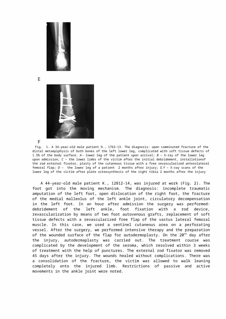
A 34-year-old male patient D., 1763-13, has suffered as a result of a traffic accident (Fig. 1). The patient got an open comminuted fracture of the distal metaepiphysis of both bones of the left lower leg, complicated with soft tissue defects of 1.5% of the body surface. In 3 hours after the admittance of the victim, we performed the

debridement, installation of an external rod fixator, and the replacement of soft tissue defects with a free revascularized graft from the vast lateral muscle of the thigh. On the 6th day after the operation, the marginal necrosis area of about 1.5 cm2 was observed, the necrotic tissue was excised, and local ointment dressings were performed. On the 15th day after the injury, we performed autodermaplasty of the flap surface. The wounds healed. On the 35th day after the injury, plate osteosynthesis of distal metaepiphysis of both right lower leg bones was performed. Postoperative wounds healed by primary intention. There was a consolidation of fractures; the patient walked fully leaning on the injured limb.
A
B
C

D
E
F Fig. 1. A 34-year-old male patient D., 1763-13. The diagnosis: open comminuted fracture of the distal metaepiphysis of both bones of the left lower leg, complicated with soft tissue defects of 1.5% of the body surface. A – lower leg of the patient upon arrival; B – X-ray of the lower leg upon admission; C – the lower limbs of the victim after the initial debridement, installationof the rod external fixator, plasty of the cutaneous tissue with a free revascularized anterolateral femoral flap; D – the lower leg of a patient 2 months after injury; E, F – X-ray scans of the lower leg of the victim after plate osteosynthesis of the right tibia 2 months after the injury
A 44-year-old male patient K., 12812-14, was injured at work (Fig. 2). The foot got into the moving
mechanism. The diagnosis: incomplete traumatic amputation of the left foot, open dislocation of the right foot, the fracture of the medial malleolus of the left ankle joint, circulatory decompensation in the left foot. In an hour after admission the surgery was performed: debridement of the left ankle, foot fixation with a rod device, revascularization by means of two foot autovenous grafts, replacement of soft tissue defects with a revascularized free flap of the vastus lateral femoral muscle. In this case, we used a sentinel cutaneous area on a perforating vessel. After the surgery, we performed intensive therapy and the preparation of the wounded surface of the flap for autodermoplasty. On the 20th day after the injury, autodermaplasty was carried out. The treatment course was complicated by the development of the seroma, which resolved within 3 weeks of treatment with the help of punctures. The external rod fixator was removed 45 days after the injury. The wounds healed without complications. There was a consolidation of the fracture, the victim was allowed to walk leaning completely onto the injured limb. Restrictions of passive and active movements in the ankle joint were noted.
A
B
C

D
E
F
G

H Fig. 2. A patient K, 44 years old, 12812-14. Diagnosis: incomplete traumatic amputation of the left foot, open dislocation of the right foot, the fracture of the medial malleolus of the left ankle joint, circulatory decompensation in the left foot. A – left lower leg upon admission; B – the lower leg and foot after reposition; C, D – X-rays of the victim after the repositioning; E – lower leg and foott of the victim after the primary surgical treatment of wounds, installation of the rod external fixato, replacement of soft tissue defects with a free revascularized anterolateral flap, F – X-ray of the injured after osteosynthesis inner malleolus with rods and wire, installation of an external rodfixator; G – the lower leg and foot of the victim after the surgery for correction of muscle flap 15 days after the injury; H – lower leg and foot after autodermoplasty 27 days after the injury.
RESULTS The surgery lasted for 3 hours in the 1st patient and 3.5 hours in the 2nd case. After the surgery, impairment of
blood circulation in the flaps was not observed. Revision of microanastomosis was not required. No deep wound infection was observed. In one case, a minimal marginal flap necrosis of about 1cm 2 developed; after excision of necrotic tissue and local treatment, the wound healed within 10 days of local treatment. In all patients, the cutaneous tissues were completely restored, which allowed the lower extremity internal fixation to be performed in the first patient.
For comparison, we report the clinical example of a patient who urgently underwent replacement of soft tissue defects with a free revascularized flap of the latissimus dorsi muscle.
A 21-year-old male patient K., 5919-12 had an injury as a result of a heavy object falling on the foot. The diagnosis: open fracture of both bones of the distal right lower leg metaepiphysis, open fracture of the talus, complicated by extensive skin crush the lower leg and foot. The patients underwent debridement, osteosynthesis of talus with wires. After excision of crushed cutaneous tissue, the defect of about 1.5% of the body surface developed. The replacement of the soft tissue defect free revascularized flap of the latissimus dorsi muscle was performed. We also performed: end-to-end anastomosis of a flap artery and the anterior tibial artery and end-to-end flap vein anastomosis with the comitant vein. Duration of the replacement was 8 hours. The postoperative venous thrombosis was marked in the single flap vein, which required exploration and re-anastomosis. On the 25th day after the injury we perfrmed autodermaplasty of the flap surface. Patients reported difficulty in walking with crutches due to the presence of scar in the underarm area. The wounds healed, but the delayed wound healing was observed, which we associate with ischemic damage of the graft due to the thrombosis of the graft vein. There was a consolidation of lower leg fractures. The patient walked with fully leaning onto the injured limb. Limitation of movement in the ankle joint was observed.
We believe that the long duration of operation in this observation is associated with the necessity of turning the victim on his side, impossibility to work in the same operative field. The development of venous thrombosis in this case was facilitated by the fact that the flap of the latissimus dorsi muscle on the vascular pedicle consisted of one vein, while the anterolateral femoral flap on the vascular pedicle consisted of two veins, the probability of thrombosis in the postoperative period of two veins is significantly lower. Also, in this study a significant loss of blood (hemoglobin before surgery – 119 g/l, after primary and revision surgery – 40 g/l) was noted, which was associated with the duration of the surgery, the development of venous flap hypertension, the need to explore anastomoses in the first hours after transplantation.
DISCUSSION Our results are generally consistent with those of other authors on the lowest rate of complications during the
urgent replacement of soft tissue defects in patients with severe lower limb trauma. Many authors present data on the significant reduction in the frequency of deep wound infection, flap necrosis and duration of treatment when using this approach [2, 8].
There have been recent reports on the use of local perforating flaps in the treatment of patients with soft tissue defects of the lower extremities [9, 10]. However, these methods do not work in conditions of high-intensity trauma and extensive damage to the surrounding tissue. The use of this type of grafts is also limited with symptoms of shock during the first hours after the injury due to the high risk of spasm, and peripheral vascular necrosis of the flap. The frequency of complications associated with the violation of perfusion of the flap when using this technology even in delayed surgeries, according to some, is about 30% [11].
The use of free revascularized muscle flaps to replace soft tissue defects of the lower limbs is considered to be the "gold standard." Authors report on the flap of the latissimus dorsi and the anterolateral femoral flap for this purpose [4, 10]. According to Collins, no need to turn the patient on his side and the ability to work on a single

operating field, as well as the possibility of surgery under regional anesthesia make anterolateral femoral flap the method of choice when performing urgent intervention [12].
CONCLUSION There have been many reports of the plastic soft tissues of the lower limb with a free anterolateral femoral
muscle flap recently. The advantages of this method are the great length of the vascular pedicle, large diameter of vessels, a wide area of the coverage, no need for turning the victim on his side and the possibility of carrying out the operation under the regional anesthesia. The presence of two veins in the flap pedicle is suitable for anastomosis, reduces the probability of postoperative complications related to the violation of the outflow of blood from the transplanted graft. These advantages make the anterolateral femoral flap the most convenient and reliable for restoration of extensive damage to tissues covering the lower limbs urgently in the first hours after the injury. This analysis of two clinical observations confirm these data.
REFERENCES1. Tikhilov R.M., Kochish. A.Yu, Rodomanova L.A., et al. Sovremennye tendentsii plastiki loskutami s osevym tipom
krovosnabzheniya na nizhney konechnosti [Modern trends in plastic flaps with axial type of blood supply to the lower limb]. Vestnik travmatologii i ortopedii im NN Priorova. 2007; 2: 71–75. (In Russian).
2. Shibaev E.Yu. Pervichnye rekonstruktivnye operatsii na konechnostyakh s ispol’zovaniem metodov mikrokhirurgicheskoy autotransplantatsii [Primary reconstructive surgery on the limbs using techniques microsurgical autotransplantation]. Annaly plasticheskoy, rekonstruktivnoy i esteticheskoy khirurgii. 1998; 3: 112. (In Russian).
3. Breugem C.C., Strackee S.D. Is there evidence-based guidance for timing of soft tissue coverage of grade III B tibia fractures? Int J Low Extrem Wounds. 2006; 5 (4): 261–270.
4. Clough T.M., Bale R.S. Audit of open tibialdiaphyseal fracture management at district accident centre. Ann R Coll Surg Engl. 2000; 82 (6): 436–440.
5. Collins J., Ayeni O., Thoma A. A systematic review of anterolateral thigh flap donor site morbidity. Can J Plast Surg. 2012; 20 (1): 17–23.
6. Gopal S., Giannouds P.V., Murray A., et al. The functional outcome of severe, open tibia fractures managed with early fixation and flap coverage. J Bone Joint Surg Br. 2004; 86 (6) 861–867.
7. Gopal S., Majumder S, Batcjelor A.G.B., et al. Fix and Flap: the radical orthopaedic and plastic treatment of severe open fractures of the tibia. J Bone Joint Surg Br. 2000; 82 (7): 959–966.
8. Liau J.E., Pu L.L. Reconsruction of a large upper tibial wound extending to knee with a free latissimusdorsi flap: optimizing the outcomes.Microsurgery. 2007; 6: 548–552.
9. Mehrotra S. Perforator plus flaps: Optimizing results while preserving function and esthetics. Indian J Plast Surg. 2010; 43 (2): 141–148.
10.Pribaz J.J., Orgill D.P., Epstein M.D., et al. Anterolateral thigh free flap.Ann Plast Surg. 1995; 34 (6): 585–592.11.Song Y.G., Chen G.Z., Song Y.L. The free thigh flap: a new free flap concept based on the septocutaneous artery. Br J Plast Surg.
1984; 37 (2): 149–159.12.Tos P., Innocenti M., Artiaco S., et al. Perforator-based propeller flaps treating loss of substance in the lower limb. J Orthop
Traumatol. 2011; 12 (2): 93–99.
Article received on 13 April, 2015
For correspondence:Aleksandr V. Nevedrov,
Researcher of the Emergency Department Plastic and Reconstructive Surgery N.V. Sklifosovsky Research Institute for Emergency Medicine of the Moscow Healthcare Department, Moscow, Russian Federation
e-mail: [email protected]