Embed Size (px)
Citation preview

FINITE ELEMENT MODELLING OF THE MITRAL VALVE REPAIR
USING AN IMPLANTABLE LEAFLET PLICATION CLIP M. D. Stepanov1*, O.S. Loboda1,4, Y. V. Novozhilov2, N. V. Vasilyev3
1Peter the Great Saint-Petersburg Polytechnic University, 29 Polytechnicheskaya, St. Petersburg 195251, Russia 2«CADFEM-CIS», 15 Kondratyevsky Prospekt, St. Petersburg 195197, Russia
3Boston Children’s Hospital, Harvard Medical School, 300 Longwood Avenue, Boston, Massachusetts, 02115,
USA 4 Institute of Problems in Mechanical Engineering, Bolshoy pr., 61, V.O., Saint-Petersburg, Russia, 199178
*e-mail: [email protected]
Abstract. We report the results of numerical simulation of the mitral valve in human heart. The beam-shell geometry model was created based on anatomical atlases and taking into account the heterogeneity of distribution of the mitral valve’s leaflets thickness. The full cycle of the mitral valve opening and closure was simulated using the finite element analysis software ANSYS Mechanical. The method of data processing from a computer tomography in a solid CAD model was implemented and tested. Keywords: mitral valve, finite element simulation, computed tomography. 1. Introduction Thorough understanding of the mitral valve (MV) mechanics is needed for surgical decision making such as choosing the type of surgical valve repair applicable for particular patient [1]. Several mathematical models [2, 3 and 4] of the MV have been developed that allowed simulating the valve opening and closure under different conditions. In this study, in addition to modeling normal MV function, we analyzed the leaflet motion in the presence of mitral valve prolapse (MVP) and its repair using novel plication device [5]. 2. Mitral valve anatomy The MV consists of the annulus, posterior and anterior leaflets, chordae tendineae that are connected to papillary muscles. Chordae tendineae are tendinous connective fibers that bond the leaflets of the MV with the papillary muscles located on the inner surface of the left ventricle. The main function of the MV is to control the blood flow from the left atrium to the left ventricle. During normal left ventricular diastole, the MV is open and blood flows from the left atrium into the left ventricle. Then, during left ventricular systole the MV closes, and blood is ejected into the aorta. During contraction of left ventricle, papillary muscles contract and tether the chordae preventing prolapse of the MV leaflets.
2.1 Mitral valve prolapse. MVP is a disease in which there is the displacement of MV leaflet into the left atrium during left ventricular systole. It is accompanied by the appearance of the blood backflow into the left atrium. Significant amount of the blood in the backflow leads to heart failure over time, which requires surgical correction of MVP.
MVP can be repaired on a beating heart using an implantable device, the Leaflet Plication Clip that has been developed at Boston Children’s Hospital [5]. The Clip is attached to the
Materials Physics and Mechanics 32 (2017) 383-392 Received: November 18, 2017
© 2017, Peter the Great St. Petersburg Polytechnic University © 2017, Institute of Problems of Mechanical Engineering
diseased leaflet and thereby prevents displacement of the leaflet toward the left atrium and reduces mitral regurgitation.
Prior to surgical operation, patient specific MV anatomy needs to be analyzed using high-resolution computed tomography or three-dimensional echocardiography. Then, the imaging data is processed into a 3D model of the MV. Finally, a physician analyzes the model with the purpose of choosing the optimal method of MV surgical repair.
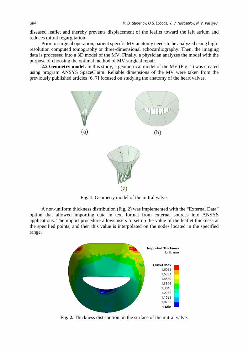
2.2 Geometry model. In this study, a geometrical model of the MV (Fig. 1) was created using program ANSYS SpaceClaim. Reliable dimensions of the MV were taken from the previously published articles [6, 7] focused on studying the anatomy of the heart valves.
Fig. 1. Geometry model of the mitral valve.
A non-uniform thickness distribution (Fig. 2) was implemented with the “External Data”
option that allowed importing data in text format from external sources into ANSYS applications. The import procedure allows users to set up the value of the leaflet thickness at the specified points, and then this value is interpolated on the nodes located in the specified range.
Fig. 2. Thickness distribution on the surface of the mitral valve.
384 M. D. Stepanov, O.S. Loboda, Y. V. Novozhilov, N. V. Vasilyev

2.3 Finite element model. According to the geometry model of the MV described above, finite element model (Fig. 3) was created with the following features: element size – 0.5 mm, number of nodes – 5 402, number of elements - 4 794, type of elements: quadrilateral and triangular for modeling of the leaflets(SHELL181) and beam elements for the chordae tendineae (BEAM188).
Fig. 3. Finite-element model.
2.4 Natural and essential boundary conditions. Initial and boundary conditions were
simulated taken into account the actual conditions of MV function. At the initial moment of calculation, the valve is in the unstressed state. This corresponds to the transition from the filling phase to the phase of left atrial systole. Based on the pressure curves from the left atrium and the left ventricle [7], the resulting pressure curve was obtained (Fig. 4).
Fig. 4. Resulting pressure curve.
Boundary conditions are shown in figure 5. The mitral annulus is fixed in three
translational degrees of freedom (A). In addition, points (B) of the lower part of chords are fixed at the place of attachment to the papillary muscle in the left ventricle. Pressure is applied to the surface (C) from the left ventricle and provides closure of the leaflets.
-202468
1012141618
0 0,2 0,4 0,6 0,8 1
Pres
sure
, MPa
Time, s
Finite element modelling of the mitral valve repair using an implantable leaflet plication clip 385

Fig. 5. Natural and essential boundary conditions.
2.5 Constitutive model of the mitral valve tissue. MV leaflets consist of collagen,
elastin and glycosaminoglycan. The relationship between collagen and elastin determine the mechanical behavior of the tissue. Angle measured between the collagen fibers describes that the fiber orientation depends on the considered region and symmetrical to the central radial axis of each MV leaflet.
Most of the biological materials are anisotropic, i.e. their deformation depends on the direction of displacement. Such fiber-reinforced composite material with a single preferred direction is called transversely isotropic material. A network of crimped collagen fibers represents tissue of the MV, particularly in the central region. The angle of these fibers is relatively uniform within the considered experimental region. Therefore, it is assumed that the tissue of the MV can be modeled as a transversely isotropic material.
We assumed local tissue homogeneity, although there is some heterogeneity due to the complicated structure of the valve leaflets. Tissue of the MV consist mainly of water and has got a reduced perfusion (blood supply). Based on these structural and mechanical observations, it can be assumed that the tissue of the MV can be modeled as a hyperplastic incompressible material that is initially and locally transversely isotropic relative to the axis of the collagen fibers.
The strain energy function is a short description of the material of this type. Several types of strain energy functions were proposed in order to account for the transversal isotropy of the soft tissues. Following the method Humphrey [8], it is possible to make an assumption about the subclass of transversely isotropic materials in which the strain energy function W presumably depends only on the two coordinate invariant measures of finite deformation (i.e., the first invariant of strain and elongation along the fiber direction α): 𝑊𝑊 = 𝑊𝑊(𝐼𝐼1,𝛼𝛼), (1) where 𝐼𝐼1 = 𝑡𝑡𝑡𝑡𝑪𝑪 = 𝑡𝑡𝑡𝑡𝑩𝑩 and 𝜶𝜶 = 𝑵𝑵 ∙ 𝑪𝑪 ∙ 𝑵𝑵.
𝑪𝑪 = 𝑭𝑭𝑇𝑇 ∙ 𝑭𝑭, 𝑩𝑩 = 𝑭𝑭 ∙ 𝑭𝑭𝑇𝑇 are the right and left Cauchy-green deformation tensor, respectively, and N is a unit vector that defines the presumed direction of the fibers of the material in the undeformed configuration. F is the deformation gradient tensor, det (F) =1, due to incompressibility of the material. The expression of Cauchy stress tensor for a material of this type can be expressed as:
𝑻𝑻 = −𝑝𝑝𝑰𝑰 + 2𝑊𝑊1𝑩𝑩 + (𝑊𝑊𝛼𝛼/𝛼𝛼) 𝑭𝑭 ∙ 𝑵𝑵⊗𝑵𝑵 ∙ 𝑭𝑭𝑇𝑇, (2) where 𝑝𝑝 is the multiplier that provides incompressibility, 𝑰𝑰 – identity tensor,
𝑊𝑊1 = 𝜕𝜕𝑊𝑊/𝜕𝜕𝐼𝐼1, 𝑊𝑊𝛼𝛼 = 𝜕𝜕𝑊𝑊/𝜕𝜕𝛼𝛼, ⊗ denotes tensor product. Partial derivatives 𝑊𝑊1 and 𝑊𝑊𝛼𝛼 can be calculated directly from the measured stress and
strain taking into account the angle φ of the collagen fibers. This formulation means that in the special case when one of the strain invariants is alternately held constant while the other is
386 M. D. Stepanov, O.S. Loboda, Y. V. Novozhilov, N. V. Vasilyev

varied, i.e., a set of experiments with constant invariant can be used to determine the functional form of W.
However, to use this type of material we would need the series of experiments to determine mechanical properties of the leaflets. In this study, a linear isotropic model of the MV leaflet material was used. Values for the stiffness matrix (in the isotropic case is the young's modulus and Poisson's ratio) were taken from the article M. A. Hisham [9] devoted to computer modeling of the leaflets of the MV under the action of the systolic pressure.
− material of leaflets: 𝐸𝐸п = 2 𝑀𝑀𝑀𝑀𝑀𝑀; 𝐸𝐸з = 1 𝑀𝑀𝑀𝑀𝑀𝑀; 𝜈𝜈 = 0.49 − material of chords: 𝐸𝐸 = 250 𝑀𝑀𝑀𝑀𝑀𝑀; 𝜈𝜈 = 0.488
3. Results 3.1 Initial configuration. The distribution of values of the principal stresses on the leaflets of the MV at different time points are shown in figure 6. The highest stresses are observed during the transition from tension phase to the expulsion phase in 0.302 sec calculation. At this moment the resulting pressure of 16 kPa acts on the leaflets, this moment is called a full closure of the valve. At the time of full closing of the leaflets oscillation occurs, which is caused by sharply decrease of the blood flow speed not allowing to overcome the closed valve.
Fig. 6. The values of principal stresses at different time points.
Finite element modelling of the mitral valve repair using an implantable leaflet plication clip 387

Closure of the MV is confirmed by checking the status of contact elements at the moment of peak stress (Fig.7). The tight closing of the MV can be judged by the image of the middle cross-section (Fig.8).
Tension on the anterior leaflet is greater than the pressure arising at the posterior. In general, stress values vary in the range of 13 kPa during the filling period of the ventricle to 637 kPa during the period of complete closure of the valve. This result is aligned with the data obtained in the articles [2, 3 and 4] devoted to studies of the MV.
Fig. 7. The middle cross-section of the valve at the moment of full closure of the valve.
Fig. 8. The contact pressure at the moment
of maximum closure of the valve.
3.2 Modified configuration of the mitral valve with the “Clip” on the posterior leaflet. The analysis of influence of the Leaflet Plication Clip device on the MV during normal operation was executed additionally in this study. “Clip” was modeled as a point mass. The device was installed on the posterior leaflet in the center (Fig.9).
Fig. 9. The location of the mass point on the
surface of the posterior leaflet.
Fig. 10. Line along the surface of the
leaflet.
The weight of the device was calculated by the formula: 𝑚𝑚 = 4𝑙𝑙𝑐𝑐𝜌𝜌𝜌𝜌𝑡𝑡2, (3) where 𝑙𝑙𝑐𝑐 – the maximum distance from the mitral annulus to the free edge of the leaflet, ρ = 6.4 g/cm3 is the density of the material of the device, r is the radius of the clip.
Thus, the weight of the clip can vary from 0.1 to 0.4 grams. This Study considers three cases: a) the mass of the “Clip” - 0.1 g., which corresponds to a wire radius of 0.5 mm; b) mass of the “Clip” - 0.23 g., the radius is 0.75 mm; с) mass of the “Clip” - 0.4 g., the radius is 1 mm.
388 M. D. Stepanov, O.S. Loboda, Y. V. Novozhilov, N. V. Vasilyev

The calculation was executed for those three types of the “Clip” and obtained results were compared with the case without the “Clip”.
Analysis of displacements along the line indicated in the figure showed that the maximum divergence of the results is 0.18% for the case without the “Clip” and the case с) Mc = 0.4 g. This suggests that the installation of clip does not significantly affects the movement of MV leaflets during normal operation.
Chart 11 shows the values of the stresses on the installing line of the “Clip” (Fig. 10) for three cases in comparison with the solution without “Clip” on the leaflet.
Fig. 11. The tension dependence of the coordinates on the line for three types of “Clip” and
case without it.
For cases a) and b) large discrepancies were not observed. However, there is divergence of results in the upper region of the valve with the mass increase. The maximum value of the divergence of results is 5.11% detected in case c) when the mass of a “Clip” is equal to 0.4 g. In article [10], the value of the dynamic ultimate tensile strength is 0.9 MPa for the material of the leaflets of the mitral valve. Thus, we can conclude that “Clip” does not entail the appearance of additional tensile stresses that can lead to the destruction of the material of the leaflets.
4. Processing data from a computer tomography One of the objectives of this study was the creation of 3D model using data obtained from computer tomography. We did not have access to high resolution valve images. The workflow was tested on images of the spinal cord. This process can be divided into two stages: a) Converting data from DICOM format to STL format; b) Creation of solid model using STL model.
4.2 Converting data from DICOM format to STL format. Pictures from the computer tomography (figure 12) are the visualization of DICOM data obtained during the survey. Using the software package 3D Slicer, by processing the DICOM files was created STL model of the human spine (figure 13).
0
0,05
0,1
0,15
0,2
0,25
0,3
0,35
0 5 10 15 20
Stre
ss, M
Pa
Coordinate, mm
without "Clip" a) Mc = 0.1 г b) Mc = 0.23 г c) Mc = 0.4г
Finite element modelling of the mitral valve repair using an implantable leaflet plication clip 389

Fig. 12. Computer tomography image.
Fig. 13. Spine STL model.
The created model has a lot of extra surfaces and irregularities caused by the noise effects during survey. Using the program MeshLab model was filtered out (figure 14).
Fig. 14. Comparison of number of surfaces before and after filtration.
4.2 Converting the STL into a solid model. STL format is widely used for storing three-
dimensional models of objects for use it in technologies of rapid prototyping. Information about the object is stored as a list of triangular facets that describe the surface and their normal lines. However, for use in the calculations in the engineering software packages required to create solid geometry CAD model. Thus, with the help of the program ANSYS SpaceClaim above STL model was converted to a CAD model. This operation allows us to use this geometry model in the calculations of biological structures using finite element method.
390 M. D. Stepanov, O.S. Loboda, Y. V. Novozhilov, N. V. Vasilyev

5. Limitations of the Study In carrying out the described analysis we made several assumptions. At first, the material model of chordae tendineae and the leaflets should possess the properties of anisotropic hyperelastic material. In the case of chords, the definition of BEAM188 elements does not allow simulation of hyperelasticity. In general, creating a high quality anisotropic hyperelastic material model requires a series of experiments to determine the constants included in the expression for the description of such a model. In this work, the material of the leaflets and chordae tendineae was modeled as a linear hyperelastic at the first order approximation.
At second, detailed studying the behavior of the MV and determining the position of “Clip” on the leaflet requires an anatomically accurate model of the valve obtained from the high resolution computer tomography. Since such equipment was not available to us, geometry model of the MV was constructed according to anatomical atlases with the dimensions confirmed with that data published in articles focused at studying the anatomy of the MV.
6. Conclusions In this study, the numerical simulation of the MV in the human heart was conducted. Based on anatomical atlases beam-shell geometry model was created taking into account the heterogeneity of distribution of the MV leaflets thickness. The full cycle of the MV opening and closure was simulated using the finite element analysis software ANSYS Mechanical. For the numerical solution of this problem transient structural (non-stationary structural) analysis type allowing to determine time-varying displacements, strains, stresses and internal forces in the body under the influence of unsteady loads was selected. For modeling, the material of the MV leaflets was chosen as linearly elastic isotropic model.
Despite the limitations and assumptions chosen for material model, the obtained results for stresses on the leaflets coincide with the data obtained from the articles [2, 3, and 4] devoted to modeling of the MV. In addition, in the framework of numerical simulation it was proven that the valve closes tightly during the transition from phase of tension to the phase of expulsion in 0.302 sec calculation, which coincides with the data for cycle of the mitral valve operation [7].
In addition to modeling the normal functioning of the MV, the simulation of the the MV function with the device “Clip” implanted on the leaflet was executed. Analysis of the obtained results permits to state that “Clip” does not entail the additional tensile stresses that can lead to the destruction of the material of the MV leaflets.
Moreover, in this study we implemented and tested a method of processing data with a computer tomography in a solid model on the example of the spine that can be later used for calculations in software systems of finite element analysis. This method will allow in the future creating of an anatomically accurate model of the MV.
In the future, we plan to use an incompressible, hyperelastic transversely isotropic material and construct the geometrical model of the MV derived from computed tomography images for more precise studies of the valves behavior.
References [1] N.A. Tenenholtz, P.E. Hammer, R.J. Schneider, N.V. Vasilyev, R.D. Howe, On the Design
of an Interactive, Patient-Specific Surgical Simulator for Mitral Valve Repair. In: IEEE/RSJ International Conference on intelligent Robots and Systems (IROS) (2011) 1327.
[2] W. Qian, S. Wei // Annals of Biomedical Engineering 41(1) (2013) 142. [3] M.S. Sacks, He Z., L. Baijens, S. Wanant, P. Shah, H. Sugimoto, A.P. Yoganathan // Annals
of Biomedical Engineering 30 (2002) 1281. [4] M.Stevanella, F. Maffessanti, C. A. Conti, E. Votta, A. Arnoldi, M. Lombardi, O. Parodi,
E. G. Caiani, A. Redaelli // Cardiovascular Engineering and Technology 2(2) (2011) 66.
Finite element modelling of the mitral valve repair using an implantable leaflet plication clip 391

[5] E.N. Feins, H. Yamauchi, G.R. Marx, F.P. Freudenthal, H. Liu, P.J. del Nido, N.V. Vasileyv // The Journal of Thoracic and Cardiovascular Surgery 147(2) (2014) 783.
[6] D.S. Kotovich, I.N. Stakan // Voennaya medicina [Military medicine] 2 (2010) 58. (In Russian).
[7] K.V. Sudakov, In: Physiology. Fundamentals and functional systems (Medicine, Moscow, 2000) p. 327. (In Russian).
[8] J.D. Humphrey, R.K. Strumpf, F.C.P. Yin // ASME Journal of biomechanical engineering 112 (1990) 333.
[9] M. A. Hisham, M. Adib, K. Osman, N. Hazreen, M. Hasni, O. Maskon, F. Naim, Z. Ahmad, I. Sahat1and A. N. Mu’tasim. In: International Conference on Environment and BioScience (IACSIT Press, Singapore, 2012), Vol.21, p. 6.
[10] Anwarul Hasan // Journal of Biomechanics 47(9) (2014) 1949.
392 M. D. Stepanov, O.S. Loboda, Y. V. Novozhilov, N. V. Vasilyev