Embed Size (px)
Citation preview

FINAL PROGRAM BOOK

TABLE OF CONTENTS
37TH ESOPRS ANNUAL MEETING 2018 2
Welcome 2
General Information 3
ESOPRS Committee 3
Venue 4
Social Events Venues 5
Faculty 6
Mustarde Lecture 6
Keynote Speakers 7
Program 10 Pre-Meeting Course 10 Annual Meeting Day 1 11 Annual Meeting Day 2 13
Abstracts 16 E-Posters 51
Videos 63
Sponsors 65
It is our greatest joy to welcome you to Bucharest for the 37th annual meeting of the European Society of Ophthalmic Plastic and Reconstructive Surgery.
We are very fortunate this year to have 5 World-renowned key-note speakers and 58 esteemed faculty from 3 different conti-nents. We also have a well-balanced esthetic and reconstructive program covering most topics encountered in todays academic or private oculoplastic practice. Our Advanced Pre-meeting Course is designed to teach innovative esthetic and reconstructive tech-niques that will be useful to both our younger and more senior oculoplastic colleagues.
The venue is situated in the historic downtown of Bucharest, with-in walking distance from most sightseeing, cultural and nightlife attractions. The Welcome Reception will take place at the ARCUB center situated in the heart of the bustling Old Town. The Gala Dinner this year will be “A Night at the Palace” and will be hosted in the Palace of Parliament (the famous People’s House).
Bucharest is not far away from many famous castles and medieval towns such as the Dracula and Peles Castles and the towns of Bra-sov, Sibiu, Sighisoara and Cluj. We offer guided pre and post-meet-ing tours to most of these attractions. We are not only very excited but also committed to making your stay in Romania both scientifically productive and culturally en-riching. We simply want you to have a blast!
Dan Georgescu, MD, PhD Daniela Cioplean, MD
LOCAL ORGANIZERS
WELCOME
www.esoprs2018.ro

37TH ESOPRS ANNUAL MEETING 2018
ESOPRS EXECUTIVE COMMITTEE
PresidentHaraldur [email protected]
TreasurerDavid Verity
SecretaryDion Paridaens
President ElectFranz Josef Steinkogler
Immediate Past PresidentGeoffrey Rose
BOARD MEMBERS
FranceThierry Malet
GermanyLeonard Holbach
ItalyLelio Baldeschi
United KingdomDaniel Ezra
SpainMiguel Gonzalez Candial
TurkeyMehmet Unal
GreeceIoannis Mavrikakis
GENERAL INFORMATION
3
Meeting Objectives• To provide the venue for the presentation of new data, techniques and
concepts in the field of reconstructive and cosmetic oculofacial plastic surgery in order to increase knowledge and competence. Our goal is to promote excellence in patient care and improve outcomes in all area of oculofacial plastic, orbital and lacrimal surgery.
• To present those areas in oculofacial plastic surgery where clinical and basic science research have led to substantial advancements and to highlight areas where further research is desirable.
• To provide the venue where colleagues from around the World can meet and directly exchange knowledge and share their experiences with the latest techniques and devices used to treat orbital, lacrimal and oculofacial conditions.
• To further our knowledge, improve skills and enhance results in the field of cosmetic oculofacial plastic surgery.
Target AudienceThe ESOPRS Annual Meeting welcomes all interested physicians such as oculoplastic surgeons, oculofacial plastic surgeons, general ophthalmolo-gists, dermatologists, otorhinolaryngologists, facial plastic surgeons and oromaxilofacial surgeons, whether they are already practicing or in train-ing.
Photography and Social Media PolicyThe European Society of Ophthalmic Plastic and Reconstructive Surgery would like to advise all participants to the 37th Annual Meeting to abide by the following rules concerning photography and social media:• Non-flash photography is allowed for personal use. • Photography taking must be done in a non-disruptive manner to the
rest of the audience.• Sharing of any identifiable photographic information on social media
and videotaping are strictly prohibited.• Respect presenters who do not wish their slides or content be photo-
graphed or shared on social media.
Liability DisclaimerIn the event of industrial disruption or other unforeseen circumstances, the event Organizers accept no responsibility for loss of money incurred by delegates. The Organizers accept no liability for injuries/losses of whatever nature incurred by participants and/or accompanying persons, nor for loss or damage to their luggage and/or personal belongings. Delegates should make their own arrangements with respect to personal insurance.
Travel and Program Disclaimers In the event of any travel disruptions, the Organizers will not be held re-sponsible for any losses incurred by delegates at or en route to or from the event.The program is correct at the time of publishing, but the Organizers re-serve the right to alter the program as necessary.
CME CreditsThe 37th Annual Meeting the European Society of Ophthalmic Plastic and Reconstructive Surgery, Bucharest, Romania, 13/09/2018 - 15/09/2018 has been accredited by the European Accreditation Council for Continuing Medical Education (EACCME®) with 19 European CME credits (ECMEC®s).Through an agreement between the Union Européenne des Médecins Spé-cialistes and the American Medical Association, physicians may convert EACCME® credits to an equivalent number of AMA PRA Category 1 Credit-sTM. Information on the process to convert EACCME® credit to AMA credit can be found at www.ama-assn.org
37TH ESOPRS ANNUAL MEETING 2018
VENUE
4
1
2
3.A
3.C
4 6 7 8
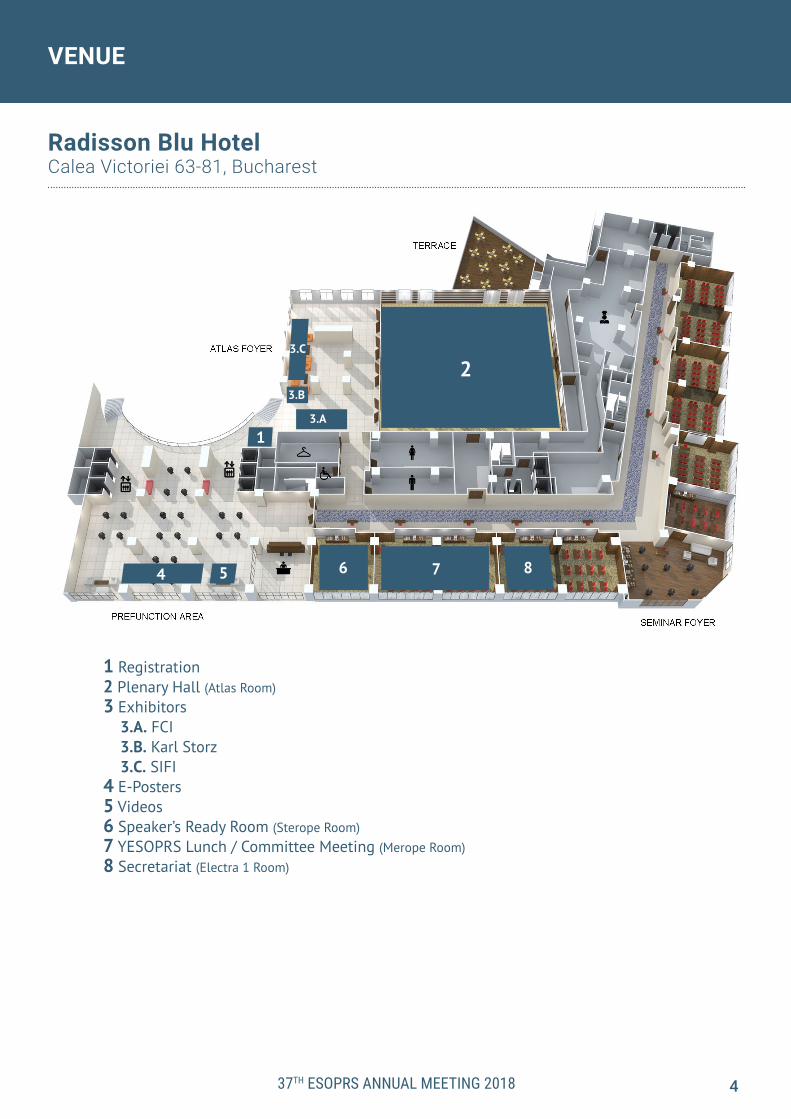
Radisson Blu HotelCalea Victoriei 63-81, Bucharest
1 Registration2 Plenary Hall (Atlas Room)3 Exhibitors 3.A. FCI 3.B. Karl Storz 3.C. SIFI4 E-Posters5 Videos6 Speaker’s Ready Room (Sterope Room)7 YESOPRS Lunch / Committee Meeting (Merope Room)8 Secretariat (Electra 1 Room)
5
3.B

SOCIAL EVENT VENUES
WELCOME COCKTAILSeptember 13, 2018 19:00-21:00 hrs
ARCUB Cultural CentreStr. Lipscani 84 - 90
GALA DINNERSeptember 14, 2018 20:00-24:00 hrs
Palace of the ParliamentStr. Izvor 2 - 4
Radisson Blu HotelCalea Victoriei 63-81
37TH ESOPRS ANNUAL MEETING 2018 5

37TH ESOPRS ANNUAL MEETING 2018
FACULTY
SCIENTIFIC COMMITTEEDaniela CiopleanDan GeorgescuRamón MedelDion ParidaensUlrich SchaudigVladimir Thaller
KEYNOTE SPEAKERS Richard Anderson (Salt Lake City, United States)Jonathan Hoenig (Los Angeles, United States)Dion Paridaens (Rotterdam, Netherlands)Patrick Tonnard (Ghent, Belgium)Hunter Kwok-Lai Yuen (Hong Kong)
MUSTARDE LECTURERMichele Beaconsfield (London, United Kingdom)
PRE-MEETING COURSE SPEAKERSChris Alabiad (Miami, United States)Bijan Beigi (Norwich, United Kingdom)Francesco Pietro Bernardini (Genova, Italy)Elin Bohman (Stockholm, Sweden)Altug Cetinkaya (Ankara, Turkey)George C. Charonis (Athens, Greece)Philip Custer (St Louis, United States)Daniel Ezra (London, United Kingdom)Tamara Fountain (Chicago, United States)Suzanne K. Freitag (Boston, United States)Olivier Galatoire (Paris, France)Miguel Gonzalez-Candial (Barcelona, Spain)Christoph Hintschich (Munich, Germany)David Jordan (Ottawa, Canada)Matthew Kay (Miami, United States)Pierre Keller (Paris, France)Ioannis Mavrikakis (Athens, Greece)Ramon Medel (Barcelona, Spain)Alessandra Modugno (Rome, Italy)Jean-Marie Piaton (Enghien, France)Geoffrey Rose (London, United Kingdom)Marco Sales-Sanz (Madrid, Spain)Ulrich Schaudig (Hamburg, Germany)Pari N. Shams (London, United Kingdom)Bazil Stoica (Madrid, Spain)Diego Strianese (Riyadh, Saudi Arabia)Dario Surace (Mestre, Italy)Sara Wester (Miami, United States)Vivian T. Yin (New York, United States)
Michele Beaconsfield, MD (London, United Kingdom)
Saturday, September 15, 2018 12:40 PMCellular Mechanisms of Targeted Cancer Therapy: Present and Future
Consultant Ophthalmic & Oculoplastic Sur-geon for over 25 years (already!) with particu-lar expertise in surgical rehabilitation (ocular cicatricial disease, thyroid ophthalmopathy, tumor defect reconstruction). Spearheaded 10 years ago the specialized Lid Oncology Ser-vice at Moorfields, which runs with consultants (including Mohs), a dedicated Fellow and Can-cer Nurse Specialist, and linked to a multidis-ciplinary team of pathologists, radiologists, oncologists and radiotherapists. Education-al supervisor and Trainee Mentor. Founding member of BOPSS (British Oculoplastic Sur-geons Society), examiner for the European Board of Ophthalmology and Royal College of Ophthalmologists. Past President of the UEMS Ophthalmic Section (union européenne de médecins spécialistes) and past Treasurer of ESOPRS. ■
2018 MUSTARDE LECTURE
6
The Mustardé lecture is an invited lecture in the honor of Dr. Jack Mustardé, the first president of the ESOPRS. A new speaker is selected each year by the ESOPRS committee. The invited speaker is both an ESOPRS member and a leader in our field.

KEYNOTE SPEAKERS
Richard Anderson, MD, FACS (Salt Lake City, United States)
Friday, September 14, 2018 2:00 PMTips Learned in 40 Years of Oculoplastic Practice
Dr. Anderson is an award winning internationally renown eyelid and facial plastic surgeon. He was honored with the American Society of Ophthalmic Plastic and Re-constructive Surgery (ASOPRS) Outstanding Contributions Award.
Dr. Anderson is a world renowned author and speaker. He has published over 300 scientific journal articles, over 100 book chapters, and 3 books on eyelid, orbital and facial plastic and cosmetic surgery. He has delivered over 1,000 papers at scientific meetings. He is the founder of many modern day techniques in cosmetic and recon-structive surgery of the eyelids and face. He has performed surgery on many digni-taries including Kings and Princes around the world.
He has been an Editorial Board member of 11 journals in the field, including Archives of Facial Plastic Surgery, Archives of Ophthalmology and Ophthalmic Plastic and Re-constructive Surgery. He was one of the original Botox investigators with over 30 years of experience and a Board member for the Benign Essential Blepharospasm Foundation and Orbital Society. ■
Jonathan Hoenig, MD (Los Angeles, United States)
Friday, September 14, 2018 3:20 PMHeading South: Rejuvenation of the Lower Face and Neck
Dr. Hoenig limits his practice to plastic surgery of the face, eyes, and neck, having performed close to 20,000 thousand procedures over the past 15 years on the face alone. He is one of the few cosmetic surgeons who is a member of the American So-ciety of Ophthalmic Plastic and Reconstructive Surgery (ASOPRS) and the American Board of Ophthalmology, as well as a diplomat of the American Board of Cosmetic Surgery.
TrainingDr. Hoenig’s completed four separate fellowships in his quest to master the art of facial plastic and reconstructive surgery. He first focused on oculoplastic surgery at New England Medical Center in Boston Massachusetts. He then pursued further training through a second fellowship in oculo-facial cosmetic surgery at the Jules Stein Eye Institute. Dr. Hoenig then completed a third fellowship in full body Cosmet-ic Surgery, mastering the techniques of Facial Plastic Surgery, liposuction, fat trans-fer, laser skin resurfacing, chemical peels as well as other cosmetic procedures of the body. In addition, Dr Hoenig completed a Mohs fellowship for skin cancer excision and reconstruction. To date he has performed over 25,000 Moh’s excision and Re-constructive cases.
TeachingDr. Hoenig has authored numerous scientific papers and medical textbook chap-ters, and is a regular lecturer at the annual meetings of the American Academy of Ophthalmology, the UCLA Jules Stein Eye Institute, and the American Society of Oph-thalmic Plastic and Reconstructive Surgery (ASOPRS). He is frequently requested to present his techniques and methodologies at meetings of general plastic, facial plas-tic, and oculoplastic surgeons alike. He is the past Director of the Oculofacial Plastic Surgery at the Albert Einstein College of Medicine. He is currently an ASOPRS Fellow-ship Director in Oculofacial Plastic Surgery in Beverly Hills and UCLA Medical Center in Los Angeles California. ■
37TH ESOPRS ANNUAL MEETING 2018 7

37TH ESOPRS ANNUAL MEETING 2018
KEYNOTE SPEAKERS
Dion Paridaens, MD (Rotterdam, The Netherlands)
Friday, September 14, 2018 10:10 AMNew Options in the Medical Treatment of Thyroid Eye Disease: Is there a Role for Nanotechnology
Following his medical training in the University of Utrecht, Dion Paridaens performed a two-year oncology fellowship in Moorfields Eye Hospital and the Institute of Oph-thalmology, London. He later completed his Ph.D. thesis on periocular melanoma in 1993 and was awarded the FC Donders Prize in 1995 for his scientific contributions.Since 1996 he has worked as a consultant surgeon in the Oculoplastic & Orbital Ser-vice of the Rotterdam Eye Hospital, The Netherlands, a high-volume tertiary care institution.
In 1997 he was a visiting orbital fellow in Vancouver, British Columbia. He has been a honorary Consultant to the Erasmus Medical Center Rotterdam and was a part-time Professor at the Hopitaux Universitaires de Geneve, Switzerland between 2011 and 2016. Dr. Paridaens has been an invited lecturer and guest surgeon in various coun-tries and has published over 170 publications, several book chapters and education-al DVDs and iPhone application on oculoplastic surgery and orbital decompression.
His research focuses on periocular and intraocular tumors, eyelid- & lacrimal surgery and Graves Orbitopathy and has supervised several PhD students. He has been the clinical director of the oculoplastic fellowship in the Rotterdam Eye Hospital since 1991 and has trained 1-2 fellows per year since then.
Between 1997 and May 2010 he was Editor-in-Chief of the international Journal OR-BIT. He is a regular reviewer for several scientific journals. Since 2012 he has been on the Committee of the European Society of Ophthalmic Plastic & Reconstructive Sur-gery (ESOPRS). Since 2014 he has served as the Secretary of ESOPRS. He is a founding member of the Dutch Orbital Society and of the Rotterdam Thyroid Center. He has served as a medical advisor for several patient organisations in the Netherlands. Dion Paridaens is married and is the proud father of three teenagers. ■
Patrick Tonnard, MD (Ghent, Belgium)
Saturday, September 15, 2018 10:30 AMAugmentation Blepharoplasty: A Review of 500 Consecutive Patients
Dr. Patrick L. Tonnard graduated in medicine from Ghent University in 1987. He spent three years as an assistant surgeon to Professor Derom at Ghent University Hospital and was then appointed as an assistant in plastic surgery at the Hôpital du Tondu in Bordeaux, France. From 1990 to 1993 he worked as a plastic surgeon alongside Pro-fessor Matton at Ghent University Hospital. He then spent six months as an assistant to Dr. Ortiz-Monasterio in Mexico City. He eventually set up his practice in Ghent.
Since 1994 Dr. Tonnard is consultant plastic surgeon at the General Hospital St-Lucas in Gent, assistant clinical professor at the University Hospital of Gent, founder of the Coupure Centre for Plastic Surgery in 1996 and the Medical Centre ‘t Zwin in Oost-burg (Netherlands) in 2005. In 2007 he raised together with his associate Dr. Alexis Verpaele the Esthetic Medical Centre 2 (E:MC²), a private surgical centre for esthetic surgery in St-Martens-Latem. Dr. Tonnard is a member of the Royal Belgian Society for Surgery, the Belgian and Dutch Association for Plastic, Reconstructive and Aesthetic Surgery, the ISAPS (In-ternational Society of Aesthetic Plastic Surgeons) and the ASAPS (American Society
8

KEYNOTE SPEAKERS
of Aesthetic Plastic Surgeons). He has also been appointed Fellow of the Collegium Chirurgicum Plasticum and Fellow of the European Board of Plastic Surgery.
Dr. Tonnard is also the author of approximately fifty scientific publications, has pre-sented over two hundred scientific papers at national and international conferences, and made numerous contributions to scientific articles and books. He is much in de-mand as a speaker and moderator at international conferences and is an authority on surgical rejuvenation of the face. Together with Dr. Verpaele he developed the revolutionary MACS lift, a minimally invasive facelift technique that aims to produce the best and most natural result with rapid recovery and minimal complications. This achievement has earned him and Dr. Verpaele an international reputation. In 2004 and 2007 they contributed two scientific chapters about this surgical technique, written for plastic surgeons all over the world: The MACS-lift Short Scar Rhytidecto-my, 2004, St Louis; and Short Scar Face-lifting, Operative Strategies and Techniques, 2007, St Louis.
Dr. Tonnard has also written for a less specialized public: he contributed to the book Van top tot teen - alles over plastische chirurgie [From top to toe - all about plastic surgery] by Hubert Tygat and in 2000 he published Schone Schijn. Een wegwijzer in de plastische chirurgie [Looking Beautiful. A guide to plastic surgery] which he wrote with the journalist Laurens de Keyser.
Dr. Tonnard and Dr. Verpaele set up ‘See and Smile’ in 2006. This is an association of Flemish plastic surgeons and ophthalmologists who travel to developing coun-tries at regular intervals to perform operations. They carry out basic plastic surgery procedures (especially treatment for hare lip and cleft palate) and eye treatments (especially for cataracts). ■
Hunter Kwok-Lai Yuen, FRCS, FRCOphth (Hong Kong)
Friday, September 14, 2018 12:40 PM Hybrid Operation for Orbital Venous Malformation
Dr. Hunter Yuen is the Consultant Ophthalmologist and Head of Oculoplastic and Orbital Service at Hong Kong Eye Hospital. He has published more than 70 peer re-viewed articles and book chapters. He is the current president of Asia Pacific Society of Ophthalmic Plastic and Reconstructive Surgery (APSOPRS), Secretary General of Asia Pacific Society of Ocular Oncology and Pathology (APSOOP), vice president of International Society of Dacryology and Dry Eye (ISDDE). Dr. Yuen has been invited as scientific committee members in numerous international meetings and has deliv-ered numerous presentations.
His awards include Best Oral Presentation in 2001 Annual Scientific Meeting Hong Kong, Best Oral presentation of the session in 4th International Symposium of Oph-thalmology, One of the best of the posters at American Academy of Ophthalmology 2005 Annual Meeting. He is also the co-authors of three other best poster awards (Best poster 1st prize at the 23rd European Society of Oculoplastic and Reconstruc-tive Surgery, Best poster at the 2005 Annual Scientific Meeting Hong Kong, Best post-er at 16th Annual meeting of Oculoplasty Association of India).
Dr. Yuen was elected as one of the Ten Young Outstanding Person in Hong Kong in 2007. He was also awarded with the APAO Yasuo Tano Travel Grant in 2010, APAO Distinguished Service Award, APAO Achievement Award in 2012 and the APAO Out-standing Service in the Prevention of Blindness award in 2017. With the outstanding involvement and achievements in numerous NGOs and charity activities, Dr. Yuen was awarded with the Hong Kong Volunteer Award in 2011. ■
37TH ESOPRS ANNUAL MEETING 2018 9

37TH ESOPRS ANNUAL MEETING 2018
PROGRAM
WELCOME & OPENING REMARKSEYELIDS SESSION Moderators: Horatiu Manole, MD & Geoffrey Rose, MD
COFFEE BREAK with Exhibitors
ORBIT SESSION Moderators: Constantin Grigoras, MD & Ioannis Zacharopoulos, MD
LUNCH BREAK
LACRIMAL SESSION Moderators: Sara Wester, MD & Robert Goldberg, MD
AESTHETICS SESSION I Moderators: Madalina Totir, MD & Catherine Hwang, MD
COFFEE BREAK with Exhibitors
AESTHETICS SESSION II Moderators: Adina Grigorescu, MD & Richard Anderson, MD
WELCOME RECEPTION at ARCUB
Müller-Muscle Conjunctival Resection (MMCR): Algorithm Dilemmas and Lessons Learned from Phenylephrine Testing - Altug Cetinkaya, MDMinimally Invasive or Classic Approach for External Levator Resection: Best Choice for Each Situation - Olivier Galatoire, MDTherapeutic Options for Correction of Post-lower Blepharoplasty Ectropion - Ulrich Schaudig, MDEyelid Reconstruction after Histologically Controlled Tumor Excision: Principles and Pearls - Christoph Hintschich, MDFrontalis Muscle Sling for Congenital Ptosis Correction - Ramon Medel, MDFacial Reanimation after Palsy - Diego Strianese, MDUpper Eyelid Myectomy for Apraxia of Lid Opening in Blepharospasm - Richard Anderson, MD, FACSDiscussion
Management of Orbital Floor Fracture Repair: Why Delayed? - Bijan Beigi, MDThe Spectrum of Prostaglandin Orbithopathy - Phillip Custer, MDSurgical Approach to Orbital Vascular Lesions - Marco Sales Sanz, MDPain and Discomfort in the Anophthalmic Socket - Elin Bohman, MDAnophthalmia, Microphthalmia and Cyst: Clinical Features, Prosthetic and Surgical Management - Alessandra Modugno, MDCurrent Trends in Orbital Decompression Surgery: An Overview - Ioannis Mavrikakis, MDThyroid Optic Neuropathy: Medical Treatments that Work - Matthew Kay, MDDiscussion
Congenital and Complex Lacrimal Anomalies - Geoffrey Rose, MDExternal DCR Pearls - Suzanne Freitag, MDNasal Endoscopic Surgery in Adult-onset Epiphora for Complete Stenosis of Hasner’s Valve: Indications, Technique and Results - Pierre Keller, MD & Jean-Marie Piaton, MDLester Jones Tubes: Tips and Tricks for Success - Daniel Ezra, MDManagement of Functional Epiphora Following an Anatomically Successful DCR - Pari Shams, MDDiscussion
Advanced Botox Techniques - Sara Wester, MDFiller Migration: A Number of Mechanisms to Consider - Bazil Stoica, MD
Fat Grafting to the Periocular Region: Indications, Technique and Results - Francesco Bernardini, MDFiller Complications and Management - Dario Surace, MDDiscussion
Managing Complications of Upper Blepharoplasty - Hunter Yuen, FRCS, FRCOphth
Surgical Options to Lower Eyelid Rejuvenation - Miguel Gonzalez Candial, MD
Adjunctive Procedures in Upper Blepharoplasty - George Charonis, MDThe Spiel on Peels - Tamara Fountain, MDFiller, Filler, You’re The Star: How I Wonder Where You Are - David Jordan, MD
Discussion / Closing Remarks
8:25 - 8:308:30 - 10:30
10:30 - 10:45
10:45 - 12:45
12:45 - 13:45
13:45 - 14:45
14:45 - 15:45
15:45 - 16:00
16:00 - 17:45
19:00 - 21:00
8:30
8:45
9:009:15
9:309:45
10:0010:15
10:4511:0011:1511:3011:45
12:0012:1512:30
13:4513:5514:05
14:1514:2514:35
14:4514:5515:0515:1515:2515:35
16:0016:15
16:3016:4517:00
17:3017:15 Laser Assisted Drug Delivery for Skin Rejuvenation - Chrisfouad Alabiad, MD
PRE-MEETING COURSE | THURSDAY, SEPTEMBER 13, 2018
10
REGISTRATION OPENS7:00
What You Absolutely Need to Know Before Building Your Filler Practice: Evidence on Managing Filler Complications - Vivian Yin, MD
YESOPRS RAPID FIRE Moderators: Oana Andrei, MD & Eva Dafgard Kopp, MD
WELCOME & OPENING REMARKS
8:30 - 9:00
9:00 - 9:05
9:05 - 10:10
ANNUAL MEETING | DAY 1 | FRIDAY, SEPTEMBER 14, 2018
PROGRAM
EYELID SESSION I Moderators: Daniela Selaru, MD & Haraldur Sigurdsson, MD
Operative versus Post-Fixation Temporal Artery Biopsy Length: A Potential Predictor of Giant Cell Arteritis - G. Fincham, R. Ford, H. Garrott
8:30
8:33
8:36
8:39
8:42
8:45
8:48
8:51
High Precision 3D Image Guided Removal of Large Orbital Osteoma Extending to Orbital Apex R. Ford, I. Pereni, M. Teo, D. PorterAtraumatic Amputation Neuroma Inside Extraocular MuscleM. Vulpe, N. Chisty, D. GeorgescuCryolite Glass Prosthetic Eyes – The Response of the Anophthalmic SocketA.C. Rokohl, W. Adler, K.R. Koch, J.M. Mor, N. Bjisterbosch, M. Trester, N.S. Pine, K.R. Pine, L.M. HeindlGun Trauma and Ophthalmic OutcomesA. Wu, N. Chopra, K. Gervasio, B. KaloszaA Novel, Personalised Artificial Eye Service Using Digital PhotographyT. Gout, T. Zoltie, P. Bartlett, E. Walshaw, S. Pavitt, G. Kalantzis, B. Chang
Discussion
Histological Findings of Levator Muscle in Unilateral Congenital PtosisF. Quaranta Leoni, S. Nardoni, S. Verrilli, A. Leonardi
9:05
9:11
9:17
9:23
Bad Boy of Ptosis Surgery? The Levator Tuck ProcedureE. Bohman, K. Bernsten, A. Nilsson, E. DafgårdCorneal Topographic Changes after Ptosis SurgeryG.O. KarabulutIn Depth Analysis of Phenylephrine Testing in Ptosis PatientA. Cetinkaya
9:29 Fatty Degeneration of the Upper Eyelid Müller Muscle Is an Under-Investigated Ethological Factor of Acquired Ptosis - Z. Dzagurova, M. Kataev, M. Zaharova, A. Shahmatova, A. Shatskikh
9:35 Association Between Eyelid Laxity and Obstructive Sleep ApneaA. Wu, T. Fox, J. Schwartz, A. Chang, C. Yim, F. Parvin-Nejad, S. Feinsilver
9:41 Transmission of the Frontal Muscle Strength to the Eyelid in the Frontal Muscle Flap: A New ConceptM.E. Correa Perez, J.C. Arboleda Hurtado, L.M. Vasquez Gonzalez, R. Medel Jimenez
9:47 Non-Inferiority Study of IncobotulinumtoxinA Compared to OnabotulinumtoxinA for Essential Blepharospasm - J. Bladen, M. Favor, A. Litwin, R. Malhotra
9:53 Management Recommendations for Refractory Blepharospasm Caused by Deep Brain Stimulation Treatment in Parkinson’s Disease - A. Manta, C. DaCosta, F. Murta, D. Ezra
9:59 Discussion
37TH ESOPRS ANNUAL MEETING 2018 11
10:10 - 10:30 KEYNOTE SPEAKER New Options in the Medical Treatment of Thyroid Eye Disease: Is there a Role for Nanotechnology Dion Paridaens, MD
COFFEE BREAK with Exhibitors/E-Posters10:30 - 10:45
ORBIT SESSION I Moderators: Diego Strianese, MD & Suzanne Freitag, MD10:45 - 12:15The Hook and Release Technique During Enucleation SurgeryD. Jordan, B. Stoica
10:45
10:51
10:57
Platelet-Rich Plasma to Rescue an Ulcerated Orbital Dermal Fat GraftR. Secondi, T. Chaparro Tapias, A. Diaz Diaz, J.C. Sánchez España, H. Coy Villamil, J. Castellar CerpaThree-Dimensional Surface Image for Clinical Trials: Accuracy and Reproducibility of Orbital Volume Measurements in Ocular Prosthesis Users - A. Borba
11:03 Method of Individualized 3D Conformer Design and Print for the Treatment of Congenital (Anophthalmia/Microphthalmia) and Acquired Complex SocketsD. Hartong, J. Remmers, Pim de Graaf, A. Groot, D. Mourits, P. Saeed
REGISTRATION OPENS7:00
Postoperative Levator Function Change in Patients with Unilateral Myogenic versus Aponeurotic Blepharoptosis S. Shahrzad, M.B. Kashkouli, P. Abdolalizadeh, A. Amirsardari, H. Esmaeilkhanian, F. Moradpasandi

PROGRAM
Three-dimensional Reconstruction of the Retrobulbar Orbital Fat Septa: A Comparative StudyA. Cheung, H. Naveed, J. Uddin, P. Adds
11:09
11:15
11:21
Management of Acute Retrobulbar Haemorrhage: A Survey of United Kingdom Non-Ophthalmic Emergency Department Physicians - M. Edmunds, A. Shirodkar, K. Jamalapuram, A. Haridas, D. MorrisRadiographic Analysis of Fat Infiltration of the Extraocular Muscles in Thyroid Eye DiseaseL. Cohen, M.E. Cunnane, M. Yoon
11:27 Facial Expression Analysis Software in the Objective Assessment of Perceived Emotional State in Thyroid Eye Disease - M. Edmunds, S. Draman, C. Dayan, D. Morris, A. HaridasComparison of Different Methods to Measure the Intraocular Pressure in Thyroid-Associated-OrbitopathyA.G. Kuebler, L. Reznicek, C. Wiecha, K. Halfter, S. Priglinger, C. Hintschich
11:33
11:39
11:45
Three-Year Serial TSH-Receptor Antibody levels and the Impact of Smoking, Radio-Iodine and Thyroidectomy in Thyroid Eye Disease - J. Roos, V. Paulpandian, R. MurthyOrbital Decompression for Thyroid Eye Disease: The Outcomes of 120 Consecutive ProceduresO. Vonica, K. Vahdani, D. Verity
11:51 Deep Lateral Orbital Decompression Ab Externo. Results and ComplicationsY. Grusha, D. Ismailova
11:57 Periosteal Muscle Anchoring for Large Angle Incomitant SquintK. Vahdani, S. Hull, T. Gupta, S. Sobti, G. Rose, G. Adams, D. Verity
12:03 Discussion
12:15 - 12:40 DEBATE Moderator: Peerooz Saaed, MD Thyroid Optic Neuropathy: Therapeutic Options and Dilemmas Matthew Kay, MD vs Francesco Quranta Leoni, MD
12:40 - 13:00 KEYNOTE SPEAKER Hybrid Operation for Orbital Venous Malformation Hunter Yuen, FRCS, FRCOphth
LUNCH BREAK13:00 - 14:00YESOPRS Lunch hosted by Jonathan Roos, PhD, FRCOphth 13:00 - 13:15 A Hodgepodge of Tips and Tricks - Ilse Mombaerts, MD 13:15 - 13:30 Fillers: Filling the Gap in Your Clinical Tool Kit - Rachna Murthy, FRCOphthLunch Symposium (Industry Sponsored)
14:00 - 14:20 KEYNOTE SPEAKER Tips Learned in 40 Years of Oculoplastic Practice Richard Anderson, MD, FACS
AESTHETIC SESSION I Moderators: Francesco Bernardini, MD & Tamara Fountain, MD14:20 - 15:20Blepharoplasty and Facial Asymmetry: Evaluating the Relationship between Brow and Ear PositionD. Meyer
14:20
14:26
14:32
Direct Brow Lift: A Simple and Precise Method to Lift and Shape the EyebrowsR. MigliardiUpper Blepharoplasty; When and How to Reposition a Lacrimal Gland ProlapseM.B. Kashkouli
14:38 Periocular Applications with Plasma Exeresis Technology: Is It Worth Adding to Our Oculoplastic Practice? - A. CetinkayaComparison of Vision-Related Quality of Life in Nonsurgical Upper Blepharoplasty and Surgical Upper Blepharoplasty - A.E Kocakaya, E. Eriş
14:44
14:50
14:56 Periocular appearance of cosmetics and fillers on magnetic resonance imagingR. Ford, S. Hunt, H. Garrott, M. Williams
15:02 Minimal Incisions Vertical Endoscopic Lifting (MIVEL) for the Management of Lateral Canthal and Lower Eyelid Malposition - F. Bernardini, B. Skippen, A. Zambelli
15:08
37TH ESOPRS ANNUAL MEETING 2018 12
Discussion
15:20 - 15:40 KEYNOTE SPEAKER Heading South: Rejuvenation of the Lower Face and Neck Jonathan Hoenig, MD
ORBIT SESSION I - continued
Update on Vascular Filler ComplicationsC. Hwang, J. Perry

PROGRAM
COFFEE BREAK with Exhibitors/E-Posters15:40 - 16:00
LACRIMAL SESSION Moderators: Raluca Nitescu, MD & Julian Perry, MD16:00 - 17:15The Lacrijet: A New Device in the Treatment of Tearing in InfantsJ. Ruban, B. Katowitz, J. Katowitz, D. Bremond-Gignac, E. Racy, B. Fayet
16:00
16:06
16:12
The Association Between Gastro Esophageal Reflux and Primary Acquired Nasolacrimal Duct ObstructionJ. Harvey, A. Hussain, S. MehtaA Retrospective Study of Patients with First-Onset DacryocystitisK. Engelsberg
16:18 Comparing Postoperative Infection Rate After Dacryocystorhinostomy with and without the Use of Systemic Antibiotic Prophylaxis - L. Jiang, J. BowyerIs Antibiotic Prophylaxis in Transcanalicular Laser Dacryocystorhinostomy Really Necessary?A. Marta, N. Silva, V. Lages, A. Friande, M. Araujo
16:24
16:30
16:36
Our Conception of Lacrimal Stents Using in Endonasal Endoscopic DacryocystorhinostomyN. Krakhovetskiy, V. Yartsev, E. At’kovaMitomycin С in Dacryocystorhinostomy: Problems and SolutionsV. Yartsev, A. Root
16:42 Are We Ready for LAWS (Local Anesthesia Without Sedation) for External Dacryocystorhinostomy? J. Kusmierczyk, I. Mombaerts
16:48 Dacryocystorhinostomy and (Wegener’s) Granulomatosis with Polyangiitis: Experiences of a Tertiary Referral Centre - P. Glasman, F. Mehmood, M. Seewoodharry, A. Berry-Brincat, J. Burns, R. SampathOutcomes of Application of TCL-DCR ECLAD and EEDCR Methods in GeorgiaE. BregvadzeLester-Jones Tubes: A Novel Technique for Cleaning And MaintenanceE. Hawkes, A. Pearson
16:54
17:00
17:06 DiscussionFULL MEMBER MEETING17:15 - 18:00
GALA DINNER at the Palace of the Parliament20:00 - 24:00
9:00 - 9:30
ANNUAL MEETING | DAY 2 | SATURDAY, SEPTEMBER 15, 2018
Malignant Pathologies Masquerading in Patients with Graves Ophtalmopathy, Three Unusual CasesE. Farah, M. Callet, M. Zmuda, A. Leclerc, O. Galatoire
9:00
9:03
9:06
9:09
9:12
9:15
9:18
9:21
Free Overlapped Grafts Technique for Inferior Eyelid and External Cantus ReconstructionC. GrigorașKaposi Sarcoma of the Caruncle in an HIV Negative PatientM. Vulpe, N. Chisty, D. GeorgescuRetroauricular Myoperiosteal Graft for Exposed Orbital Implant CoverageJ.C. Arboleda, M.E. Correa, L.M Vásquez, M.V. Cicinelli, J.C. Sanchez, A. Tapia, R. MedelEyelid Ulceration as First Manifestation of Type A lymphomatoid Papulosis in a Young ManR. Secondi, J.C. Sánchez EspañaIntraoperative Customized Prosthesis as a New Method for Early Rehabilitation of Patients with Contracted Sockets - A. Awara, O. ShalabyReconstruction of the Mucosa, Bone and Skin Defect Developed at the Incision Site Following External Dacryocystorhinostomy with Bilobed Flap Technique in a Patient with Rheumatoid ArthritisM.S. Mangan, C. Arici, P. Kaynak
YESOPRS RAPID FIRE Moderators: Gabriela Barlea, MD & Jonathan Roos, PhD, FRCOph
Discussion9:24
9:30 - 10:30 AESTHETIC SESSION II Moderators: Miguel Gonzalez Candial, MD & Chrisfouad Alabiad, MD
37TH ESOPRS ANNUAL MEETING 2018
Are You Rejecting Me After All This Time? Immune-Mediated Reaction to Periocular Hyaluronic Acid Beyond the Expected Filler Lifespan - R. Murthy, B. Beigi, J. Roos
9:30
9:36 Treatment of Hyaluronic Acid Complications in the Periorbital AreaR. Migliardi
13
Post Ptosis Repair Change in Lower Eyelid Retraction in Unilateral Myogenic and Aponeurotic Blepharoptosis - A. Heiratiasbagh, M.B. Kashkouli, Y. Hadi, P. Abdolalizadeh, A. Amirsardari, M. Ghazizadeh

PROGRAM
9:42
9:48
9:54
10:00
10:06
Orbital and Ocular Ischemic Syndrome with Blindness after Facial Filler InjectionS. Leibowitz, D. Fiaschetti, R. Goldberg, S. Ramesh
The Pursuit of Perfection: Lipofilling and Nanolipofilling in Oculoplastic SurgeryS. Roata, M. TazartesManagement of Complications Following Periocular Fat Transfer: Towards an Evidence Based ApproachM. Malik, V. Kit, H. HendersonInterest of the Malar Lift in the Management of the Look after a Facial PalsyA. Ferron
10:12 Simultaneous Aesthetic Eyelid Surgery and Orbital Decompression for Rehabilitation of Thyroid Eye Disease: The One-Stage Approach - B. Skippen, A. Zambelli, B. Riesco, M. Devoto, F. Bernardini
10:30 - 10:50 KEYNOTE SPEAKER Augmentation Blepharoplasty: A Review of 500 Consecutive Patients Patrick Tonnard, MD
Discussion10:18
COFFEE BREAK with Exhibitors/E-Posters10:50 - 11:10
11:10 - 12:10 EYELID SESSION II Moderators: Alexandra Muresan, MD & Albert Wu, MD, PhD11:10
11:16
11:22
11:28
11:34
Upper Lid Ptosis Surgery: What Is the Optimal Interval for the Postoperative Review? A Retrospective Review of 300 Cases - A. Manta, A. Porteus, A. Haridas, R. Collin, D. VerityA Combined Approach for the Correction of Long-Existing and Complicated Paralytic LagophthalmosI. Filatova, S. ShemetovThe Hatchet Flap: Where Have You Been All My Career?P. Custer, R. MaamariThe Botulinum Toxin Use to Reduce the Free Skin Graft Contraction after Reconstruction of Upper EyelidM. Zakharova, M. Kataev, F. KhulamkhanovaManagement Options Followed in Patients Attending External Disease Clinic with Stevens Johnson Syndrome Related Keratopathy - A. Grixti, F. Tacea, A.R. Chaudhuri, I. Sian, S. Ahmad
11:40 Role of Orbicularis Muscle Excision in the Management of Severe Trachomatous Cicatricial Upper Lid Entropion - O. Shalaby, A. AwaraPrognostic Factors for Recurrence Following Surgical Treatment of Basal Cell Eyelid Carcinoma: A Multicenter Retrospective Study - G. Grimaldi, G. Midena, U. De Vico, R. Bernardo, A. Iuliano, G. Savino
11:46
11:52 National Incidence of Eyelid Tumours in Ireland 2005 - 2015C. Quigley, E. Hughes, E. McElnea, S. Chetty, S. Deady, Z. Lina
11:58 Discussion
12:10 - 12:40 PEDIATRIC CONTROVERSIES Moderator: Onur Konuk, MD
The Management of Congenital Nasolacrimal Duct Obstruction after Probing Failure William Katowitz, MD vs Jean-Marc Ruban, MD
12:40 - 13:00 MUSTARDE LECTURECellular Mechanisms of Targeted Cancer Therapy: Present and Future Michele Beaconsfield, MD
LUNCH BREAK13:00 - 14:00
14:00 - 15:05 ORBIT SESSION II Moderators: Bazil Stoica, MD & Dion Paridaens, MD
14:00
14:06
14:12
14:18
14:24
For Your Eyes Only: How Does James Bond Avoid Traumatic Eye Injury?C. Malone, K. Vigneswaran, S. ChettyPatients’, Globe, and Vision Survivals in Rhino-Orbito-Cerebral MucormycosisM.B. Kashkouli, P. Abdolalizadeh, M. Oghazian, Y. Hadi, N. Karimi, M. GhazizadehPCR Can Trace Aspergillus in Inconclusive Histology and Deliver Resistance Information Against AzoleA. Eckstein, M. Lever, F. Grabellus, R. Pförtner, N. Bechrakis, P. RathOrbital Mycoses in an Adult Subtropical PopulationA. Lee, P. Lee, T. Smith, T. SullivanClassification for Mild, Moderate, and Severe Microphthalmia Based on Axial LengthA. Groot, J. Remmers, A. Gilani, D. Mourits, Pim de Graaf, P. Saeed, D. Hartong
37TH ESOPRS ANNUAL MEETING 2018
AESTHETIC SESSION II - continued
14
Lunch Symposium My Approach to Endonasal DCR - Jean-Marie Piaton, MD and Pierre Keller, MD
Management of Unilateral Superior Sulcus Deformity with Dermis-Fat GraftA. Cetinkaya

37TH ESOPRS ANNUAL MEETING 2018
PROGRAM
14:30
14:36
14:42
14:48
14:54
Xanthogranulomatous Inflammation of the Orbit - A Clinicopathologic Study of 28 PatientsL. Holbach, R. Meiller, J. Weller, A. Bergua, C. Huchzermeyer, F. Kruse, A. Agaimy, A. HartmannGalloping SarcomaK. Vahdani, G. Rose, I. Hutchison, D. VerityA Safe Primary Surgical Approach to Orbital LymphangiomasK. ChaloupkaClinical Differentiation of Non-Hodgkin Orbital Lymphoma and Idiopathic Orbital InflammationK. Laban, R. Van Aarle, R. KalmannDiscussion
15:05 - 16:15 ONCOLOGY ROUNDTABLE Hosted by: Bita Esmaeli, MD, Michelle Beaconsfield, MD & Robert Goldberg, MD
15:05
15:11
15:17
15:23
Long Term Outcome of Eyelid MelanomaJ. Bladen, F. Lawson, A. Litwin, R. MalhotraPrognostic Factors of Sebaceous Gland Carcinoma: Evaluation of (AJCC) Cancer Staging System in Predicting the Management Outcome - D. StrianeseMerkel Cell Carcinoma of the Eyelid: Prognostic Relevance of Eyelid Carcinoma Classification T Category for Its Management According to the 7th Edition Staging Manual of American Joint Committee on CancerM. Dubois, M. Zmuda, E. Farah, P. Jacomet, O. Galatoire
15:29
15:35
15:41
Modified Cheek Advancement Flap for Lower Eyelid and Infraorbital Cheek Reconstruction: A Case SeriesG. Albanese, L.C. AbercrombieUltra Low Dose Radiation for Orbital LymphomaBita Esmaeli, MDRoundtable DiscussionJUNIOR ESOPRS AWARD16:15 - 16:30CLOSING REMARKS16:30 - 16:35
ORBIT SESSION II - continued
Visual Outcomes after Endoscopic Endonasal Transsphenoidal Resection of Pituitary Adenomas: Our Institutional Experience C. Eenhorst, M. Essen Van, I. Muskens, H. Gosselaar, G.J. Amelink, M. Broekman, T.P. Doormaal
15

ABSTRACTS
37TH ESOPRS ANNUAL MEETING 2018
YESOPRS RAPID FIRE I Friday, September 14, 20188:30 AM - 9:00 AM
RF001
Operative versus Post-Fixation Temporal Artery Biopsy Length: A Potential Predictor of Giant Cell ArteritisGregory Fincham1, Rebecca Ford1, Helen Garrott1
1Bristol Eye Hospital, Bristol, United Kingdom8:30 AM - 8:33 AM
Introduction.– Post-fixation temporal artery biopsy (TAB) length of at least 7mm has been shown to have the highest sensitivity for the histological diagnosis of giant cell arteritis (GCA).¹ We investigated the differenc-es in TAB length following fixation compared to ex-vivo operative measurements, and assessed if differences in length were predictive of GCA.Methods.– Prospective six month audit comparing ex-vi-vo operative versus post-fixation TAB length in cases diagnosed with and without GCA.Results.– Twenty six of 29 consecutive TAB cases had ex-vivo operative and post-fixation measurements and were included for analysis. Average post-fixation length (21.2mm [9mm - 31mm]) was 8.5% shorter than ex-vivo operative length (23.5mm [14mm - 35mm]), (p = 0.07). Subgroup analysis suggested the average change in post-fixation shortening in 11 (42.3%) samples diag-nosed with GCA (0.9mm [-10mm - 5mm]) was less than the 15 (57.7%) samples without GCA (3.3mm [-1mm - 10mm]); this trend was not statistically significant (p = 0.07).Conclusion.– Ex-vivo operative TAB samples in our se-ries shrunk by approximately 8.5% following fixation; this should be taken into account when surgically har-vesting samples to ensure the recommended post-fixa-tion length of at least 7mm is available for pathological processing.¹ This pilot study also suggests GCA positive TAB samples contract less after fixation than those without GCA. This presumed reduced contractile com-pliance in the artery walls of cases with active inflam-mation and may prove to be an additional prognostic indicator in diagnosing GCA, but would require greater study numbers to confirm. ■
RF002
High Precision 3D Image Guided Removal of Large Orbital Osteoma Extending to Orbital ApexRebecca Ford1, Ioana Pereni1, M Teo2, David Porter2 1University Hospitals Bristol, NHS Foundation Trust, Bristol Eye Hospital, Bristol, United Kingdom, 2North Bristol NHS Foundation Trust, Department of Neurosurgery, Bristol, United Kingdom8:33 AM - 8:36 AM
Introduction.– A 57 year old lady presented to our orbit-al centre with 18 months history of left gradual onset 6mm proptosis and a heavily calcified superonasal or-bital mass 2.5x4cm. Visual function was unaffected and she had no clinical signs of optic nerve compression, diplopia or pain. CT and MRI scans suggested an ossify-ing lesion. Sinuses were not involved.Methods.– Excisional biopsy was recommended due to progressive proptosis and proximity of the mass to optic nerve and apical structures. Contrast CT images showed extension of the bony mass to the orbital apex suggesting significant risk of surgical complications. To decrease this risk, excision was performed under general anaesthetic in the neurosurgical theatre, using an image guidance system set up by our neurosurgi-cal colleagues. 3D image guidance was used to identify intraoperative positioning within the orbit on CT scan displays in real time. The tumour was removed via an upper lid skin crease using a combination of surgical burrs, rongeurs and osteotomes. An orbital drain was left in situ.Results.– The surgical planning software and 3D ‘wand’ allowed continuous intraoperative monitoring of surgi-cal excision, and guidance of positioning of cuts. The fixed bony nature of the tumour made it particularly suitable for this technique. It was possible to fully excise the entire 2.5x 4 cm mass without any access osteoto-my, breach into the anterior cranial cavity, or damage to orbital apical structures. Histopathology confirmed the diagnosis of orbital osteoma. No postoperative complications occurred and final BCVA was 6/6.Conclusion.– Orbital osteomas are rare bone-like tu-mours, usually originating in the frontal (80%) or para-nasal sinuses but in this case arising primarily from the orbital roof (1). Use of state-of-the-art 3D precision sur-gical planning software and image guidance equipment can aid in safe excision of very large osteomas without damage to surrounding structures. ■
RF003
Atraumatic Amputation Neuroma Inside Extraocular MuscleMihnea-Ilie Vulpe1, Naja Chisty2,3, Dan Georgescu1,2
1Oculoplastic Institute, Bucharest, Romania, 2Nova Southeastern University, Miami, USA, 3Larkin Community Hospital, Miami, USA8:36 AM - 8:39 AM
Objectives.– To describe a unique case of amputation neuroma arising in an extraocular muscle without prior history of trauma or surgery.Methods.– Retrospective chart review.Results.– A 69 year old caucasian male presented with a two year history of progressive left eye proptosis and double vision. There was no ocular pain and no history of prior trauma or surgery. MRI of the orbits showed diffuse enhancement and thickening of the left inferior rectus muscle. An orbital biopsy from the left inferior
16

ume, and viscosity of discharge were associated with higher frequency of cleaning (p≤0.001). There were no associations of conjunctival inflammation or discharge with cause of eye loss, years of wearing a prosthesis, and wearing habits overnight.Conclusion.– Discharge severity associated with pros-thetic eye wear was positively correlated with more conjunctival inflammation, higher cleaning frequency and less hand washing before handling. The results suggest that cryolite glass eyes should not be removed daily for cleaning and that further research should be undertaken to develop a standardized treatment proto-col for managing inflammation and mucoid discharge. This protocol would advise hand washing before han-dling cryolite glass eyes and recommend a minimum period of wear between cleaning sessions. ■
RF005
Gun Trauma and Ophthalmic OutcomesAlbert Wu1, Nitin Chopra2, Kalla Gervasio2, Brittany Kalosza2 1Stanford University, Palo Alto, United States of America, 2Icahn School of Medicine at Mount Sinai, New York, United States of America8:42 AM - 8:45 AM
Purpose.– This retrospective cohort study assesses the visual outcomes of patients who survive gunshot wounds to the head.Methods.– The Elmhurst City Hospital Trauma Registry and Mount Sinai Data Warehouse were queried for gun trauma resulting in ocular injury over a 16-year period. Thirty-one patients over 16 years of age were found who suffered a gunshot wound to the head and resul-tant ocular trauma: orbital fracture, ruptured globe, foreign body, or optic nerve injury. Gun types included all firearms and air guns. Nine patients were excluded due to incorrect coding or unavailable charts. Statistical analysis was performed using a simple bivariate analy-sis (χ2).Results.– Of the 915 victims of gun trauma to the head, 27 (3.0%) sustained ocular injuries. Of the 22 patients whose records were accessible, 18 survived. Eight of the 18 surviving patients (44%) suffered long-term vi-sual damage, defined as permanent loss of vision in at least one eye to the level of counting fingers or worse. Neither location of injury (P = 0.243), nor type of gun used (P = 0.296), nor cause of gun trauma (P = 0.348) predicted visual loss outcome. The Glasgow Coma Scale eye response score on arrival to the hospital also did not predict visual loss outcome (P = 0.793).Conclusion.– There has been a dearth of research into gun trauma and even less research on the visual out-comes following gun trauma. Our study finds that sur-vivors of gun trauma to the head suffer long-term visual damage 44% of the time after injury. ■
37TH ESOPRS ANNUAL MEETING 2018
rectus muscle showed S100 positive branching nerve twigs, consistent with traumatic neuroma. Due to the infiltrative nature of the lesion and the absence of dis-ease progression, no further treatment was required during the first 6 months following the biopsy.Conclusions.– An amputation neuroma, also known as traumatic neuroma or pseudo-neuroma is an aber-rant, non-neoplastic nerve regeneration process that can occur after previous trauma to a peripheral nerve. Damage to the peripheral nerve leads to a fibro-inflam-matory, disorganized mass-like lesion. Patients often present with focal pain or tenderness and history of pri-or trauma or surgery in which the involved nerve was transected or injured. This is the first reported case of a diffuse, painless traumatic neuroma arising in an ex-traocular muscle in the absence of prior orbital trauma or surgery. ■
RF004
Cryolite Glass Prosthetic Eyes – The Response of the Anophthalmic SocketAlexander C. Rokohl1, Werner Adler2, Konrad R. Koch1, Joel M. Mor1, Niklas Bjisterbosch1, Marc Trester3, Nico-la S. Pine4, Keith R. Pine5, Ludwig M. Heindl11University of Cologne, Cologne, Germany, 2Friedrich-Alexander University Erlangen-Nürnberg, Erlangen, Germany, 3Trester-Insti-tute for Ocular Prosthetics and Artificial Eyes, Cologne, Germany, 4Auckland District Health Board, Auckland, New Zealand, 5Uni-versity of Auckland, Auckland, New Zealand8:39 AM - 8:42 AM
Purpose.– To investigate mucoid discharge and the in-flammatory response of anophthalmic sockets to cryo-lite glass prosthetic eye wear.Methods.– One hundred and one cryolite glass pros-thetic eye wearers used visual analogue scales (0-10) to measure the frequency, color, volume, and viscosity of mucoid discharge associated with their prosthesis. Standardized photographs of the conjunctiva of their anophthalmic sockets were taken and conjunctival in-flammation was semi-quantitatively graded (0-4). All characteristics of discharge and conjunctival inflamma-tion were analyzed with eye loss cause, years wearing a prosthesis, hand washing behavior, cleaning regimes, and wearing habits overnight as explanatory variables.Results.– Mean mucoid discharge characteristics (0-10 scale) were frequency 5.3±2.8, color 4.8±3.2, volume 4.9±3.0 and viscosity 5.1±3.2. The mean conjunctival inflammation score (0-4 scale) was 2.1±1.0. There was a positive correlation between the grade of conjunc-tival inflammation and the frequency (p=0.010), color (p<0.001), volume (p<0.001), and the viscosity of mu-coid discharge (p=0.004). More conjunctival inflamma-tion was associated with higher frequency of cleaning (p<0.001) and lower frWequency of hand washing be-fore removal (p<0.001). Higher frequency, color, vol-
ABSTRACTS
17
RF006
A Novel, Personalised Artificial Eye Service Using Digital PhotographyTaras Gout1, Timothy Zoltie2, Paul Bartlett3, Emma Walshaw3, Sue Pavitt2, George Kalantzis3, Bernard Chang3
1York Teaching Hospital, York, United Kingdom, 2University of Leeds, Leeds, United Kingdom, 3Leeds Teaching Hospitals, Leeds, United Kingdom8:45 AM - 8:48 AM
Objectives.– Following enucleations, eviscerations or ex-enterations many patients suffer from anxiety and de-pression associated with their perceived disfigurement. We are developing a novel, personalised, high defini-tion artificial eye service in a one-stop clinic in order to improve the rehabilitation pathway, quality of life and patient experience.Methods.– The novel manufacture process is simple, reproducible, and creates a life-like prosthesis. This is achieved through digital photography of the unaffected eye and printing onto an adhesive paper that is pressed over the ocular prosthesis. Currently the most com-monly used method of production of artificial eyes is by hand painting. This is the technique used by the Nation-al Artificial Eye Service (NAES) in the United Kingdom. We compared our novel digital photographic technique to the NAES gold standard in terms of iris colour, iris definition, pupil margin, limbus, vasculature, scleral definition.Results.– The manufacturing process has been gradually developed over several years. This has enabled evalu-ation and improvements in digital photography, colour calibration, inkjet printing and the laboratory fabrica-tion process. The service provision process is being refined into a one-stop clinic model through close co-operation between the medical illustration and maxil-lofacial prosthetics departments. This model enables a personalised service with patient feedback encouraged throughout the process. Comparison to the gold stan-dard is favourable in terms of iris colour, iris definition, pupil margin, limbus, vasculature, scleral definition.Conclusions.– Initial results demonstrate favourable outcomes for the novel manufacture in terms of patient satisfaction, prosthetic appearance, and manufacturing turnaround time. Further research is required to for-mally evaluate these factors. This should help us fur-ther improve of our ocular prosthetic service in terms of rehabilitation pathway, quality of life and patient ex-perience. ■
RF007
Postoperative Levator Function Change in Patients with Unilateral Myogenic versus Aponeurotic Blepharoptosis
ES001
Histological Findings of Levator Muscle in Unilateral Congenital PtosisFrancesco Quaranta Leoni1, Stefano Nardoni2, Sara Verrilli1, Antonella Leonardi31Orbital And Adnexal Service - Villa Tiberia Hospital - GVM Care & Research, Roma, Italy, 2Department of Pathology - Ospedale
37TH ESOPRS ANNUAL MEETING 2018
Seyedsahabaldin Shahrzad1, Mohsen B Kashkouli1, Parya Abdolalizadeh1, Anahita Amirsardari1, Houri Esmaeilkhanian1, Farideh Moradpasandi11Eye Research Center, Rassoul Akram Hospital, Iran Univer-sity of Medical Sciences, Tehran, Iran8:48 AM - 8:51 AM
Objectives.– To compare the change in levator function (LF) after levator resection in patients with unilateral myogenic (MP) and aponeurotic ptosis (AP).Methods.– In a prospective study, patients of >5 years old were included from June 2015 to April 2017. Oth-er types of ptosis, associated strabismus and previ-ous eyelid surgery were excluded. Eyelid examination and photography were performed before and at least 6 months after surgery. Success was defined as Mar-gin reflex distance 1 (MRD1) of within 0.5 mm of the non-ptotic side. All procedures (levator resection) were performed by or under supervision of one oculo-facial plastic surgeon.Results.– There were 58 patients in the MP (mean age: 19.2 years) and 20 in the AP (mean age: 49.5) group with median follow up of 10 months. LF was significantly im-proved from 5.8 to 7.3 mm in the MP and from 11.8 to 13.6 mm in the AP group. LF improvement was not ob-served in 17.2% of MP and 25% AP group (P=0.5). Mean preoperative LF was significantly (Odd ratio=1.8) higher in patients with than without LF improvement in the MP group. It was significantly reverse in the AP group (Odd ratio=0.38). A significantly positive (r=0.30) and negative (r=-0.72) correlations were observed between preoper-ative LF and LF improvement in the MP and AP groups, respectively. Success was observed in 90% of AP and 84.5% of MP group. In order of frequency, undercorrec-tion, overcorrection, and contour abnormality were the reasons for failure. No variable significantly affected the success rate in either group.Conclusion.– Majority of MP and AP showed postoper-ative LF improvement. While higher preoperative LF was significantly associated with LF improvement in the MP, it was reverse in the AP group. Success rates were almost the same in both groups with no factor signifi-cantly affecting them. ■
ABSTRACTS
EYELID SESSION I Friday, September 14, 20189:05 AM - 10:10 AM
18

this procedure for aponeurotic ptosis at a tertiary ocu-loplastic eye center.Method.– A retrospective chart review was conducted of 133 procedures on eyelids with aponeurotic ptosis (levator function >10mm, MRD-1 ≤2mm). Operations were performed between January 2011 and December 2012 by a single surgeon and charts were revised until February 2018. Indication for reoperation, time from primary surgery and intraoperative findings at 2nd op-eration were noted.Results.– Ten eyelids in six patients required a second operation (reoperation rate 7.5%). Indication was un-der-correction in seven eyelids (four patients) and height difference between eyelids in three eyelids (two patients). In one patient, the prolene suture cheese wired tarsus in both eyelids. This was discovered ear-ly and reoperation carried out at 12 days. In all other patients, the sutures were intact, in expected position and redo-surgeries were performed 3-49 months after initial operation. No patient needed more than one re-operation in the same eyelid.Conclusion.– The levator tuck procedure is a reliable op-eration. Despite previous doubts about its lasting result only 7.5% needed a reoperation during the 5 year fol-low-up. ■
ES003
Corneal Topographic Changes after Ptosis SurgeryGamze Ozturk Karabulut1
1University of Health Sciences, Beyoglu Eye Research and Education Hospital, Istanbul, Turkey, Istanbul, Türkiye9:17 AM - 9:23 AM
Objective.– To evaluate corneal refractive and topo-graphical changes after various ptosis surgery types on patients with congenital and aponeurotic ptosis.Methods.– 71 eyes of 71 patients underwent frontalis sling, anterior levator complex tightening and Müller’s muscle conjunctival resection surgery. Visual acuity, margin reflex distance and cycloplegic refraction were analyzed preoperatively and at first, third and sixth months postoperatively. Changes in corneal topogra-phy were analyzed with Sirius System (CSO, Florence, Italy) using parameters including corneal astigmatism, average simulated keratometry value, apical keratom-etry front, symmetry index front and central corneal thickness at the same intervals postoperatively.Results.– After ptosis surgery the mean increase in best-corrected visual acuity (p=0.04, p<0.01 and p<0.01, respectively) and margin reflex distances (p<0.01) were significant at all controls postoperatively. Corneal astig-matism, axis, central corneal thickness and symmetry index front did not show significant differences be-tween preoperative and postoperative measurements. Significant decreases were found in apical keratometry front at third and six months (p=0.003 and p<0.01, re-spectively) and in average simulated keratometry value
37TH ESOPRS ANNUAL MEETING 2018 19
San Filippo Neri, Roma, Italy, 3Oftalmoplastica Roma, Roma, Italy9:05 AM - 9:11 AM
Introduction.– A previous study demonstrated that the outcome of surgical correction for unilateral congeni-tal ptosis might be influenced by the age of the opera-tion, as results appear to be better if levator resection is performed in the range of 2 to 4 years. In this study it was evaluated the different degree of muscle atrophy in specimens of levator muscle of patients operated on for unilateral congenital ptosis, as related to the age of the patient.Methods.– Histological analysis of the specimen of le-vator muscle of 17 patients who underwent a levator muscle resection from February 2014 to April 2018 was performed. The study population was divided into two different groups according to the age of surgery: group 1 included 10 children from 2 to 4 years; group 2 includ-ed 7 children from 4.1 to 11 years.Results.– Levator muscle of most patients of group 1 showed mild to moderate degree of muscle atrophy, with striated muscle fibers separated by thin fibrous septa incorporating groups of cells with peripheral nu-clei and non-hyalinized cytoplasm (Masson’s trichrome stain). Levator muscle showed in most cases of group 2 severe atrophy, with discontinuous striated muscle fibers separated by thick fibrous septa including cells with centralization of nuclei, hyalinization of cytoplasm (Masson’s trichrome stain) and fatty infiltration.Conclusion.– Myofibers found in specimens of levator muscle obtained following levator resection for con-genital ptosis show characteristics of a degenerative process. A previous study showed that fat amount or atrophy in the levator muscle from congenital ptosis appeared not to be related to age, sex, or levator mus-cle function. This study seems instead to demonstrate that atrophy of the muscle tends to be more evident in older children with congenital ptosis, as in these cases histology shows signs of more severe atrophy of levator muscle. ■
ES002
Bad Boy of Ptosis Surgery? The Levator Tuck ProcedureElin Bohman1, Kristina Bernsten2, Anna Nilsson1, Eva Dafgård1
1Sankt Erik Eye Hopsital , Stockholm, Sweden, 2Östersund General Hospital, Östersund, Sweden9:11 AM - 9:17 AM
Introduction.– The levator tuck procedure for eyelid ptosis is fast, simple, requires minimal dissection, gives little postoperative edema and is suitable for more severe ptosis than normally considered for conjunk-tivomullerectomy. However, it has been questioned whether it provide the anatomical prerequisites for a lasting result and whether the suture is intact over time. We assess the reoperation rate and cause of failure of
ABSTRACTS

at six month (p<0.01).Conclusion.– The pressure of upper eyelid in patients with ptosis appeared to have resulted in steepening of the superior cornea along this axis. The surgical correc-tion of ptosis induces modification of anterior corneal surface, restores corneal symmetry to a more regular state and results increase in visual acuity. ■
ES004
In Depth Analysis of Phenylephrine Testing in Ptosis PatientAltug Cetinkaya1
1Dunyagoz Ankara Hastanesi, Ankara, Turkey9:23 AM - 9:29 AM
Objectives.– Phenylephrine (PE) test in ptosis patients is simply scored as negative or positive and its correlation with other eyelid functions has not been studied exten-sively. This study aims to analyze the relation of PE test with the etiology, severity of ptosis and levator function.Methods.– This prospective study included ptosis pa-tients examined between November 2016 and May 2018. Demographic data, ptosis etiology, MRD before PE test, MRD at 2, 5 and 10 minutes of PE test, and leva-tor function were recorded.Results.– The study included 208 eyes of 162 patients aged 67.521.1 years. Congenital & neurogenic pathol-ogy was evident in 53 eyes. Allergy, trauma, contact lenses, prolonged inflammation, Horner’s syndrome, and involution were responsible in 155 eyes. Response at 2 minutes revealed 63 poor, 81 moderate, 47 good and 17 excellent results, whereas these values were 27, 73, 67, and 41 at 5 minutes, and 21, 70, 69, and 48 at 10 minutes, respectively. Among 155 eyes, good-excel-lent response was seen in %45 of severe ptosis, in %70 of moderate ptosis, in %44 of mild ptosis cases, and in %50 vs %59.7 vs %64.9 of poor vs good vs excellent le-vator function cases, consecutively. Almost half of pa-tients in the congenital group showed good-excellent response irrespective of levator function.Conclusions.– PE test results may assertively be inter-preted at 5 minutes. Patients with moderate ptosis and better levator function demonstrated better response to PE. Positive PE response was observed in almost half of congenital cases irrespective of levator function, therefore PE testing should not be ignored in these pa-tients. Levels of response greater than 2.5mm (up to 4.5mm) was recorded in 27.7% of cases which may en-able the surgeon to lift the eyelid more than 2mm in several patients that was once believed to be the upper limit. ■
ES005
Fatty Degeneration of the Upper Eyelid Müller Muscle Is an Under-Investigated Ethological Factor of Acquired Ptosis
Zarina Dzagurova1, Mikhail Kataev1, Maria Zaharova1, Anastasia Shahmatova1, Anna Shatskikh1
1S. Fyodorov Eye Microsurgery Federal State Institution, Moscow, Russian Federation9:29 AM - 9:35 AM
Purpose.– To prove that the fatty infiltration of the up-per-eyelid Müller muscle (mM) is an unexplained cause of blepharoptosis.Methods.– A biopsy of the “conjunctiva-mM” complex of 69 patients (79 eyes) with acquired blepharoptosis was studied under a light microscope (mean MRD1= -0.1 mm, min -3mm, max + 2mm). The average age of wom-en is 55 years (min 19, max 86, SD 19.5); men - 55 years (min 17, max 82, SD18.6). Cases with congenital and acquired ptosis of traumatic, neurogenic and myogenic etiology are excluded. The biopsies were prepared by the method of paraffinization and stained with H&E, Van Gison and Mallory methods. At the pre-operative stage, the following parameters were determined: mar-ginal reflex distance (MRD), the width of the ocular gap, the height and symmetry of the orbitopalpebral fold. Statistical processing: Statistica software v.10.0.Results.– After receiving informed consent, patients un-derwent surgery for blepharopathy with transcutane-ous access. In 19 patients (21 eyes), the average age of 45 years (min 17, max 76, SD 20.5), a significant thick-ening of mM was intraoperatively determined. The al-tered muscle was resected with conjunctiva (6-8 mm). Remote mM was marked by increased density, rigidity, increased thickness, yellowish color. Histopathological examination revealed a morphological picture of par-tial adipose degeneration of smooth muscle tissue. In-vasion of lipocytes in the thickness of the mM led to its deformation, separation into fascicles, the dispersion of smooth muscle fibers. In the remaining 50 patients (58 eyes) with aponeurotic ptosis, mM had a clear struc-ture, compact arrangement and absence among the fascicles of smooth muscle fibers of adipose tissue.Conclusion.– The study uncovers an unexplained cause of blepharoptosis. It is proved that the adipose degen-eration of mM can cause the acquired blepharoptosis. Resection of the dystrophic muscle produces a per-sistent positive clinical result. ■
ES006
Association Between Eyelid Laxity and Obstructive Sleep ApneaAlbert Wu1,Timothy Fox2, Jeffrey Schwartz2, Aimee Chang2, Cindi Yim2, Fatemeh Parvin-Nejad2, Steven Feinsilver2
1Stanford University, Palo Alto, USA, 2Icahn School of Medicine at Mount Sinai, New York, USA9:35 AM - 9:41 AM
Objective.– To evaluate the association between OSA and quantitative markers of eyelid laxity or secondary ocular surface disease in a sleep clinic population.
37TH ESOPRS ANNUAL MEETING 2018
ABSTRACTS
20

Design.– This investigation was a cross-sectional obser-vational study at the Center for Sleep Medicine at Icahn School of Medicine at Mount Sinai. Participants were individuals referred for overnight polysomnography from March 1 to August 30, 2015.Main Outcomes and Measures.– Eyelid laxity and ocular surface disease were assessed on bedside ophthalmo-logic examination. The presence and severity of OSA were determined from polysomnography results. Ini-tial correlation between OSA and ocular surface and eyelid markers was calculated through bivariate linear regression analysis, and the association between ocu-lar symptoms was obtained through bivariate ordered logistic regression.Results.– In total, 201 individuals (402 eyes) were en-rolled in the study. Their mean (SD) age was 53.2 (13.5) years, 43.3% (n = 87) were female, 56.7% (n = 114) were of white race/ethnicity, 26.9% (n = 54) were black/Afri-can American, 4.0% (n = 8) were Asian, 8.0% (n = 16) were multiracial or other, and 4.5% (n = 9) were of un-known race/ethnicity, with 21.9% (n = 44) of all individ-uals self-identifying as Hispanic and 75.1% (n = 151) self-identifying as non-Hispanic. After adjustment, no association was observed between OSA severity and an eyelid laxity score or an ocular surface score. Through subset analysis, male sex was associated with a higher ocular surface score, while older age and diabetes were associated with a higher eyelid laxity score. Only one patient (0.5%) exhibited findings of floppy eyelid syn-drome.Conclusions.– Among individuals referred for overnight polysomnography, quantitative markers of eyelid lax-ity were not associated with the presence or severity of OSA. Subset analysis suggests that prior studies may have been limited by confounding variables or the tech-nique of identifying eyelid laxity. ■
ES007
Transmission of the Frontal Muscle Strength to the Eyelid in the Frontal Muscle Flap: A New ConceptMaria Encarnacion Correa Perez1, Johana Catalina Ar-boleda Hurtado1, Luz Maria Vasquez Gonzalez1, Ramon Medel Jimenez1
1Instituto de Microcirugia Ocular (IMO), Barcelona, Spain9:41 AM - 9:47 AM
Introduction.– Frontalis flap surgery is indicated in blepharoptosis with poor levator function. We describe the new concept of transmission of the frontal muscle strength to the upper eyelid. The Frontal-Eyelid-Trans-mission (FET) is the % of the strength of the frontalis muscle conduced to the superior eyelid, specially use-ful in patients with high risk of postoperative exposure keratitis.Methods.– We performed a retrospective study of 119 eyes (94 patients) that underwent direct frontal flap
surgery in our institution with a minimum follow up of 1 year after the surgery, organized in age groups in or-der to analyze. The FET was calculated as the percent-age of millimeters of the maximum eyebrow elevation (frontal muscle function, FF) transmitted to the eyelid as an increasing from the basal superior Margin-Re-flex-Distance (basal MRD1) to the maximum with fron-tal function (maximum MRD1). The used formula was: [maximum MRD1 - basal MRD1] x 100 / FF.Results.– There were no statistical differences between the pre and postoperative FF in all groups. The aver-age preoperative MRD1 was -0,56 mm. After 1 year from the surgery, basal MRD1 was +2,4 mm, maximum MRD1 +5,86 mm and FF was then +8,83 mm. The global FET increased along the follow up with a maximum val-ue of 35,7% after 1 postoperative year.Conclusion.– The main advantage of the frontal flap pro-cedure is the direct action of the frontal muscle on the upper eyelid, without another material. The patients can control their eyelid, specially important in cases of ophthalmoplegia and neurogenic blepharoptosis, where a ptosis under correction is searched in the sur-gery because of the risk of corneal exposure. The FET give the patients the tool to regulate their upper eyelid height based on the frontalis contraction and a better quality of life in these patients with neurological pathol-ogies. ■
ES008
Non-Inferiority Study of Incobotulinum-toxinA Compared to OnabotulinumtoxinA for Essential BlepharospasmJohn Bladen1, Maribel Favor1, Andre Litwin1, Raman Malhotra1
1Queen Victoria Hospital, East Grinstead, United Kingdom9:47 AM - 9:53 AM
Objectives.– To evaluate the impact of switching from onabotulinumtoxinA (Botox) to incobotulinumtoxinA (Xeomin) in the treatment of essential blepharospasm (EB).Methods.– A prospective, interventional, non-inferiority case series audit assessing the switch over from Botox® to Xeomin® was performed. Patients were masked to the switchover as the product was always referred to as Botulinum A toxin. A 1:1 unit dose ratio was utilized. Ef-ficacy assessments were completed contemporaneous-ly at each visit. Validated objective assessments includ-ed blepharospasm disability score (BDS) and Jankovic score (JS). Subjective evaluation comprised treatment satisfaction (using a percentage rating scale of 0-100 with 100% as very satisfied), duration of maximum ef-fect (DME) in weeks and complications. A minimum of 3 Xeomin® and 3 Botox® regimens were administered to 20 patients over a minimum of 2 years.Results.– Twenty patients with EB received 60 Botox®
37TH ESOPRS ANNUAL MEETING 2018 21
ABSTRACTS

and 60 Xeomin® treatments. Subjective outcome re-vealed treatment satisfaction 80% and 85%, DME of 10 and 12 weeks for Botox® and Xeomin® respectively. Objective outcomes showed a median BDS of 7 and 6 and JS of 2 and 2 for Botox® and Xeomin® respec-tively. Minor complications occurred at a similar rate in both treatments with bruising (3 Botox®, 2 Xeomin®) followed by ptosis (1 Botox®, 1 Xeomin®) as the most common. There was an overall cost saving with the use of Xeomin®.Conclusions.– Switching from Botox® to Xeomin® did not result in an inferior outcome for the treatment of essential blepharospasm. Moreover, this had a cost saving implication for the service. ■
ES009
Management Recommendations for Refractory Blepharospasm Caused by Deep Brain Stimulation Treatment in Parkinson’s DiseaseAlexandra Manta1, Claudia DaCosta1, Fabiola Murta1, Daniel Ezra1Moorfields Eye Hospital NHS Trust, London, United Kingdom9:53 AM - 9:59 AM
Introduction.– Patients with Parkison’s Disease who receive subthalamic nucleus - deep brain stimulation (STN-DBS) can develop a severe form of involuntary eye-lid closure with a significant apraxia component. STN-DBS has proven effective in the symptomatic treatment of advanced Parkinson’s and should be considered in patients who do not respond to medical approaches. In these cases where blepharospasm and lid apraxia are worsened or triggered by this treatment, involuntary eyelid closure is sometimes present for more than 50% of the time, dramatically affecting the patient’s quality of life.Methods.– We report 5 cases of patients with Parkinson’s disease who developed severe blepharospasm and/or apraxia of lid opening after STN-DBS. Assessment of the patients’ improvement of symptoms and quality of life was done with the aid of the blepharospasm disability score index (BDI) and the blepharospam Jankoviv score (BJS) 4-6 weeks post initiation of treatment.Results.– We found that extending the botulinum toxin injection treatment to all periorbital muscles, including pretarsal, preseptal and orbital orbicularis, procerus and corrugator muscles and shorter injection spacing has given good results in these cases, improving the BDI to an average of 12/24 and the BJS to 6/8 or better after 4-6 weeks. Apraxia of the eyelid can be addressed with brow suspension although this does not reduce the need for regular botulinum toxin injections.Conclusion.– The management of these cases can be very challenging most of them needing combined treat-
37TH ESOPRS ANNUAL MEETING 2018
OS001
The Hook and Release Technique During Enucleation SurgeryDavid Jordan1, Bazil Stoica2
1University Of Ottawa Eye Institute, Ottawa, Ontario, Canada, 2Centro Oftalmologico y Oculoplastico de Madrid , Madrid, Spain10:45 AM - 10:51 AM
Purpose.– To describe the results and potential benefit of direct muscle release from the globe during enucle-ation surgery without identifying sutures; a technique referred to as the “hook and release technique”.Methods.– Single center, retrospective, interventional case series. A chart review of 35 patients undergoing enucleation with direct removal of the recti muscles without identifying sutures between 2011 and 2014 was carried out. All patients met the inclusion criteria of primary enucleation without previous strabismus sur-gery and at least 6 months follow-up. Forty consecutive enucleation patients had direct release of their extraoc-ular muscles without identifying sutures prior to releas-ing them from the globe. The recti muscle were easily located and reconnected to the orbital implant wrap. The oblique muscles were not reconnected. This study was performed with Institutional Review Board Approv-al and in compliance with the Declaration of Helsinki.Results.– In each of the 35 patients, the extraocular muscles were easily located by gently applying traction superiorly at the conjunctiva/Tenons edge using double pronged skin hooks. There was no instance of a “lost or slipped muscle” during the study. The recti muscles are held in place by the fibrous connective tissue frame-work.Conclusion.– The hook and release technique is a simple and effective method to remove the extraocular mus-cles from the globe and still easily locate them. Con-trary to popular belief, the recti muscles do not retract into the orbit but remain in place due to the connective tissue framework and the extraocular muscle pulley system. The hook and release technique has been par-ticularly helpful teaching resident staff how to do enu-cleation surgery and not worry about globe penetration during a potential intraocular tumor case. It avoids the more time consuming placement of double armed lock-ing sutures through the muscle insertions with risk of globe penetration. ■
ABSTRACTS
ORBIT SESSION I Friday, September 14, 201810:45 AM - 12:15 AM
22
ment with toxin botulinum and surgery. Patients’ symp-toms and quality of life can be significantly improved by using this approach. ■
OS002Platelet-Rich Plasma to Rescue an Ulcerated Orbital Dermal Fat GraftRoberto Secondi1, Tania Chaparro Tapias2, Alberto Diaz Diaz2, Juan Carlos Sánchez España3, Helena Coy Villamil2, Johnny Castellar Cerpa3
1Sapienza University, Rome, Italy, 2Fundación Oftalmológica de Santander, Bucaramanga, Colombia, 3Hospital General, Granollers, Spain10:51 AM - 10:57 AM
Objectives.– Central graft ulceration is a rare compli-cation of an orbital dermal fat graft (DFG) caused by diminished blood supply to the implant. This study re-ports on the efficacy and safety of the use of a single subconjunctival injection of autologous platelet-rich plasma (PRP) to rescue an ulcerated orbital DFG.Methods.– Prospective, non-comparative, interventional case series of ulcerated DFG treated with a 2 ml PRP injection from March 2017 to September 2017.Results.– The patients treated were 2 men and 1 woman who had undergone autologous DFG implant as treat-ment for anophthalmic socket. In the preoperative ex-amination, all the patients presented an epithelial de-fect of the DFG. The PRP injection was introduced into the exposed graft margins. One month later, the chron-ic epithelial defect had resolved and the graft tissue ap-peared integrated within the orbital tissues in all cases. There were no major complications such as necrosis or infection. All patients were referred for artificial eye placement assessment.Conclusion.– Although further work is needed, our find-ings suggest that a single subconjunctival PRP injection could be an effective, safe and economic alternative to surgery to rescue an ulcerated orbital DFG. ■
OS003
Three-Dimensional Surface Image for Clinical Trials: Accuracy and Reproducibility of Orbital Volume Measurements in Ocular Prosthesis UsersAndre Borba1
1University of São Paulo, São Paulo, Brasil10:57 AM - 11:03 AM
Introduction.– Numerous devices can evaluate the vol-ume of the orbit and the evaluation of the effectiveness of these devices is based on qualitative comparisons of previous photographs and a patient’s appearance and clinical exam. The current standard for measur-ing enophthalmos in ocular prosthesis users involves computed tomography, exophthalmometry and ocular prosthesis volume analysis. These measures present possible human error and may incorrectly refute the ef-fectiveness of a device or procedure. Our objective was to compare the accuracy and reproducibility of manual
measurement (exophthalmometry) versus 3D photog-raphy of the normal orbit and ocular prosthesis user.Materials and Methods.– Fifty patients were analyzed. Each patient were evaluated with exophthalmometry of the normal and enophthalmic orbit, in addition to measurement of ocular prosthesis volume. The sub-jects were also photographed and measured by the 3D system.Results.– The variance for the 3D photography system were analyzed versus the measurements performed with the exophthalmometer.Conclusion.– A promising alternative to manual mea-surements is three-dimensional (3D) photography. This technology allows the comparison of measurements of orbital volumes in normal or enophthalmic orbits, as well as analysis of facial symmetry using 3D digital models. ■
OS004Method of Individualized 3D Conformer Design and Print for the Treatment of Congenital (Anophthalmia/Microphthal-mia) and Acquired Complex SocketsDyonne Hartong1, Jelmer Remmers, Pim de Graaf, Annabel Groot, Daphne Mourits, Peerooz Saeed1Amsterdam Medical Center, Amsterdam, The Netherlands11:03 AM - 11:09 AM
Introduction.–To introduce a method to design and print individualized conformers for the treatment of complex sockets (congential microphthalmia, anophthalmia, ac-quired contracted sockets).Methods.– 3D facial scanning or magnetic resonance im-aging (MRI), in combination with data from a digitalized impression of the socket are used to design a conform-er that is adapted to the individual requirements.Results.– In anophthalmic and microphthalmic cases it is possible to print a correct fit of the conformer. Sub-sequent conformers for continuous growth stimulation can be adapted from the original design. In acquired contracted sockets, the needed postoperative conform-er size can be produced, and the model (anterior part) is adjusted to the fellow eye, to enable direct transition to the subsequent ocular prosthesis. Adaptations such as holes in the conformer or a central extension can be used for fornix creation or conformer fixation during the healing process. Conformers left in situ for up to 4 months in previously failed surgeries, resulted in ade-quate fornix formation and subsequent retention of a cosmetic prosthesis. The design may also involve the print of a personalized facial night mask for compres-sive effects where socket surgery is not (yet) indicated.Conclusion.– 3D conformer design and print using data from MRI scans facilitates the treatment of congenital anophthalmia and microphthalmia, and 3D conformer print using 3D facial photography aids in the treatment of severely contracted sockets. ■
37TH ESOPRS ANNUAL MEETING 2018 23
ABSTRACTS
OS005
Three-dimensional Reconstruction of the Retrobulbar Orbital Fat Septa: A Comparative StudyAlison Cheung1, Hasan Naveed, Jimmy Uddin, Philip Adds1Institute of Medical and Biomedical Education (Anatomy), St. George’s University of London, United Kingdom11:09 AM - 11:15 AM
Introduction.– The human orbit contains a scaffold of adipose tissue that is interspersed by a connective tis-sue network. This configuration is vital in supporting the intraorbital structures and coordinating eye move-ments. However, difficulty in visualising the connective tissue fat septa prevents investigations into their archi-tectural variations. This novel anatomical study used serial histological sections to generate high-resolution three-dimensional (3D) reconstructions of orbital septa to enable morphometric comparisons.Methods.– Eight formalin-fixed and soft-fixed human orbit specimens from four individuals (aged 55-88; 1:3 male/female ratio), whose bodies were donated to St George’s Hospital Medical School Anatomy Department under the Human Tissue Act (2004), were dissected and impregnated with Biodur® E12 epoxy resin. Sections of 0.3 mm were cut with a Buehler slow-speed diamond saw, and stained with a modified Gomori’s trichrome. In-dividual histological sections were then photographed, and the images were used to render high-resolution 3D reconstructions of the orbital connective septa using BioVis3D software. Results.– The project generated four 3D reconstructions of the connective septa, in which each structure was available for dynamic viewing by utilising rotation, iso-lation and manipulation on all axes. This study demon-strates that orbits from the same individuals share a similar arrangement and condensations of septa. The 3D models have shown the immense intricacy of the connective tissue apparatus, while the serial histologi-cal sections showed areas of condensation, connective tissue sleeves surrounding the rectus muscles, and oth-er variations of septa among different orbits.Conclusion.– Although sharing similar prominent fea-tures, the orbital septa of different individuals display a wide range of variations in thickness, density and fine organisation. The results and methodology from this study could serve as groundwork for defining the nor-mal anatomy of the septa, and investigations into the clinical and surgical implications of these variations. ■
OS006
Management of Acute Retrobulbar Haemorrhage: A Survey of United Kingdom Non-Ophthalmic Emergency Department Physicians
Matthew Edmunds1, Amy Shirodkar1, Kasyap Jamalapuram2, Anjana Haridas1, Daniel Morris1
1University Hospital Of Wales, Cardiff, United Kingdom, 2Birmingham Children’s Hospital, Birmingham, United Kingdom11:15 AM - 11:21 AM
Introduction.– Acute retrobulbar haemorrhage (aRBH) is a sight-threatening ophthalmic emergency necessi-tating expeditious treatment with lateral canthotomy/ inferior cantholysis (LC/IC). However, such cases may present to non-ophthalmic emergency departments (ED) out-of-hours, when specialist intervention may not be available. We completed a survey of United Kingdom (UK) ED physicians, with no specialist ophthalmic train-ing, to explore experiences of aRBH and confidence in undertaking LC/IC.Methods.– An online survey was sent to ED physicians of all training grades in 7 UK locations. The survey com-prised a case vignette of a patient presenting with clin-ical features of aRBH, with multiple choice questions on the diagnosis, management and onward referral of such cases. Additional questions explored experience of aRBH, LC/IC and perspectives on current and future training of ED physicians in this area.Results.– 190 ED doctors completed the survey (re-sponse rate 70%). While 83% correctly diagnosed aRBH, and 96% recognised irreversible visual loss as a con-sequence of untreated aRBH, 83% indicated that they would initially undertake imaging rather than perform-ing LC/IC. Only 15% had previously encountered a case of aRBH and only 36% would perform LC/IC themselves, with 92% indicating that this was due to lack of training. 95% felt that more training was required for ED physi-cians in aRBH management.Conclusion.– While cases of aRBH are infrequent, it is crucial that aRBH management with LC/IC is added to the UK’s Royal College of Emergency Medicine training curriculum. At present, though the majority of UK ED physicians can identify aRBH, the minority are willing or able to undertake LC/IC, potentially risking irreversible but avoidable visual loss. ■
OS007
Radiographic Analysis of Fat Infiltration of the Extraocular Muscles in Thyroid Eye DiseaseLiza Cohen1, Mary Elizabeth Cunnane1, Michael Yoon1
1Massachusetts Eye And Ear Infirmary, Harvard Medical School, Boston, MA, USA11:21 AM - 11:27 AM
Objectives.– The radiographic finding of fatty infiltration of the extraocular muscles has been described in pa-tients with thyroid eye disease (TED), yet it has not been studied on a large scale. Our purpose was to define and characterize this entity in patients with TED.
37TH ESOPRS ANNUAL MEETING 2018
ABSTRACTS
24

Methods.– An Institutional Review Board approved ret-rospective review of medical records from April 2015 to May 2018 from one institution identified patients with a diagnosis of TED. Patients with at least one CT orbits were included. Scans were reviewed in soft tis-sue window in the coronal plane under the guidance of a neuroradiologist. Quantitative analysis consisted of defining the presence of fatty infiltration in each rec-tus muscle by calculating the average tissue density of a 2 mm² elliptical region of interest area in Houndsfield Units (HU), using the range for fat as -200 to -30 HU. Qualitative analysis included laterality, muscles affect-ed, and pattern of infiltration.Results.– Of 252 patients, 36 (14.3%) had fatty infiltration of the extraocular muscles. Twenty patients (55.6%) ex-hibited unilateral and 16 (44.4%) bilateral infiltration. The average number of muscles affected per patient was 2.1 (SD 1.6, range 1-7). The inferior rectus was most commonly involved and of 76 total muscles with fatty infiltration accounted for 47 (61.8%). The next most fre-quently involved muscle was the lateral rectus (19.7%), then superior rectus (11.8%) and medial rectus (6.6%). The majority (67/76, 88.2%) of muscles exhibited a heterogeneous pattern of infiltration, compared to dif-fuse infiltration (9/76, 11.8%). The average density of fat infiltration was -40.4 HU (SD 10.2, range -78.1 to -30.0).Conclusions.– This is the first study to define and charac-terize the radiographic finding of fatty infiltration of the extraocular muscles in patients with TED. Our findings raise additional questions, including if there are differ-ences in clinical outcomes in this subset of patients. ■
OS008
Facial Expression Analysis Software in the Objective Assessment of Perceived Emotional State in Thyroid Eye DiseaseMatthew Edmunds1, Shazli Draman1, Colin Dayan1, Daniel Morris1, Anjana Haridas1
1University Hospital Of Wales, Cardiff, United Kingdom11:27 AM - 11:33 AM
Introduction.– Thyroid Eye Disease (TED) is an inflam-matory condition of the orbit and periocular tissues, associated with significant psychosocial morbidity. TED patients are often concerned that their altered facial appearance may be negatively interpreted by others. We aimed to objectively assess the perceived emotion-al state of TED patients using commercially available facial expression analysis software.Methods.– Full face, neutral expression photographs of 80 TED patients and 40 age- and sex-matched healthy controls were analysed with FaceReader (version 7.1) software. All images were taken by clinical photogra-phers under standardised conditions. FaceReader is a robust, automated system for the recognition of a number of specific affective states derived from static
facial images, including six basic expressions: ‘happy’, ‘sad’, ‘angry’, ‘surprised’, ‘scared’, and ‘disgusted’. This software has previously been widely used in consumer behaviour and market research.Results.– FaceReader analysis outputs for photographs from TED subjects were significantly associated with greater recognition of the affective states ‘surprised’, ‘scared’ and ‘disgusted’ compared with healthy con-trols. Photographs from healthy controls were signifi-cantly associated with ‘neutral’ affective state outputs. Individual TED subjects undergoing rehabilitative sur-gery gained greatest benefit to ‘neutral’ assessments with upper eyelid lowering.Conclusion.– Facial expression analyses determined that the clinical appearance of TED is associated with exter-nal perception of negative emotional states such as ‘surprised’, ‘scared’ and ‘disgusted’. This study demon-strates that facial expression analyses contribute to (1) more quantitative, objective means of describing ap-pearance changes in TED and (2) the development of a TED disfigurement index or score. ■
OS009
Comparison of Different Methods to Measure the Intraocular Pressure in Thyroid-Associated-OrbitopathyAylin Garip Kuebler1, Lukas Reznicek2, Caroline Wie-cha1, Kathrin Halfter3, Siegfried Priglinger1, Christoph Hintschich1
1Ludwig-Maximilians-University, Department of Ophthalmology, Munich, Germany, 2Professor Lachenmayr & PD Reznicek Private Practice, Munich, Germany, 3Ludwig Maximilans University, The Institute for Medical Information Processing, Biometry, and Epidemiology, Munich, Germany11:33 AM - 11:39 AM
Purpose.– To evaluate the correlation of the intraocular pressure measurements (IOP) with non-contact tonom-eter Corvis Scheimpflug Technology (Corvis ST), Gold-mann applanation tonometry (GAT), Ocular Response Analyzer (ORA), and iCARE rebound tonometer in pa-tients with Thyroid Associated Orbitopathy (TAO) and eye-healthy subjects (control group).Methods.– Twenty-nine consecutive patients with TAO (79% female) and 30 eye healthy subjects (60% female) were included in this prospective, age- and sex-matched study. The IOP measurement with Corvis, ORA, GAT, iCARE, and central corneal thickness (CCT) with Corvis were obtained from all study participants.Results.– The mean age of the patients was 51 ± 10 years in patients with TAO, and 56 ± 13 years in the control group. The mean IOP measurements with GAT, Corvis, ORA, and iCARE were 15.93 ± 4.42 mmHg, 18.10 ± 7.54 mmHg, 18.40 ± 7.93 mmHg and 16.61 ± 7.96 mmHg in patients with TAO and 14.52 ± 3.02 mmHg, 14.48 ± 3.38 mmHg, 15.29 ± 4.64 mmHg and 14.13 ± 3.85 mmHg in
37TH ESOPRS ANNUAL MEETING 2018 25
ABSTRACTS

the control group (P= 0.157; P= 0.004; P= 0.017; and P= 0.176 respectively). The mean CCT was 547.5 ± 39.2 μm in patients with TAO and 560.8 ± 49.8 μm in the control group (P= 0.261).Conclusions.– The data collected shows an agreement between the iCARE and GAT IOP measurements in TAO patients and in eye-healthy patients. However, the mean value of IOP measurements with Corvis and ORA was significantly higher in patients with TAO in compar-ison to the control group (P=0.044 and P=0.029 respec-tively). Therefore, according to our results, iCARE seems to be acceptable and interchangeable in patients with TAO, so that iCARE can be an alternative to GAT in daily practice. However, caution should be exercised using the Corvis and ORA due to the possible overestimation of the IOP in patients with TAO. ■
OS010
Three-Year Serial TSH-Receptor Antibody levels and the Impact of Smoking, Radio-Iodine and Thyroidectomy in Thyroid Eye DiseaseJonathan Roos1, Vignesh Paulpandian1, Rachna Murthy1,2
1Ipswich Hospital, Ipswich, United Kingdom, 2Cambridge University Hospitals, Cambridge, United Kingdom11:39 AM - 11:45 AM
Background.– The TSH Receptor Antibody (TRAb) is thought to play a key role in the pathogenesis of TED. Recent therapeutic studies have sought to use changes in proptosis as an indicator of efficacy.Aim.– 1) To study the relationship between TED activity and TRAb levels. 2) To determine the effect of smoking and endocrine treatments on the TRAb profile.Methods.– Retrospective review of the medical records of 105 TED patients over a 3-year period. The clinical features, Clinical Activity Score (CAS), MRI signal intensi-ty, TSH levels, TRAb levels and treatments were record-ed for each patient.Results.– There was a positive correlation between initial TRAb and CAS score (p<0.0001) and a positive correla-tion between MRI changes and CAS score (p<0.0001). Importantly, there was no statistically significant cor-relation between proptosis and CAS score (p=0.3705). The mean time for the TRAb to normalise was 18.5 +/- 6.5 months in the non-smokers and 32.5 +/- 8.5 months in smokers.Post-thyroidectomy the TRAb normalised in 7.2 +/- 3.3 months. Post-radioiodine, the mean time for the TRAb to normalise was 12.6 +/-6.6 months, though 2 patients developed recurrent TED.Conclusions.– We demonstrate prolonged persistence of TRAb in smokers with TED. Whilst TRAb levels and MRI changes are predictors of clinical activity, propto-sis is not and does not indicate response to treatment. TRAb levels and smoking may allow stratification of risk
of eye disease and guide prognosis and thyroid gland treatment. Our results should inform future therapeu-tic trial design. ■
OS011
Orbital Decompression for Thyroid Eye Disease: The Outcomes of 120 Consecutive ProceduresOana Vonica1, Kaveh Vahdani1, David Verity1
1Orbital Clinic, Moorfields Eye Hospital, London, United Kingdom11:45 AM - 11:51 AM
Purpose.– To evaluate the safety and effectiveness of orbital decompression surgery in thyroid eye disease (TED) patients in a tertiary referral center.Methods.– A retrospective consecutive case series of all patients who underwent orbital decompression in one institution by one surgeon, between 2008 to 2018. Patients with minimum of six months follow-up were included. Reviewed parameters included: Snellen and Ishihara acuity, pupil reactions, exophthalmometry and surgical complications.Results.– A total of 120 orbits of 118 patients were stud-ied. One third of the patients had bilateral simultane-ous surgery. Fifty-two percent of the patients had sin-gle-wall surgery (lateral), 8 % had medial one-and-a-half wall and 40% had balanced decompression surgery. The indications for surgery were: aesthetic (73%), dys-thyroid optic neuropathy (DON) (14%) and orbital con-gestion without optic neuropathy (‘hydraulic’ orbitopa-thy) (13%). The mean reduction of proptosis was 3.23 mm ± 1.03 for lateral wall decompression and 5.36 mm ± 1.57 for balanced medial and lateral wall decompres-sion. No visual loss was noted post-operatively. In DON group, 80% of eyes recovered a normal Ishihara colour test performance at first post-operative visit. The re-mainder had pre-existing non-TED related colour vision loss. None of the patients developed per- or postopera-tive orbital haemorrhage, orbital cellulitis, suture-relat-ed infection or imploding antrum. Eight percent of the patients reported new onset of post-operative diplopia. Two patients presented with post-operative subcon-junctival haemorrhage and chemosis, respectively.Conclusion.– Orbital decompression surgery (both se-quential and simultaneous bilateral) is a safe proce-dure, providing durable reduction of proptosis. Rapid visual recovery can be achieved in patients with DON undergoing medial wall decompression. ■
OS012
Deep Lateral Orbital Decompression Ab Externo. Results and ComplicationsYaroslav Grusha1,2, Dilyara Ismailova1
1Institute of Eye Diseases, Moscow, Russia, 2First Moscow State Medical I.M. Sechenov University, Moscow, Russia11:51 AM - 11:57 AM
37TH ESOPRS ANNUAL MEETING 2018
ABSTRACTS
26

37TH ESOPRS ANNUAL MEETING 2018 27
Aim.– to analyze results and complications of deep lat-eral orbital decompression.Method.– 77 patients (122 orbits) with TED were en-rolled into the study: 57 female and 20 male. Mean age was 55,4±8,7 y.o. (from 32 to 76 y.o.). Deep lateral wall decompression via external approach was performed in all patients. In 45 patients intervention was bilater-al. Mean duration of the disease was 25,2±31,9 months (from 2 months to 11 years), mean CAS was 2,9±3,6. All the patients underwent routine ophthalmological examination (visometry, tonometry, perimetry, biomi-croscopy, ophthalmoscopy), exophthalmometry, color and contrast sensitivity testing. Optic coherence to-mography was used to assess optic nerve head and RN-FL-thickness. The main outcome measures were best corrected visual acuity (BCVA), reduction of proptosis, keratopathy one month after surgery and complication rate.Results.– The indications for surgery were disfiguring exophthalmos (77 orbits), optic neuropathy (34 or-bits) and corneal damage (11 orbits). BCVA was sta-ble: 0,84±0,87 preop and 0,91±0,62 postop (р>0,05). There was a mean reduction in proptosis of 2,8±1,9mm (р<0,05). In all cases of keratopathy or corneal ulcer im-provement and epithelization were achieved. The rate of serious complications was low, transient temporal numbness in 34 cases (27,9%), corneal erosion in the early postoperative period in 13 cases (10,6%), 1 case of new onset diplopia (0,8%), 1 case of wound infec-tion with fistula formation (0,8%), 9 cases of obvious temporal hollowing (4,9%), 4 cases of dural tear with CSF leak (3,3%), transient gaze-evoked pain in 6 cases (4,9%). There were no cases of permanent visual loss or decrease of visual acuity, anterior cranial fossa dura damage, overcorrection, orbital haematoma formation, oscillopsia, reduction of jaw opening. Сosmetically sig-nificant scar occurred in 7 cases (5,7%).Conclusion.– deep lateral orbital decompression is an effective and safe option for treatment of patients with TED with low complication rate. ■
OS013
Periosteal Muscle Anchoring for Large Angle Incomitant SquintKaveh Vahdani1, S Hull2, Tarang Gupta1, S Sobti2, Geoff Rose1, Gill Adams2, David Verity1
1Orbital Clinic, Moorfields Eye Hospital, London, United Kingdom, 2Strabismus Clinic, Moorfields Eye Hospital, London, United Kingdom11:57 AM - 12:03 PM
Introduction.– To describe the evolution of a modified surgical technique for correcting large angle incomitant exodeviations.Methods.– A consecutive series of 26 patients with pre-dominantly third nerve palsy (n=21, medial rectus pal-sy=2, exotropia=2, Moebius=1) were operated on be-
tween 2005-2017 by a joint Strabismus/Adnexal team. Retrospective analysis included prism dioptre (PD) de-viations and complications.Results.– Three patients missed follow up leaving 23 pa-tients mean age 37.8 years (range 4-79). All had mini-mal medial rectus function. Twelve had undergone pri-or surgery. Pre operative exotropia ranged from 45 to >115 PD. There were 29 operations (19 patients=1, 4 patients >1) with the medial rectus insertion anchored to retrocaruncular periosteum. The lateral rectus was disinserted then fixated to the lateral orbital rim except for 2 recessions and 5 botulinum toxin injections (4 per-formed 2005-6). Medial traction sutures were inserted in 21 of 29 surgeries (8 without performed 2005-9) for a mean of 5 weeks (range 2-8). Final review was at an average 32 months (range 2 to 130) with a mean reduc-tion in deviation of 44 PD (range 10 to 79). The 5 toxin procedures had a mean reduction of 22 PD. All 4 pa-tients requiring further surgery had initial procedures in which only one rectus muscle was fixated.There were no complications.Discussion.– Large angle incomitant exodeviations pres-ent a difficult surgical challenge. The surgical approach has evolved with lateral rectus disinsertion preferred and traction sutures routinely inserted.Conclusion.– We advocate a combined bi-rectus fixation approach with traction sutures to hold the globe in the primary position. ■
ABSTRACTS
AESTHETIC SESSION I Friday, September 14, 20182:20 PM - 3:20 PM
AS001
Blepharoplasty and Facial Asymmetry: Evaluating the Relationship between Brow and Ear PositionDale Meyer1,2
1Ophthalmic Plastic Surgery, Albany (Slingerlands), USA, 2Lions Eye Institute-Albany Medical Center, Albany (Slingerlands), USA2:20 PM - 2:26 PM
Introduction.– This study analyzed associated brow and ear position in patients presenting for blepharoplasty evaluation; including the relative symmetry/asymmetry between these facial structures.Methods.– Retrospective chart review was performed for consecutive patients presenting for blepharoplasty evaluation during the 17-month study period. Associ-ated brow position and ear position were assessed, in-cluding the prevalence of brow ptosis and noting which structure was lower (left or right); or similar. Chi-square analysis and the z-test of proportions were used to sta-tistically assess these relationships.Results.– A total of 133 patients met study inclusion cri-teria; with an age range of 43-91 years (mean=66); 80%

ly believed that transcutaneous browlift procedures cause visible scars. Direct brow lift requires accurate planning, preservation of subcutaneous volume, lim-ited undermining. When these requirements are ful-filled, the authors have found that direct brow lift does not leave a visible scar and is the easiest, most accurate and reliable procedure for brow shaping. No patients complained about scars which were visible in just one case. The direct browlift surgery is an easy and mini-mally invasive technique safe and effective for the ap-propriate patient. ■
AS003
Upper Blepharoplasty; When and How to Reposition a Lacrimal Gland ProlapseMohsen B Kashkouli1
1Eye Research Center, Rassoul Akram Hospital, Iran University of Medical Sciences, Tehran, Iran2:32 PM - 2:38 PM
Objectives.– To demonstrate signs suggesting lacrimal gland prolapse (LGP) and technique for its reposition during upper blepharoplasty procedure.Methods.– Subjects who were seeking for upper bleph-aroplasty with and without eyebrow/forehead lifting were preoperatively examined for any sign of LGP. The eyelid was then everted to observe the down displace-ment of the LG. Intraoperatively, LG was dissected from the pre-aponeurotic fat pad and levator muscle and its anterior part was repositioned into the periosteum of the lacrimal fossa. Sub-cutaneous tension relieving su-tures were used to approximate the wound at the later-al third of incision and skin was closed.Results.– Bulging of LG and double eyelid crease/fold are the presenting signs of LGP. LGP presents itself more clear on supine rather than upright position. Reposi-tioning the LGP during upper blepharoplasty procedure help have a smoother lateral eyelid and no hooding af-ter upper blepharoplasty procedure.Conclusion.– A good preoperative eyelid examination to detect the LGP and proper surgical technique in reposi-tioning the LG are crucial for any surgeon dealing with upper blepharoplasty procedures. ■
AS004
Periocular Applications with Plasma Exeresis Technology: Is It Worth Adding to Our Oculoplastic Practice?Altug Cetinkaya1
1Dunyagoz Ankara Hastanesi, Ankara, Turkey2:38 PM - 2:44 PM
Objectives.– Plasma Exeresis is gaining popularity among non-surgeon physicians, however Pubmed search re-veals no clinical studies on indications or outcomes of this intervention for eyelid conditions. This study aims
37TH ESOPRS ANNUAL MEETING 2018
female and 20% male. Some degree of brow ptosis was noted in 83% of patients. Brow asymmetry was found in 88% of patients, and ear asymmetry in 77%. Of those patients who had asymmetry, 61% had the right brow lower and 75% had the right ear lower; and further-more 73% of patients had the brow and ear lower on the same side (p < .001). Only 8 patients (6%) had “com-plete” symmetry (brows/ears both at similar positions).Conclusions.– Some degree of facial asymmetry is thought to be present in the general population. To the authors’ knowledge, no prior study has specifically quantified the relationship between eyebrow position and other structures in the same region of the face, in-cluding the ear. In this study, brow ptosis and asymme-try were found to be quite common. In addition, the side of the lower brow correlated strongly with the side of the lower ear; and right side structures were more often lower than left. Patients presenting for blepharo-plasty evaluation may have an element of generalized facial asymmetry which includes the brows and ears. These findings can be important for pre-operative plan-ning, counseling and managing patient expectations. ■
AS002
Direct Brow Lift: A Simple and Precise Method to Lift and Shape the EyebrowsRenata Migliardi1
1Clk Policlinico Di Monza, Torino, Italy2:26 PM - 2:32 PM
Objectives.– The upper third of the face is integral to our perception of youth and beauty. While the eyelids un-derpin this facial cosmetic unit, the eyebrows are intrin-sically linked to the eyelids, and their position and tex-ture play an important role in creating pleasing eyes as well as conveying mood and youth. The most common browlifts are performed with endoscopic visualization. Yet, this technique requires special equipment and a prolonged learning curve. Our objective wasthe evaluation of the aesthetic outcome of the direct browlift technique and hits potential side effects.Methods.– We analisez the outcome of 23 patients from september 2014 to april 2018. All underwent a direct brow lift on the lateral part of the brow associated to upper blepharoplasty. All patients were evaluated clin-ically and by means of pre- and postoperative photo-graphs. Patients completed questionnaires indicating scar quality and satisfaction with the results.Results.– Transcutaneous direct brow lift results in a sig-nificant browlift with secondary smoothing of the fore-head topography. Aside from bruising and swelling, it results in minimal side effects. Scar visibility was low, and patients expressed a high level of satisfaction with the aesthetic results of direct brow liftConclusions.– Browlifts are an important procedure in rejuvenating the upper third of the face and improving the overall facial aesthetic appearance. It is erroneous-
ABSTRACTS
28

to provide the first clinical report on the use of Plasma Exeresis for various oculoplastic conditions to analyze whether this technique may be a viable option in the oculoplastic practice.Methods.– All cases treated with Plasma Exeresis be-tween November 2017 and May 2018 were investigat-ed. Patient demographics, indications for treatment, outcomes, crusting and edema duration, complications and patient/physician satisfaction were analysed.Results.– A total of 43 patients were treated during the study period: 15 for eyelid masses, 13 for non-surgical blepharoplasties, 8 for asymmetric eyelid skin and/or crease adjustments or management of lower eyelid wrinkles after previous surgeries, 4 for lower eyelid skin tightening in combination to tear trough fillers, and 3 for skin rejuvenation during transconjunctival lower lid blepharoplasties. Wide field treatments resulted in an average of 7 days of crusting and 3 days of edema. Only complication was erythema longer than 2 months in the lower lids in 2 patients who did not use UV-block-ing agents as recommended. The most satisfied group of patients were the ones with eyelid masses, and the revision cases after initial surgeries. The least satisfied group included non-surgical blepharoplasty group, al-most half requiring a second session.Conclusions.– Plasma Exeresis is an easy, fast and blood-less treatment which appeals to many patients for not requiring operative room setting, an incision or periop-erative blood, stiches and post-surgical ecchymosis. It is very useful especially in the management of marginal eyelid masses, crow’s feet, wrinkles and for postopera-tive minor extra skin and eyelid crease revisions, how-ever despite very careful patient selection, it does not seem to provide similar satisfaction for non-surgical blepharoplasty cases in a single session. ■
AS005
Comparison of Vision-Related Quality of Life in Nonsurgical Upper Blepharoplasty and Surgical Upper BlepharoplastyArife Esra Kocakaya1, Erdem Eriş2
1Gozakademi Eye Hospital, Department of Ophthalmology, Denizli, Turkey, 2Beyoglu Eye Research and Training Hospital, Istanbul, Turkey2:44 PM - 2:50 PM
Introduction.– Plexr is a hand operated device that trans-fers heat to the treated skin tissues. It could use dif-ferent voltages. Plexr uses plasma energy with heating a gas or subjecting it to a strong electromagnetic field applied with a laser or microwave generator. We used plasma energy as a non-invasive upper blepharoplasty method.In this study we aimed to compare quality of life in patients with nonsurgical upper blepharoplasty and surgical upper blepharoplasty.
Methods.– Patients were divided into two groups of nonsurgical upper blepharoplasty or surgical upper blepharoplasty. Patients included 50 who underwent nonsurgical upper blepharoplasty, 50 who underwent surgical upper blepharoplasty. We used plasma energy as a noninvasive upper blepharoplasty. The mean age of the patients was 32.9 years (range 24 to 54 years). Mean postoperative follow-up was 6 months.The na-tional eye institute visual function questionnaire (NEI-VFQ-9) was administered. In follow up period initial, post op in first week, first month and sixth month all patients underwent full ophthalmic examination and patient satisfaction assessment with quality of life sur-vey (QLS).Results.– In group 1, mean QLS in initial, first week, first month, sixth month visits were 27.3 ± 3.15, 20.84 ± 1.69, 32.74 ± 1.61 and 42.5 ± 3.17.In group 2, mean QLS in initial, first week, first month, sixth month visits were 27.9 ± 3.02, 20.75 ± 1.92, 33.02 ± 1.92 and 43.85 ± 3.61.Conclusion.– Our study showed that two treatment methods had nearly equally results. And we found pa-tients had nearly equal pleasured in both groups ac-cording to quality of life questionnaire. ■
AS006
Update on Vascular Filler ComplicationsCatherine Hwang1, Julian Perry1
1Cleveland Clinic Foundation, Cleveland OH, USA2:50 PM - 2:56 PM
Objectives.– To update current treatment protocol and research on vascular filler complicationsMethods.– Review of literature specifically presenting data from animal research on vascular complications from author.Results.– For vascular skin ischemia associated with filler injection, the only proven treatment is high dose hyaluronidase. For blindness associated with filler in-jections, no treatment has been proven. Retrobulbar hyaluronidase has been suggested, however, recent animal research by the author does not support this. Data will be presented.Conclusions.– Vascular Filler complications are increas-ingly reported in the literature and discussed at meet-ings around the world. Various treatment algorithms have been proposed, still with some misinformation. We will present the treatment recommendations for both skin necrosis and blindness based on the liter-ature and science available. Specifically, nitroglycer-in paste has no scientific backing for it’s use and high dose hyaluronidase is the only proven treatment for skin ischemia. In addition, blindness and fillers will be reviewed including animal research on the utility of ret-robulbar hyaluronidase as well as future directions. ■
37TH ESOPRS ANNUAL MEETING 2018 29
ABSTRACTS

AS007Periocular Appearance of Cosmetics and Fillers on Magnetic Resonance ImagingRebecca Ford1, Samantha Hunt1, Helen Garrott1, Mandy Williams2 1Bristol Eye Hospital, Bristol, United Kingdom, 2Bristol Royal Infirmary, Bristol, United Kingdom2:56 PM - 3:02 PM
Objectives.– To illustrate the signs and artefacts of cos-metics and aesthetic fillers on MRI of the face and orbit and encourage clinicians to consider aesthetic treat-ments as a differential for unexplained pathology. To demonstrate why clinicians should advise patients not to wear mascara during orbital magnetic resonance imaging in order to reduce avoidable artefacts that may impact interpretation.Methods.– We present two cases where occult presence of periocular aesthetic products made interpretation of MRI scans challenging. Both young women, the first patient was investigated for proptosis and suspected thyroid eye disease; her orbital MRI images were dis-torted by artefact from ferrous particulates within her mascara. The second woman was investigated for re-current swollen eyelids; STIR imaging revealed high sig-nal extending from her cheeks to infraorbital rims and masseters. She subsequently confirmed she had facial fillers of an unknown brand some years previously. Results.– Metallic particles, particularly in iron oxide, within mascara cause a paramagnetic effect, altering the local magnetic field of tissues, causing distortion or artefact on MRI that can mimic pathology. Similar effects have been reported with compounds in other cosmetics or pigments used to create eyeliner tattoos. Facial fillers produce different intensity patterns on MRI depending on composition, and granulomatous reac-tion to fillers may enhance with gadolinium contrast. Conclusions.– Cosmetics such as mascara are in wide-spread use. Semi-permanent and permanent cosmetic procedures, including eyelid tattooing, eyebrow tattoo-ing and cosmetic fillers are largely unregulated in the UK but thought to be increasing in prevalence. Not all patients disclose cosmetic procedures when discussing medical issues; as such clinicians need to have a high index of suspicion when interpreting images in order to distinguish fillers and artefacts from true pathologies. We remind clinicians requesting magnetic resonance imaging of the orbits to advise patients not to wear mascara to prevent avoidable artefacts. ■
37TH ESOPRS ANNUAL MEETING 2018
ABSTRACTS
AS008
Minimal Incisions Vertical Endoscopic Lifting (MIVEL) for the Management of Lateral Canthal and Lower Eyelid MalpositionFrancesco Bernardini1, Brent Skippen, Alessandra Zambelli1Oculoplastica Bernardini, Genova and Milano , Italy3:02 PM - 3:08 PM
Introduction.– The purpose of this article is to de-scribe the necessary steps to correct both primary and post-blepharoplasty lateral canthal and lower eyelid malpositions at the time of endoscopic face lifting.Materials and Methods.– Retrospective, single centre study including 63 consecutive patients who underwent MIVEL (minimal incisions vertical endoscopic lifting) for management of both primary and post-blepharoplasty lateral canthal and lower eyelid malpositions. The sur-gical technique is described in detail.Results.– Mean patient age in the study group was 54; 94% of the patients were female and minimum fol-low-up time was 9.6 months, with average follow-up of 13.2 months. All patients underwent MIVEL for aesthetic rejuvenation of the periocular aesthetic unit in the pre-vious two years. 24 female patients had primary involu-tional changes of the lower eyelid and lateral canthus, 10 patients (6 female and four male) had post-bleph-aroplasty lower eyelid and lateral canthal malposition and 29 female patients with no lower eyelid or lateral canthal malposition formed the control group. Overall subjective satisfaction was high in 92% of patients and there were no long term side effects or complications in this series.Discussion.– Endoscopic surgery is traditionally consid-ered only for brow and forehead lifting. However in our practice we have successfully expanded the indications of the MIVEL technique from being primarily indicated for forehead/brow elevation to also treating both pri-mary and secondary lower eyelid and lateral canthal malpositions. This has eliminated the need for direct lateral canthal manipulation in primary cases and any need for open canthoplasty and posterior spacers in secondary cases. ■
30

LS001
The Lacrijet: A New Device in the Treatment of Tearing in InfantsJean-Marc Ruban1, Bill Katowitz2, Jim Katowitz2, Dominique Bremond-Gignac3, Emmanuel Racy3, Bruno Fayet3
1University of Lyon, France, 2University of Philadelphia, USA, 3University of Paris, USA4:00 PM - 4:06 PM
Introduction.– We present a new technique using a new device (the Lacrijet) in the treatment of lacrimal tearing in infants, using a pre-loaded instrument designed for a “pushed” monocanalicular nasolacrimal intubation procedure.Methods.– The Lacrijet is a pre-loaded Monoka™ silicone tube attached at right angle to the punctal plug which is contained entirely inside a metallic introducer connect-ed to a piston. The procedure begins with intubation of the nasolacrimal duct with the metallic introducer of the handpiece. Traction on the piston retracts the metallic introducer inside the handpiece. This relative shortening progressively ejects the stent, starting with its free end at the bottom of the introducer. 37 preload-ed Monoka™ stents were placed consecutively, involv-ing 37 nasolacrimal duct intubations in 25 children (37 sides). The procedures were performed in 2 centers (Paris and Lyon) between October 2016 and April 2018The mean age was 2,8 years (range from 1.2 to 13.3 years).Results.– The overall success rate was 90.6% (29/32), The mean follow up was 17 weeks, range from 8 to 53 weeks. The main difficulties encountered were:-At the end of nasolacrimal duct intubation, access to the lacrimal punctum was problematic in 4 cases.-At the beginning of stent placement, premature ejec-tion of the punctal plug within the end of the introducer occurred in 6 cases.-At the end of insertion, retention of the punctal plug in the introducer occurred in 2 cases.Complications.– No cases of intraoperative or postoper-ative epistaxis were observed in this series. We observed 7 loss, 6 extrusions and 1 migration of the silicone tube.Conclusions.– This new method of treating congenital nasolacrimal ducts obstructions in chidren seem to be very encouraging. The Lacrijet’s design’s modification will improve the reproducibility and reliability of the technique, thus reducing strongly the encountered side effects. ■
LACRIMAL SESSION I Friday, September 14, 20184:00 PM - 5:15 PM
ABSTRACTS
LS002
The Association Between Gastro Esophageal Reflux and Primary Acquired Nasolacrimal Duct ObstructionJohn Harvey1, Ahsen Hussain1, Sonul Mehta1
1McMaster University, Hamilton, Ontario, Canada4:06 PM - 4:12 PM
Authors have postulated that their might be an asso-ciation between Primary Acquired Nasolacrimal Duct Obstruction (PANDO) and gastro-esophageal reflux (GERD).We have carried out a controlled randomized prospec-tive trial and have shown a statistically significant asso-ciation between these 2 conditions.Furthermore we have shown an association with GERD and failed DCR (dacryocystorhinostomy) surgery. ■
LS003
A Retrospective Study of Patients with First-Onset DacryocystitisKarl Engelsberg1
1Skane University Hospital, Lund, Sweden4:12 PM - 4:18 PM
Introduction.– Dacryocystitis is a common disease at the ophthalmic emergency room. A lot of studies have been done concerning the different surgical options and their results. However, there is a lack of knowledge about the prognosis after a first-onset dacryocystitis. We were interested to investigate the prognosis after a first-onset dacryocystitis. Do all patients with a first-on-set dacryocystitis need an operation and what opera-tion is to be expected?Methods.– 52 Patients with first-onset dacryocystitis occurring during the years 2010-2013 were retrospec-tively followed for three years after their episode of dacryocystitis. Factors studied were: age at the onset of dacryocystitis, gender, choice of antibiotic, epipho-ra, recurrence of dacryocystitis, surgery and choice of operation.Results.– The average age of the patients when they got their first-onset dacryocystitis was 51,6 years. 73,1% were women and 26,9 men. All patients were given per oral antibiotics and the most common used was Flu-cloxacillin.18 (34,6%) of the patients were operated. DCR was the most common (50%) surgical procedure. Probing and intubation were performed in 7 patients (39%). 2 patients (11%) were operated with dacryocys-tectomy. One patient was reoperated.Conclusion.– First-onset Dacryocystitis is effectively treated with antibiotics and after it settles no surgery is needed in the majority of patients. The most common surgical procedure was DCR. ■
37TH ESOPRS ANNUAL MEETING 2018 31

LS004
Comparing Postoperative Infection Rate After Dacryocystorhinostomy with and without the Use of Systemic Antibiotic ProphylaxisLi Jiang1, Jeremy Bowyer1
1University Hospital North Midlands, Stoke-on-Trent, UK4:18 PM - 4:24 PM
Purpose.– Currently there is no clear consensus on the use of post-operative systemic oral antibiotic prophy-laxis following external dacryocystorhinostomy (DCR). We aim to compare the postoperative infection rate retrospectively with and prospectively without routine post-operative systemic antibiotics.Methods.– Retrospective review of case notes of 112 cases of consecutive external DCR procedures between January 2013 to February 2018 performed by the senior author at a single centre. All cases received intra-oper-ative intravenous antibiotics (1.2 gram Co-amoxiclav) intraoperatively and topical Maxitrol eye drops (dexa-methasone 0.1%, neomycin 3.5 mg and polymixin B sulphate 10,000 units) in a reducing course postoper-atively. Group 1: 83 cases received post-operative oral antibiotic (Co-amoxiclav 375 mg three times a day for 5 days). Group 2: 29 patients received no systemic oral antibiotics on discharge.Results.– Both groups reported no superficial wound in-fection or dacryocystitis (p=0.000135). Rate of epistaxis or secondary haemorrhage was 2 (4.4%) and 1 (3.6%) for Group 1 and Group 2 respectively. Success rates by anatomical patency (on syringing), physiological pa-tency (positive fluoroscein dye test on nasal endosco-py) and symptomatic relief were comparable between the two groups (p=0.00222). All 12 patients in Group 2 presenting with recurrent dacryocystitis or mucocoeles had complete success with no complications. Patients with common canalicular pathology reported less fa-vourable success rates (66.7%).Conclusions.– There is no significant difference between each group in post-operative infection rate, second-ary haemorrhage and surgical success rate. Our prac-tice of withholding post-operative systemic antibiotics prophylaxis has not resulted in any complications and we observe good and comparable symptomatic, phys-iological and anatomical success rates. Post-operative systemic antibiotic prophylaxis may still be warranted in selected high risk cases. These results though sug-gest that routine use of postoperative systemic anti-biotic prophylaxis in uncomplicated primary external DCR is not justified in our practice. ■
LS005Is Antibiotic Prophylaxis in Transcanalic-ular Laser Dacryocystorhinostomy Really Necessary?
37TH ESOPRS ANNUAL MEETING 2018 32
ABSTRACTS
Ana Marta1, Nisa Silva1, Vânia Lages1, António Friande1, Maria Araújo1
1Centro Hospitalar Porto, Oporto, Portugal4:24 PM - 4:30 PM
Objective.– Transcanalicular laser dacryocystorhinos-tomy (TLDCR) is a minimally invasive surgery. Postop-erative use of prophylactic antibiotics is controversial. According to World Health Organization, antibiotic re-sistance is growing, with an incorrect use accelerating the phenomenon and compromising treatment of in-fections. The purpose was to analyze the use of antibi-otic prophylaxis after TLDCR in patients without signs of infection during the procedure.Methods.– A retrospective study of post-surgical infec-tions in patients with epiphora submitted to TLDCR in a central hospital, from January 2014 to April 2018 was performed and statistically analyzed. Target population was characterized by sex, age, symptoms and signs be-fore surgery, history of previous infections, antibiotic prophylaxis, infectious post-surgical complications and end of follow-up. Patients with follow-up, lesser than one month or medicated with intravenous antibiotic during surgery by purulent secretions in nasolachrymal ducts were excluded.Results.– This study included 104 eyes, 49% right and 51% left. Women had a higher rate of surgery (68% vs 32%). The mean age was 64.2±12.2 years. The end of follow-up was spread evenly (32.7% 1-3 months, 35.6% 3-6 months and 31.7% >6 months). 30 eyes were infect-ed (red eye and discharge) and 74 non-infected after surgery. Previous infections were observed in 22 eyes (21%), but weren’t associated with a more infections af-ter surgery (p>0.01). Most patients (61%) didn’t do an-tibiotic prophylaxis, 20% did topical and 19% did both oral and topical. These 3 groups of patients had similar infection and failure rate after surgery (p>0.01), but in-fections manifested earlier in patients without antibiot-ic prophylaxis than in patients who did (p<0.01). The fol-low-up was longer and surgical failure was higher in the group of patients with post-surgical infection (p<0.01).Conclusion.– This study indicates the limited action of antibiotic prophylaxis in the prevention of infections after TLDCR without differences in infection rate, fol-low-up or failure rate. ■
LS006
Our Conception of Lacrimal Stents Using in Endonasal Endoscopic Dacryocystorhi-nostomyNikolay Krakhovetskiy1, Vasiliy Yartsev1, Evgenia At’kova1
1The Research Institute of Eye Diseases, Moscow, Russia4:30 PM - 4:36 PM

Introduction.– The problem of the necessity of insertion of lacrimal stents after endoscopic endonasal dacryo-cystorhinostomy (eDCR) is still unsolved. The purpose of this study was to determine the indications for lacri-mal stents using after eDCR.Methods.– We have followed 116 patients (126 cases) with acquired nasolacrimal duct obstruction. The pa-tients underwent eDCR using modified techniques: Group1 (26 patients, 31 cases) – eDCR with plastic os-tium creation; Group 2 (31 patients, 33 cases) – eDCR with removal of lacrimal mucosal flaps; Group 3 (29 patients, 32 cases) – eDCR with bicanalicular lacrimal stenting; Group 4 (30 patients, 30 cases) – eDCR with both plastic ostium creation and bicanalicular lacrimal stenting. All four groups were comparable by age and gender distribution, as well as by anatomical criteria. The follow-up period was 12 months after surgery. The results were assessed as positive in case of “recovery” or “improvement” and as negative in case of “recur-rence” of the disease.Results.– The best results were obtained in Groups 1 (70.9% «recovery», 19.4% «improvement», 9.7% «recur-rence») and 4 (73.3% «recovery», 16.7% «improvement», 10.0%«recurrence»); these indices were significantly higher in comparison with Groups 2 (39.4% «recov-ery», 30.3% «improvement», 30.3% «recurrence») and 3 (46.9% «recovery», 43.7% «improvement», 9.4% «recur-rence») (p<0.05).Conclusion.– The effectiveness of eDCR with plastic os-tium creation is higher than with the removal of lacri-mal mucosal flaps. The use of lacrimal stents in eDCR with the removal of mucosal flaps decreases the rate of postoperative recurrence – 30.3% in Group 2 and 9.4% in Group 3. However the use of lacrimal stents during eDCR with plastic ostium creation does not increase the effectiveness of the operation. ■
LS007
Mitomycin С in Dacryocystorhinostomy: Problems and SolutionsVasily Yartsev1, Anna Root1
1Scientific Research Institute Of Eye Diseases, Moscow, Russia4:36 PM - 4:42 PM
Introduction.– Despite surgery technique development, current dacryocystorhinostomy (DCR) recurrence rate reaches 20%. Antifibrotic treatment may play a benefi-cial role in increasing surgical success rate.Methods.– Group I included 42 patients (48 cases) who underwent endoscopic DCR with injection of mitomycin C (MMC) into the nasal mucous lining and the lacrimal sac wall. Group II comprised 43 patients (49 cases) with MMC applications during the endoscopic DCR. Patients in Group III (45 patients, 51 cases) underwent a stan-dard procedure DCR. Mucous lining biopsy of the oper-ation site for histological examination was performed
37TH ESOPRS ANNUAL MEETING 2018
ABSTRACTS
in 16 cases in Group I, 15 cases in Group II, 18 cases in Group III, on days 2, 5, 7, 10, 14, 21, 28, and 60. We per-formed high-performance liquid chromatography-mass spectrometry to measure MMC concentration at the in-jection/application site in groups I (32 cases) and II (34 cases). Clinical effect was assessed in 8 months.Results.– Histological examination revealed that patients in Group I showed signs of collagen genesis slowdown due to collagenous extracellular matrix formation inhi-bition, mitosis abortion and fibroblast transformation suppression, all of which were not present in patients of Groups II and III. MMC concentration at the injection site in Group I patients equaled 385±10 µg/g right af-ter the procedure, 123±19 µg/g 30 minutes after and non-present the next day. MMC levels in Group II pa-tients at corresponding timepoints were 0,626 ±0,176 µg/g, 0,23±0,06 µg/g and also non-present. High clinical efficiency was 93.8% of cases in Group I, 87.8% in Group II and 82.4% in Group III.Conclusion.– MMC injection into the nasal mucous lin-ing and the lacrimal sac wall is a useful procedure for endoscopic DCR efficiency enhancement. MMC applica-tions show a low efficacy since they are not sufficient to reach a required cytostatic concentration in tissues. ■
LS008
Are We Ready for LAWS (Local Anesthesia Without Sedation) for External Dacryo-cystorhinostomy? Jaroslaw Kusmierczyk1, Ilse Mombaerts1University Hospitals Leuven, Leuven, Belgium4:42 PM - 4:48 PM
Introduction.– While local anesthesia has been proven to be effective in dacryocystorhinostomy (DCR), most patients receive sedation during the procedure. Pain experienced during DCR under local anesthesia without sedation (LAWS) has not been investigated.Aim.– To study subjective pain in patients undergoing DCR with LAWS.Methods.– Prospective data collection from all patients who underwent external DCR with LAWS performed at the single academic center by the same surgeon be-tween January 2016 and January 2018. LAWS consisted of an infraorbital and infratrochlear nerve block, local infiltration of the incision site with 2% lidocaine with 1:100 000 epinephrine, and intranasal lidocaine/co-caine-soaked packs. Using a verbal numeric rating scale (vNRS), pain was scored during 3 stages of the proce-dure: injection, osteostomy, and creation of the flaps. The global pain score, i.e. vNRS and visual analogue scale (VAS) of the entire procedure, was measured in the immediate postoperative period.Results.– 58 patients (47 women, 11 men) with a mean age of 67 years old (range, 38-97; median, 65), under-went DCR with LAWS (44 unilaterally and 14 bilaterally,
33

of whom only the first surgical site was included). 19% of patients had previously undergone unsuccessful DCR. The mean duration of surgery was 22 minutes (range, 12-44). The added mean vNRS score of the 3 stages was 7,69 (range 0-16) with the highest score measured at the stage of the injection (mean, 3,59; range 0-7,5). The global mean pain score was measured with vNRS (range 0-8) and 2,36 with VAS (range 0-6,6). Pain did not statistically differ between men and women and did not correlate to the duration of the surgery. At mean fol-low-up of 5 months, the lacrimal system was patent in 78% of cases.Conclusion.– In selected patients, DCR under LAWS is a well-tolerated procedure, with the experienced pain mainly related to the anesthetic injection. ■
LS009
Dacryocystorhinostomy and (Wegener’s) Granulomatosis with Polyangiitis: Experiences of a Tertiary Referral CentrePeter Glasman1,2, Faizan Mehmood2, Mansha See-woodharry2, Antonella Berry-Brincat2, Joyce Burns2, Raghavan Sampath2
1Manchester Royal Eye Hospital, Manchester, United Kingdom, 2Leicester Royal Inirmary, Leicester, United Kingdom4:48 PM - 4:54 PM
Objectives.– The treatment of patients with Granuloma-tosis with Polyangiitis (GPA, formerly known as Wege-ner’s granulomatosis) and nasolacrimal duct obstruc-tion represents a challenge in view of the co-existing systemic vasculitis which, even if quiescent, may pre-dispose to complications or failure. Since there is no consensus on the best approach to management, this study aims to better inform clinical decision making for these patients.Methods.– A retrospective case review. Records were examined, noting clinical features and prior treatment, disease course, salient biochemical values and the timing of surgery in relation to disease activity, as well as follow up. Dacryocystorhinostomy (DCR) was per-formed by three surgeons using standard technique and placement of silicone stents which were left in situ for six weeks. Topical postoperative steroids were pre-scribed for one month.Results.– We report five patients who underwent nine DCRs. Seven DCRs were performed externally and two via the endonasal approach. All patients were treated with either rituximab or azathioprine for GPA. Six out of nine procedures (66%) were successful with a minimum of one year follow up. Three of the five patients (60%) required re-do procedures, one of which had multiple procedures for fistula formation.Conclusions.– Our experience suggests surgery is a vi-able option but patients must be adequately immu-nosuppressed and appropriately counselled before
37TH ESOPRS ANNUAL MEETING 2018 34
ABSTRACTS
proceeding, since complications are more likely. Both external and endonasal approaches resulted in succes-sul surgery amongst our cohort. ■
LS010
Outcomes of Application of TCL-DCR ECLAD and EEDCR Methods in GeorgiaEliso Bregvadze1
1New Hospitals, Tbilisi, Georgia4:54 PM - 5:00 PM
Objective.– To compare two endoscopic DCR techniques: TCL-DCR ECLAD and EEDCR performed in Georgia and to analyze success rate, post-operative state, and re-covery time for each method.Methods.– We have operated 130 patients of all age groups with EEDCR method over the course of 19 months and 350 patients of all age groups with ECLAD method (with 980 mm diode laser and 360 micron optic fibers) over the 48 months period. All the patients were operated under general anesthesia, syringing of naso-lacrimal duct with betadine and 0.04% 0,1 mg. Injection of mitomycin C around the sac was performed immedi-ately after the surgery. As the last step, in case of EED-CR we inserted a bicanalicular silicon stent, whereas in case of ECLAD, we have used monocanalicular silicone stent intubation from upper punctum. All of the EEDCR procedures were observed with 0 degree Karl Stortz en-donasal endoscope and camera. For the group of EED-CR follow-up period was 17 months, for ECLAD group follow-up period was 48 months.Results.– Application of ECLAD over four years yielded in 99.5% success rate. Out of 350 procedures performed only five resulted in minor complications – two (0.57%) with epiphora and three (0.86%) with granulation tis-sue, easily corrected under endoscope by removing crust, granulation tissue. The average ECLAD procedure took 7-10 minutes. Syringing with mitomicin C was per-formed from lower punctum once a week during two months. In the case of EEDCR success rate was 100%. The average procedure time was 20-40 minutes. For both case series silicon stents were removed after two months.Conclusions.– ECLAD and EEDCR methods application in Georgia demonstrates that both are minimally invasive, atraumatic procedures with minimum complications or side effects. There is no post-operative bleeding and scars and patient recovery time is short in both. The success rate of both methods is close to 100%. ■
LS011Lester-Jones Tubes: A Novel Technique for Cleaning And MaintenanceElizabeth Hawkes1, Andrew Pearson1
1Royal Berkshire NhS Foundation Trust, Reading, UK5:00 PM - 5:06 PM

Purpose.– Lester Jones (LJ) tubes are frequently used in the context of canalicular scarring and failure of primary dacryocystorhinostomy surgery. Due to the deposition of tear salts or mucus crusting resulting in obstruction, patient maintenance and annual specialist oculoplastic clinic appointments are required. We describe a novel approach to clean a LJ tube.Methods.– Using a readily available over-the-counter brush with the identical diameter and length of a LJ tube. Brush dynamics were tested ex vivo to ensure a snug but non-sticking fit that did not require excessive force to enter the tube. We have trailled cleaning of the tube using the brush in patients in the oculoplastic clinic at the Royal Berkshire NHS Foundation Trust, Reading, UK. All patients received topical anaesthetic and count-er traction was applied to the tube using non-toothed forceps to allow brush entry and provide stability.Results.– Cleaning with the new brush was well tolerated with no discomfort during the procedure. There were no incidents of LJ tube damage, migration or brush snapping within the LJ tube. We employed a direct vi-sualisation technique using a rigid nasal endoscope in the clinic to see the distal portion of the tube during the cleaning procedure. We illustrate with photographs the successful dislodging of a mucus crust, which could be easily missed using conventional techniques.Conclusion.– We have described a novel, practical and easily adoptable technique for efficiently cleaning LJ tubes. Distal tube mucus obstructions are easily tack-led with the brush, whereas they could easily remain following conventional techniques leading to the build-up of biofilm and potential inflammation and infection. This has direct consequences on the long-term success of LJ tube patency and patient satisfaction. ■
37TH ESOPRS ANNUAL MEETING 2018
ABSTRACTS
YESOPRS RAPID FIRE IISaturday, September 15, 20189:00 AM - 9:30 AM
RF008
Malignant Pathologies Masquerading in Patients with Graves Ophtalmopathy, Three Unusual CasesEdgard Farah1, Marie Callet1, Mathieu Zmuda1, Augustin Leclerc1, Olivier Galatoire1
1Fondation Ophtalmologique A De Rothschild, France9:00 AM - 9:03 AM
Objective.– Thyroid eye disease is the most common cause of proptosis in adults ,among the other less com-mon causes Orbital lymphoma and orbital metastasis should be considered in the differential diagnosis.Methods.– Three patients with history of thyroid eye dis-ease. The first case a 42 years old patient who present-
ed to the emergencies with axial proptosis and acute visual loss with RAPD in two weeks time. Second patient a 78 years old bedridden woman presented with severe corneal exposure, pain and major lagophtalmos and acute visual loss. Third case a 67 years patient with bi-lateral acute visual loss, RAPD ,optic disc oedema.Results.– Orbital MRI showed enlarged extra ocular muscles and compressive optic neuropathy. The Biop-sy of the first case revealed a B cell type non Hodgkin Lymphoma, the second case breast cancer metastasis, the third case MALT and follicular Lymphoma.Conclusions.– Even when elements clearly indicate the presence of TED , unsatisfactory results or disease de-terioration should raise a suspicion and always lead to surgical biopsy to exclude malignancy. ■
RF009
Free Overlapped Grafts Technique for Inferior Eyelid and External Cantus ReconstructionConstantin Grigoraș1,2,3
1”St.Spiridon” Hospital, Iassy, Romania, 2Sanoptic Clinic, Iassy, România, 3”Providența” Hospital, Iassy, România9:03 AM - 9:06 AM
Purpose.– to demonstrate that free tarsoconjunctival and myocutaneous grafts have a good survival rate when used together in overlapped technique to recon-struct inferior eyelid and external cantus defects.Method.– the prospective study enroled 82 patients with subtotal or total inferior eyelid defects with or without external cantus involvement, resulted from trauma or tumor excision, who underwent reconstruction be-tween march 2013 and may 2018.Results.– The posterior and anterior lamelae were re-constructed separately with free tarsoconjunctival and myocutaneous grafts respectively, which were har-vested from the homologous superior eyelid. The free grafts were applied in an overlapped manner to repair the defect. After 3 to 5 days from the intervention, all the tarsoconjunctival grafts were fully vascularised. The most of the myocutaneous grafts survived, with full vas-cularisation after 7 to 10 days from the reconstruction. In six cases the myocutaneous grafts underwent necro-sis and this resulted from ischemia or poor hygiene. Af-ter removing this grafts we used myocutaneous flaps to cover the tarsoconjunctival viable grafts beneath and correct the defect. The risk factors for graft failure that resulted from our study are poor periocular hygiene and uncontroled diabetes melitus.Conclusion.– Free overlapped grafts technique offer an excellent alternative for inferior eyelid and external cantus reconstruction. ■
35

37TH ESOPRS ANNUAL MEETING 2018 36
ABSTRACTS
RF010
Kaposi Sarcoma of the Caruncle in an HIV Negative PatientMihnea-Ilie Vulpe1, Naja Chisty2,3, Dan Georgescu1,2
1Oculoplastic Institute, Bucharest, Romania, 2Nova Southeastern University, Miami, USA, 3Larkin Community Hospital, Miami, USA9:06 AM - 9:09 AM
Objectives.– To describe a unique case of Kaposi sarco-ma of the caruncle in an HIV negative, HHV8 positive patient.Methods.– Retrospective chart review.Results.– A 68 year old woman of African descent pre-sented with a rapidly growing mass in the inner corner of the right eye that started one month prior. There was no history of trauma or prior eye surgery. The patient had a Kaposi sarcoma removed from the right side of the neck 4 months prior without recurrence. HIV testing performed at that time was negative. The patient also suffered from chronic asthma and was using Advair (Fluticasone/Salmeterol) twice daily. Examination showed an ulcerated, highly vascularized right caruncular lesion that was excised in the office un-der local anesthesia. Pathology analysis showed ulcer-ated Kaposi sarcoma, near-completely excised with one positive margin. The tumor cells were HHV8 positive, CD31 positive, S100 negative and Desmin negative. No recurrence was observed up to 8 months from surgery.Conclusions.– Kaposi sarcoma associated with HHV8 in HIV negative patients living in Equatorial Africa is called “Endemic KS” or “African KS” and is thought to be asso-ciated with a weakened immune system from malnu-trition, chronic infections and malaria. The Endemic KS tends to affect younger people, in the their 40s.This is the first reported case of Kaposi sarcoma of the caruncle in an HIV negative, HHV8 positive patient of African descent on chronic steroid inhalers living in the US. It is possible that the combination of HHV8 positivity and chronic steroid use were responsible for the Kaposi sarcoma lesions found in our patient. With proper asth-ma treatment adjustment, no recurrence of the neck and right caruncle Kaposi sarcoma lesions was seen at the last postoperative visit, 12 and 8 months later, re-spectively. There were no new lesions detected. ■
RF011
Retroauricular Myoperiosteal Graft for Exposed Orbital Implant CoverageJohana Catalina Arboleda1, Maria Encarnacion Cor-rea1, Luz Maria Vásquez1, Maria Vittoria Cicinelli2, Juan Carlos Sanchez1, Alejandra Tapia1, Ramon Medel11Instituto De Microcirugia Ocular (IMO), Barcelona, Spain, 2Department of Ophthalmology University Vita-Salute, Scientific Institute San Raffaele, Milan, Italy9:09 AM - 9:12 AM
Objetives.– To document our experience with the man-agement of orbital implant exposure with retroauricu-lar myoperiosteal graft.Methods.– Retrospective review of clinical records of all consecutive patients with anophthalmic socket and exposed orbital implant who underwent retroauricular myoperiosteal graft, treated at Instituto de Microcirugia Ocular (Barcelona, Spain) from 2007 to 2017.Demographic data, primary diagnosis, surgical tech-nique, postoperative complications, additional proce-dures and time of follow up were analized.Results.– 27 eyes of 27 patients with implant exposure have been reviewed; among these, 18 patients were managed with retroauricular myoperiosteal graft with a mean age at the time of surgery of 48.4 ± 16 years.Implant exposure was diagnosed after a mean of 123.6 months (range 2-393) of primary surgery; five out of 18 patients (27.8%) presented with early exposure (<1 year). After the retroauricular myoperiosteal graft, 15 patients achieved successful implant coverage with no dehiscence, good cosmetic appearance and motility of prosthesis and without complications in the donnor site. Only three patients required additional treatment after the first graft : Two patients experienced implant re-exposure (3 months and 3 years ) , after the first pro-cedure, requiring re-grafting with the same technique in both patients. Another patient presented implant re-exposure 9 months later and was successfully man-aged medically with topic chloramphenicol ointment.Conclusions.– Myoperiosteal graft is a reliable technique for the management of exposed orbital implants. The autologous nature of the graft, the accessibility of the donor site associated with low morbidity, and the pos-sibility of re-grafting in case of failure represent consis-tent advantages in favor of this technique. ■
RF012
Eyelid Ulceration as First Manifestation of Type A lymphomatoid Papulosis in a Young ManRoberto Secondi1, Juan Carlos Sánchez España2
1Sapienza University, Rome, Italy, 2Hospital General, Granollers, Spain9:12 AM - 9:15 AM
Objectives.– To report the ingravescent eyelid ulceration as first manifestation of type A lymphomatoid papulo-sis (LP) in a young man.Methods.– Case report.Results.– A 37-year-old man presented with a growing up, ulcerated lesion of the lateral third of the left upper eyelid . The lesion was previously treated with oral an-tibiotic and topical steroid during three weeks, without any clinical response. Ocular examination showed an indurated and ulcerated lesion with pearled edges and telangiectasias, that measured 23 x 11 mm. It was asso-

ciated with a preseptal cellulitis. No other ocular abnor-malities were noted. No other skin or mucosal lesions, nor local or systemic lymphadenopathy were observed. All laboratory and microbiologic exams resulted nor-mal. The patient was scheduled for an incisional biop-sy. The histopathological examination revealed the di-agnosis of type A lymphomatoid papulosis. The patient was sent to the dermatologist for a systemic evaluation. During the following month the eyelid lesion resolved spontaneously, but the patient presented several simi-lar lesions in the extremities and trunk.Conclusions.– Type A lymphomatoid papulosis is a rare primary cutaneous CD30+ lymphoproliferative disor-der. To our knowledge, this is the second such case re-ported of eyelid ulceration as presenting sign of type A LP in a young man. Eyelid biopsy is essential for the diagnosis. A multidisciplinary systemic evaluation is crucial to stage and follow up the illness, due to the higher risk of developing malignant nodal or cutane-ous lymphoid neoplasms. The role of the oculoplastic surgeon is to prevent potential eyelid complications as secondary malpositions due to scars. It is therefore rec-ommendable, at least during the first months after the remission of the lesion, to perform periodic evaluations in order to prevent such complications, especially for lesions involving the lower eyelid or the eyelid margin. ■
RF013
Intraoperative Customized Prosthesis as a New Method for Early Rehabilitation of Patients with Contracted SocketsAmr Awara1, Osama Shalaby1
1Tanta University, Faculty of Medicine, Tanta, Egypt9:15 AM - 9:18 AM
Contracted socket surgery may be carried out in stages with lengthy period of time without an ocular prosthe-sis. Such period can have profound effects on patients’ behavior.Aim.– To allow early rehabilitation of patients with dif-ferent grades of contracted socket and creation of an adequate socket that safely retains a customized pros-thesis with good cosmetic appearance.Patients & Methods.– Prospective randomized controlled study included 40 patients with moderately to severe-ly contracted sockets. After correction of volume and surface defects (by oral mucous membrane graft or dermis fat graft according to the severity of the condi-tion) each patient was randomly assigned to one of 2 groups: Group A, 20 cases where a custom made arti-ficial prosthesis was previously prepared matching the other eye, its size re-adjusted intraoperative according to socket surface area, drilled by 4 holes to enable its fixation by two double armed polypropylene 4/0 su-tures to the upper and lower orbital margin , Group B, 20 cases treated by traditional clear shell conformer for
37TH ESOPRS ANNUAL MEETING 2018
ABSTRACTS
the early postoperative period before final adaptation of the cosmetic shell. All cases were followed up for 12 months postoperatively, early by quality of life and sat-isfaction questionnaire, aesthetic appearance and late for socket re-contraction.Results.– Patients in group A had significant early reha-bilitation (2-3 weeks) and satisfaction more than group B. However being the prosthesis fixed, short-term care wasn’t a problem with easy handled few complications (slightly shifted prosthesis from the center 10%, sub-cutaneous granuloma related to embedded suture 5%). Higher percentage of patients suffered late socket re-contraction in group B,30%, than in group A,15% .Conclusion.– Intraoperative customized prosthesis with periosteal fixation provides rapid cosmetic rehabilita-tion with anatomic results better than clear shell con-former. It acts as a pressure conformer deepening the fornices with less possibility for re-contraction especial-ly in severely contracted sockets. ■
RF014
Reconstruction of the Mucosa, Bone and Skin Defect Developed at the Incision Site Following External Dacryocystorhinostomy with Bilobed Flap Technique in a Patient with Rheumatoid ArthritisMehmet Serhat Mangan1, Ceyhun Arici2, Pelin Kaynak3
1Health Sciences University, Okmeydani Education and Re-search Hospital, Istanbul, Turkey, 2Istanbul University, Cer-rahpasa School of Medicine, Istanbul, Turkey, 3Rufus Laser and Ophthalmic Surgery Center, Istanbul, Turkey9:18 AM - 9:21 AM
Rheumatoid arthritis may affects tissue healing. Here, mucosa, bone and skin defect at the incision site after external dacryocystorhinostomy (Ext-DCR) surgery was diagnosed and treated. 71 years-old female patient who underwent Ext-DCR surgery 3 years ago. She ap-plied to hospital with hole in the skin located lacrimal sac area. Post-operatively she had a defect in incision site which is 8x4 mm. Primer resuturation was applied for 3 times to the insicion area which has never healed. We decided to use bilobed flap technique for treatment which is for its usefulness to cover a defect. We have planned surgery under elective conditions. Interest-ingly, one day before the surgery, she said she has lost vision of her left eye for two days. Her left vision was hand motion. On biomicroscopy examination, 1-2 mm area of the cornea was perforated. Seidel test was pos-itive. Operation procedure changed from electivity to the emergency. First step, we treated the corneal perfo-ration area with cyanoacrylate tissue adhesive. Second step was defect reconstruction with bilobed flap from nasal area. Post-surgery first week; vision increased to 0.7, seidel test was negative and flap’s perfusion was very well. Topical and oral cyclosporin treatment have given to the patient who has already used leflunomide.
37

Because her disease was active as we consulted depart-ment of rheumatology. Post-surgery first month; defect site completely closed and patient had no complaints. Post-surgery one year there was minor scar on the flap’s area. There is no such complication has been report-ed in literature after Ext-DCR surgery. We would like to emphasize the importance of strictly suturing lacrimal diaphragm and periosteum before closing the skin in cases with a disease that can delay wound healing like collagen tissue disorders. Bilobed flap technique was found to be effective method for incision defect repair after Ext-DCR operation. ■
RF015
Post Ptosis Repair Change in Lower Eyelid Retraction in Unilateral Myogenic and Aponeurotic BlepharoptosisAbtin Heiratiasbagh 1, Mohsen B Kashkouli1, Yasaman Hadi1, Parya Abdolalizadeh1, Anahita Amirsardari1, Mahya Ghazizadeh1
1Eye Research Center, Rassoul Akram Hospital, Iran University of Medical Sciences, Tehran, Iran9:21 AM - 9:24 AM
Objectives.– To compare associated lower lid retraction (LLR) with control fellow eyelid in patients with unilat-eral myogenic (MP) and aponeurotic (AP) ptosis before and after the ptosis repair and analyze factors affecting them.Methods.– Patients of >5 years old were included from June 2015 to April 2017. Other types of ptosis, associat-ed strabismus and previous eyelid surgery were exclud-ed. Eyelid examination, lower lid margin reflex distance (MRD2), and photography were performed before and at least 6 months after ptosis repair. MRD2 of >0.5mm from the control eyelid was considered as LLR. All pro-cedures (levator resection) were performed by or un-der supervision of one oculo-facial plastic surgeon.Results.– Seventy-eight cases with MP (58) and AP (20) with mean age of 19.2 (MP) and 49.5 (AP) years and median follow up of 10 months were included. Mean MRD2 was 5.5 mm in MP (5 on the non-ptotic side) and 5.6 in AP (4.8 on the non-ptotic side) (P=NS). LLR was observed in 56.9% (33/58) of MP and 80% (16/20) of AP (P=0.06). Preoperative MRD2 was significantly (P=0.01) and negatively (r=-0.3) correlated with MRD1. Mean MRD2 was significantly (P=0.001) decreased from 5.5 to 5 in the MP and 5.6 to 4.9 mm in the AP group. All MP (33/33) and 80% (15/16) of AP group showed ≥0.5 mm improvement in MRD2 at last follow up (P=NS). No vari-able was significantly associated with mean post-oper-ative MRD2 as well as its success.Conclusion.– LLR are commonly associated with both MP and AP in which the more severe the ptosis the higher the LLR. LLR was improved in all MP and majority of AP, postoperatively. ■
37TH ESOPRS ANNUAL MEETING 2018 38
ABSTRACTS
AESTHETIC SESSION IISaturday, September 15, 20189:30 AM - 10:30 AM
AS009
Are You Rejecting Me After All This Time? Immune-Mediated Reaction to Periocular Hyaluronic Acid Beyond the Expected Filler LifespanRachna Murthy2,3, Bijan Beigi1, Jonathan Roos1
1Norfolk & Norwich University Hospital, Norwich, UK, 2Cambridge University Hospitals, Cambridge, UK, 3Ipswich Hospitals NHS Trust, Ipswich, UK9:30 AM - 9:36 AM
Background.– The European dermal filler market is ex-panding rapidly and set to reach $10 billion by 2023. However this growth has been coupled with a marked rise in reported complications, usually occurring within minutes, hours or days due to vascular occlusion, in-flammation or infection. However a delayed-type hy-persensitivity is now increasingly reported.Aim.– To 1) report a hyaluronic acid (HA) filler rejection episode occurring three years post first treatment and 15 months post last treatment; 2) describe the imaging characteristics; 3) introduce a treatment algorithm for the diagnosis and management of such rejection epi-sodes.Methods.– Case report & literature review.Case.– An otherwise healthy 40-year-old female under-went tear trough treatment using HA filler, for aesthetic purposes by an experienced surgeon, under sterile con-ditions with repeat treatment a year later. 17 months later, following uneventful peri-ocular Botulinum toxin injection away from the tear troughs and a self-limit-ing respiratory tract infection, she developed an acute, firm, red, non-fluctuant, painful swelling in the tear trough area. HA filler was dissolved with hyaluronidase. Subsequent Magnetic Resonance Imaging confirmed high signal consistent with persistence of inflammation and filler. Further treatment with hyaluronidase, sys-temic oral tetracycline and steroid resulted in complete resolution of signs and symptoms.Conclusions.– We report an immune-mediated reaction to filler 17 months post last treatment and beyond the filler’s expected lifespan. This likely occurred due to a distant immunogenic trigger. and not and not as a re-sult of biofilm or contamination. Oculoplastic surgeons will be called on to recognise and treat such complica-tions with increasing frequency given the market ex-pansion. The practitioner should ask about a history of filler placement when faced with facial inflammation. Future studies may consider whether filler degradation products accumulating over years are more immuno-genic than the newly prepared product. ■

37TH ESOPRS ANNUAL MEETING 2018
ABSTRACTS
AS010
Treatment of Hyaluronic Acid Complications in the Periorbital AreaRenata Migliardi1
1CLK Policlinico di Monza, Torino, Italy9:36 AM - 9:42 AM
Objectives.– Hyaluronic Acid (HA) has been used in the periorbital area for over 20 years. Appropriate selection and placement of products helps avoid complications. However, apparent incorrect or excessive placement of product may not always appear so initially and may present as a complication when patients request fur-ther repeat or top-up treatments some time later. Our objective is to evaluate HA complications around the eyes and their treatment.Materials and Methods.– We present 17 patients with HA complications related to incorrect or excessive place-ment in the periorbital area. All were female patients. All patients presented requesting improvement of con-tour irregularities with a translucid mass under the skin.Eight patients denied having fillers in this area. Four admitted to having received “revitalization” products. The remaining 5 admitted to problems appearing after sevral HA injection in the periorbital area in all cases at least 3 mounths after the injection (late complecations). One patients had problems 2 years after a second injec-tions after having autoimmune problems. Ecography showing foreign material in all cases. Due to the accu-mulation without signs of inflammation and/or infec-tion. All patients received Hyaluronidase and reviewed a day-3.Results.– Seven patients required repeat hyaluronidase injection. One patient had changes in skin quality with a depression requiring filler injection to achieve a final good result. All patients had good results that were well seen on the pictures and were documented by ecogra-phy. All were very satisfied.Conclusion.– Periorbital HA related over-fill, or promi-nence can appear late as irregularities or greyish disc-olouration. It is important to consider and be familliar with the use of hyaluronidase for such cases. ■
AS011
Orbital and Ocular Ischemic Syndrome with Blindness after Facial Filler InjectionSteven Leibowitz1, Danica Fiaschetti1, Robert Goldberg1, Sathyadeepak Ramesh1
1UCLA, Los Angeles, CA, USA9:42 AM - 9:48 AM
A 23-year-old Hispanic man presented to our oculoplas-tics clinic one week after injection to the face with Juve-derm (Allergan, Irvine, CA) and subsequent vision loss. He described his most recent treatment one week prior as injection of a product putatively described as Juve-derm, with a needle obliquely onto the dorsum of the
nose to elevate the nasal bridge. He reported a “cold” sensation in his face and brow within 5 seconds of this injection, a sudden drooping of the right upper eyelid, and total darkness in his vision without any pain. He was sent to a local emergency department where he was given intravenous fluids, ocular massage, and diag-nosed with a central retinal artery occlusion by an oph-thalmologist. During his admission, he also developed pustular skin lesions across his brow, nose, and face, and was treated with intravenous steroids and antibi-otics without improvement. He presented to our clinic seven days after the injection and vision loss.At this time, he was noted to have NLP vision with a dull retrobulbar ache, crusted lesions on his forehead and nose, edematous and ptotic eyelid, conjunctival in-jection and subconjunctival hemorrhage, and anterior chamber hypopyon with a fixed, dilated pupil. Orbit-al signs included motility impairment in all gazes and 1mm proptosis with moderate resistance to retropul-sion. Sensation in the right V1 distribution was slightly decreased compared to the left side.He was diagnosed with filler-related vascular occlusion involving the ophthalmic artery. Due to his significant orbital and ocular ischemia, he was treated with 1200U of hyaluronidase. This was diluted into 15cc of normal saline and slowly injected into the orbital apex with a 1 ½” 25g needle over 2 minutes. A further 600U was diluted into 10cc of normal saline and infiltrated into the skin lesions. ■
AS012
Management of Unilateral Superior Sulcus Deformity with Dermis-Fat GraftAltug Cetinkaya1
1Dunyagoz Ankara Hastanesi, Ankara, Turkey9:48 AM - 9:54 AM
Objectives.– Unilateral superior sulcus deformity poses a significant cosmetic problem. Different methods and techniques were previously described, however per-manent management of this deformity is challenging. Successful use of dermis-fat graft for this condition will be demonstrated.Methods.– Upper eyelid blepharoplasty with limited skin excision is followed by horizontal incision of orbicularis muscle. The septum is widely incised and the fat pads are released from their attachments. Dermis-fat graft sized around 30X10X10mm is obtained from the left lower abdomen, transferred to the eyelid dermal side facing up and the graft is shaped to slightly overcor-rect the potential space. Superior portion of the graft is then sutured to the superior part of fat capsule and the inferior portion is left to hang in towards the po-tential space unsutured. The superior skin-orbicularis flap is then draped over the graft to assess the desired volume effect and fat is trimmed further if necessary. Crease reconstruction is then carried out by passing interrupted sutures from upper skin to supratarsal le-
39

37TH ESOPRS ANNUAL MEETING 2018 40
ABSTRACTS
vator aponeurosis to lower skin. Skin incision is closed with running prolenes.Results.– Postoperative antibiotic oinments are used BID for 1 week and skin sutures are removed at 1 week. Hollowness is immediately corrected after surgery and usually settles at 1 month. The postoperative course is not complicated with major adverse events, except for temporary ptosis that may range between 1-3 months.Conclusions.– Dermis-fat graft technique provides very efficient and safe permanent correction of superior sul-cus hollowness without the risk of compromising peri-orbital structures and eyelid functions. ■
AS013
The Pursuit of Perfection: Lipofilling and Nanolipofilling in Oculoplastic SurgerySorinela Roata1, Michel Tazartes1,2
1CHNO Des XV XX, Paris, France, 2Necker Hospital, Paris, France9:54 AM - 10:00 AM
Objective.– Surgical planning in various oculoplastic pro-cedures implies careful thinking about the limitations and anatomical ways of approach of classical tech-niques: superior and inferior blepharoplasty, ptosis, brow and subperiosteal lifting of the lateral canthus; are they going to fulfill the patient expectations? How to make it better? The lipo and nanolipofilling technique brings an excellent reply to our quest.Methods.– Between 2013 and 2018 lipo and nanolipo-filling techniques were discussed with 33 patients prior to planned surgery in indications such superior and in-ferior blepharoplasty, brow ptosis, senile ectropion and facial asymmetry secondary to orbit tumor ablation and orbital fractures. The technique and the quantity to be injected was planned based on clinical examination, photographies included and 3D reconstruction CT in cases of important asymmetry. The technique of lipo-suction and injection ( face, tear through, orbit, eyelid, brow ) are shown in videos and discussed in didactical purposes with pros and cons for every approach.Results.– We noticed an important improvement of our surgery results when combined with these two tech-niques. Not only the anatomical restitution is better, but the filling is constant in time and well integrated in the surrounding tissues as it brings adipocytes stem cells who help regenerating the receptor site.Conclusions.– The lipofilling technique is used more and more to restore the facial volumes lost in aging process or after ablative surgery, especially tumors. Seing the results years and years after the surgery, we noticed that something more than simple restoration of vol-umes is happening, like amelioration of skin texture and troficity, representing a real regeneration of the tissues. We emphasize the importance of adding this technique when planning the oculoplastic techniques we propose to our patients, as the results are far more better when done in the same time. ■
AS014
Management of Complications Following Periocular Fat Transfer: Towards an Evidence Based ApproachMohsan Malik1, Dr. Vivienne Kit1, Mr Hugo Henderson1
1Royal Free London, London, United Kingdom10:00 AM - 10:06 AM
Introduction.– Tides of ocular rejuvenation have swung towards fat preservation and volume restoration. Au-tologous fat injections (AFI) have evolved from the described Colman technique, with the advent of na-no-grafting and fat fluid injection; this has been associat-ed with wider use in ophthalmic plastic surgery. The aim of this review is to determine the current reported com-plications following fat transfer and their management.Method.– A literature review was performed using PUBMED and EMBASE databases. Expanded search cri-teria “Lipo* trans*” OR “Fat trans*” was combined with “periocular” OR “eyelid” AND “complication”. Articles were assessed and qualified as per Oxford Centre of Evidence-Based Medicine Levels 1-5 (1 = highest level of evidence).Results.– 46 articles matched our search criteria. AFI is associated with post-operative surface irregularities immediate and late, often can be managed conser-vatively. No studies identified rate of adverse events. Several authors advocated use of intra-lesional steroids for persistent lumps, and excision in case of lipogran-uloma formation (level 5). Forehead and upper eyelid AFI was associated occlusive disease, with ophthalmic artery occlusion carrying higher risk of stroke and poor visual prognosis (level 4). Eyelid deformity was correct-ed by lipoexcision via transconjunctival approach, and if required blepharoplasty (level 4). In cases of diffuse overcorrection, retroseptal fat was excised (level 4).Conclusion.– Many articles describe the clinical experi-ences of senior oculofacial plastic surgeons. We found majority of complications can be managed conserva-tively. Upper eyelid and brow AFI perhaps carries high-er risk of unusual complications. Patients should be informed of intractable swelling, iris colour changes, ptosis, diplopia and loss of vision. Further epidemiolo-gy study is warranted to assess the frequency of these events. Micro/ nano-grafting or superficial enhanced fat fluid injection may carry lower risk of serious adverse events. Further randomised control trial is warranted to assess benefit and risk reduction. ■
AS015
Interest of the Malar Lift in the Management of the Look after a Facial PalsyAbraham-Paul Ferron1
1Oculoplastic Clinic, Bordeaux, France

37TH ESOPRS ANNUAL MEETING 2018
10:06 AM - 10:12 AM
Introduction.– The asymmetry caused by the facial palsy is of course a cause of demand for surgery. Facial palsy has significant functional and aesthetic impact.Aim.– The autor describes a technique of malar lift in treatment of the eyelids and midface post facial palsy.Methods.– A technique for malar and lower eyelid is pro-posed. The standard subciliary lower eyelid incision is used. Through these incision, a skin flap dissection and a subperiosteal malar dissection are performed. The arcus marginalis is just raised, without being divided, avoiding eyelid retraction. The subperiosteal dissection is continued to the lower edge of the malar bone. The concentric elevation of the malar volume resulting from three suspensions distributed on the lower and lateral rims of the orbit.Results.– The concentric malar lift is a good technique to optimize the symmetry of the look for patients with after-effects of facial palsyConclusion.– We know the interest of malar lift in the correction of paralytic ectropion. The autor also wants to show the benefit of malar lift in the treatment of par-alyzed look. ■
AS016
Simultaneous Aesthetic Eyelid Surgery and Orbital Decompression for Rehabilitation of Thyroid Eye Disease: The One-Stage ApproachBrent Skippen1,2, Alessandra Zambelli3, Benjamin Ries-co5, Martin Devoto6, Francesco Bernardini3,4
1Wagga Eye Surgery, Australia, 2University of New South Wales Medical School, Wagga Wagga, Australia, 3Ocu-loplastica Bernardini, Genova, Italy, 4Department of Oph-thalmology and Department of Plastic Surgery, University of Genova, Genova, Italy, 5Oftalmologo and Cirujano Oculoplastico, Santiago, Chile, 6Dr Martin Devoto, Buenos Aires, Argentina10:12 AM - 10:18 AM
Background.– Aesthetic rehabilitation of thyroid orbi-topathy includes orbital decompression, correction of eyelid retraction and aesthetic blepharoplasty, per-formed traditionally in separate stages.Objectives.– To report the results of orbital decompres-sion surgery associated with aesthetic eyelid surgery in one stage for aesthetic rehabilitation of patients affect-ed by thyroid eye disease.Methods.– Retrospective, multi centric study including 40 consecutive patients, who underwent orbital decom-pression surgery associated with aesthetic eyelid sur-gery in two centers: Genova (Group 1) + Buenos Aires (Group 2). Surgical techniques are described in detail.Results.– Mean patient age in the study group was 41.2; 85% of the patients were female and minimum fol-
ABSTRACTS
low-up time was 12 months, with average follow-up of 27 months. All patients underwent orbital decompres-sion; at the same time, 26 patients (65%) underwent bilateral upper blepharoplasty and 32 patients (80%) underwent transconjunctival lower blepharoplasty. As-sociated upper eyelid procedures included 23 patients (58%) undergoing upper eyelid retraction repair, 9 pa-tients (23%) undergoing associated inferior retractor recession and 12 patients (30%) closed trans-canthal lateral canthopexy. Seven patients (17%) needed stra-bismus surgery for the treatment of new-onset diplopia and none required further revision eyelid surgery.Conclusions.– Shorr and Seiff suggested 4 stages of sur-gical rehabilitation: 1) orbital decompression, 2) eyemuscle surgery, 3) correction of eyelid retraction and 4) removal of excess fat and skin (1). This is the first study to suggest single-stage aesthetic rehabilitation consist-ing of combined orbital decompression and aesthetic eyelid surgery. This approach has high patient satis-faction and significant reduction in direct and indirect healthcare costs. ■
EYELID SESSION IISaturday, September 15, 201811:10 AM - 12:10 PM
ES010
Upper Lid Ptosis Surgery: What Is the Optimal Interval for the Postoperative Review? A Retrospective Review of 300 CasesAlexandra Manta1, Alastair Porteus1, Anjana Haridas1, Richard Collin1, David Verity1
1Moorfields Eye Hospital, London, United Kingdom11:10 AM - 11:16 AM
Objectives.– Correction of upper eyelid ptosis is one of the most commonly performed oculoplastic proce-dures on the NHS but there is currently no data in the literature informing the surgeon of the optimal time for the first postoperative review. Our aim was to inves-tigate how often a complication that warranted inter-vention occurred in the first 6 weeks after surgery and whether such a complication could have been predict-ed preoperatively.Methods.– A retrospective review was performed of 300 operations in 239 patients over a 9-month period at Moorfields Eye Hospital, London. Electronic medical record software was used to extract data regarding the timing of first postoperative review, complications, any return to theatre, and any underlying risk factors or co-morbidities.Results.– At 1 week 44 % (133) cases were reviewed, 30% (89) at 2 weeks, 17% (50) at 3 weeks, and 9% (28) at 4 or more weeks. The overall complication rate at any time
41

37TH ESOPRS ANNUAL MEETING 2018 42
ABSTRACTS
during the 6-week follow-up interval was 8%. The ma-jority of these complications were minor (24 eyes, 8%) and 1 was major (0.3%). Of the 25 complications, an un-derlying risk factor was identified in 14 cases.Conclusions.– These data indicate that postoperative complications are very low in the absence of preoper-ative risk factors. In our institution, as the risk of over-correction is low, most patients without risk factors for exposure (51% in this series) can safely be reviewed later than 1 week after surgery, but for those with risk factors earlier follow-up is warranted. ■
ES011
A Combined Approach for the Correction of Long-Existing and Complicated Paralytic LagophthalmosIrina Filatova1, Sergeyi Shemetov1
1Moscow Helmholtz Research Institute of Eye Diseases, Moscow, Russia11:16 AM - 11:22 AM
Paralytic lagophthalmos, caused by lesion of the facial nerve branches, leads to the development of severe corneal pathology and causes significant discomfort with prolonged exposure.Purpose.– Development of a combined method of elim-ination or maximum reduction of paralytic lagophthal-mos, with a stable effect and adequate cosmetic result.Material and methods.– The clinical group consisted of 17 patients aged 24 to 69 years (m = 41.5±11.7 years). The causes of facial nerve paralysis were neurosurgical operations -15, the trauma - 2. The combined operation was performed in all patients - levator recession with plastic implant from polytetrafluoroethylene (PTFE) with simultaneous strengthening of the lower eyelid with a similar implant. The levator aponeurosis is cut off from the tarsal plate and the implant is fixed to the tarsal plate and to aponeurosis. The implant of PTFE is fixed to the anterior surface of the tarsal plate of low-er eyelid and to the periosteum over the ligaments. In addition partial lateral tarsoraphy is performed in se-vere atony of the eyelids. Follow up period up to 3 years (m=1,3years).Results.– In all cases, positive results were obtained. Lagophthalmos decreased in m=6,3±mm, while the eye was not excluded from the act of vision and a suf-ficiently high cosmetic effect was preserved. The cases of hypo-effect were not noted. The PTFE implant does not dissolve and igrows with fibrovascular tissues. The result is achieved by a one-time reduction of the upper eyelid retraction and strengthening of the lower eyelid with PTFE implant, which is relevant for long-existing paralytic lagophthalmos, pronounced weakness and lower eyelid ectropion.Conclusion.– The developed complex of operations con-sists of a levator recession with a one-stage strength-ening of the lower eyelid, performed with the use of a
PTFE implant allows to successfully correct the long-ex-isting paralytic lagophthalmos while maintaining the maximum cosmetic effect. ■
ES012
The Hatchet Flap: Where Have You Been All My Career?Philip Custer1, Robi Maamari11Washington University, St Louis, United States11:22 AM - 11:28 AM
The hatchet flap was first described in 1977. While there are several reports of its use in facial reconstruc-tion, the technique has received little attention in the ophthalmic literature.Surgical Technique.– Theory, design, and limitations of the flap in different anatomic locations will be dis-cussed. The flap is developed in an area of tissue laxity adjacent to the defect. A curvilinear incision extends from the defect, forming a rotational flap. At the end of this incision, a cut-back is created. Closing this cut-back in a V-Y fashion mobilizes tissue toward the defect. The flap tip can either be partially excised or used to create a small transposition flap, facilitating closure of the flap donor site.Results.– Between 2016 and 2018 a hatchet flap was used to repair defects of the eyelids or mid-face in 16 procedures. Estimated surface area of the defects ranged from 0.9-23.6 cm2, with 44% being greater than 4 cm2. The flap tail was transposed in 78% of cases. Would closure was aided by a small transposition flap developed from redundant tissue near the advanced base of the hatchet flap in 6 cases and a small skin graft was used in 6 procedures. Minor complications in-cluded transient flap ischemia (n=2), suture granuloma (n=1), and hypertrophic scar responding to steroid in-jection (n=2). There were no permanent complications. All patients achieved an excellent result.Conclusions.– Since learning of the hatchet flap, we are using this technique with increasing frequency. Trans-posing the tail of the flap increases the amount of tissue that can be mobilized. The hatchet flap is particularly suited to repair defects where there is combined loss of skin, subcutaneous tissue, and muscle. In selected patients it provides a result superior to that which likely could be achieved with other techniques, such as fore-head or Mustarde flaps. ■
ES013
The Botulinum Toxin Use to Reduce the Free Skin Graft Contraction after Reconstruction of Upper EyelidMaria Zakharova1, Michail Kataev1, Fatima Khulamkhanova2
1The S. Fyodorov Eye Microsurgery Federal State Institution, Moscow, Russia, 2Morozov Children’s Clinical Hospital,

37TH ESOPRS ANNUAL MEETING 2018
Moscow, Russia11:28 AM - 11:34 AM
Purpose.– This study was performed to compare the postoperative contraction of the free skin graft after re-construction of upper eyelids with the use of botulinum toxin and without it.Methods.– The prospective randomized intervention-al study of 22 eyes (22 patients) with cicatricial upper eyelid deformations and lagophthalmos after mechan-ical injury. The area of skin grafts was measured from patient photos and using lpSquare v5.0 («Calculation area») software (LProSoft). Measurements were per-fomed on 7, 14, 30 days and 6 month after surgery. The patients were divided in two groups: the patients of group 1 received preoperative botulinum toxin, the patients of group 2 did not receive it. Preoperatively a botulinum toxin was injected into the orbicularis ocu-li muscle and levator of upper eyelid. Paired student t-test was used for analysis.Results.– 11 eyes were included in group 1 (11 patients) and 11 eyes in group 2 (11 patients). A skin graft was taken from the postauricular region in 5 cases and from the contralateral upper eyelid in 6 cases in both groups. In the first group, after 6 months, the area of skin grafts decreased from 2 to 26% compared to their area mea-sured on the 7th day after the operation. In the sec-ond group, the area of skin grafts decreased by 7-46% during the same period of observation.Conclusions.– The botulinum toxin injections into the orbicularis oculi muscle and levator of upper eyelid re-duced postoperative contraction of the free skin graft almost in half after reconstruction of upper eyelids af-ter 6 months follow-up. The orbicularis oculi muscle and levator of upper eyelid relaxed by toxin provide postoperative reduction of constrictions trends and a smoother engraftment of the graft. ■
ES014
Management Options Followed in Patients Attending External Disease Clinic with Ste-vens Johnson Syndrome Related KeratopathyAndre Grixti1, Filofteia Tacea2, Ani Ray Chaudhuri1, Indy Sian1, Sajjad Ahmad1
1Moorfields Eye Hospital, London, United Kingdom, 2Royal Liverpool Hospital, Liverpool, United Kingdom11:34 AM - 11:40 AM
Objectives.– The aim of this study is to assess the degree of adherence to internationally recognised treatment consensus for ocular complications of Stevens Johnson Syndrome (SJS).Methods.– Retrospective review of all consecutive pa-tients who have attended the external disease clinic for SJS related keratopathy at Moorfields Eye Hospital over the last 20 years. Data collected included ocular surface
ABSTRACTS
(cornea and conjunctival findings) as well as lid man-ifestations in each patient and management options followed.Results.– 200 patients attended Moorfields Eye hospi-tal with SJS related keratopathy. The most common lid manifestations included upper and lower lid entropi-on, distichiasis, lid margin keratinization and forniceal shortening. The most prevalent ocular surface findings included dry eyes with a Schirmers test <5mm, sym-blepharon, corneal and conjunctival scarring, keratini-zation, neovascularization and limbal stem cell failure. Secondary complications such as microbial keratitis leading to corneal perforation were also noted. Treat-ment options included use of topical lubricants and retinoic acid, bandage contact lenses, electrolysis/cryoptherapy or epilation of the lashes, gray line split and anterior lamellar repositioning, and forniceal re-construction with oral mucous membrane grafts. Oth-er less common surgical procedures included salivary gland transplantation and osteo-odonto-keratopros-thesis in end stage disease. Corneal transplantation tended to be unsuccessful in the presence of a very dry ocular surface and limbal stem cell failure. Systemic im-munosuppression was necessary in a small proportion of patients who developed late onset ocular surface in-flammation or a mucous membrane pemphigoid phe-notype. Topical steroids and amniotic membrane grafts were most effective in the acute setting.Conclusions.– Stevens Johnson’s Syndrome is a rare au-toimmune condition leading to widespread death of keratinocytes and epidermal necrosis which typically occurs as a result of an adverse drug reaction. Local management strategies show significant concordance with the recommendations by international consensus guidelines. ■
ES015
Role of Orbicularis Muscle Excision in the Management of Severe Trachomatous Cicatricial Upper Lid EntropionOsama Shalaby1, Amr Awara1
1Tanta University, Tanta, Egypt11:40 AM - 11:46 AM
Background.– Management of trachomatous cicatricial entropion of the upper eye lid continue to be a chal-lenging problem to manage. Long standing cicatrisation induces a spasm of orbicularis muscle that adds to the severity of the condition. We supposed that Orbicularis muscle excision if added to the standard technique of correction may improve the outcome.Method.– Tarsal fracture & everting sutures through skin incision with or without lid margin split, if associat-ed with trichiasis or not. Orbicularis muscle is dissected and excised from all over the tarsal plate to allow for free rotation.. This technique was done in 31 cases (56 eye lids) (group A). While the same technique was per-
43

37TH ESOPRS ANNUAL MEETING 2018 44
formed in 30 cases (50 eye lids) cases without orbicu-laris muscle excision (group B). All cases were followed up for 12 months postoperatively. Lid margin and eye-lash position, improvement of symptoms, aesthetic ap-pearance and over or under correction or recurrence were assessed at each visit.Results.– In all cases, the normal eyelashes rotated away from the surface of the eye and were no longer in con-tact of the eye ball in all position of gaze. All eyes had adequate lid closure and regular lid margin. Three eyes had mild over correction in group A which regressed without any surgical intervention. The recurrence rate was 0% in group A after one year follow up, while it was 3% in group B at 6 months, increased to 6% at one year.There was no case of secondary lid ectropion or retrac-tion, and the aesthetic alterations were accepted by all the patients.Conclusion.– Excision of orbicularis muscle increases long term stability of lid margin after correction yielding high patient satisfaction, better cosmetic and function-al outcome. ■
ES016
Prognostic Factors for Recurrence Follow-ing Surgical Treatment of Basal Cell Eyelid Carcinoma: A Multicenter Retrospective StudyGabriela Grimaldi1, Giulia Midena1, Umberto De Vico1, Roberta Bernardo2, Adriana Iuliano2, Gustavo Savino1
1Department of Head and Neck Surgery - Institute of Oph-thalmology, Rome, Italy, 2Department of Neuroscience and Reproductive Sciences and Odontostomatology - University of Naples Federico II, Naples, Italy11:46 AM - 11:52 AM
Purpose.– Basal cell carcinoma (BCC) is the most com-mon malignant tumor of the eyelid. Surgical excision with margin control is the gold standard for the treat-ment of periocular BCC. Despite radical surgery, post-operative clinical outcomes are variable. The aim of the study was to report a case series of patients undergo-ing surgical excisional biopsy for primary BCC and to investigate the prognostic value of tumor size, location and clinical-histological type.Methods.– Retrospective, multicenter, observational case series of 94 consecutive patients undergoing sur-gical excisional biopsy with histologic assessment of tu-mor margins for primary BCC at two institutions from January 2011 to January 2018. Patients were divided into two groups based on AJCC staging: BCC ≤ T2b (Group 1) and BCC > T2b (Group 2). Patient demographics, tu-mor, and treatment data were recorded and compared between study groups. Outcomes measured included tumor size according to AJCC staging system, tumor lo-cation, clinical-histological type and recurrence rate fol-lowing radical surgical treatment.
ABSTRACTS
Results.– Of 94 enrolled patients with BCC, 55 cases were included in Group 1 (59%), whereas 39 patients were included in Group 2 (41%). Histopathologic examina-tion showed a higher incidence of infiltrative subtype in Group 2 (16%). The most frequent tumor location was lower eyelid for both groups. Recurrence of the tumor was observed in 1 case of Group 1 (1.8%) and in 2 cases in Group 2 (5.1%).Conclusions.– Preoperative tumor size of BCC according to AJCC system and histological subtype of excised le-sions seem to have a prognostic role in BCC following radical tumor excision with margin control. Assessment of prognostic features can help personalizing patient care developing a precision medicine approach to the treatment of periocular BCC. ■
ES017
National Incidence of Eyelid Tumours in Ireland 2005 - 2015Clare Quigley1, Emily Hughes1, Elizabeth McElnea2, Shivona Chetty1, S. Deady3, Z. Lina4
1Sligo University Hospital, Ireland, 2Royal Victorian Eye and Ear Hospital, Melbourne, Australia, 3National Can-cer Registry, Ireland, 4Department of Public Health and Primary Care, Trinity College, Dublin, Ireland 11:52 AM - 11:58 AM
Aims.– We describe the incidence of eyelid tumours, and associations with demographic factors including age and sex, in Ireland over an 11-year period from 2005 to 2015.Methods.– The National Cancer Registry of Ireland iden-tified all registered eyelid basal cell carcinomas (BCC), squamous cell carcinomas (SCC), and melanomas and other tumours, during the period 2005 to 2015. Age standardised rates (ASR) were calculated using the Eu-ropean Standard Population (2013). Longitudinal data analysis using linear regression, and associations with age and sex were evaluated with the statistics program R.Results.– There were 4,824 patients diagnosed with eyelid BCC during the study period, the incidence of BCC was unchanged at mean ASR of 14.45 per 100,000 annually. There was no association with sex for eyelid BCC, whereas age was associated, the absolute number increased by a mean factor of 1.49 per decade between 45 and 75 years old. There were 528 patients diagnosed with SCC, ASR of SCC was found to increase significant-ly, from 1.28 per 100,000 in 2005 to 2.06 per 100,00 in 2015 (p=0.009). Male sex was associated with SCC; rela-tive risk of 1.2 (95% confidence interval 1-1.5). Age was exponentially associated with SCC, absolute number in-creased by a factor of 2.27 per decade between 45 and 75 years old. Melanoma and other eyelid tumours were uncommon (50 and 55 cases respectively).Conclusion.– The majority of eyelid tumours in Ireland are BCCs, BCC and SCC increase with age. Eyelid SCC are associated with male sex and are increasing in incidence. ■

37TH ESOPRS ANNUAL MEETING 2018
ABSTRACTS
ORBIT SESSION IISaturday, September 15, 20182:00 PM - 3:05 PM
OS014
For Your Eyes Only: How Does James Bond Avoid Traumatic Eye Injury?Conor Malone1, Krishanth Vigneswaran1, Shivona Chetty1
1Sligo University Hospital, Sligo, Ireland2:00 PM - 2:06 PM
Since 1962, the James Bond character has appeared in 26 films across 7 incarnations. Bond is notorious for action and combat, including head and facial trauma; however, the long-term sequelae of such repeated in-sults are never portrayed.Approximately 10% of head trauma is accompanied by eye injury. Consequences of facial trauma can include cosmetic changes, but more significantly, orbital frac-tures can lead to globe injury and visual impairment, while orbito-facial fractures can impair function, caus-ing difficulty in eating, speaking, and maintaining an airway. Traumatic brain injury (TBI) accounts for 30% of deaths from all-cause trauma and can cause diplopia, ocular dysmotility, optic neuropathy, and cranial nerve damage.Bond movies have been viewed by billions of people worldwide over almost 6 decades. We reviewed 7 mov-ies (1 for each of the 7 Bond actors), documenting the frequency, type, and extent of all head trauma shown on screen. Since the first instalment, the frequency and intensity of violence in these films has increased. Our review demonstrates that head and facial trauma are commonly depicted on screen but the inevitable mor-bidity and mortality are not acknowledged. Orbital in-jury and the ophthalmic effects of TBI should not be overlooked when considering head and facial trauma. ■
OS015
Patients’, Globe, and Vision Survivals in Rhino-Orbito-Cerebral MucormycosisMohsen B Kashkouli1, Parya Abdolalizadeh1, Mitra Oghazian1, Yasaman Hadi1, Nasser Karimi1, Mahya Ghazizadeh1
1Eye Research Center, Rassoul Akram Hospital, Iran Uni-versity Of Medical Sciences, Tehran, Iran2:06 PM - 2:12 PM
Objective.– To report the frequency and factors affecting patients’, globe, and vision survivals in rhino-orbito-ce-rebral mucormycosis (ROCM) as well as comparing the characteristics of diabetic versus non-diabetic ROCM.
Methods.– In a retrospective case series, 63 patients (79 eyes) with biopsy proven ROCM were included (2008-2016). Systemic and ophthalmic manifestations, im-aging, management, and final results were recorded. Globe survival was defined as no exenteration and vision survival as final visual acuity of light perception and more.Results.– Mean age was 55.56 (SD:12.92) years old with no gender preference. Diabetes was the most common underlying disease (68.3%). Patients’ survival was ob-served in 57.1%. Presence of frozen eye (OR=4.62), na-sal mucosal involvement (OR=7.32), and shorter dura-tion of anti-fungal therapy (OR= 1.03) were significantly associated with increased mortality rate. Exenteration did not significantly change the mortality rate in total and diabetics. Globe survival was detected in 43%. High-er white-blood-cell was associated with higher risk of exenteration (P=0.02). Vision survival was observed in 25.3% in whom older age had significantly better vision survival. Ketoacidosis was observed in 46.5% of diabet-ic patients. Significantly higher number of females and lymphocyte count were observed in diabetic ROCM.Conclusion.– Patients’, globe, and vision survivals were 57%, 43% and 25%. Exenteration did not affect the pa-tients’ survival in total nor in diabetics. While frozen eye, nasal mucosal involvement, and shorter duration of treatment were significantly associated with a higher mortality, higher WBC count significantly increased the risk of exenteration. ■
OS016
PCR Can Trace Aspergillus in Inconclusive Histology and Deliver Resistance Information Against AzoleAnja Eckstein1, Mael Lever1, Florian Grabellus1, Roman Pförtner1, Nikolaos Bechrakis1, Peter Rath1
1University Duisburg Essen, Essen, Germany2:12 PM - 2:18 PM
Case report.– An 78 year old immunocompetent female patient presented with lid swelling, moderate pain and ptosis of the left upper eyelid. Due to progression un-der local steroid therapy with development of proptosis an MRI was performed and revealed a mass lesion with inhomogeneous contrast-enhancement in the supe-rior nasal left orbit. Incisional biopsy was performed, revealing a lymphoplasmacellular infiltration without short chain restriction, which led to the diagnosis of id-iopathic orbital inflammation. The disease progressed under systemic steroid therapy and a second more extensive biopsy showed hypha of the aspergillus type with marked inflammatory infiltration. Systemic voriconazole with sufficient serum levels led to a com-plete regression of the symptoms clinically and radio-logically. Both biopsies were analysed with PCR-anal-yses (AsperGenius, Pathonostics, Maastricht, The Netherlands). Aspergillus fumigatus DNA was found in
45

37TH ESOPRS ANNUAL MEETING 2018 46
ABSTRACTS
both, in particular in the first, histologically inconclusive biopsy. Additionally, testing for mutations which are associated with resistance against azole (L98H, TR34, T289A) were negative, confirming the effectiveness of the ongoing antimycotic therapy.Take-home message.– Orbital aspergilloma is a rare en-tity in immunocompetent patients and therefore often misdiagnosed. Since sino-orbital aspergillosis is poten-tially fatal, mortality may be avoided with timely diag-nosis and treatment. PCR-analysis can be helpful in in-conclusive histology and additionally deliver resistance status to plan the right treatment. ■
OS017
Orbital Mycoses in an Adult Subtropical PopulationAllister Lee1,2, Princeton Lee1, Tai Smith3, Timothy Sullivan1,2
1Royal Brisbane and Women’s Hospital, Brisbane, Aus-tralia, 2University of Queensland, Brisbane, Australia, 3Queensland Eye Institute, Brisbane, Australia2:18 PM - 2:24 PM
Objective.– To report the spectrum of fungal infections involving the orbit encountered in an Australian sub-tropical population with respect to presentation, host risk factors, involved pathogens, treatment and out-comes.Methods.– A multicenter retrospective chart review was performed on all patients with orbital mycosis treated by the senior author (TJS) from 1986 to 2017 in a tertia-ry setting.Results.– A total of 30 cases of fungal infection involving the orbit were included in the study. Of these, 26 pa-tients had invasive disease and 4 patients had non-inva-sive disease. Causative organisms included mucormy-cosis (16), aspergillus (8), and other fungi (7). Common risk factors included haematological disorders or ma-lignancy, neutropenia, corticosteroid use and diabetes mellitus. Mucormycosis in three immunocompetent patients was caused by Apophysomyces elegans. Orbit-al apex syndrome was observed in approximately one third of patients at initial ophthalmological assessment. Amphotericin B was used in most cases of mucormyco-sis, while there was a more varied spectrum of antifun-gal use in other fungal infections. Seven patients with mucormycosis proceeded to orbital exenteration with a survival rate of 43%. No patients with other orbital fungal infections were exenterated.Conclusion.– Orbital mycoses are not only opportunis-tic but true pathogenic infections. While initial symp-toms may be varied, the development of orbital apex syndrome should raise suspicion for this condition, re-gardless of patient immune status or age. Survival and visual outcomes are often poor with invasive disease. We believe orbital exenteration is warranted in cases
with extensive necrotic, non-viable tissue. Multidisci-plinary team management with early orbital specialist involvement is essential. ■
OS018
Classification for Mild, Moderate, and Severe Microphthalmia Based on Axial LengthAnnabel Groot1, Jelmer Remmers1, Asra Gilani, Daphne Mourits, Pim de Graaf1, Peerooz Saeed1, Dyonne Hartong1
1Academic Medical Center, Amsterdam, The Netherlands2:24 PM - 2:30 PM
Introduction.– Prevailing opinion in treating children with unilateral microphthalmia is that socket expan-sion is required in an early stage to obtain a cosmeti-cally acceptable appearance later in life. There is how-ever a large difference in the clinical presentation of the microphthalmic eye and treatment is not always nec-essary to prevent facial asymmetry. Current classifica-tions in microphthalmia are usually based on aetiology, and not on indication to treat. We aim to use biometric measurements to classify subgroups. Methods.– In 28 (yet) untreated unilateral microphthal-mic children we measured the axial length (ultrasonog-raphy or MRI scan) of both the affected and unaffected side. Using a ruler, we also measured the horizontal palpebral fissure length on both sides. Expansive treat-ment with sequential conformers was started in cases with disturbing facial asymmetry. Since axial length and horizontal palpebral fissure size is dependent on gesta-tional age, we used the percentage of the affected side in comparison to the healthy fellow eye.Results.– We noted a relation between horizontal palpe-bral fissure size and axial length. Five of 28 cases pre-sented with severe facial asymmetry. Their axial lengths were less than 40% compared to the fellow eye. Thir-teen cases had no disturbing facial asymmetry. Their axial lengths were > 70%, and horizontal palpebral fissure lengths were > 80% compared with the fellow eye. For these cases no expansion was indicated. Ten cases had axial lengths between 40-70% and variable horizontal palpebral fissure length between 60 and 100%, of which most had disturbing facial asymmetry for whom treatment with expanding conformers was started. Conclusion.– we propose a classification of severe mi-crophthalmia/anophthalmia (axial length <40%), mod-erate microphthalmia (axial length between 40-70%) and mild microphthalmia (axial length >70%. Classifica-tion may be used to guide treatment indication and to compare subroups for treatment results. ■

37TH ESOPRS ANNUAL MEETING 2018
Verity1
1Orbital Clinic, Moorfields Eye Hospital, London, United Kingdom, 2Maxillofacial department, Barts and the Royal London Hospital, London, United Kingdom2:36 PM - 2:42 PM
Sarcomas are relatively uncommon, accounting for 1% of all malignancies with fewer than 3500 cases oc-curring annually in the UK. One in five sarcomas arise from bone, the rest deriving from soft tissues. Sarco-ma tumour biology is highly variable, with some lesions evolving gradually, and others demonstrating an explo-sive growth pattern.We describe the presentation and combined surgical management of a series of advanced adult mid face and orbital sarcomas, with two of the patients demon-strating galloping progression and an eight-fold in-crease in tumour volume within a matter of weeks to months, being refractory to immunotherapy. Although certain sarcomas (including chondrosarcoma) can be successfully controlled with local excision or exenter-ation if correctly managed at the outset of disease, in others palliative tumour debulking may be the only op-tion, with molecular therapy for aggressive lesions be-ing ineffective. ■
OS021
A Safe Primary Surgical Approach to Orbital LymphangiomasKarla Chaloupka1
1University Hospital Zurich, Zurich, Switzerland2:42 PM - 2:48 PM
Objectives.– Orbital lympho-venous malformations are difficult to treat. We compare the surgical versus scle-rosing therapy followed by surgery and present a safe primary surgical approach to orbital lymphangiomas.Methods.– A retrospective case series of all patients from the authors’ practice from 2007-2017. Primary surgical approach was chosen using tissue gel to form each single cystic lesion before excision. The results were compared to secondary surgical approach after failed sclerosing therapy.Results.– 16 patients were operated, half of them after primary sclerosing therapy. Seven of eight patients with primary surgical approach were recurrence free. Two patients with sclerosing therapy needed an emergen-cy decompression to prevent visual loss. All secondary surgeries were more demanding due to scar tissue, in four cases followed by recurrences.Conclusions.– We demonstrate a primary surgical ap-proach to orbital lymphangiomas allowing a safe ap-proach to these lympho-venous orbital malformations with minimal risk for complications and recurrence. ■
ABSTRACTS
OS019
Xanthogranulomatous Inflammation of the Orbit - A Clinicopathologic Study of 28 PatientsLeonard Holbach1, Ralph Meiller1, Julia Weller1, Anto-nio Bergua1, Cordt Huchzermeyer1, Friedrich Kruse1, Abbas Agaimy2, Arndt Hartmann2
1Department of Ophthalmology & Eye Hospital, University Erlangen-Nürnberg, Germany, 2Department of Pathology, University Erlangen-Nürnberg, Germany2:30 PM - 2:36 PM
Introduction.– To determine diagnostic features in 28 patients with biopsy-proven xanthogranulomatous in-flammation of the orbit.Methods.– Retrospective analysis of 28 patients (21 fe-males, 7 males; mean age at presentation 48 years, range 26-85 years) who underwent an incisional biopsy and surgical debulking. 16 lesions were unilateral, 12 bilateral. The mean duration of disease was 5.7 years (range 1-18 years). One organ was involved in 10 pa-tients, more than one in 18 patients. The mean fol-low-up was 8.7 years (range 1-16 years).Results.– Common clinical symptoms and/or signs in-cluded eyelid swelling, proptosis, downward displace-ment of the globe, blepharoptosis, yellowish eyelid infiltrates, palpable mass and/or clinical evidence of preseptal inflammation. Histopathologically, lymphop-lasmacytic infiltrates and xanthogranulomatous inflam-mation were detectable in all patients. Other common histopathologic features included sclerosing inflam-mation and lymphoid hyperplasia. The mean IgG4:IgG ratio in the biopsy specimens was 0.7 (range 0.2-1.0), the mean IgG4 serum concentration 1001 mg/dl (range 5-6620). Two patients with Erdheim-Chester disease presented with bilateral proptosis due to xanthogran-ulamotous inflammation in the intraconal orbit. One of these was treated with vemurafenib (BRAF-positive), the second (BRAF-negative) died after a follow-up of 18 months. Systemic steroids showed a prompt and efficient response in all patients, recurrences after ta-pering in 9 patients. Methotrexate, azathioprine and mycophenolate were associated with recurrences and incomplete remission. Rituximab and steroids showed in 12 patients sustained improvement and few side ef-fects.Conclusion.– Xanthogranulomatous inflammation of the orbit is a histopathologic finding, not a diagnosis and may be part of various disease entities (e.g. IgG4-relat-ed disease or Erdheim-Chester disease) that require different management strategies. ■
OS020Galloping SarcomaKaveh Vahdani1, Geoff Rose1, Iain Hutchison2, David
47

37TH ESOPRS ANNUAL MEETING 2018 48
OS022
Clinical Differentiation of Non-Hodgkin Orbital Lymphoma and Idiopathic Orbital InflammationKamil Laban1, Richard Van Aarle1, Rachel Kalmann1
1University Medical Center Utrecht, The Netherlands2:48 PM - 2:54 PM
Objective.– Non-Hodgkin orbital lymphoma (NHOL) and idiopathic orbital inflammation (IOI) can be difficult to differentiate due to overlapping clinical, radiological and laboratory features. Additionally, orbital biopsies are not always possible for deep localizations. Differen-tiation is necessary for adequate and timely treatment. In this retrospective study, we investigate the potential of clinical discriminating features within these diseases and we aim to develop a set of clinical features that can be used as a simple differentiating tool in the diagnosis of NHOL and IOI.Methods.– We retrospectively investigated clinical fea-tures of 221 adult patients diagnosed with NHOL or IOI between 2000 and 2017 in the University Medical Center Utrecht. We statistically analyzed clinical pa-tient- and disease characteristics using a multivariable logistic regression. A set of discriminating features was tested for correct classification using a receiver opera-tor characteristic curve.Results.– We included 69 patients with NHOL and 152 patients with IOI in this study. Age of disease onset, the presence of pain, eyelid edema, ptosis and proptosis showed statistically significant differences between NHOL and IOI (all p<0.01), with a combined classifica-tion power (area under the curve) of more than 90% (p<0.01).Conclusion.– Clinical features are important in the diag-nostic process of NHOL and IOI. Using a set of simple features including age, presence of pain, eyelid edema, ptosis and proptosis, a differentiation between NHOL and IOI can be made with high accuracy. A multicenter replication is needed to validate these results. ■
ABSTRACTS
OR001
Visual Outcomes after Endoscopic Endonasal Transsphenoidal Resection of Pituitary Adenomas: Our Institutional ExperienceChristine Eenhorst1, Max Essen Van1, Ivo Muskens1,3, H Gosselaar1, GJ Amelink1, JD Marike Broekman2,3, TP Doormaal1 1University Medical Center Utrecht, Utrecht, The Neth-erlands, 2University Medical Center Leiden, Leiden, The
ONCOLOGY ROUNDTABLESaturday, September 15, 20183:05 PM - 4:15 PM
Netherlands, 3Computational Neurosurgical Outcome Center (CNOC), Brigham and Women’s Hospital, Boston, United States of America3:05 PM - 3:11 PM
Objectives.– Visual dysfunction in patients with pituitary adenomas indicate a clear need for endoscopic endo-nasal transsphenoidal surgery (EETS). However, the fi-nal visual outcomes vary greatly among patients, and it remains unclear what aspects of tumor type , patient characteristics, timing and surgery contribute to post-operative outcomes.Methods.– 100 patients with pituitary adenomas who underwent EETS between January 2011 and June 2015 were retrospectively reviewed at a single institution. General patient characteristics, pre- and postoperative visual status, clinical presentation, tumor characteris-tics such as size and hormone production, radiological features, and procedural characteristics were included in the statistical analysis for association with presenting visual symptoms and visual outcomes postoperatively. Results.– 65% (65/100) of all patients showed visual field defects at time of surgery, and 82% of these patients (53/65) had visual symptoms. Visual acuity (VA) below 1,0 Snellen vision in one or both eyes was measured in 65 % (65/100) of patients and 22% had a VA below 0.5. VFD improved in 35 (35 %) patients and worsened in 4 (4 %) patients postoperatively. Mean VA improved from 0.67 Snellen vision preoperatively to 0.84 postop-eratively (p=0.04). VFD at presentation was associated with greater craniocaudal tumor size and older age. Suprasellar tumor extension (SSE) was the only factor independently associated with postoperative improve-ment of VFD. Male sex was independently associated with postoperative improvement of VA.Conclusion.– Greater craniocaudal tumor size and older age were independent predictors for VFD at presenta-tion. EETS significantly improved both visual acuityand visual field defects for most patients, although a few patients showed deterioration of visual deficits postop-eratively. No factors associated with deterioration were identified. SSE and male gender were independent pre-dictors of improvement of VFD and VA, respectively. ■
OR002
Long Term Outcome Of Eyelid MelanomaJohn Bladen1, Fiona Lawson1, Andre Litwin1, Raman Malhotra1
1Queen Victoria Hospital, East Grinstead, United Kingdom3:11 PM - 3:17 PM
Objectives.– Surgery is the mainstay of treatment with UK excision margin recommendations being too large in the periocular region. Specific management stan-dards were assessed including excision margin used, immediate re-excision rate, recurrence rate, genetic analysis in high risk/recurrent cases, advanced melano-ma therapies and vitamin D levels.

ABSTRACTS
Methods.– Retrospective case review of primary eyelid melanoma presenting to a single centre melanoma unit (QVH) over 10 years; 22 patients were identified: 7 fe-males/15 males; 13 left and 10 right eyes; 21 lower and 2 upper eyelids.Results.– Overall 10 year survival of 91% (2 deaths from nodular and lentigo maligna melanoma). Histological diagnosis: 7 melanoma in situ (MiS), and 15 invasive melanoma. Invasive subtypes included 8 lentigo malig-na, 4 nodular, 2 amelanotic & 1 desmoplastic. Mean for invasive disease Breslow of 6 mm (range 0.5-26), Clark 4 (range 3-5) mitotic rate 8 (1-30) and 2 ulcerations, 2 perineural/lymphovascular invasions, 4 brisk infiltrat-ing lymphocytes, 4 regression, 7 genetic testing with 2 tumours containing actionable genes (BRAF/NRAS). Mean excision margins used for MiS 3 mm (range 2-5 mm) and invasive 5 mm (range 2-10). Immediate re-ex-cisions were performed in 9 (41%; 2 went on to recur) and 6 recurred. Imaging occurred for node/advanced disease. Sentinel node biopsy was not performed. Ad-vanced melanoma therapy 2 cases. No vitamin D test-ing occurred.Conclusion.– Survival rates are in line with 90% overall survival in the UK, however, this includes 7 cases of MiS. Recommended Vitamin D evidence needs to be put into clinical practice. Excision margins vary with high re-ex-cision rate of 41%, which needs to be conveyed to pa-tients undergoing treatment. In addition, upstaging of MiS occurred, advocating excision rather than observa-tion. Further studies are needed to determine the opti-mal management of eyelid melanoma. ■
OR003
Prognostic Factors of Sebaceous Gland Carcinoma: Evaluation of (Ajcc) Cancer Staging System in Predicting the Management OutcomeDiego Strianese1
1King Khaled Eye Specialist Hospital, Riyadh, Saudi Arabia,3:17 PM - 3:23 PM
Aim.- To determine whether T category of the AJCC TNM staging system, 7th edition, the histopathology patterns and the tumor differentiation correlate with outcome of patients with SCC of the eyelid Methods: multicenter retrospective cohort study. Data analysis on presenta-tion, pathology, management and follow up. Results: 66 patients, 35 (53 %) female. Median age, 70; range 29-94 years. Median time of follow up: 1.15 year. TNM des-ignations: T1N0M0,3 patients (pt);T2aN0M0,4pt; T2b-N0M0, 17pt; T2bN1M0, 4pt; T3aN0M0, 9pt; T3aN1M0, 4pt; T3bN0M0, 13pt; T3bN0M0, 1pt; T3bN1M0, 3pt; T4N0M0, 3pt;T4N1M0, 1pt. T3a or greater were associ-ated with lymphnode metastasis (P<0.001). tumor larg-er size was significantly associated with poor tumor cell differentiation (P<0.001) and pagetoid spread to con-junctiva (P<0.001). While lobular SGC pattern was sig-
nificantly associated with smaller tumor size compared to other patterns (P <0.001).Conclusion: T category of the AJCC TNM staging system correlates with the risk of regional lymphnode metastasis. Histopathology pat-terns and tumor grading also well correlate with prog-nosis and should be included in future updated staging system for SCC. ■
OR004
Merkel Cell Carcinoma of the Eyelid : Prognostic relevance of Eyelid Carcinoma classification T Category for its manage-ment according to the 7th edition staging manual of American Joint Committee on CancerMathieu Dubois1, Mathieu Zmuda2, Edgard Farah2, Pierre-Vincent Jacomet2, Olivier Galatoire2
1Université de Médecine Pierre et Marie Curie, Paris, France, 2Fondation Ophtalmologique Adolphe de Rothschild, Paris, France3:23 PM - 3:29 PM
Introduction.– The purpose was to study the correlation between T category from Eyelid Carcinoma (EC) classifi-cation with outcomes of patients with Merkel Cell Car-cinoma (MCC) of the eyelid as recommended in the 7th edition staging manual of American Joint Committee on Cancer.Methods.– 13 patients treated in the Oculoplastic Unit of the Ophthalmological Foundation Adolphe de Roth-schild in France for MCC of the eyelid between January 1, 2009 and August 31, 2017 were included in a survival retrospective study. The prognostic impact of T stages from each classification was evaluate analysing their correlation with disease free survival (DFS) defined as the time between tumor removal and occurrence of local recurrence, lymph node or distant metastasis.Results.– Median age was 81.5 years and median fol-low-up was 25 months. Local invasion with EC classifi-cation was T2a for 3 patients (23%), T2b for 4 patients (31%), T3a for 5 patients (38%) and T3b for 1 patient (8%). DFS was significantly correlated with EC T-stages taken independently (P = 0.006) and tended to be sig-nificantly correlated with those of MCC (P = 0.06). More than half of the cohort (7 patients) went from T1 to T2 (T2a or T2b) with the EC staging system. The T3 stage of EC classification was significantly associated with an in-creased risk of lymph node metastasis at presentation (P = 0.029). The mean treatment time was 4 months. A delay of longer than 4 months was the strongest single predictor of shorter DFS (P = 0.005)Conclusion.– The EC classification T-Stages are signifi-cantly better associated with DFS and seems more suitable for eyelid MCC management. Early clinical sus-picion or histological diagnosis must lead to rapid man-agement in a reference centre. ■
37TH ESOPRS ANNUAL MEETING 2018 49

ABSTRACTS
37TH ESOPRS ANNUAL MEETING 2018
OR005
Modified Cheek Advancement Flap for Lower Eyelid and Infraorbital Cheek Reconstruction: A Case SeriesGiorgio Albanese1, Lorraine C. Abercrombie1
1Nottingham University Hospitals NHS Trust, Nottingham, United Kingdom3:29 PM - 3:35 PM
Objective.– To evaluate the outcomes of a modified cheek advancement flap technique for the reconstruc-tion of medium to large defects of the lower eyelid and infraorbital area secondary to Mohs micrographic sur-gery for excision of non-melanoma skin cancers.Methods.– Modified cheek advancement flap differs from the original technique described by Mustardé by having less dissection and no horizontal incision parallel to the lower eyelid margin. A retrospective notes review of the patients undergoing this procedure between 2012 and 2018 at Queen’s Medical Centre, Nottingham, UK, was undertaken. Risk factors for flap failure, com-bination with additional oculoplastic procedures and early and late complications were reviewed. Patients’ satisfaction with the cosmetic outcome was rated using a five-level Likert-type scale.Results.– 42 patients underwent the modified cheek advancement flap. Mean follow up was 2.2 years. Early complications rate was 11.6%, including infection, me-dial ectropion, webbing at the medial canthus and puck-ering at the lateral canthus. All of these settled com-pletely at a later stage. No late complications, such as cicatricial ectropion, hypertrophic scar and facial nerve damage occurred. Satisfaction in terms of cosmetic outcome was rated as “extremely high” and “high” by 79.1% and 20.9% of patients respectively.Conclusion.– Often reconstruction of the infraorbit-al cheek is addressed by facial plastic surgeons, even though possible repercussions on the lower eyelid posi-tion warrant particular care in the design of local flaps. A modified cheek advancement flap is a valuable and safe option in periocular reconstructive surgery. An un-derstanding of eyelid function and an ability to tight-en the lower eyelid, should encourage reconstructive surgeons to use this technique when repairing medium to large defects in the medial canthal and infraorbital cheek area. ■
50
37TH ESOPRS ANNUAL MEETING 2018
E-POSTERS
EYELID
P001
Eye SebaceomaRiddhi Thaker1, Tristan McMullan1
1Northampton General Hospital, United Kingdom
P002Linear Scleroderma with Focal Trichiasis Secondary to Tarsal ThinningEdith R. Reshef1, Natalie Wolkow1,2, Frederick A. Jakobiec2, Michael K. Yoon1
1Department of Ophthalmology, Ophthalmic Plastic Surgery, Massachusetts Eye And Ear Infirmary, Harvard Medical School, Boston, United States of America, 2David G. Cogan Laboratory of Ophthalmic Pathology, Depart-ment of Ophthalmology, Massachusetts Eye and Ear Infirmary, Harvard Medical School, Boston, United States of America
P003Influence of Eyelid Tattooing on Meibomian Glands and Tear FilmMartin Emesz1, Ulrike Höller1, Katarzyna Matuszak1, Christoph Laufenböck1
1Department of Ophthalmology, Tauernklinikum, Zell Am See, Austria
P004Eyelid Sporotrichosis: An Emergent Disease in BrazilAna Drumond Cassimiro1, Renata Ayres Santos Paiva1, Carolina Maciel de Oliveira1, Gustavo Vieira Rodrigues Maciel2, Danielle Pimenta Viana Trindade1, Ana Rosa Pimentel11Hospital São Geraldo HC-UFMG, Belo Horizonte, Brazil, 2Department of Pathology HC-UFMG, Belo Horizonte, Brazil P005Eyelid Block Excision with Skin-Muscle Flap: An Alternative Surgical Technique for Floppy Eyelid SyndromeSofie Caen1, Dion A.D.A Paridaens1, Willem A. Van den Bosch1
1Het Oogziekenhuis, Rotterdam, The Netherlands
P006Total Upper Eyelid Reconstruction Using Abbé’s FlapAmin Bennedjai1,2, Julia Meney1,2, José-Alain Sahel1,2, Pr. Michel Paques1,2, Dr. Julien Boumendil1,2
115-20 Institute, Paris, France, 2UPMC University, Paris, France
P007 A New Perspective in Oculoplastic Surgical Management of Symptomatic Distichiasis in Lymphedema-Distichiasis SyndromeMichelle Attzs1, Twishaa Sheth1, Katya Tambe1
1Nottingham University Hospital NHS Trust, Nottingham, United Kingdom
P008British Oculodrastics and TipsSabah Stafanous1
1Chesterfield Royal Hospital NHS Foundation Trust, Chesterfield, United Kingdom
P009The Permeability of Eyelid Skin to Topically Applied Nanoenabled Polymeric Lidocaine SolutionsMaria Sukhanenko1, Krisztina Emeriewen1, George Saleh1
1Moorfields, London, United Kingdom
P010Outcomes of Lateral Tarsal Strip in Conjunction with A Minimal Skin Muscle Excision with Cauterization in Korean patients with Involutional EntropionSungwon Yang1, Hwa Lee1, Jinhwan Park1, Sehyun Baek1
1Korea University Medical Center, Seoul, Republic of Korea
P011The Direct Brow-Lift with Periosteal Fixation Through Supra Brow ExcisionHyee Jae Yang1, Younghun Chung1, Sang Yoon Kang1, Jin Woo Jang1 1Department of Plastic Surgery, Kyung Hee University Hospital, Kyung Hee University College of Medicine, Seoul, Republic of Korea
P012Reconstructive Options after Tumor Excision in Medial Eyelid AngleZornitsa Zlatarova1, Ekaterina Softova2 1Medical University Of Varna, Varna, Bulgaria, 2Eurohospital, Varna, Bulgaria P013 Comparison of Three Methods for Correction of Involutional Lower Eyelid EntropionKaveh Vahdani1, Rebecca Ford2, Helen Garrott2, Vladimir Theodor Thaller3
1Moorfields Eye Hospital, London, United Kingdom, 2Bristol Eye Hospital, Bristol, United Kingdom, 3Royal Eye Infirmary, Derriford Hospital, Plymouth, United Kingdom
51

37TH ESOPRS ANNUAL MEETING 2018
E-POSTERS
P014Oncoplastic Surgery of the Upper Eyelid: The Blepharoplasty Myocutaneous FlapMaria Panzarella1, Alberto Bolletta1, Antonio Bulla1, Gian Vittorio Campus1 1Universita Degli Studi Di Sassari, Sassari, Italy
P015Poroma of the Eyelid: Exceptional LocationSilvia Perez Trigo1, Andrea Calle Moscoso1, Enrique Mencía Gutiérrez1, Álvaro Bengoa González1, Bianca Laslau1, Maria Dolores Lalo Llinas1, Ana Santana1 1Hospital Universitario 12 de Octubre, Madrid, Spain
P016Eccrine Poroma of the Eyelid; Case Report of a Rare Lid MassOsama Shalaby1, Amr Awara1, Wesam Shams1 1Tanta University, Egypt
P017Upper and Lower Eyelid Changes After Phacoemulsification Cataract SurgeryVictoria Marqués Fernández1, Alicia Galindo Ferreiro, María García Zamora, Rajiv Khandekar, Silvana Schellini 1Hospital Universitario Rio Hortega, Valladolid, Spain
P018Non-Glabelar Flaps for Medial Canthal ReconstructionLea Mogilnicki1 1Gh Novo Mesto, Novo Mesto, Slovenia P019Where Can We Get Substitutes for Posterior Lid LamellaLea Mogilnicki1, Peter Hudoklin1 1Gh Novo Mesto, Novo Mesto, Slovenia
P020Repair Palpebral Medial Canthus after Tumor Excision: Lasseiz Faire versus Rhomboid FlapGladys Lorena Mora Botia1
1Hospital Verge de La Cinta, Tortosa, Tarragona, Spain P021Endoscopic Corneal NeurotizationSunil Moreker1,2,3, Harshvardhan Ghorpade2, Preetha Sharma1
1Nanavati Superspeciality Hospital, Mumbai, India, 2Fortis Hiranandani Hospital, Navi Mumbai, India, 3Apollo Hospital, Belapur, Navi Mumbai, India
P022 Surgical Management of Lid Retraction in Thyroid Eye Disease - Towards Evidence Based ApproachVivienne Kit1, Mohsan Malik1, Hugo Henderson1 1Royal Free Hospital, NHS Foundation Trust, London, United Kingdom P023Serious Inflammation of the Eyelid after Usage of Japanese Mint OilMonica Lang1, Eva Schader1, Karl-Heinz Emmerich1 1Klinikum Darmstadt GmbH, Darmstadt, Germany
P024 Reconstruction of Upper Eyelid Defects Secondary to Malignant Tumors with a Newly Modified Cutler-Beard Technique with Tarsoconjunctival GraftBianca Maria Laslău1, Álvaro Bengoa González1, Maria Dolores Lago Llinás1, Enrique Mencía Gutiérrez1, Silvia Pérez Trigo1 1Department of Ophthalmology, 12 de Octubre Hospital, Complutense University, Madrid, Spain P025Eyelid Suspension Using Gracilis TendonThomas Lathiere1, Pierre-Alain Mathieu1, Christian Mabit1, Juliette Delmas1, Jean-Paul Adenis1, Pierre-Yves Robert1 1University Hospital Center Of Limoges, Limoges, France
P026Evaluation of Anxiety and Pain in Eyelid Surgery PatientsStéphanie Lemaitre2, Miguel González-Candial1 1Hospital Universitario Doctor Josep Trueta, Girona, Spain, 2IdibGi, Girona, Spain
P027Symblepharon Management StudyAnna March De Ribo1, Francesc March De Ribot1, Ariel Ceriotto2, Guillermo Salcedo2
1University Hospital, Bisbal, Spain, 2University Hospital, Mexico City, Mexico
P028Excessive Verrucous Hyperplasia of the Upper Eyelid - Results of a Function Restoring Treatment by CryotherapyUlrike Grenzebach1, Julia Termühlen1, Raphael Diener, Nicole Eter 1Deptartment of Ophthalmology, University of Muenster Medical Center, Muenster, Germany
52

E-POSTERS
P029Limbal Stem Cell Defficiency Treatment with Limbic-Conjunctival AutograftConstantin Grigoraș1
1St. Spiridon Hospital, Iași, Romania
P030Frontalis Suspension with Silicone Rods in Blepharoptosis with Poor Levator FunctionConstantin Grigoraș1
1St. Spiridon Hospital, Iași, Romania P031Steps Facial Reconstruction in the Case of Neurofibromatosis Type 1Nina Jovanovic1, Admira Dizdarevic1, Nedzad Dizdarevic2, Bruno Abramusic1, Aida Pidro1, Lamija Gafurovic2 1Canton Hospital Zenica, Ophthalmology Department, Zenica, Bosnia and Herzegovina, 2Canton Hospital Zenica, Plastic and Reconstructive Surgery Department, Zenica, Bosnia and Herzegovina P032Atypical Herpes Zoster Maxillaris with MRSA SuperinfectionDimitrios Kapantais1, Michalis Malandrakis1, Fiona Robinson1 1Ophthalmology Department, King’s College Hospital NHS Foundation Trust, London, United Kingdom
P033Pre- and Postoperative Eyelash Ptosis in Unilateral Myogenic and Aponeurotic BlepharoptosisHamed Sianati1, Mohsen B. Kashkouli1, Parya Abdolalizadeh1, Anahita Amirsardari1, Yasaman Hadi1, Maria Sharepour1, Houri Mirzakhani1 1Eye Research Center, Rassoul Akram Hospital, University of Medical Sciences, Tehran, Iran P034Surgical Correction of Lower Lid Epiblepharon Using Skin Only Resection with the New Incision LineHochang Kim1 1Dongguk University Gyeongju Hospital, Gyeongju-si, Republic of Korea
P035Floppy Eyelid Syndrome Associated to Subconjuctival Orbital Fat Prolapse - A New Entity?Silvija Delfin1, Ana Pajtler1, Krešimir Macan1, Brigita Drnovšek Olup1
1Eye Hospital Ljubljana, University Clinical Centre, Ljubljana, Slovenia
P036 Regional Anaesthesia for Subperiosteal Midface-Lift: A Safe Way to Do It!Gregory Destruhaut1, Abraham Ferron 1Oculoplastic Clinic, Bordeaux, France
P037Oculocutaneus Leishmaniasis and Eyelid Reconstruction of Leishmaniasis SequelaeSemih Doğan1, Seyhmus Arı2
1Istanbul Gelisim University, Istanbul, Turkey, 2Private Sultan Hospital, Diyarbakır, Turkey
P038Autologous Dermis Graft for Management of Lower Eyelid RetractionEdgard Farah1, Olivier Galatoire1 1Fondation Ophtalmologique A De Rothschild, Paris, France
P039Surgical Treatment of Post Burn SymblepharonNikolay Gavrylyuk1, Stanislav Yakimenko1
1The Filatov Institute of Eye Diseases and Tissue Therapy of the National Academy of Medical Sciences of Ukraine, Odessa, Ukraine P040Skin Micropigmentation as Coadyuvant Treatment after Large Periocular Reconstructive SurgeryHugo González Valdivia1,2, Tomás Yunes Abumohor2, Jon Federío Arostegi2, Teresa De La Fuente Sanchez2 1Hospital San Joan De Deu, Barcelona, Spain, 2Hospital Universitario de Álava, Vitoria Gasteiz, Spain
P041The Results of Reconstructive Surgery of Extensive Cicatricial Defects of the Eyelid Using a Composite GraftPavel Banshchikov1, Victor Egorov1, Galina Smoliakova1 1S.N. Fyodorov NMRC «MNTK «Eye Microsurgery», Khabarovsk, Russian Federation P042Secondary Intention Healing Following Mohs Micrographic Surgery for Periocular Tumours; 12 years’ Experience of a Single CentreAthanasios Bezatis1, Thomas Ha1, Cornelius Rene1 1Cambridge University Hospital, Cambridge, United Kingdom
37TH ESOPRS ANNUAL MEETING 2018 53

E-POSTERS
P043Blepharospasm and Apraxia of Eyelid Opening: Clinical Features and TherapyAndre Borba1, Suzana Matayoshi11University of São Paulo , São Paulo, Brazil P044Collision Tumor: Sebaceous Carcinoma and Merkel-Cell Carcinoma of the Upper Eyelid - Case ReportJanez Bregar1, Brigita Drnovšek Olup1 1Eye Hospital, UMC Ljubljana, Ljubljana, Slovenia P045Ectropion of the Lower Eyelid: New Treatment by CO2 LaserAlexia Brehon1 1Montelimar, France P046The Treatment of Lower Eyelid Malposition: A Practical ApproachAntonio Bulla1, Alberto Bolletta1, Maria Panzarella1, Gian Vittorio Campus1 1Università Degli Studi Di Sassari, Sassari, Italy
P047The Use of In Vivo Confocal Microscopy to Detect the Effects of Oral Mucosal Graft Aiming to Correct Lid Margin Pathologies in Cicatricial Ocular Surface DiseasesWei-Li Chen1 1Department Of Ophthalmology, National Taiwan University Hospital, Taipei, Taiwan P048Autologous Tissue for Correction of Complex Upper Lid MalpositionAdriana Chilinska1, Gordon Lau1, Marta Perez-Lopez1, Lucy Clarke1 1Newcastle Eye Centre, Royal Victoria Infirmary, Newcastle upon Tyne, United Kingdom P049Second Intention Healing for Wounds of the Medial Canthus: A Retrospective Study of 96 PatientsAdriana Chilinska1, Stamatina Verykiou2, Lucy Clarke1, Clifford Lawrence2, Thomas Oliphant2, David Brass2, Christopher Matthews1, Eric Barnes1, James Langtry2 1Newcastle Eye Centre, Royal Victoria Infirmary, Newcastle upon Tyne, United Kingdom, 2Dermatology Department, Royal Victoria Infirmary, Newcastle upon Tyne, United Kingdom
P050Posterior Approach for Congenital Ptosis Correction. A Case SeriesGiorgio Albanese1, Ricardo De Sousa Peixoto1, Katya Tambe1 1Nottingham University Hospitals NHS Trust, Nottingham, United Kingdom P051Modified Lazy-T Procedure for Correction of Total Tarsal Ectropion of the Lower EyelidGiorgio Albanese1, Alexander J. Foss1 1Nottingham University Hospitals NHS Trust, Nottingham, United Kingdom P052Fresh Frozen Plasma (Octaplas®) and Topical Heparin in the Management of Ligneous Conjunctivitis: A Multicenter, Collaborative StudyAbdullah Al-Mujaini1, Patrick Watts2
1Sultan Qaboos University, Muscat, Oman, 2University Hospital Cardiff, Cardiff, United Kingdom P053Intraocular Pressure after Lower Lid Tightening with Lateral Tarsal StripSarju Athwal1, Valerie Juniat1, Meriam Islam1, Rajagopal Govindan1, Ali Hassan1, Mona Khandwala1 1Maidstone and Tunbridge Wells NHS Trust, Maidstone, United Kingdom
37TH ESOPRS ANNUAL MEETING 2018
LACRIMAL
P054Instrument Tracking as a Performance Metric in Endoscopic Dacryocystorhinostomy SurgeryMaria Sukhanenko1, Nisha Nesaratnam2, Krisztina Emeriewen1, George Saleh1, James Wawrzynski4, Tim Hoare4, Phil Smith3
1Moorfields Eye Hospital, London, United Kingdom, 2Addenbrooke’s Hospital, Cambridge, United Kingdom, 3University of Surrey, Surrey, United Kingdom, 4Moorfields Eye Hospital, Bedford, United Kingdom
P055Clinico-Bacteriological Study of Chronic Dacryocystitis in EgyptMohamed Yasser Farag1, Sahar Negm2
1Magrabi Hospital, Riyadh, Saudi Arabia, 2Research Institute of Ophthalmology, Giza, Egypt
54

E-POSTERS
P056Audit of 3 Snip ProcedureSabah Stafanous1
1Chesterfield Royal Hospital, Chesterfield, United Kingdom
P057 Meatoplasty - New Possibilities for the Correction of Nasolacrimal Duct Ostial StenosisVasily Yartsev1, Evgenia At’kova1, Nikolay Krakhovetskiy1 1The Research Institute Of Eye Diseases, Moscow, Russian Federation
P058 Surgical Resolution of the Obstruction of LD in the Ophthalmologic Clinic in Witting CFR 1 BucharestMaria-Steluta Niculescu-Radu1
1Spitalul Witting CFR Nr.1, Bucharest, Romania
P059 Clinical Features and Treatment Outcomes of Patients with Tearing after ChemotherapyJinhwan Park1, Sungwon Yang1, Hwa Lee1, Sehyun Baek1
1Department of Ophthalmology, Korea University College of Medicine, Seoul, Republic of Korea
P060Actinomyces Israelii Canaliculitis: An Underdiagnosed Condition in Primary Care?Cornelia Poitelea1, Alasdair Kennedy1, Sam Kanavati1 1Western Sussex Hospitals Trust, Worthing, United Kingdom
P061Dacryoendoscopy and Endocanalicular Laser Dacryoplasty: Preliminary ResultsSorinela Roata1, Agnès Guerre2
1CHNO des XV XX, Paris, France, 2Pitié Salpetriere Hospital, Paris, France
P062 The Role of Intraoperative Mitomycin-C in External Dacryocystorhinostomy - From the State of the Art to Our ExperienceAna Marta1, Pedro Manuel Baptista1, Luisa Malheiro1, António Friande1, Maria Araújo1 1Centro Hospitalar E Universitário Do Porto, Porto, Portugal P063Repeat Transcanalicular Diode Laser-Assisted Dacryocystorhinostomy (DCR) after the Failure of a First ProcedureStéphanie Lemaitre2, Miguel González-Candial1 1Hospital Doctor Josep Trueta, Girona, Spain, 2IdibGi, Girona, Spain
P064Management of Complete Loss of Lacrimal Canaliculus - Case Report and Review of LiteratureGregor Hawlina1 1Eye Hospital, University Medical Centre, Ljubljana, Slovenia
P065How Reliable Is the Lacrimal Scintigraphy Report? An Inter- and Intra-Observer Agreement StudyMohsen B. Kashkouli1, Navid Abolfathzadeh1, Parya Abdolalizadeh1, Nasser Karimi1, Raheleh Hedayati2, Samira Jafari1, Amirpooya Alemzadeh1 1Iran University Of Medical Sciences, Eye Research Center, Rassoul Akram Hospital, Tehran, Iran, 2Iran University of Medical Sciences, Nuclear Medicine Department, Rassoul Akram Hospital, Tehran, Iran P066Probing and Baloon Dilatation Results and Best Timing for ProceduresVolkan Dericioglu1, Eren Cerman1, Sena Sümmen2, Mehmet Orkun Sevik1, Muhsin Eraslan1 1Marmara University Faculty of Medicine, Istanbul, Turkey, 2Ardahan State Hospital, Ardahan, Turkey
P067 Comparison of Nasolacrimal Duct Obstruction (Either Completely or Relative) and Bacteriological Evaluation in Patients with Acute DacryocystitisMohammad Etezad Razavi1 1Eye Research Center of Mashhad University of Medical Sciences, Masshhad, Iran P068Analysis of New Epiphora Referrals to a Tertiary Oculoplastics ClinicRebecca Ford1, Samantha Hunt1, Helen Garrott1 1Bristol Eye Hospital, University Hospitals Bristol NHS Foundation Trust, Bristol, United Kingdom
P069A Lacrimal Paradox: Enlarged Lacrimal Sac with Patent Drainage SystemIrene Gil-Hernandez1, Raul Martinez-Belda1, Ana Lopez Montero1, Antonio Duch-Samper1 1Hospital Clinico Universitario de Valencia, Valencia, Spain
P070The Complications Caused by Smartplug Insertion for the Treatment of Dry Eye Syndrome
37TH ESOPRS ANNUAL MEETING 2018 55

E-POSTERS
P074Characteristics and Management of Congenital Anophthalmos and Microphthalmos at a Tertiary Eye HospitalAlicia Galindo-Ferreiro1, Victoria Marques-Fernandez1, Fatimah Al Hammad2, Laila AlGhafri2, Manar AlWohaibi2, Dalal AlEssa2, Sahar ElKhamary2, Rajiv Khandekar2, Silvana Schellini31Rio Hortega University Hospital, Valladolid, Spain, 2Rio Hortega University Hospital, Valladolid, Spain, 3King Khaled Eye Specialist Hospital, Riyadh, Saudi Arabia
P075Orbital Infantile Haemangioma: Radiological Features and Treatment. A Case SeriesGiorgio Albanese1, Padma Mohandas1, Louise Wells1, Jane Ravenscroft1, Jothsana Srinivasan1, Shery Thom-as1, Timothy Taylor1, Katya Tambe1
1Nottingham University Hospitals NHS Trust, Nottingham, United Kingdom
P076 Congenital Orbital Neuro-Ectodermal Lesion: A Rare Entity with Diagnostic Challenges
Chien-Jong Huang1, Wei-Li Chen1 1Department Of Ophthalmology, National Taiwan University Hospital, Taipei, Taiwan P071Efficacy and Quality of Informed Consent for Dacryocystorhinostomy - A Prospective StudyLaura Ahkye1, Anna Ginter2, Tessa Fayers1, Ahmad Aziz1 1Western Eye Hospital, London, United Kingdom, 2Moorfields Eye Hospital, London, United Kingdom P072The Applicability of AS-OCT in Diagnosing Punctal StenosisWalaa Alturkistany1, Juliette Delmas1, Thomas Lathière1, Pierre-Yves Robert1
1Limoges University Hospital, Limoges, France
P073Acute Sterile Silicone Canaliculitis: A Case Report and Review of LiteratureEmily Li1, Alberto Distefano1, Mahsa Sohrab1
1Yale School Of Medicine, New Haven/Connecticut, United States
PEDIATRICS
Diego Strianese1
1King Khaled Eye Specialist Hospital, Riyadh, Saudi Arabia
P077Congenital Bilateral Cystic Eyes - From Birth to Ocular ProsthesisVladimir Bogdanovic1, Djoko Obućina1, Miroljub Bogdanovic1, Milica Bogdanovic1 1Gamma Medicine, Belgrade, Serbia
P078Comparison of Frontalis Muscle Transposition with Frontalis Muscle Sling in Patients with Congenital PtosisFarzad Pakdel1, Abolfazl Kassai1, Yoosef Shabani1, M. Tofighi1, A. Banafsheafshan1, Nilofar Pirmarzdashti 1
1Farabi Hospital, Tehran University of Medical Sciences, Tehran, Iran
P079 Arthrogryposis Multiplex CongenitalAnna March De Ribot1, Francesc March De Ribot1, Jose Gabriel Ruiz Valadez2 1University Hospital, Bisbal, Spain, 2University Hospital, Mexico City, Mexico
P080Orbital Mycoses in a Paediatric Subtropical Population: A Case Series and Review of the LiteratureAllister Lee1, Princeton Lee1, Tai Smith1, Timothy Sullivan1,2 1Department of Ophthalmology, Royal Brisbane and Women’s Hospital, Brisbane, Queensland, Australia, 2University of Queensland, Brisbane, Queensland, Australia
P081Orbital Aneurysmal Bone Cyst in an Infant: A Case Report and Review of the LiteratureGregory Griepentrog1, Smith Ann Chisholm1 1Medical College of Wisconsin, Wisconsin, United States of America P082Bilateral Intranasal Congenital Dacryocystocele: A Rare Cause of Neonatal Respiratory DistressGregory Fincham1, Rebecca Ford1 1Bristol Eye Hospital, Bristol, United Kingdom
P083Mueller’s Muscle-Conjunctival Resection Combined with Tarsectomy in the Treatment of Congenital PtosisInbal Gazit1, Lior Or1, Cat N. Burkat2, Morris E. Hartstein1
37TH ESOPRS ANNUAL MEETING 2018 56

E-POSTERS
ONCOLOGY
P087Neoadjuvant Intraarterial Cytoreductive Chemotherapy in Lacrimal Gland Adenoid Cystic CarcinomaEhud Reich1,2, Idit Dan1,2, Iftach Yassur1,2, Eliyahu Perlow2,3
1Rabin Medical Center, Petach Tikva, 2Tel Aviv University, Sackler School of Medicine, Tel Aviv, Israel, 3Invasive Ra-diology, Rabin Medical Center, Petach Tikva, Israel
P088Dermis-Fat Graft after Extended Enucleation for Choroidal Melanoma Recurrence Following EndoresectionBiljana Kuzmanović Elabjer1, Mladen Bušić1, Borna Šarić1, Daliborka Miletić1, Mirjana Bjeloš1
1University Eye Clinic, Faculty of Dental Medicine and Health Care Osijek, Faculty of Medicine Osijek, University Josip Juraj Strossmayer in Osijek, University Hospital Sveti Duh, Zagreb, Croatia
1Assaf Harofeh Medical Center, Zerifin, Israel, 2Oculoplas-tic, Orbital, & Cosmetic Facial Surgery Department of Ophthalmology & Visual Sciences, University of Wisconsin - Madison, United States of America
P084Significance and Possibilities of Prosthetic Treatment with Oculoplastic Reconstructive Treatment in Rehabilitation of Congenital MicrophtalmosMiroljub Bogdanovic1, Djoko Obucina1, Milica Bogdanovic1, Vladmir Bogdanovic1 1Gamma Medicine, Belgrade, Serbia P085New Classification of Congenital Lacrimal FistulaKarla Chaloupka1, Reili Rebane2 1University Hospital Zurich, Zurich, Switzerland, 2Tallinn Eye Clinic, Tallinn, Estonia P086To Report Our Outcomes with Ritleng Monocanalicular Stent Intubation (RMSI) in Children with Congenital Nasolacrimal Duct Obstruction (C -NLDO)Michelle Attzs1, Giorgio Albanese1, Katya Tambe1 1Nottingham University Hospital NHS Trust, Nottingham, United Kingdom
P089Malignant Neoplasms of the Eyelids and Periocular Region - Our ResultsAdam Kopecky1,2,3, Jan Nemcansky1, Konrad R Koch2, Georgia Avgitidou2, Alexander Rokohl2, Frantisek Benda1, Ludwig M. Heindl21University Hospital Ostrava, Ostrava, Czech Republic, 2University Hospital Cologne, Cologne, Germany, 3First Faculty of Medicine, Charles University Prague, Prague, Czech Republic
P090Do Tumour Board Influence Ophthalmic Cancer OutcomesIrfan Jeeva1, Sidra Masud1, Nadeem Abbasi1, Ayesha Butt1, Zehra Fadoo1, Kiran Hilal1, Amna Majeed1, Nau-reen Mushtaq1, Sadaf Altaf1
1Aga Khan University Hospital, Karachi, Pakistan
P091Natural History of Breast Cancer Metastatic to the Eyelids: A Case Report and Literature ReviewBrent Skippen1,2, William Boneham3
1Wagga Eye Surgery, Wagga Wagga, Australia, 2Univer-sity of New South Wales Medical School, Wagga Wagga, Australia, 3University of Sydney Medical School, Sydney, Australia
P092 Missed and Misdiagnoses in Ocular Surface Squamous Neoplasia: A Case SeriesSai Vijitha Vempuluru1, Anasua Kapoor1, 1L. V. Prasad Eye Institute, Vijayawada, Andhra Pradesh, India P093Ocular Surface Squamous Neoplasia with Concomitant Microbial KeratitisSai Vijitha Vempuluru1, Anasua Kapoor1, 1L. V. Prasad Eye Institute, Vijayawada, Andhra Pradesh, India
P094Conjunctival Involvement in Rosai Dorfman Disease: A Report of Two Atypical CasesSai Vijitha Vempuluru1, Anasua Kapoor1, 1L. V. Prasad Eye Institute, Vijayawada, Andhra Pradesh, India
P095Sebaceous Carcinoma of the Eyelid and Muir- Torre SyndromeOgnjen Zrinšćak1, Karla Ranđelović1, Renata Iveković1, Zoran Vatavuk1, Tamara Žigman2 1University Hospital Center Sestre Milosrdnice, Department of Ophthalmology, Zagreb, Croatia, 2University Hospital
37TH ESOPRS ANNUAL MEETING 2018 57

E-POSTERS
Center Sestre Milosrdnice, Department for Genetic Councel-ing, Zagreb, Croatia
P096 IgG4-Related Disease with Orbital Pseudotumors Treated with Rituximab Combined with Palpebral SurgeryMathieu Zmuda1, Alexandre Marill1, Mathieu Dubois1, Iman Aouidad2, Pierre Schneider2, Jeremy Gotlieb2, Manuelle Viguier2, Edgard Farah1, Pierre Vincent Ja-comet1, Olivier Galatoire1 1Rothschild Foundation Hospital, Paris, France, 2Depart-ment of Dermatology, Assistance Publique-Hôpitaux de Paris (APHP), Saint-Louis Hospital, Paris, France
P097Primary Orbital Yolk Sac Tumor with Retinal LesionTieu Vy Nguyen1, Liza Cohen2, Efren Gonzalez1, Ankoor Shah1 1Department of Ophthalmology, Boston Children’s Hospital, Boston, United States of America, 2Department of Ophthalmology, Massachusetts Eye and Ear Infirmary, Harvard Medical School, Boston, United States of America
P098Two Cases of Warthin’s TumorKoh-Ichi Ohshima1 1Okayama Medical Center, Okayama, Japan
P099 Ancient Orbital SchwannomaAnna March De Ribot1, Francesc March De Ribot1 1University Hospital, Bisbal, Spain
P100 Orbital Smooth Muscle TumorAnna March De Ribot1, Francesc March De Ribot1, Ariel Ceriotto2, Guillermo Salcedo2 1University Hospital, Bisbal, Spain, 2University Hospital, Mexico City, Mexico
P101 Sudden Onset Eyelid Tumor Secondary to Melanoma MetastasisAnna March De Ribot1, Francesc March De Ribot1, Carmen Sánchez1, Carmen Jurjo1 1University Hospital, Department of Ophthalmology, Oculoplastic Unit, Bisbal, Spain
P102 Epidermoid Carcinoma of the Lacrimal Sac Masquerading as DacryocystitisAlexandre Marill1, Chafik Keilani1,2,3, Olivier Galatoire1 1Adolphe De Rothschild Fondation, Paris, France, 2Faculty
of Medicine Pierre et Marie Curie, Paris, France, 3Assistance Publique - Hôpitaux de Paris, Paris, France
P103 Treatment Options for Periocular Lentigo Maligna: Four Clinical CasesMaria Mas Castells1, Marta Calsina Prat1, Yasmin Arely Cartagena Guardado1, Ana Martínez Palmer1 1Hospital de La Esperança, Barcelona, Spain
P104 Precision Medicine and Sebaceous CarcinomaSonul Mehta1, Lisa Lin1 1Hospital of the University of Pennsylvania, Philadelphia, United States of America
P105 Excision of Benign Mixed Tumor of the Lacrimal Gland Via Lid Crease Trans-Septal Orbitotomy: A 17 Year StudyMohammad Abd El-Nasser1, Ahmad Abd El-Nasser1 1Assiut University, Assiut, Egypt
P106Surprise Sebaceous Carcinoma in a Post Operative Case of Brow Ptosis Presenting as Excoriating Canthus and Multidrug Resistant Bacterial UlcerSunil Moreker1,2,3, Preetha Sharma1, Harshvardhan Ghorpade3
1Nanavati Superspeciality Hospital, Mumbai Maharash-tra, India, 2Apollo Hospital Belapur, Navi Mumbai, India, 3Fortis Hiranandani Hospital Vashi, Navi Mumbai, India
P107Atypical Cystoid, Cutaneous and Intraorbital Metastasis of a Squamous Cell Carcinoma of the BladderInga Neumann1, Gerd Geerling1, Nektaria Simiantonaki2, Maria Borrelli1 1Ophthalmology Department, University Düsseldorf, Düesseldorf, Germany, 2Pathology Department, University Düsseldorf, Düesseldorf, Germany
P108IgG4-Related Orbital Disease: Clinical Features and Therapeutic Outcomes in 19 CasesOnur Konuk1, Berçin Tarlan1, Gökçen Gürpınar1 1Gazi University Medical School Deptartment of Ophthalmology, Ankara, Turkey P109MicroRNA Differentiation and Classification of Non-Hodgkin Orbital Lymphoma and Non-
37TH ESOPRS ANNUAL MEETING 2018 58
E-POSTERS
P1175- and 10-Year Outcome of Patients Diagnosed with Lymphoma Via an Orbital BiopsyDavid Shahnazaryan1, Sreedhar Jyothi1, Lien Brett1, Robert Blizzard1, George Kalantzis1, Nabil El-Hindy1, Bernard YP Chang1
1Leeds Teaching Hospitals NHS Trust, Leeds, United Kingdom
P118Concerns of Anophthalmic Patients - A Comparison Between Cryolite Glass and Polymethyl Methacrylate Prosthetic Eye WearersAlexander C. Rokohl1, Konrad R. Koch1, Werner Adler2, Marc Trester3, Wolfgang Trester3, Nicola S. Pine4, Keith R. Pine5, Ludwig M. Heindl11University Of Cologne, Cologne, Germany, 2Friedrich-Alex-ander University Erlangen-Nürnberg, Erlangen, Germany, 3Trester-Institute for Ocular Prosthetics and Artificial Eyes, Cologne, Germany, 4Auckland District Health Board, Auck-land, New Zealand, 5University of Auckland, Auckland, New Zealand
P119Contributing Factors to Outcome of Optic Nerve Sheath Fenestration in Patients with Idiopathic Intracranial HypertensionFarzad Pakdel1, Askar Ghorbani2, Mostafa Soltan Sanjari3, Raziyeh Mahmoudzadeh4, Mohamad Rohani5, Sam Habibollahi4, Niloofar Pirmarzdashti5, Mehdi Moghadassi5, Samira Yadegari21Oculofacial Department, Farabi Hospital, Tehran University of Medical Sciences, Tehran, Iran, 2Neurology Department, Shariati Hospital, Tehran University of Medical Sciences, Tehran, Iran, 3Ophthalmology Department, Rasoul Akram Hospital, Iran University of Medical Scienc-es, Tehran, Iran, 4Ophthalmology Department, Farabi Hospital, Tehran University of Medical Sciences, Tehran, Iran, 5Neurology Department, Rasoul Akram Hospital, Iran University of Medical Sciences, Tehran, Iran
Specific Orbital Inflammation; Preliminary Data of a Discovery CohortKamil Laban1, Rachel Kalmann1 1University Medical Center Utrecht, Utrecht, The Netherlands
P110 Immunoglobulin G4-Related Orbital Disease: A Review of 16 CasesBianca Maria Laslău1, Álvaro Bengoa González1, Maria Dolores Lago Llinás1, Enrique Mencía Gutiérrez1, Silvia Pérez Trigo1 1Department of Ophthalmology, 12 de Octubre Hospital, Complutense University, Madrid, Spain
P111 Eyelid Sebaceous Carcinoma: Validation of the 8th Edition of the American Joint Committee on Cancer T Staging System and the Prognostic Factors for Local Recurrence, Nodal Metastasis, and SurvivalShu Lang Liao1 1Department of Ophthalmology, National Taiwan University Hospital, Taipei, Taiwan
P112The Use of In-Vivo Reflectance Confocal Microscopy for Periocular Lentigo Maligna and Lentigo Maligna MelanomaAlexandra Manta1, Hannah Timlin1, Emma Craythorne2, Rakesh Patalay2, Claire Daniel1,2 1Moorfields Eye Hospital NHS Foundation Trust, London, United Kingdom, 2Guy’s and St Thomas’ NHS Foundation Trust, London, United Kingdom
P113A Case of Orbital Granulomatous Inflammation in Sarcoidosis Mimicking Demyelinating Disease and Optic Nerve GliomaGijsbert Hötte1, Dion Paridaens1 1Rotterdam Eye Hospital, Rotterdam, The Netherlands
P114Physician Consensus Study for Ocular Side Effects of Imatinib MesylatePeter Kally1, Irina Belinsky1, Habeeb Ahmad1
1New York University Langone Medical Center, New York City, United States of America
P115Be Aware of Unilateral SiccaKarla Chaloupka1 1University Hospital Zurich, Zurich, Switzerland
ORBIT
37TH ESOPRS ANNUAL MEETING 2018 59
P116Ocular Adnexal Lymphomas: A Retrospective Analysis Leading to a More Personalized ApproachGiulia Midena1, Remo Battendieri1, Gabriela Grimaldi1, Pasquale Napolitano2, Vittoria Lanni2, Adriana Iuliano2, Gustavo Savino1
1Fondazione Policlinico Gemelli IRCCS, Roma, Italia, 2Università degli Studi di Napoli Federico II, Napoli, Italia

E-POSTERS
P127Custom Made Patient Specific Implants in Orbital Floor Reconstruction: Conformity of Presurgical Planing and Postsurgical OutcomeOksana Petrenko1 1P.L. Shupyk National Medical Academy of Postgraduate Education, Kiev Ukraine P128Medial Orbital Fractures: Expect the Unexpected!Cornelia Poitelea1, Diana Colta1, Alasdair Kennedy1
1Western Sussex Hospitals Trust, Worthing, United Kingdom P129Muscle Injury and Strabismus in Oculoplastic SurgeriesAbolfazl Rahimi1, Hadi Samimi2 1Islamic Azad University, Tehran Medical Sciences Branch, Tehran, Iran, 2Tehran University of Medical Sciences, Tehran, Iran P130Twenty-Year Retrospective Review: The Incidence of Graves’ Orbitopathy Referrals to a Specialist Ophthalmic Clinic and the Impact of Pathway RefinementJonathan Roos1, Vignesh Paulpandian1, Clive Edelsten1, Rachna Murthy1,2 1Ipswich Hospital, Ipswich, United Kingdom, 2Cambridge University Hospitals, Cambridge, United Kingdom P131Radiation Recall Dermatitis Presenting as Acute Peri-Orbital CellulitisJonathan Roos1, Bijan Beigi1, Rachna Murthy2,3 1Norfolk & Norwich University Hospital, Norwich, United Kingdom, 2Cambridge University Hospitals, Cambridge, United Kingdom, 3Ipswich Hospitals NHS Trust, Ipswich, United Kingdom
P132Olivari’s Technique for Orbital Decompression in Thyroid OrbitopathyPhilipp Schwember1, Roberto Castro Seco1, Fernando Rodríguez Alvarez1 1Hospital de la Santa Creu I Sant Pau, Barcelona, Spain
P133Clinical, Radiological, Pathological Features, Treatment and Follow up of Periocular and/or Orbital Amyloidosis: Report of 6 Cases and Literature ReviewArnaud Martel1, Nathalie Tieulie2, Aurélie Oberic1,
P120The Farnsworth Panel D-15 Colour Vision Test in Thyroid Eye Disease: Preliminary ResultsBernard YP Chang1, Danielle Guy1, Tarab Ajjan2, Lien Brett1, David Shahnazaryan1, George Kalantzis1, Nabil El-Hindy1, Sreedhar Jyothi1, Ramzi Ajjan1
1Leeds Teaching Hospitals NHS Trust, Leeds, United Kingdom, 2Department of Sciences, Georgia Institute of Technology, Atlanta, United States of America
P121 Measuring IOP on Lateral Gaze in Patients with Thyroid Eye Disease Related MyopathyGalton Vasconcelos1, Aline Ribeiro1, Ana Rosa Figueiredo1
1Universidade Federal De Minas Gerais, Belo Horizonte, Brazil
P122Using Multi-layered Resorbable Implants via Superomedial Sulcus Approach in Medial Orbital Wall ReconstructionHyee Jae Yang1, Younghun Chung1, Sang Yoon Kang1, Jin Woo Jang1 1Department of Plastic Surgery, Kyung Hee University Hospital, Kyung Hee University College of Medicine, Seoul, Republic of Korea
P123 Various Fixation of the Insertion Plate in Reconstruction of Orbital Wall FractureSeong-Won Yang1, Hwa-Rang Lim1
1Department of Ophthalmology, Chosun University School of Medicine, Gwangju-Metropolitan City, Republic of Korea
P124Patient Specific Implants for Orbital ReconstructionMichael Yoon1, Larissa Habib1, 1Massachusetts Eye and Ear Infirmary, Boston, United States of America P125Percutaneous Sclerosing Therapy of Orbital Lymphangioma in Three CasesJose Nieto Nieto1,2, Jesús Torres3, Jaume Sampere2
1Hospital Moises Broggi, Barcelona, Spain, 2Centro Médico Teknon, Barcelona, Spain, 3Hospital de Viladecans, Viladecans, Spain
P126 Orbital Apex Syndrome in Disseminated Invasive AspergillosisSilvia Perez Trigo1, Andrea Calle Moscoso1, Enrique Mencía Gutiérrez1, Álvaro Bengoa González1, Bianca Laslau1 1Hospital Universitario 12 de Octubre, Madrid, Spain
37TH ESOPRS ANNUAL MEETING 2018 60

E-POSTERS
Alexandre Moulin1, Mehrad Hamedani1 1Oculoplastic Department, Jules Gonin Eye Hospital, Lausanne, Switzerland, 2Ophthalmology Department, University Hospital of Nice, Nice, France
P134 Rehabilitation of Anophthalmic Patients: Results of a SurveyChristina V. Miller1, Susanna König1, Christoph Hintschich1 1Department of Ophthalmology, LMU University Hospital, Munich, Germany P135 Orbital Mucormycosis Treatment Without ExenterationSunil Moreker1, Pritha Sharma1, Amol Patil1 1Nanavati Superspeciality Hospital, Mumbai, India
P136 Small Incision Mini Orbitotomy for Deep Large Dermoid ExcisionSunil Moreker1,2,3,4, Pritha Sharma1, Khushboo Jain2, S. Panchami2 1Nanavati Superspeciality Hospital, Mumbai, India, 2Conwest Jain Hospital, Mumbai, India, 3Fortis Hiranandani Hospital, Mumbai, India, 4Apollo Belapur Hospital, Navi Mumbai, India
P137Surgical Outcomes Following Removal of Intraconal Cavernous Hemangioma: Evaluation According to LocationMin Ho Kim1, Ji Hyun Kim2, Sung Eun Kim2, Suk-Woo Yang2
1Department of Ophthalmology, Bucheon St. Mary’s Hospital, College of Medicine, The Catholic University of Korea, Bucheon, Republic of Korea, 2Department of Ophthalmology and Visual Science, Seoul St. Mary’s Hospital, College of Medicine, The Catholic University of Korea, Seoul, Republic of Korea
P138 Endoscopic Transnasal versus Transcaruncular Reconstruction in Isolated Medial Orbital Wall FracturesJong Woo Kim1, Sung Mo Kang1 1Department of Ophthalmology, Inha University School of Medicine, Incheon, Republic of Korea
P139Retrobulbar Filler Injection for Orbital Volume AugmentationChanghyun Koh1, Juwan Park1 1Yeouido St. Mary’s Hospital, College of Medicine, The Catholic University of Korea, Seoul, Republic of Korea
P140 Aspergillosis as a Cause of Orbital Apex SyndromeAmparo Lanuza-Garcia1, Amparo Perez-Mestre1, Maria Dolores Alvarez-Diaz2 1Hospital Arnau De Vilanova, Valencia, Spain, 2Hospital Arquitecto Marcide, El Ferrol, Spain
P141Deep Lateral Wall Orbital Decompression in Spontaneous Globe Subluxation Associated with Shallow Orbits and Eyelids LaxityBianca Maria Laslău1, Maria Dolores Lago Llinás1, Álvaro Bengoa González1, Elena Salvador2 1Department of Ophthalmology, 12 de Octubre Hospital, Complutense University, Madrid, Spain, 2Department of Radiology, 12 de Octubre Hospital, Complutense University, Madrid, Spain
P142Comparison of Orbital Volume Measured by Three Different Software Programs Based on CTJeong Kyu Lee1, Na Ri Park1 1Chung-Ang University Hospital, Seoul, Republic of Korea P143A Case of Primary Localized Orbital AmyloidosisChih-Chung Lin1 1Department of Ophthalmology, Taipei City Hospital, Taipei, Taiwan P144Lateral Orbitotomy for Intraconal and Extraconal TumorsConstantin Grigoraș1 1St. Spiridon Hospital, Iași, Romania P145Orbit Venolymphatic Malformation Complicated with Compressive HematomaConstantin Grigoraș1, Nicoleta Anton1 1St. Spiridon Hospital, Iași, Romania P146Histopathological Investigation of Orbital Medial Wall Bowing in TEDYaroslav Grusha1,2, Anatoliy Fedorov1, Alexandra Kolodina1, Petr Kochetkov2 1State Eye Institute, Moscow, Russian Federation, 2Sechenov University, Moscow, Russian Federation P147The Target of a Fish: OrbitH Deniz Ilhan1, Tulay Cagatay2, Kamil Karaali1 1Akdeniz University, Faculty of Medicine, Antalya, Turkey, 2Akdeniz University, Faculty of Fisheries, Antalya, Turkey
37TH ESOPRS ANNUAL MEETING 2018 61

E-POSTERS
P148 MicroRNA-27a Inhibits Adipogenesis in Orbital Fibroblast from Patients with Graves’ OrbitopathySunyoung Jang1 1Department of Ophthalmology, Soonchunhyang University Bucheon Hospital, Soonchunhyang University College of Medicine, Bucheon, Republic of Korea P149Sarcoidosis - When the Front Meets the Back (of the Eye)Sam Kanavati1, Alasdair Kennedy1, Cornelia Poitelea1 1Western Sussex Eye Unit - NHS, Shoreham, United Kingdom P150Surgical Outcomes of Endoscopic Medial Orbital Wall DecompressionSungmo Kang1 1Inha University Hospital, Incheon, Republic of Korea P151Subjective versus Objective Dry Eye Disease in Patients with Moderate-Severe Thyroid Eye DiseaseBagher Abtahi1, Mohsen B. Kashkouli1, Amirpooya Alemzadeh1, Hossein Aghaei1, Farzad Pakdel1, Parya Abdo-lalizadeh1, Mahya Ghazizadeh1, Farideh Moradpasandi1 1Iran University of Medical Sciences, Tehran, Iran P152The Manchester Experience of Using Azathioprine in the Treatment of Thyroid Eye Disease (TED)Anne Cook1, James Young1, Sophia Stephanides3, Aruna Dharmasena1, Claire Higham2 1Manchester Royal Eye Hospital, Manchester, United Kingdom, 2Christies Hospital, Manchester, United Kingdom, 3University of Manchester, United Kingdom
P153Orbital Decompression Surgery for Restoring Visual Fields in a Patient with Van Buchem DiseaseAdriaan Coumou1, Irene Notting1, Radboud Koot1, Stijn Genders1 1Leiden University Medical Center, Leiden, The Netherlands
P154 MRI Scanning in Graves OrbitopathyAnnikki De Niet1, Shahzad Tavakoli Rad1, Maartje de Win1, Peerooz Saeed1 1Amsterdam University Medical Center, Orbital Center, Amsterdam, The Netherlands
P155A Quality of Life Survey in Patients with Long-Term Silicone Oil or Phthisis BulbiJennifer Doyle1, Yunfei Yang1, Jonathan Norris1, Sher Aslam1 1John Radcliffe Hospital, Oxford, United Kingdom P156Orbital Lymphangioma and Pregnancy: A Case Report and a Brief Review of LiteratureAna Drumond Cassimiro1, Tábata Passos Ferreira1, Danielle Pimenta Viana Trindade1, Rosa Pimentel1 1Hospital São Geraldo HC-UFMG, Belo Horizonte, Brazil
P157Associated Malformations in Anophthalmia and Microphthalmia PatientsKorhan Fazil1, Ziya Akingol2, Safak Karslioglu2 1Beyoglu Eye Research and Training Hospital, Istanbul, Turkey, 2Maltepe University Faculty of Medicine, Ophthalmology Department, Istanbul, Turkey
P158Illustrative Case Study: Personalised Digital Ocular ProsthesisTaras Gout1, Paul Bartlett2, Tim Zoltie3, Emma Walshaw3, Sue Pavitt3, Bernard Chang2, George Kalantzis2
1York Teaching Hospital, York, United Kingdom, 2Leeds Teaching Hospitals, Leeds, United Kingdom, 3University of Leeds, Leeds, United Kingdom P159Orbital Emphysema Following Vitreoretinal Surgery with Undiluted Perfluoropropane Gaseous Tamponade Leads to Orbital Compartment Syndrome and Loss of Vision Despite Recognised Management with Lateral Canthotomy and Cantholysis and DecompressionAhmad Aziz1, Rajni Jain1, Tessa Fayers1, Sonali Nagendran1, Vickie Lee1 1Imperial College Healthcare NHS Trust, London, United Kingdom P160A Novel Method Using Irradiated Homologous Cadaveric Rib Graft to Repair an Orbital Roof Defect Following Resection of a Frontal Ossifying FibromaAhmad Aziz1, Vickie Lee, Rajni Jain, A. Khanna, Hesham Saleh 1Imperial College Healthcare NHS Trust, London, United Kingdom
37TH ESOPRS ANNUAL MEETING 2018 62

P161Orbital Steroid Injection for Active Thyroid OphthalmopathyAbbas Bagheri1, Mohammad Abbaszadeh1, Mohadeseh Feizi1, Mojdeh Zonoubi11Ophthalmic Research Center, Shahid Beheshti University of Medical Sciences, Tehran, Iran P162Experimental and Clinical Results of Application of Technique of Modified EviscerationAlisa Barash1 1Gomel State Medical University, Gomel, Belarus P163Skull Transorbital Wound with a Foreign Body in Wound CanalsAnna Chmil1, Natalia Rozumiy1, Rimma Skrypnik2, Tetiana Pivneva1, Iryna Klyuyko1 1Oleksandrivska Clinical Hospital, Kiev, Ukraine, 2Bogomolets National Medical University, Kiev, Ukraine
37TH ESOPRS ANNUAL MEETING 2018
E-POSTERS & VIDEOS
P164 Oculoplastic, Lacrimal and Orbital Theatre Debriefing: Lessons LearnedStefania Giulvezan1, Huw Oliphant1, Saul Rajak1
1Sussex Eye Hospital, Brighton and Sussex University Hospitals NHS Trust, Brighton, United Kingdom
P165Clinical Coding of Oculoplastic ProceduresSarju Athwal1, Valerie Juniat1, Rajagopal Govindan1, Henry Smith1, Mona Khandwala1 1Maidstone And Tunbridge Wells Hospitals, Maidstone, United Kingdom
P166Lexical Analysis in Oculoplastics: Plotting Cultural Change and Adoption of Preferred Scientific TerminologyJonathan Roos1 1Norfolk & Norwich University Hospital, Norwich, United Kingdom
VARIOUS
V001Where Did the Eye Go? Management of a Severely Fractured Orbit that Barned a Buried Globe Altug Cetinkaya1
1Dunyagoz Ankara Hastanesi, Ankara, Turkey
V002Simplified Internal Ptosis Repair: Aesthetic Patient CaseAndre Borba1
1University of São Paulo, Brazil
V003Management of Anophthalmic SocketAnna March De Ribot1, Francesc March De Ribot1, Ariel Ceriotto2, Guillermo Salcedo2
1University Hospital, Bisbal, Spain, 2University Hospital, Mexico City, Mexico
V004Urgent Approach to a Retrobulbar Hemorrhage Secondary To BlepharoplastyAnna March De Ribot1, Francesc March De Ribot1
1University Hospital, Bisbal, Spain
V005Lacrimal Deformity ManagementAnna March De Ribot1, Francesc March De Ribot1, Ariel Ceriotto2, Guillermo Salcedo2
1University Hospital, Bisbal, Spain, 2University Hospital, Mexico City, Mexico
V006GoPro for Dummies: How to Enhance Your Surgery Videos by Using the Head Strap GoPro Cyrielle Bela1, 2
1Private Practice, Geneva, Switzerland, 2CHNO XVXX, Paris, France
V007Direct Orbital Endoscopic Assisted Surgery - Old Tools for a Better View During Orbital Lesion Removal, Fracture Repair, and Foreign RemovalEhud Reich1,2, Ethan Soudry1,2, Iftach Yassur1,2
1Rabin Medical Center, Petach Tikva, Israel, 2Tel Aviv University, Sackler School of Medicine, Tel Aviv, Israel
VIDEOS
63
VIDEOS
37TH ESOPRS ANNUAL MEETING 2018
V008Traumatic Lower Eyelid Avulsion with Tissue Loss Repaired by Hughes Flap, Free Skin Transplantation and Reconstruction of Inferior Canaliculus with Surrounding Tissue and Monocanalicular Silicone StentingGregor Hawlina1
1Eye Hospital, University Medical Centre, Ljubljana, Slovenia
V009Extraconic Tumors of the Anterior-Mid and Posterior Orbit: Mini-Invasive Transperiostal Biopsy by Using MicroscopeGustavo Savino1, Remo Battendieri1, Martina Maceroni1, Gabriela Grimaldi1, Giulia Midena1
1Fondazione Policlinico Gemelli IRCCS, Rome, Italy
V010The Sutureless MullerectomyInbal Gazit2, Juliana Gildener-Leapman1, Iyar Sheps1, Ran Stein1, Oren Binyamini1, Morris Hartstein1
1Yitzhak Shamir Medical Center, Tzrifin, Israel, 2Assaf Harofeh Medical Center, Rishon Le Zion, Israel
V011Outcome of Endoscopic Dacryocystorhinostomy (DCR) in Asian PopulationIrfan Jeeva1, Sidra Masud1, Sohail Awan1, Hadees Murad1
1Aga Khan University Hospital, Karachi, Pakistan
V012Lower Eyelid Spacer: An Easy Option in the Treatment of LagophthalmosMaria Araujo1, Antonio Friande1
1Serviço de Oftalmologia, Centro Hospitalar Universitário do Porto, Porto, Portugal
V013Cadaveric Lower Eyelid Surgical Dissection TechniquesOzgun Melike Gedar Totuk1, Kerem Kabadayi1, Safak Karslioglu2
1Bahcesehir University, Faculty of Medicine, Istanbul, Turkey, 2Maltepe University, Faculty of Medicine, Istanbul, Turkey V014Suture Fixation of Lower Eyelid Skin Graft BolstersPhilip Custer1, Robi Maamari11Washington University, St. Louis, USA
V015Ergonomics in Ophthalmic Plastic Surgery: The Need of the Hour Sai Vijitha Vempuluru1, Anasua Kapoor1 1L.V. Prasad Eye Institute, Vijayawada, Andhra Pradesh, India
V016Sindoor Related Hyper Pigmentation and Inflammation in a Case with Brow - Lid Complex Avulsion, Canalicular Tear, Angle Recession and Optic Nerve Injury Due to Floor Fracture Posteriorly Sunil Moreker1,2,3, Harshvardhan Ghorpade1, Preetha Sharma2
1Fortis Hiranandani Hospital, Navi Mumbai, India2Nanavati Super Speciality Hospital, Mumbai, India3Apollo Navi Mumbai Hospital, Belapur, Navi Mumbai, India
64

37TH ESOPRS ANNUAL MEETING 2018 65
SPONSORS
PARTNERS
MAIN MEDIA PARTNER
REGIONAL MEDIA PARTNER
MEDIA PARTNERS
GOLD SPONSOR
BRONZE SPONSOR
SPONSOR

Come & visit FCI booth to discover our latest innovations in lacrimal surgery!#ESOPRS Bucarest
RITLENG®+Self-Retaining Ritleng® Intubation
www.fciworldwide.com
Your Patients,Our Expertise
NEW & EXCLUSIVE
FCI18
/027
Ind.
A -
Juill
et 2
018
- ©
Isto
ck
37TH ESOPRS ANNUAL MEETING 2018 66

37TH ESOPRS ANNUAL MEETING 2018 67

37TH ESOPRS ANNUAL MEETING 2018 68
Date: Friday, September 14, 2018
Time: 13:00-14:00 (during the lunch break) Room: Plenary Hall
Trans Endoscopic Lacrimal Duct Recanalization
Speaker:
Reynaldo M. Javate, M.D., F.I.C.S. Professor and Chairman UST Hospital Eye Institute Chief, Lacrimal, Orbital and Oculofacial Plastic Surgery Section Manila, The Philippines
European Society of Ophthalmic Plastic and Reconstructive Surgery September 13-15, 2018, Bucharest, Romania

Ralcom Exhibitions Srl20 A Raspantiilor St. 020548 Bucharest
Mobile: +40 722 349 523Phone: +40 21 2106540; +40 21 2105814
Fax: +40 21 2122702http://www.ralcom.ro
25 years of passion and innovation for events.
since 1993