Embed Size (px)
Citation preview

F I B R O B L A S T S A N D M A C R O P H A G E S O F M I C E
W I T H T H E C H E D I A K - H I G A S H I - L I K E S Y N D R O M E
M I C R O T U B U L E S A N D A C T I N C A B L E S
H A V E
FRED R. FRANKEL, ROBERT W. TUCKER, JENNIFER BRUCE, and
RICHARD STENBERG
From the Department of Microbiology, University of Pennsylvania School of Medicine, Philadelphia, Pennsylvania 19104 and the Laboratory of Biochemistry, The National Cancer Institute, Bethesda, Maryland 20014. Dr. Tucker's present address is The Sidney Farber Cancer Institute, Boston, Massachusetts 02115.
ABSTRACT
Cells of the beige mouse contain abnormally large lysosomes and show enhanced capping of concanavalin A. It has been suggested that these phenomena may be secondary to a defect in microtubule polymerization. We have examined the cytoskeleton of beige mouse cells by indirect immunofluorescence and find the number and distribution of microtubules and actin cables to be indistinguishable from those of normal control cells.
KEY WORDS beige mouse capping - cytoskeleton �9 immunofluorescence lysosomes
The Chediak-Higashi syndrome is an autosomal recessive disease that occurs in man and has analogues in mouse, mink, and cattle. It is char- acterized by defective leukocyte function, the presence of abnormally large lysosome-like organ- elles in granule-containing cells, partial oculocu- taneous albinism, and, in man, a tymphoma-like malignancy that is seen during accelerated stages of the disease. It has been reported that polymor- phonuclear leukocytes (PMNs) from affected mice (7) and humans (6) spontaneously form surface caps after the binding of concanavalin A (Con A), whereas normal PMNs form caps only after treat- ment with colchicine. This suggested that Con A capping in normal cells may be inhibited by intact microtubules, and that Chediak-Higashi cells may therefore have fewer polymerized microtubules. Treatment of affected cells with cyclic guanosine monophosphate or cholinergic agonists or ascorbic
acid, either in vivo or in vitro, reduced sponta- neous Con A cap formation (6, 7), prevented giant granule formation (5), and restored bacterial cell killing and chemotaxis (2). The latter effects seemed to be correlated with a dramatic decrease in an abnormally high concentration of cyclic adenosine monophosphate in one patient's PMNs. These effects on cyclic nucleotide metabolism have also been interpreted as consistent with an alteration of microtubule function in affected cells, since there is some indication that cyclic AMP may reduce and cyclic GMP may enhance the number of microtubules surrounding the cen- trioles of PMNs as seen by electron microscopy (12). Because a direct relationship between cyto- plasmic microtubule polymerization and Con A mobility on the surface of cells has not been definitively established, and because cyclic nucleo- tides may alter cell morphology and may thereby alter the geometric relationship of microtubules and centrioles, we have chosen to examine the presence of microtubules in cells from the beige
J, CELL BIOLOGY �9 The Rockefeller University Press �9 0021-9525/78/1101-040151.00 Volume 79 November 1978 401-408
401
Dow
nloaded from http://rupress.org/jcb/article-pdf/79/2/401/1267001/401.pdf by guest on 24 N
ovember 2021

mouse by an independent method-immunofluo- rescence microscopy. Since actin-containing struc- tures have also been implicated in the movement of membrane receptors into caps (10), the same ceils from beige mice have also been examined for their distribution of actin.
MATERIALS AND METHODS
Cells Macrophages were obtained from the peritoneum of
male and female beige mice (C57/6J, bg/bg) or normal mice (C57/6J, + / + ) 3 days after i.p. injection of 2 ml of thioglycolate medium. The cells were washed in Dul- becco's modified Eagle's medium, t0% fetal calf serum
and allowed to attach and spread onto glass cover slips for ~3 h in that medium.
Fibroblasts were obtained either by subculturing a trypsin digest of the livers of adult beige or normal mice or by trypsinizing whole embryos derived from the mating of male and female C57/6J, bg/bg mice or male and female C57/6J, + / + mice. All cells were grown in Dulbecco's modified Eagle's medium with 10% fetal calf serum and antibiotics in a humidified 8% CO2 incubator.
Granule Morphology Fibroblasts on cover slips were incubated in acridine
orange, 2 /xg/ml, in phosphate-buffered saline at 37~ for 2-5 rain (5, 11). The cells were immediately exam- ined, without prior fixation, in a Zeiss microscope equipped with epifluorescent illumination.
FmURE 1 The morphology and distribution of lysosomes in sparsely seeded cultures of embryonic beige mouse fibroblasts (top) and embryonic normal mouse fibroblasts (bottom). Cells ingested acridine orange for 3 min at 37~ and were then immediately examined in a Zeiss fluorescence microscope. The lysosomes in the mutant cells are large and cluster in the perinuclear region of the ceils. Those of the normal cells are much smaller, more numerous, and spread more uniformly throughout the cell cytoplasm, x 3,000.
402 THE JOURNAL OF CELL BIOI.O~Y - VOLUME 79, 1978
Dow
nloaded from http://rupress.org/jcb/article-pdf/79/2/401/1267001/401.pdf by guest on 24 N
ovember 2021

FIGURE 2 The distribution of microtubules in embryonic beige mouse fibroblasts. The cells were stained specifically with rabbit anti-tubulin antiserum followed by a second layer of rhodamine-conjugated goat anti-rabbit IgG immunoglobulin. Substitution of normal rabbit serum or no serum for the first layer yielded no cell staining. Incubation of the first antiserum with column-purified bovine tubulin prevented staining. • 3,000.
Microtubules and Act in Cables
Microtubules and actin cables were observed by im- munofluorescence microscopy. The anti-tubulin anti- body, prepared against native porcine brain microtubule protein (3), was shown to specifically stain tubulin (9). The anti-actin antibody, prepared against polyacryl- amide gel electrophoresis-purified guinea pig actin, spe- cifically stained actin (9). Cells were fixed in 3.7% formaldehyde in phosphate-buffered saline and dehy- drated in -20~ acetone, as described previously (3). After incubation for 30 min at 37~ with 1:100 dilutions of the antisera, the cover slips were washed and incu- bated with a rhodamine conjugate of goat anti-rabbit IgG (N. L. Cappel Laboratories Inc., Cochranville, Pa.). The cover slips, after washing and mounting, were observed with a Zeiss 63• planapochromatic (NA 1.4) objective in a Zeiss epifluorescence microscope.
R E S U L T S
Cultured embryonic fibroblasts from a homozy- gous mating of beige mice show the presence of
abnormal giant granules (Fig. 1) characteristic of those seen in granule-containing cells of Chediak- Higashi individuals. The granules of normal cells are also shown in Fig. 1. This difference in granule morphology in beige mouse fibroblasts was first demonstrated by Oliver, Krawiec, and Berlin (5). The predominant acridine-orange-containing granules of > 8 0 % of the mutant cells were unu- sually large and clustered in the perinuclear area of the cells, whereas the much smaller granules of the normal cells were spread more uniformly throughout their cytoplasm.
In contrast to the previous report (5), this abnormal morphology and distribution of granules was similar in both sparse and confluent cell populations. This allowed optimum visualization of the cytoskeleton by immunofluorescence mi- croscopy in sparse, well-isolated cells. Such cells produce a larger area of thin lamellar cytoplasm in which microtubules are most readily seen (9).
FRANKEL ET AL. Microtubules and Actin Cables in Beige Mouse Cells 403
Dow
nloaded from http://rupress.org/jcb/article-pdf/79/2/401/1267001/401.pdf by guest on 24 N
ovember 2021

Also, the presence of overlapping regions of cyto- plasm in a layer of confluent cells results in a background of excessive fluorescent staining against which individual microtubules and bundles are difficult to distinguish.
The microtubules of mutant and normal embry- onic fibroblasts were examined by indirect immu- nofluorescence using an anti-microtubule protein antiserum shown previously to specifically stain tubulin (9). Fig. 2 shows the fluorescent images of microtubules in mutant cells, and Fig. 3, those of the control normal cells. Both types of cells con- tained normal-appearing microtubules. The mi- crotubules were most concentrated in the endo- plasm of the cells, where they probably originated at the cell centrioles (3). They then spread into the thin lamellar cytoplasm where they could be seen as individual fibers or small bundles of fibers. Unlike the slides showing the acridine-orange- stained granules, the normal and mutant cultures could not be distinguished by their fluorescent microtubule images. The four mutant cells shown in Fig. 2 reflect a somewhat biased sampling since
regions of cells which contained relatively few tubules were selected to more clearly show the presence of intact individual fibers.
Fig. 4 shows the results of staining fixed mutant embryonic fibroblasts with specific rabbit anti-ac- tin antiserum. The control normal cells similarly treated are shown in Fig. 5. Again, the images were indistinguishable. Heavily-stained, thick, ac- tin-containing cables stretched in parallel or inter- secting arrays across the surface of the cells adjoin- ing the substratum. A diffuse uniform staining of the cytoplasm was also seen in most of the cells.
Fibroblasts obtained by trypsinizing livers of adult mutant and normal mice were also examined for the presence of microtubules and actin cables. The mutant cells appeared normal in both respects (not shown).
Since a principal symptom of the Chediak-Hi- gashi syndrome is the frequent occurrence of severe pyogenic infections, it was desirable to examine the cytoskeleton of phagocytic cells. Un- fortunately, we have not successfully been able to examine the microtubules of polymorpho-
FIGURE 3 The distribution of microtubules in embryonic normal mouse fibroblasts. See legend to Fig. 3 for details, x 3,000.
404 THE JOURNAL OF CELL BIOLOGY' VOLUME 79, 1978
Dow
nloaded from http://rupress.org/jcb/article-pdf/79/2/401/1267001/401.pdf by guest on 24 N
ovember 2021

FmuR~ 4 The distribution of actin in embryonic beige mouse fibroblasts. The cells were stained specifically with rabbit anti-actin antiserum followed by a second layer of rhodamine-conjugated goat anti- rabbit IgG immunoglobulin. Preincubation of the anti-actin antiserum with purified human leukocyte actin abolished staining, as did substitution with normal rabbit serum. • 3,000.
nuclear leukocytes by immunofluorescence light microscopy, perhaps because of the great abun- dance of granules as well as the paucity of microtubules. However, macrophages are phago- cytic cells that may differentiate from the same reticuloendothelial stem cells as PMNs but do contain clear microtubules when examined by fluorescence microscopy. Also, the relatively sparse microtubules of well-spread macrophages show an interesting and characteristic distribution in which they originate at one edge of the nucleus and travel more or less directly toward the cell periphery (3). We therefore compared the fluores- cent images obtained by staining the microtubules of peritoneal macrophages from mutant and nor- mal mice. The appearance of the microtubules in both populations of cells was the same (Fig. 6).
DISCUSSION
Our studies have demonstrated that two different cell types (fibroblasts and macrophages) from the
beige mouse contained cytoplasmic microtubules and actin cables when assayed by immunofluores- cence microscopy. Furthermore, these compo- nents of the cytoskeleton of mutant cells appeared qualitatively similar in morphology, number, and distribution to those from normal cells. Therefore, the abnormal granule morphology, and possibly the defective leukocyte function and enhanced Con A capping of the mutant cells as well, cannot be secondary to a defect in the polymerization of the bulk of cytoplasmic microtubules. This is not to say that some functions or aspects of the ultrastructure of the cytoplasmic microtubules, not detectable by this technique, may not be affected in the mutant cells. For example, a small subpopulation of microtubules whose structure is otherwise similar to that of bulk microtubules could be absent in the mutant cells. A small difference in the total number of microtubules would probably not be detected by immunofluo- rescence or by any other technique of visualiza-
FRANtC~L ET AL. Micrombules and Actin Cables in Beige Mouse Cells 405
Dow
nloaded from http://rupress.org/jcb/article-pdf/79/2/401/1267001/401.pdf by guest on 24 N
ovember 2021
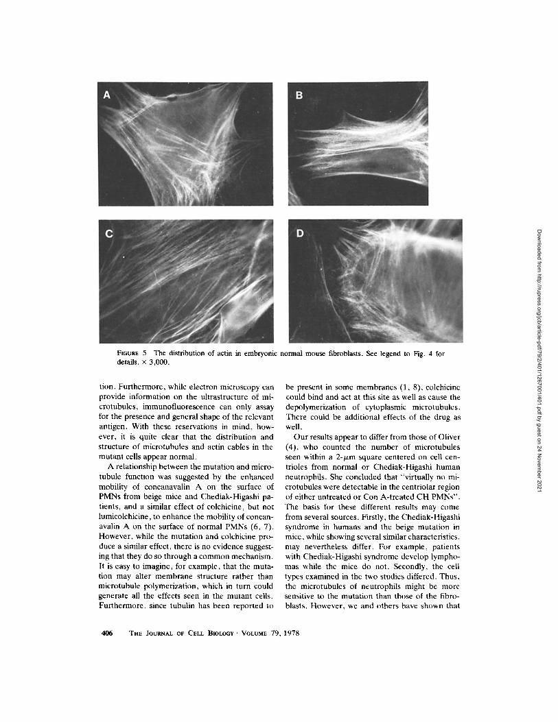
FIGuv.E 5 The distribution of actin in embryonic normal mouse fibroblasts. See legend to Fig. 4 for details, x 3,000.
tion. Furthermore, while electron microscopy can provide information on the ultrastructure of mi- crotubules, immunofluorescence can only assay for the presence and general shape of the relevant antigen. With these reservations in mind, how- ever, it is quite clear that the distribution and structure of microtubules and actin cables in the mutant cells appear normal.
A relationship between the mutation and micro- tubule function was suggested by the enhanced mobility of concanavalin A on the surface of PMNs from beige mice and Chediak-Higashi pa- tients, and a similar effect of colchicine, but not lumicolchicine, to enhance the mobility of concan- avalin A on the surface of normal PMNs (6, 7). However, while the mutation and colchicine pro- duce a similar effect, there is no evidence suggest- ing that they do so through a common mechanism. It is easy to imagine, for example, that the muta- tion may alter membrane structure rather than microtubule polymerization, which in turn could generate all the effects seen in the mutant cells. Furthermore, since tubulin has been reported to
be present in some membranes (1, 8), colchicine could bind and act at this site as well as cause the depolymerization of cytoplasmic microtubules. There could be additional effects of the drug as well.
Our results appear to differ from those of Oliver (4), who counted the number of microtubules seen within a 2-~tm square centered on cell cen- trioles from normal or Chediak-Higashi human neutrophils. She concluded that "virtually no mi- crotubules were detectable in the centriolar region of either untreated or Con A-treated CH PMNs". The basis for these different results may come from several sources. Firstly, the Chediak-Higashi syndrome in humans and the beige mutation in mice, while showing several similar characteristics, may nevertheless differ. For example, patients with Chediak-Higashi syndrome develop lympho- mas while the mice do not. Secondly, the cell types examined in the two studies differed. Thus, the microtubules of neutrophils might be more sensitive to the mutation than those of the fibro- blasts. However, we and others have shown that
406 THE JOURNAL OF CELL BIOLOGY �9 VOLUME 79, 1978
Dow
nloaded from http://rupress.org/jcb/article-pdf/79/2/401/1267001/401.pdf by guest on 24 N
ovember 2021

FIGV~ 6 The distribution of microtubules in peritoneal exudate cells of a beige mouse (top) and a normal mouse (bottom). See legend to Fig. 2 and Materials and Methods for details. • 3,000.
fibroblasts and other cell types do show the dra- matic morphological change in granule structure caused by the mutation. Finally, the examination of thin sections by electron microscopy can reveal only the concentration of microtubules in the volume of the section and may not only result in gross underestimates of the total number of tu- bules in a cell, but more importantly may be sensitive to small changes in the internal architec- ture or volume of the cell, which conceivably might be affected by the Chediak-Higashi condi- tion. While the striking increase in the number of microtubules seen by electron microscopy in cells treated with cyclic GMP and Con A is provoca- tive, it seems premature to ascribe it to a reversal of a defect in microtubule polymerization.
In conclusion, the examination of beige mouse fibroblasts and macrophages by immunofluores- cence microscopy revealed microtubules and actin cables in normal numbers, distribution and ap- pearance. Thus, the abnormal granule morphol-
ogy seen in these beige mouse cells cannot be ascribed to a significantly decreased polymeriza- tion of cytoplasmic microtubules or actin cables. It will be desirable to extend these studies to Chediak-Higashi cells of humans. In fact, a recent electron microscopic study of fibroblasts cultured from human patients indicates the presence of normal numbers of microtubules in these cells as well (Hinds and Danes. Unpublished observa- tions).
Received for publication 15 February 1978, and in revised form 6 June 1978.
REFERENCES
1. BLITZ, A. L., and R. E. FrNE. 1974. Muscle-like contractile proteins and tubulin in synal~osomes. Proc. Natl. Acad. Sci. U. S. A. 71:4472- 4476.
2. BoxEIt, L. A., A. M. WATm'~Am~, M. RlfftE1, H. R. BI~CH, J. AI.t.I~N, and R. L. BAErI~EII. 1976. Correction of leukocyte function in Chediak-Higashi syndrome by ascorbate. New Engl. J. Med. 295:1041-1045.
3. F10~KEL, F. R. 1976. Organization and energy-dependent gl'owth of
FRANKEL ET AL. Microtubules and Actin Cables in Beige Mouse Cells 407
Dow
nloaded from http://rupress.org/jcb/article-pdf/79/2/401/1267001/401.pdf by guest on 24 N
ovember 2021

microtubules in cells. Proc. Natl. Acad, Sci. U. S. A. 73:2798-2802. 4. OuvE1. J. M. 1976. Impaired microtubule function correctable by
cyclic GMP and cholinergic agonists in the Chediak-Higeshi Syndrome. Am. J. Pathol. S.~:395-412.
5. OuvEt, J. M., J. A. KaAwmc, and R. D. Bv.auN. 1976. Carbamyb choline prevents giant granule formation in cultured fibroblasts from beige ( ~ - l - F t g a s l f i ) mice.J. Cell Biol. 69:205-210.
6. OLIVER, J. M., and R. B. ZURIER. 1976. Correction of characteristic abnormalities of microtubule function and granule morphology in Cbediak-Higeshi syndrome with cholinergic agonists. J. Clln. Invest. $7:1239-1247.
7. OuvEa, J. M., R. B. Zutma, and R. D. Bv.m~. 1975. Concanavalin A cap formation on polymorphomr, lear leukocytes of normal and beige (Chediak-Higashi) mice. Nature (Lond.). 7~3:471-473.
8. S~t, tmNS, R. E. 1977. Major membrane protein differences in cilia and flagella: evidence for a membrane-associated tubulin. Biochemistry.
16:2047-2058. 9. Tuck .a .R. W,, K. K. S~roan, and F. R. F l ~ g ~ , . 1978. Tubulin
and actin in paired normeoplsstic and spontaneously tcam~rmed neol~astir cell lines/n v/~,o: fluorescent antibody studies. Cell. 13:629- 642.
10. UNAnI.E, E. R., and M. J. KM~OVSIY. 1974. LipBd-lnduced movement of lymphocyte membrane macromolecules. V. Capping. cell movement, and microtubular function in normal and lectin-treated lymphocytes. J. Exp. Meal. 14@:.1207-1220.
11. ZE~rm~. A. V. 1966. Fluorescence microscopy of lysosomes and related structures in living cells. Nature (Lond.). 111:425-426.
12. Z',.aun, R. B., G. W ~ , S. Hon~s'nB~', S. K ~ , w g , i, and H. H. T~d. 1974. Mechanisms of ~ m a l enzyn~ release from human leukocytes. II, Effects of cAMP, cGMP, autonomic asonists and agents which affect microtubule function. J. C/in. Invest. $3:297- 309.
40~ THE JOURNAL OF CELL BIOLOGY" VOLUME 7 9 , 1 9 7 8
Dow
nloaded from http://rupress.org/jcb/article-pdf/79/2/401/1267001/401.pdf by guest on 24 N
ovember 2021