Embed Size (px)
Citation preview

nutrients
Article
Immune-Enhancing Effects of a High MolecularWeight Fraction of Cynanchum wilfordii Hemsley inMacrophages and Immunosuppressed Mice
Mi Jang 1,2,†, Tae-Gyu Lim 1,†, Sungeun Ahn 2, Hee-Do Hong 1, Young Kyoung Rhee 1,Kyung-Tack Kim 1, Eunjung Lee 1, Jeong Hoon Lee 3, Yun Ji Lee 3, Chan Sik Jung 3,Dae Young Lee 4 and Chang-Won Cho 1,*
1 Traditional Food Research Center, Korea Food Research Institute, Seongnam 13539, Gyeonggi, Korea;[email protected] (M.J.); [email protected] (T.-G.L.); [email protected] (H.-D.H.); [email protected] (Y.K.R.);[email protected] (K.-T.K.); [email protected] (E.L.)
2 Department of Oriental Medicinal Biotechnology, College of Life Sciences, Kyung Hee University,Yongin 17104, Gyeonggi, Korea; [email protected]
3 Department of Herbal Crop Research, National Institute of Horticultural and Herbal Science, RDA,Eumseong 27709, Chungbuk, Korea; [email protected] (J.H.L.); [email protected] (Y.J.L.);[email protected] (C.S.J.)
4 Herbal Crop Utilization Research Team, National Institute of Horticultural and Herbal Science, RDA,Eumseong 27709, Chungbuk, Korea; [email protected]
* Correspondence: [email protected]; Tel.: +82-31-780-9312† These authors contributed equally to this work.
Received: 26 July 2016; Accepted: 13 September 2016; Published: 27 September 2016
Abstract: The objective of this study was to investigate the immune-enhancing activity of a highmolecular weight fraction (HMF) of Cynanchum wilfordii in RAW 264.7 macrophages and thecyclophosphamide (CYC)-induced mouse model of immunosuppression. To identify the bioactivesubstances of HMF, a crude polysaccharide (HMFO) was obtained and treated with sodium periodate(an oxidation agent) or digested with protease. In macrophages, HMF treatment enhanced theproduction of nitric oxide (NO) and cytokines (tumor necrosis factor alpha (TNF-α), interleukin 6(IL-6), and interleukin 1β (IL-1β)), as well as phagocytic ability. In CYC-immunosuppressed mice,HMF improved relative spleen and thymus weights, natural killer (NK) cell activity, and spleniclymphocyte proliferation. These increases in NO and cytokines were mediated by up-regulation ofnuclear factor kappa B (NF-κB) and mitogen-activated protein kinase (MAPK) signaling pathways.Periodate treatment, but not protease treatment, decreased the immune-enhancing activity of HMFO,suggesting that polysaccharides are the active ingredients in C. wilfordii extract.
Keywords: Cynanchum wilfordii; polysaccharide; immunostimulatory activity; cytokine; nitricoxide; cyclophosphamide
1. Introduction
The immune system is the host’s defense against various infectious organisms, such as bacteria,viruses, parasites, and fungi, and the immune response triggered by these infectious sources can causedisease [1]. The level of the immune response can be adjusted and maintained to a desired levelby immunomodulation, which is very important for promoting health. There is a close relationshipbetween the immune system and disease [2]. Cytokines, which are produced by immune cells and haveimmunomodulatory and anti-inflammatory abilities, play an important role in the immune responseand allow for the integration of the behavior of cells in time. Activated macrophages, T lymphocytes,and natural killer (NK) cells mainly produce the cytokines such as tumor necrosis factor alpha (TNF-α),
Nutrients 2016, 8, 600; doi:10.3390/nu8100600 www.mdpi.com/journal/nutrients
Nutrients 2016, 8, 600 2 of 19
interleukin 6 (IL-6), and IL-1β, and they play an important role in the cellular immune process byaiding the elimination of abnormal cells [3]. Suppression of the immune system induces significantchanges in the human body. Immune-suppression has been shown to be caused by various externalstresses, including pesticides, alcohol and tobacco abuse, antibiotics, chemotherapy, birth control pills,cortisone, and other drug therapies [4–6]. Recently, herbal extracts have been studied extensively fortheir potential therapeutic effects in immune function. The immunomodulatory effects of traditionalherbal medicines, such as echinacea, ginseng, and astragalus, have been investigated as agents againstinfections and neoplastic diseases [7].
As per the Korean Pharmacopoeia [8], Cynanchi wilfordii Radix is the root of Cynanchum wilfordiiHemsley. The root of C. wilfordii has been used widely as a traditional herbal medicine, and previousstudies have demonstrated that it possesses properties as a blood tonic, with vital essence enrichingand immune-enhancing activities [9]. In addition, antitumor, anti-oxidative, anti-diabetes mellitus,and anti-inflammatory activities have been attributed to the root of C. wilfordii [9–14]. The roots ofC. wilfordii contain several biologically active compounds, such as gagaminine and its glycosides,wilfosides and cynauricuosides, as well as sarcotine, penupogenin, and cynandione A [15]. Previously,it was reported that acetophenones, cynandione A, and its derivatives exhibited neuroprotective andanti-tumor activities [11,16,17]. Although enhancement of the immune response by C. wilfordii hasbeen reported, its related immune-modulatory activities in vitro and in vivo remain unclear. In thisstudy, we investigated the immunostimulatory activity of a high-molecular-weight fraction (HMF)of Cynanchum wilfordii Radix in RAW 264.7 macrophages and a cyclophosphamide (CYC)-inducedimmunosuppressed mouse model. For further study, we also explored the immunobioactive substanceof high molecular weight fraction HMF, a crude polysaccharide (HMFO) obtained from HMF, and itsimmunostimulatory effect and underlying molecular mechanisms in RAW 264.7 macrophages.
2. Materials and Methods
2.1. Chemicals and Reagents
Dulbecco’s Modified Eagle’s Medium (DMEM), fetal bovine serum (FBS), penicillin, andstreptomycin were obtained from Life Technologies (Carlsbad, CA, USA). The primary antibodiesagainst inducible nitric oxide synthase (iNOS), total p38, phosphorylated-c-Jun N-terminal kinase(JNK), phosphorylated-extracellular signal regulated kinase (ERK)1/2, total JNK, total ERK1/2,and β-actin were purchased from Santa Cruz Biotechnology (Dallas, TX, USA). Antibodiesagainst phosphorylated-inhibitor of kappa B (IκB)-α, phosphorylated-IκB kinase (IKK)-α/β, andphosphorylated-p38 were purchased from Cell Signaling Technology, Inc. (Beverly, MA, USA).Enzyme-linked immunosorbent assay (ELISA) kits for TNF-α and IL-6 were obtained from BDBiosciences (San Jose, CA, USA). An OptEIA mouse ELISA kit for IL-1β was obtained from R&DSystems (Minneapolis, MN, USA).
2.2. Preparation of a High Molecular Weight Fraction of Cynanchum wilfordii
The roots of Cynanchum wilfordii (CW) were cultivated in Jeongseon, Gangwon Province, Korea,for 2 years, harvested in August 2015, and authenticated by Dr. Jung Hoon Lee, National Instituteof Horticultural and Herbal Science (NIHHS), Rural Development Administration (RDA) of Korea.The dried roots of CW (1 kg) were extracted twice with 44 L of water solution at 95 ◦C for 6 h.After extraction, the solution was filtered and concentrated using a rotary evaporator under vacuumat 55 ◦C. The filtered concentrate was lyophilized with a freeze dryer at −35 ◦C, and the yield of thedried powder was approximately 25.1% (w/w). Commercial ultrafiltration was used to collect andconcentrate a high molecular weight fraction. The dried powder of CW extract in distilled water(DW) (20 g/4 L) was centrifuged at 6350× g for 5 min and then the supernatant was processedthrough polyethersulfone ultrafiltration (UF) membranes with a molecular weight cut-off (MWCO) of30 kDa (Sartocon 3081465902E-SG; Sartorius, Göttingen, Germany) in a cross-flow filtration system.

Nutrients 2016, 8, 600 3 of 19
The ultra-filtered retentates were freeze-dried, and the high molecular weight fraction, named HMF,was used for the experiments.
2.3. Extraction of a Crude Polysaccharide
A crude polysaccharide (HMFO) was obtained from the HMF of CW. The HMF was precipitatedby the addition of four volumes of 95% ethanol. The mixture was allowed to stand at 4 ◦C overnight andthen was centrifuged at 6350× g for 20 min to obtain the precipitate. The precipitate was lyophilizedto produce HMFO.
2.4. Chemical Analyses
Total sugar content was determined by the phenol-sulfuric acid method [18] using glucose asa standard. Uronic acid content was measured by the m-hydroxydiphenyl sulfuric acid method asmodified by Taylor and Buchanan-Smith [19] using galacturonic acid as the standard. Protein contentwas determined with the bicinchoninic acid (BCA) protein assay kit (Pierce Biotechnology, Rockford,IL, USA), using bovine serum albumin (BSA; Sigma-Aldrich, St. Louis, MO, USA) as a standard [20].The content of 2-keto-3-deoxy-D-manno-2-octulosonic acid (KDO) was determined colorimetrically bythe modified thiobarbituric acid (TBA) method [21] using 2-keto-3-deoxyoctonate ammonium salt asa standard.
2.5. Monosaccharide Composition and Molecular Weight Distribution
In order to carry out H2SO4-hydrolysis, the samples were mixed with 72% H2SO4 at 30 ◦C for 2 hand then the reaction mixtures with added DW were hydrolyzed at 120 ◦C for 1 h. The monosaccharidecomposition was identified and quantified using high-performance anion exchange chromatographycoupled with pulsed amperometric detection (HPAEC-PAD). The hydrolysates (25 µL) were appliedonto a Dionex ICS-5000 system fitted with a CarboPac PA1 analytical column (4 mm × 250 mm, DionexCo., Sunnyvale, CA, USA) combined with a Dionex CarboPac PA1 Guard Column (4 mm × 50 mm,Dionex Co.). The monosaccharides were separated isocratically using 18 mM NaOH and a constantflow rate of 1.0 mL/min. A mixture of fucose, rhamnose, arabinose, galactose, glucose, xylose, mannose,and fructose was used as a standard. In addition, separation and quantification of galacturonic acidand glucuronic acid was performed in a Dionex ICS-5000 system as described above, except the mobilephase was 100 mM NaOH and 100 mM NaOAc. Molecular weight distribution was determined usinghigh performance gel permeation chromatography (HPGPC) equipped with a refractive index detector(RID). The sample solution was injected on two serially linked Shodex OHpak SB-803 HQ and SB-805HQ (8.0 mm × 300 mm, Showa Denko Co., Tokyo, Japan). The injection volume was 20 µL, and theflow rate of 0.1 M sodium chloride eluent was 0.5 mL/min at 30 ◦C. The MW was calculated by thecalibration curve obtained by using a Shodex standard pullulan kit with P-800 (MW 853,000), P-400(MW 380,000), P-200 (MW 186,000), P-100 (MW 100,000), P-50 (MW 48,000), P-20 (MW 23,700), P-10(MW 12,200), and P-5 (MW 4,800) (Showa Denko Co.) as standards.
2.6. Protease Treatment and Periodate Oxidation of HMFO
For protease treatment, HMFO (50 mg) was dissolved in 10 mM sodium acetate buffer with5 mM calcium acetate at pH 7.5, and 50 mg of protease (protease type XIV from Streptomyces griseus,Sigma-Aldrich) was added. After incubation at 37 ◦C for 48 h, the digest was heated in a boiling waterbath for 5 min to stop the enzymatic reaction. The solution was centrifuged at 400× g for 15 min,and the supernatant was lyophilized after dialysis. To conduct periodate oxidation, 50 mg of HMFOwas dissolved in 30 mL of 50 mM acetate buffer (pH 4.5) and then 75 mM sodium periodate (10 mL)was added. After the reaction mixture was incubated at 4 ◦C in the dark for 96 h, 5 mL of ethyleneglycol was added to destroy excess periodate and then the mixture was dialyzed against DW for 72 h.Non-dialyzable solution was concentrated to 20 mL, and 20 mg of sodium tetrahydridoborate was

Nutrients 2016, 8, 600 4 of 19
added to the concentrate. The reaction mixture was stirred for 12 h at room temperature and thenneutralized with acetic acid. The oxidized product was obtained as a lyophilizate after dialysis.
2.7. Cell Culture
The RAW 264.7 murine macrophage cells were purchased from the Korean Cell Line Bank (KCLB,Seoul, Korea) and cultured in DMEM medium supplemented with 10% FBS, penicillin (100 units/mL),and streptomycin sulfate (100 µg/mL) at 37 ◦C in a humidified incubator (5% CO2).
2.8. Cell Viability Assay
Cell viability was measured using the CCK-8 (Cell Counting Kit, Dojindo, Tokyo, Japan)-basedcolorimetric assay. RAW 264.7 cells were seeded in a 96-well plate at a density of 1 × 10⁴ cells per welland were treated with various concentrations of HMF or HMFO for 24 h. Cells were incubated withthe CCK-8 reagent for 2 h, and optical density (OD) was determined at 450 nm (Infinite M200, TecanTrading AG, Männedorf, Switzerland). The OD of the samples was compared to that of the untreatedcontrol to obtain the percentage viability.
2.9. Measurement of Nitric Oxide (NO)
The presence of nitrite, a stable oxidized product of NO, was determined in cell culture mediaby Griess reagent. RAW 264.7 macrophages (1 × 10⁵ cells/mL) were cultured in 24-well platesand stimulated with HMF, HMFO, protease-treated HMFO, or periodate-oxidized HMFO for 24 h.One hundred microliters of culture supernatant were collected and mixed with an equal volume ofGriess reagent (0.1% N-(1-naphthyl) ethylenediamine dihydrochloride, 1% sulfanilamide, and 2.5%H3PO4). After incubation for 15 min at room temperature, the OD was measured at 540 nm using amicroplate reader. Nitrite concentrations in the supernatants were determined by comparison with asodium nitrite standard curve.
2.10. Determination of TNF-α, (IL-6) and IL-1β Production
RAW 264.7 macrophages (1 × 10⁵ cells/mL) were cultured in 24-well plates and stimulated withHMF, HMFO, protease-treated HMFO, or periodate-oxidized HMFO. Supernatants were collectedafter 24 h, and the levels of TNF-α, IL-6 and IL-1β were measured by ELISA kits (R&D Systems,Minneapolis, MN, USA) according to the manufacture’s protocols.
2.11. Phagocytosis Assay in RAW 264.7 Cells
The phagocytic ability of RAW 264.7 macrophages was detected using the CytoSelect™ 96-wellphagocytosis assay kit (Cell Biolabs Inc., San Diego, CA, USA), following the manufacturer’sinstructions. RAW 264.7 macrophages (1 × 10⁴ cell/well) were plated in 96-well plates and incubatedovernight at 37 ◦C to allow adherence to the plate. The cells were pre-incubated with a series ofconcentrations of HMF (final concentrations of 50, 100, and 200 µg/mL) as well as a negative control(complete DMEM) and positive control (lipopolysaccharide (LPS), 1 µg/mL) for 24 h. Subsequently,non-opsonized zymosan particles were added incubated at 37 ◦C for 2 h. The amount of engulfedzymosan particles was determined using a colorimetric assay at an absorbance of 405 nm.
2.12. Western Blot Analysis
The cells were incubated with HMFO (50, 100, and 200 µg/mL) for 24 h, collected by centrifugation,and washed once with phosphate buffered saline (PBS). Washed cell pellets were lysed for 30 minon ice with Pro-Prep™ protein extraction solution according to the protocol provided. Cell lysateswere centrifuged at 15,700× g at 4 ◦C for 5–10 min, and clear supernatants were collected in newtubes and stored at −70 ◦C. For immunoblotting, proteins in cell lysates were resolved by sodiumdodecyl sulfate (SDS)-polyacrylamide gel electrophoresis (PAGE) on 8%–10% gels and then transferred

Nutrients 2016, 8, 600 5 of 19
to nitrocellulose membranes (Millipore, Billerica, MA, USA) for 1.5 h. Immunoblots were incubatedfor 1 h with blocking solution (5% skim milk) at room temperature and then incubated overnightwith a 1:1000 dilution of primary antibody at 4 ◦C. Blots were washed three times with Tween20/Tris-buffered saline (T/TBS) and then incubated with a 1:2000 dilution of horseradish peroxidase(HRP)-conjugated secondary antibody (Santa Cruz Biotechnology Inc.) for 90 min at room temperature.Finally, blots were visualized using an enhanced chemiluminescence system (Amersham BiosciencesInc., Piscataway, NJ, USA) followed by exposure to X-ray film (Fuji Photo Film Co., Ltd., Tokyo, Japan).
2.13. Animals and Treatments
Female BALB/c mice (8 weeks old, 18–20 g) were purchased from Koatech Animal Inc.(Pyeongtaek, Korea). The mice were housed at 22 ± 1 ◦C, with 12 h-light/12-h dark cycle, 50%–60%relative humidity, and given free access to food and water during the experiments. All animal studieswere performed in accordance with the Guiding Principles for the Care and Use of Laboratory Animalsof the Ethics Committee of the Korea Food Research Institute.
Animal experiments were conducted using immunosuppressed mice. Mice were randomlydivided into five groups (eight mice in each group). Normal control mice did not receive any treatmentfor immunosuppression. The remaining four groups were injected intraperitoneally with 150 mg/kgbody weight CYC, a known immunosuppressant, on days 8, 9, and 10 after administration of therespective treatment. The immunosuppressed control group mice received normal saline for a periodof 28 days. The immunosuppressed model positive control mice were orally administered 200 mg/kgbody weight/day CVT-E002™ for 28 days. CVT-E002™ is an immunostimulatory polysaccharide-richextract of the root of North American ginseng (Panax quinquefolius) [22]. The two HMF treatmentgroups were orally administered 100 or 200 mg/kg body weight/day HMF for 28 days. At the endof the study, mice were weighed and sacrificed by cervical dislocation. Relative thymus and spleenweights were calculated according to the following formula: Relative (%) = (thymus or spleen weights(g)/body weight (g)) × 100. The collected spleen samples were used to measure the splenocyteproliferation and NK cell activity.
2.14. Preparation of Mouse Splenocytes
The spleen was gently homogenized, and the cell suspension was passed through a 100-µm nyloncell strainer (BD Falcon, San Jose, CA, USA) to obtain single cell suspensions. After centrifugation,the red blood cells were lysed in Red Cell Lysis Buffer (Hybri-Max™, Sigma-Aldrich). After washingtwice with serum-free Roswell Park Memorial Institute (RPMI) 1640 medium, the splenocytes wereharvested and resuspended in RPMI 1640 medium (with 10% FBS, 100 unit/mL penicillin, and100 µg/mL streptomycin).
2.15. Splenocyte Proliferation Assay
Splenocyte proliferation was assessed using the CCK-8 kit (Dojindo Laboratories, Tokyo, Japan)according to manufacturer’s instructions. The cell number was briefly adjusted to a densityof 3 × 10⁶ cells/mL of medium, and 1 mL of spleen cell suspension was seeded in 24-well platesand cultured with 75 µL of the mitogen solutions or sterile DW for control culture. The mitogens wereconcanavalin A (Con A) and LPS, which were dissolved in sterile DW to a concentration of 100 µg/mLand 400 µg/mL, respectively. Con A and LPS were used for measuring the proliferation of T andB lymphocytes in splenocyte cultures, respectively. After incubation at 37 ◦C in a humidified incubatorwith 5% CO2 for 72 h, aliquots (100 µL) of CCK-8 reagent were added to the cells and then cultured inthe incubator for 4 h. The absorbance at 450 nm was measured on a microplate reader.
2.16. Flow Cytometric Analysis
Flow cytometry-assisted analysis was performed using 1 × 10⁶ splenocytes suspended in 100 µLof staining buffer (0.5% BSA, 0.04% EDTA, 0.05% sodium azide in PBS), which was pre-incubated with

Nutrients 2016, 8, 600 6 of 19
anti-mouse Fc Block FcR blocking reagent (BD PharMingen, San Diego, CA, USA) for 20 min. Surfacestains included FITC-conjugated anti-mouse CD3 (eBioscience, San Diego, CA, USA), PerCP-Cy™5.5-conjugated anti-mouse CD8 (BD PharMingen), and APC-conjugated anti-mouse CD4 antibodies(BD PharMingen). The cells were then fixed and permeabilized with BD Cytofix/Cytoperm solution(BD Biosciences) for 20 min at 4 ◦C and washed once. Stained cells were analyzed by flow cytometryusing a FACSCanto II flow cytometer (Becton Dickinson, Mountain View, CA, USA). The acquired datawere analyzed with CellQuest PRO software. For analysis of T cells, cells were gated to be CD3+, andthis population was analyzed for expression of CD4 and CD8. This is particularly true for antibodiesto CD3, a marker commonly used to identify T cell receptor-bearing cells. The results were expressedas a percentage of CD3 + CD4 + or CD3 + CD8 + cells.
2.17. NK Cell Activity
YAC-1 lymphoma cells (KCLB NO. 40160) were purchased from the Korean Cell Line Bank.The cell lines were grown in RPMI 1640 medium with 10% FBS and 1% penicillin-streptomycin andincubated at 37 ◦C in 5% CO2. YAC-1 cells were used as the target cells, while splenocytes werethe effector cells. Two different ratios of effector to target cell were used. The NK cell activity ofthe splenocytes was determined by cytotoxicity against NK cell-sensitive YAC-1 cells using a lactatedehydrogenase (LDH) leakage assay [23]. The amount of LDH in the cultured medium was an indicatorof cytotoxicity. After the end point of the experiment, the LDH activity of the cells in the culturemedium was determined, following the instructions of the LDH cell cytotoxicity assay kit (DoGen,Seoul, Korea). The NK activity of effector cells was calculated by the following formula: cytotoxicity(%) = 100 × {(A − B) − (C − D)}/(E − F); where A is the experimental release, B the spontaneousrelease of effector cells, C the spontaneous release of target cells, D the blank for the spontaneousrelease of effector cells, E the maximum release of target, and F is the blank for the maximum release.
2.18. Statistical Analysis
All data are presented as the mean ± standard deviation (SD). Data were analyzed using one-wayanalysis of variance (ANOVA) followed by Scheffe’s test to detect intergroup differences using SPSSsoftware version 20.0 (SPSS Inc., Armonk, NY, USA); p < 0.05 was considered statistically significant.
3. Results
3.1. Effects of HMF on NO Production and Cytokine and Phagocytic Activity
To confirm the immunostimulatory activity of HMF, NO production was evaluated afterRAW 264.7 macrophages were treated with HMF. HMF treatment (50, 100, 200 or 400 µg/mL)dose-dependently increased NO concentration compared to untreated cells (Figure 1B). HMF (upto 400 µg/mL) did not affect cell viability of RAW 264.7 macrophages (Figure 1A), showing thatincreases in NO production were not due to any cytotoxic effect of HMF. To assess the effect of HMFon immune stimulation, we measured the production of these cytokines in RAW 264.7 macrophages.As seen in Figure 1C–E, treatment of HMF increased the production of all immunostimulatory cytokines(TNF-α, IL-6, and IL-1β) examined compared to untreated cells. As shown in Figure 1F, HMF treatmentfor 24 h enhanced the phagocytic activity of RAW 264.7 cells. Phagocytosis in macrophages treatedwith HMF (200 µg/mL) was 1.3-fold higher than that of zymosan-only treated control cells.
3.2. Effects of HMF on Decreased Immune Function in CYC-Treated Mice
To confirm the immune-enhancing activity of HMF under physiological conditions, an in vivostudy was performed. HMF was orally administered to CYC-immunosuppressed mice, and theeffects on average body weight and the relative weights of thymus and spleen were determined(Table 1). CYC-treated mice exhibited a significant reduction in relative spleen and thymus weightscompared with the normal group mice. HMF treatment at 100 or 200 mg/kg body weight/day,

Nutrients 2016, 8, 600 7 of 19
however, markedly restored relative spleen and thymus weights to normal levels and to levels evenhigher than the CVT-treated group, the positive control.
Nutrients 2016, 8, 600 7 of 18
compared with the normal group mice. HMF treatment at 100 or 200 mg/kg body weight/day,
however, markedly restored relative spleen and thymus weights to normal levels and to levels even
higher than the CVT‐treated group, the positive control.
Figure 1. Effects of a high molecular weight fraction (HMF) of Cynanchum wilfordii on nitric oxide
(NO), tumor necrosis factor alpha (TNF‐α), interleukin (IL)‐6, and IL‐1β production, and phagocytic
activity in macrophages. RAW 264.7 macrophages were stimulated with the HMF (50, 100, 200, or 400
μg/mL) or with lipopolysaccharide (LPS; 1 μg/mL). LPS was used as the positive control. (A) Cell
viability was determined by cell counting kit (CCK)‐8 assay; (B) NO production was determined by
measuring nitrite accumulation in culture medium. The productions of (C) TNF‐α; (D) IL‐6; and (E)
IL‐1β were measured using enzyme‐linked immunosorbent assays (ELISAs); (F) Phagocytic activity
was determined using a phagocytosis assay. Values with the different letters are significantly different
(p < 0.05).
Figure 1. Effects of a high molecular weight fraction (HMF) of Cynanchum wilfordii on nitric oxide (NO),tumor necrosis factor alpha (TNF-α), interleukin (IL)-6, and IL-1β production, and phagocytic activityin macrophages. RAW 264.7 macrophages were stimulated with the HMF (50, 100, 200, or 400 µg/mL)or with lipopolysaccharide (LPS; 1 µg/mL). LPS was used as the positive control. (A) Cell viability wasdetermined by cell counting kit (CCK)-8 assay; (B) NO production was determined by measuring nitriteaccumulation in culture medium. The productions of (C) TNF-α; (D) IL-6; and (E) IL-1β were measuredusing enzyme-linked immunosorbent assays (ELISAs); (F) Phagocytic activity was determined using aphagocytosis assay. Values with the different letters are significantly different (p < 0.05).

Nutrients 2016, 8, 600 8 of 19
Table 1. Effect of HMF on terminal body weight, absolute and relative organ weights of mice.
Group Initial BodyWeight (g)
Terminal BodyWeight (g)
Spleen Weight Thymus Weight
Absolute (g) Relative (%) Absolute (g) Relative (%)
Negative control 19.11 ± 0.75 a 20.03 ± 0.65 c 0.103 ± 0.009 a 0.512 ± 0.027 ab 0.047 ± 0.004 a 0.233 ± 0.017 a
CYC 18.82 ± 0.94 a 18.10 ± 0.57 a 0.085 ± 0.005 b 0.471 ± 0.022 a 0.041 ± 0.005 a 0.225 ± 0.023 a
HMF (100 mg/kg) 18.38 ± 0.52 a 18.41 ± 0.53 ab 0.100 ± 0.002 a 0.544 ± 0.014 b 0.042 ± 0.003 a 0.230 ± 0.012 b
HMF (200 mg/kg) 19.11 ± 0.34 a 19.52 ± 0.63 bc 0.108 ± 0.007 a 0.554 ± 0.021 b 0.045 ± 0.003 a 0.232 ± 0.009 b
Positive control 19.32 ± 0.64 a 19.69 ± 0.85 c 0.100 ± 0.008 a 0.510 ± 0.037 ab 0.047 ± 0.005 a 0.238 ± 0.022 b
Values represent Mean ± SD; abc Means with different superscripts in the same column are significantly differentat p < 0.05. CYC, cyclophosphamide; CVT, immunostimulatory polysaccharide-rich extract of the root of NorthAmerican ginseng (Panax quinquefolius); HMF, high-molecular-weight fraction of Cynanchum wilfordii.
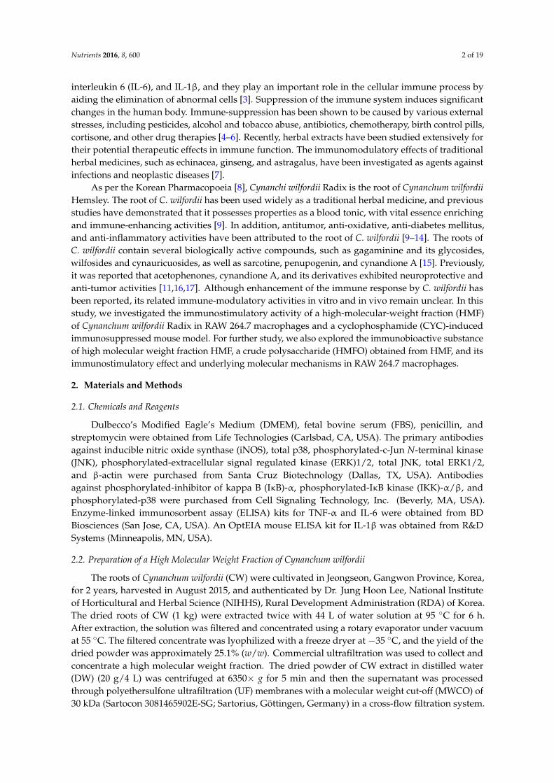
At all effector-to-target (E:T) ratios, treatment with CYC significantly decreased the activity ofsplenic NK cells compared with mice in the normal control group. However, a dose of 200 mg/kgHMF restored NK cell activity to normal control levels in CYC-treated mice (Figure 2A). As shownin Figure 2B,C, proliferation of T and B lymphocytes in the CYC-treated group was significantlydecreased compared with the normal control group. Treatment with 100 and 200 mg/kg HMFenhanced proliferation of T and B lymphocytes significantly compared with the CYC-treated group.Con A and LPS were used to stimulate T and B lymphocyte proliferation, respectively.
To identify phenotypically distinct subsets of CD4 and CD8 T cells, we carried out flow cytometryanalysis. The percentage of helper (CD3+CD4+) T cells was decreased in CYC-treated mice comparedto mice in the normal control group: the helper T cell population was 41.7% in normal controlsversus 27.7% in CYC-treated group (Figure 3). Treatment with HMF increased the percentage ofhelper T cells compared with mice in the CYC-treated group. In addition, the percentage of cytotoxic(CD3+CD8+) T cells in the CYC-treated mice was decreased compared with the normal control group,while HMF-treated mice had a higher percentage of cytotoxic T cells than CYC-treated mice.
3.3. Effects of a Crude Polysaccharide of HMF on the Immunostimulatory Activity in RAW 264.7 Macrophages
We prepared a crude polysaccharide (HMFO) from HMF by ethanol precipitation and investigatedits immunostimulatory effect and underlying molecular mechanisms in RAW 264.7 macrophages.
3.3.1. Effects of Chemical and Enzymatic Treatments of HMFO on Immunostimulatory Activity inRAW 264.7 Macrophages
To identify the immunobioactive substance of HMFO, HMFO was treated with sodium periodateto degrade the carbohydrate units and digested with protease to hydrolyze proteins. Protease-treatedHMFO significantly increased the production of NO and cytokines (TNF-α, IL-6, and IL-1β),but periodate-oxidized HMFO did not affect the levels of NO and all measured cytokines (Figure 4A–D).Periodate oxidation of HMFO decreased the immunostimulatory activities in RAW 264.7 macrophagesof HMFO by oxidative depolymerization of polysaccharides. Protease-treated HMFO enhancedimmunologic macrophage activation, likely because of the existence of structural moieties required forexpression of stimulating activity on macrophages.
Nutrients 2016, 8, 600 9 of 19Nutrients 2016, 8, 600 9 of 18
Figure 2. Effects of HMF on natural killer (NK) cell activity and splenic lymphocyte proliferation in CYC‐induced mice. HMF (100 mg/kg or 200 mg/kg) or CVT (200 mg/kg)
was administered orally once daily for 28 days to CYC‐induced mice. (A) NK cell activity was determined by lactate dehydrogenase (LDH) assay as described in Materials
and Methods. The E:T ratio indicates ratio of effector cells (splenocytes) and target cells (YAC‐1 cells); (B) Concanavalin A (Con A)‐induced T‐lymphocyte proliferation;
(C) LPS‐induced B‐lymphocyte proliferation. Cell proliferation was measured by using CCK‐8 kit. Values with the different letters are significantly different (p < 0.05).
CYC, cyclophosphamide; CVT, immunostimulatory polysaccharide‐rich extract of the root of North American ginseng (Panax quinquefolius); HMF, high‐molecular‐weight
fraction of Cynanchum wilfordii.
Figure 2. Effects of HMF on natural killer (NK) cell activity and splenic lymphocyte proliferation in CYC-induced mice. HMF (100 mg/kg or 200 mg/kg) orCVT (200 mg/kg) was administered orally once daily for 28 days to CYC-induced mice. (A) NK cell activity was determined by lactate dehydrogenase (LDH)assay as described in Materials and Methods. The E:T ratio indicates ratio of effector cells (splenocytes) and target cells (YAC-1 cells); (B) Concanavalin A (ConA)-induced T-lymphocyte proliferation; (C) LPS-induced B-lymphocyte proliferation. Cell proliferation was measured by using CCK-8 kit. Values with the differentletters are significantly different (p < 0.05). CYC, cyclophosphamide; CVT, immunostimulatory polysaccharide-rich extract of the root of North American ginseng(Panax quinquefolius); HMF, high-molecular-weight fraction of Cynanchum wilfordii.

Nutrients 2016, 8, 600 10 of 19Nutrients 2016, 8, 600 10 of 18
Figure 3. Flow cytometric analyses of spleen CD4+ and CD8+ T cell subsets. Groups of
cyclophosphamide, HMF, and CVT mice were immunosuppressed as indicated in the Materials and
Methods section. Spleen cells were gated to be CD3+ for analysis of T cells, and this population was
analyzed for expression of CD4 and CD8 by flow cytometry. Results were expressed as percentages
of CD4+ and CD8+ T cell subsets.
Figure 3. Flow cytometric analyses of spleen CD4+ and CD8+ T cell subsets. Groups ofcyclophosphamide, HMF, and CVT mice were immunosuppressed as indicated in the Materialsand Methods section. Spleen cells were gated to be CD3+ for analysis of T cells, and this populationwas analyzed for expression of CD4 and CD8 by flow cytometry. Results were expressed as percentagesof CD4+ and CD8+ T cell subsets.
Nutrients 2016, 8, 600 10 of 18
Figure 3. Flow cytometric analyses of spleen CD4+ and CD8+ T cell subsets. Groups of
cyclophosphamide, HMF, and CVT mice were immunosuppressed as indicated in the Materials and
Methods section. Spleen cells were gated to be CD3+ for analysis of T cells, and this population was
analyzed for expression of CD4 and CD8 by flow cytometry. Results were expressed as percentages
of CD4+ and CD8+ T cell subsets.
Figure 4. Cont.

Nutrients 2016, 8, 600 11 of 19Nutrients 2016, 8, 600 11 of 18
Figure 4. Effects of chemical and enzymatic treatments of HMFO on immunostimulatory activity in
RAW 264.7 macrophages. RAW 264.7 macrophages were stimulated with HMFO, protease‐treated
HMFO, periodate‐oxidized HMFO (50, 100, or 200 μg/mL), CVT (200 μg/mL), or LPS (1 μg/mL) for
24 h. The production of NO (A) was determined by measuring nitrite accumulation in culture medium.
Amounts of TNF‐α (B); IL‐6 (C); and IL‐1β (D) released into culture media were determined by ELISA.
Values with the different letters are significantly different (p < 0.05). CVT, immunostimulatory
polysaccharide‐rich extract of the root of North American ginseng (Panax quinquefolius); HMFO, a
crude polysaccharide of high molecular weight fraction; Protease, protease treated‐HMFO; Periodate,
periodate‐oxidized HMFO.
3.3.2. Effects of HMFO on NO Production and iNOS Expression in RAW 264.7 Macrophages
HMFO significantly and concentration‐dependently increased the level of NO compared to
control (Figure 5A). To determine if this increase in NO secretion was related to modulation of iNOS
expression, we measured the protein levels of iNOS in the presence of HMFO (50, 100, or 200 μg/mL)
by western blotting. Although iNOS protein was not detectable in unstimulated RAW 264.7
macrophages, HMFO treatment significantly and dose‐dependently increased iNOS protein
expression (Figure 5B). In addition, HMFO (up to 400 μg/mL) did not affect cell viability of RAW
264.7 macrophages (data not shown), showing that increases in NO production via iNOS expression
were not due to any cytotoxic effect of HMFO. Polymyxin B (an LPS inhibitor) markedly inhibited
NO production induced by LPS. However, it had no effect on HMFO‐induced NO production (Figure
5C). These results indicate that enhanced NO production by HMFO was not the result of endotoxin
contamination.
Figure 4. Effects of chemical and enzymatic treatments of HMFO on immunostimulatory activity inRAW 264.7 macrophages. RAW 264.7 macrophages were stimulated with HMFO, protease-treatedHMFO, periodate-oxidized HMFO (50, 100, or 200 µg/mL), CVT (200 µg/mL), or LPS (1 µg/mL)for 24 h. The production of NO (A) was determined by measuring nitrite accumulation in culturemedium. Amounts of TNF-α (B); IL-6 (C); and IL-1β (D) released into culture media were determinedby ELISA. Values with the different letters are significantly different (p < 0.05). CVT, immunostimulatorypolysaccharide-rich extract of the root of North American ginseng (Panax quinquefolius); HMFO,a crude polysaccharide of high molecular weight fraction; Protease, protease treated-HMFO; Periodate,periodate-oxidized HMFO.
3.3.2. Effects of HMFO on NO Production and iNOS Expression in RAW 264.7 Macrophages
HMFO significantly and concentration-dependently increased the level of NO compared tocontrol (Figure 5A). To determine if this increase in NO secretion was related to modulation of iNOSexpression, we measured the protein levels of iNOS in the presence of HMFO (50, 100, or 200 µg/mL) bywestern blotting. Although iNOS protein was not detectable in unstimulated RAW 264.7 macrophages,HMFO treatment significantly and dose-dependently increased iNOS protein expression (Figure 5B).In addition, HMFO (up to 400 µg/mL) did not affect cell viability of RAW 264.7 macrophages (data notshown), showing that increases in NO production via iNOS expression were not due to any cytotoxiceffect of HMFO. Polymyxin B (an LPS inhibitor) markedly inhibited NO production induced by LPS.However, it had no effect on HMFO-induced NO production (Figure 5C). These results indicate thatenhanced NO production by HMFO was not the result of endotoxin contamination.
Nutrients 2016, 8, 600 11 of 18
Figure 4. Effects of chemical and enzymatic treatments of HMFO on immunostimulatory activity in
RAW 264.7 macrophages. RAW 264.7 macrophages were stimulated with HMFO, protease‐treated
HMFO, periodate‐oxidized HMFO (50, 100, or 200 μg/mL), CVT (200 μg/mL), or LPS (1 μg/mL) for
24 h. The production of NO (A) was determined by measuring nitrite accumulation in culture medium.
Amounts of TNF‐α (B); IL‐6 (C); and IL‐1β (D) released into culture media were determined by ELISA.
Values with the different letters are significantly different (p < 0.05). CVT, immunostimulatory
polysaccharide‐rich extract of the root of North American ginseng (Panax quinquefolius); HMFO, a
crude polysaccharide of high molecular weight fraction; Protease, protease treated‐HMFO; Periodate,
periodate‐oxidized HMFO.
3.3.2. Effects of HMFO on NO Production and iNOS Expression in RAW 264.7 Macrophages
HMFO significantly and concentration‐dependently increased the level of NO compared to
control (Figure 5A). To determine if this increase in NO secretion was related to modulation of iNOS
expression, we measured the protein levels of iNOS in the presence of HMFO (50, 100, or 200 μg/mL)
by western blotting. Although iNOS protein was not detectable in unstimulated RAW 264.7
macrophages, HMFO treatment significantly and dose‐dependently increased iNOS protein
expression (Figure 5B). In addition, HMFO (up to 400 μg/mL) did not affect cell viability of RAW
264.7 macrophages (data not shown), showing that increases in NO production via iNOS expression
were not due to any cytotoxic effect of HMFO. Polymyxin B (an LPS inhibitor) markedly inhibited
NO production induced by LPS. However, it had no effect on HMFO‐induced NO production (Figure
5C). These results indicate that enhanced NO production by HMFO was not the result of endotoxin
contamination.
Figure 5. Cont.

Nutrients 2016, 8, 600 12 of 19Nutrients 2016, 8, 600 12 of 18
Figure 5. Effects of HMFO on NO production and iNOS expression in RAW 264.7 macrophages. RAW
264.7 macrophages were stimulated with HMFO (50, 100, or 200 μg/mL) and LPS (1 μg/mL) for 24 h.
The production of NO (A) was determined by measuring nitrite accumulation in culture medium; (B)
Total proteins were isolated from cells stimulated with HMFO (50, 100, or 200 μg/mL) or LPS (1 μg/mL)
for 24 h. β‐Actin was used as an internal loading control; (C) Cells were pretreated with polymyxin B
(1 μg/mL), followed by stimulation with HMFO (200 μg/mL) or LPS (0.5 μg/mL) for 24 h. NO
production was determined by measuring nitrite accumulation in culture medium. Values with the
different letters are significantly different (p < 0.05). HMFO, polysaccharides of high‐molecular‐
weight fraction.
3.3.3. Effects of HMFO on the Phosphorylation of IκB‐α and IKK‐α/β in RAW 264.7 Macrophages
We investigated whether HMFO enhanced nuclear localization of NF‐κB and the
phosphorylation of IκB‐α and IKKα/β in RAW 264.7 macrophages by western blotting. HMFO
treatment significantly elevated nuclear translocation of NF‐κB and the levels of phosphorylated
IκB‐α and IKKα/β (Figure 6). Our findings indicate that HMFO regulates NF‐κB activation by
increasing the nuclear translocation of p65 via IKK‐α/β‐dependent IκB phosphorylation.
Figure 5. Effects of HMFO on NO production and iNOS expression in RAW 264.7 macrophages.RAW 264.7 macrophages were stimulated with HMFO (50, 100, or 200 µg/mL) and LPS (1 µg/mL)for 24 h. The production of NO (A) was determined by measuring nitrite accumulation in culturemedium; (B) Total proteins were isolated from cells stimulated with HMFO (50, 100, or 200 µg/mL) orLPS (1 µg/mL) for 24 h. β-Actin was used as an internal loading control; (C) Cells were pretreatedwith polymyxin B (1 µg/mL), followed by stimulation with HMFO (200 µg/mL) or LPS (0.5 µg/mL)for 24 h. NO production was determined by measuring nitrite accumulation in culture medium.Values with the different letters are significantly different (p < 0.05). HMFO, polysaccharides ofhigh-molecular-weight fraction.
3.3.3. Effects of HMFO on the Phosphorylation of IκB-α and IKK-α/β in RAW 264.7 Macrophages
We investigated whether HMFO enhanced nuclear localization of NF-κB and the phosphorylationof IκB-α and IKKα/β in RAW 264.7 macrophages by western blotting. HMFO treatment significantlyelevated nuclear translocation of NF-κB and the levels of phosphorylated IκB-α and IKKα/β (Figure 6).Our findings indicate that HMFO regulates NF-κB activation by increasing the nuclear translocation ofp65 via IKK-α/β-dependent IκB phosphorylation.
Nutrients 2016, 8, 600 12 of 18
Figure 5. Effects of HMFO on NO production and iNOS expression in RAW 264.7 macrophages. RAW
264.7 macrophages were stimulated with HMFO (50, 100, or 200 μg/mL) and LPS (1 μg/mL) for 24 h.
The production of NO (A) was determined by measuring nitrite accumulation in culture medium; (B)
Total proteins were isolated from cells stimulated with HMFO (50, 100, or 200 μg/mL) or LPS (1 μg/mL)
for 24 h. β‐Actin was used as an internal loading control; (C) Cells were pretreated with polymyxin B
(1 μg/mL), followed by stimulation with HMFO (200 μg/mL) or LPS (0.5 μg/mL) for 24 h. NO
production was determined by measuring nitrite accumulation in culture medium. Values with the
different letters are significantly different (p < 0.05). HMFO, polysaccharides of high‐molecular‐
weight fraction.
3.3.3. Effects of HMFO on the Phosphorylation of IκB‐α and IKK‐α/β in RAW 264.7 Macrophages
We investigated whether HMFO enhanced nuclear localization of NF‐κB and the
phosphorylation of IκB‐α and IKKα/β in RAW 264.7 macrophages by western blotting. HMFO
treatment significantly elevated nuclear translocation of NF‐κB and the levels of phosphorylated
IκB‐α and IKKα/β (Figure 6). Our findings indicate that HMFO regulates NF‐κB activation by
increasing the nuclear translocation of p65 via IKK‐α/β‐dependent IκB phosphorylation.
Figure 6. Cont.

Nutrients 2016, 8, 600 13 of 19Nutrients 2016, 8, 600 13 of 18
Figure 6. HMFO‐induced nuclear factor kappa B (NF‐κB) activation by the phosphorylation of
inhibitor of kappa B (IκB)‐α and IκB kinase (IKK)‐α/β in RAW 264.7 macrophages. Cells were
stimulated with HMFO (50, 100, or 200 μg/mL) or LPS (1 μg/mL). (A) Nuclear extracts (N) were
isolated from cells stimulated with HMFO or LPS. Level of p65 in the nuclear fractions was
determined by immunoblotting analysis; (B,C) Phosphorylated IκB‐α and IKKα/β isolated from cell
lysates were determined by immunoblotting analysis. β‐actin was used as an internal control. Values
with the different letters are significantly different (p < 0.05).
3.3.4. Effects of HMFO on Mitogen‐Activated Protein Kinase (MAPK) Phosphorylation in RAW
264.7 Macrophages
To determine whether HMFO activates the production of NO and cytokines (TNF α, IL‐6, and
IL‐1β) through MAPKs signaling pathways, RAW 264.7 macrophages were cultured as described
above, and phosphorylated MAPKs were analyzed using western blot analysis. As shown in Figure
7, HMFO concentration‐dependently induced the phosphorylation of ERK, JNK, and p38.
Figure 7. HMFO induced mitogen‐activated protein kinase (MAPK) phosphorylation in RAW 264.7
macrophages. Cells were stimulated with HMFO (50, 100, or 200 μg/mL) or LPS (1 μg/mL).
Phosphorylated‐c‐June N‐terminal kinase (JNK), phosphorylated extracellular signal regulated
kinase (ERK), phosphorylated p38, and β‐actin isolated from cell lysates were determined by
immunoblotting analysis. Values with the different letters are significantly different (p < 0.05).
Figure 6. HMFO-induced nuclear factor kappa B (NF-κB) activation by the phosphorylation of inhibitorof kappa B (IκB)-α and IκB kinase (IKK)-α/β in RAW 264.7 macrophages. Cells were stimulated withHMFO (50, 100, or 200 µg/mL) or LPS (1 µg/mL). (A) Nuclear extracts (N) were isolated from cellsstimulated with HMFO or LPS. Level of p65 in the nuclear fractions was determined by immunoblottinganalysis; (B,C) Phosphorylated IκB-α and IKKα/β isolated from cell lysates were determined byimmunoblotting analysis. β-actin was used as an internal control. Values with the different letters aresignificantly different (p < 0.05).
3.3.4. Effects of HMFO on Mitogen-Activated Protein Kinase (MAPK) Phosphorylation inRAW 264.7 Macrophages
To determine whether HMFO activates the production of NO and cytokines (TNF α, IL-6, andIL-1β) through MAPKs signaling pathways, RAW 264.7 macrophages were cultured as describedabove, and phosphorylated MAPKs were analyzed using western blot analysis. As shown in Figure 7,HMFO concentration-dependently induced the phosphorylation of ERK, JNK, and p38.
Nutrients 2016, 8, 600 13 of 18
Figure 6. HMFO‐induced nuclear factor kappa B (NF‐κB) activation by the phosphorylation of
inhibitor of kappa B (IκB)‐α and IκB kinase (IKK)‐α/β in RAW 264.7 macrophages. Cells were
stimulated with HMFO (50, 100, or 200 μg/mL) or LPS (1 μg/mL). (A) Nuclear extracts (N) were
isolated from cells stimulated with HMFO or LPS. Level of p65 in the nuclear fractions was
determined by immunoblotting analysis; (B,C) Phosphorylated IκB‐α and IKKα/β isolated from cell
lysates were determined by immunoblotting analysis. β‐actin was used as an internal control. Values
with the different letters are significantly different (p < 0.05).
3.3.4. Effects of HMFO on Mitogen‐Activated Protein Kinase (MAPK) Phosphorylation in RAW
264.7 Macrophages
To determine whether HMFO activates the production of NO and cytokines (TNF α, IL‐6, and
IL‐1β) through MAPKs signaling pathways, RAW 264.7 macrophages were cultured as described
above, and phosphorylated MAPKs were analyzed using western blot analysis. As shown in Figure
7, HMFO concentration‐dependently induced the phosphorylation of ERK, JNK, and p38.
Figure 7. HMFO induced mitogen‐activated protein kinase (MAPK) phosphorylation in RAW 264.7
macrophages. Cells were stimulated with HMFO (50, 100, or 200 μg/mL) or LPS (1 μg/mL).
Phosphorylated‐c‐June N‐terminal kinase (JNK), phosphorylated extracellular signal regulated
kinase (ERK), phosphorylated p38, and β‐actin isolated from cell lysates were determined by
immunoblotting analysis. Values with the different letters are significantly different (p < 0.05).
Figure 7. HMFO induced mitogen-activated protein kinase (MAPK) phosphorylation inRAW 264.7 macrophages. Cells were stimulated with HMFO (50, 100, or 200 µg/mL) or LPS (1 µg/mL).Phosphorylated-c-June N-terminal kinase (JNK), phosphorylated extracellular signal regulated kinase(ERK), phosphorylated p38, and β-actin isolated from cell lysates were determined by immunoblottinganalysis. Values with the different letters are significantly different (p < 0.05).

Nutrients 2016, 8, 600 14 of 19
3.4. Chemical Compositions and Molecular Weight Distribution
The percentages of neutral sugar, uronic acid, KDO, protein content, and the componentsugars of HMFO are summarized in Table 2. More specifically, the monosaccharide compositionof HMFO was investigated by HPAEC-PAD. The main monosaccharide components of HMFO wereglucose, arabinose, galactose, rhamnose, and galacturonic acid. For HMFO, there were severalpredominant monosaccharides that contained component sugars characteristic of pectic substances,such as arabinose, galactose, rhamnose, and galacturonic acid. The molecular weight range of HMFOwas estimated to be between 11.8 and 520.4 kDa based on the calibration with pullulan standards(Figure S1).
Table 2. The chemical and monosaccharide composition of HMFO.
HMFO
Chemical composition (%)Neutral sugar 1 63.8 ± 0.9
Uronic acid 2 33.1 ± 1.9KDO 3 0.3 ± 0.1
Protein 4 2.9 ± 0.2
Component monosaccharide (mg/g)Arabinose 86.0 ± 1.6Galactose 76.5 ± 2.9Rhamnose 23.0 ± 0.4
Xylose 2.2 ± 0.6Glucose 241.2 ± 6.5
Mannose 9.0 ± 1.6Fucose 5.7 ± 0.2
Fructose ND 5
Galacturonic acid 178.8 ± 2.5Glucuronic acid 4.9 ± 0.2
Values represent Mean ± SD; 1 Phenol sulfuric acid method; 2 m-Hydroxydiphenyl sulfuric acid method; 3 KDO,2-keto-3-deoxy-D-manno-octulosonic acid; 4 Bradford method; 5 ND, not detected.
4. Discussion
In this study, we demonstrated the immunostimulatory effects of HMF and HMFO on RAW 264.7macrophages as well as its enhancement of immune function in CYC-immunosuppressed mice.
Activated macrophages secrete various immune mediators, such as NO, TNF-α, IL-1β, andIL-6 [24]. Our results showed that HMF significantly increased the production of NO andimmunostimulatory cytokines (TNF-α, IL-1β, and IL-6) in RAW 264.7 macrophages. We alsofound that HMF enhanced phagocytic activity, which is the initial step of macrophage responseto invading antigens. NO production has been linked with many biological functions, includingvasodilatation, neurotransmission, immune response, and platelet aggregation [25]. Activatedmacrophages, T lymphocytes, and NK cells mainly produce TNF-α, IL-6, and IL-1β, and thesecytokines play an important role in the cellular immune process by aiding in the elimination ofabnormal cells [3]. Macrophages are known to be the most important phagocytes, and phagocytosisby macrophages represents the first and imperative step in the immune response [26]. Macrophagesprotect the host from infectious agents by phagocytosis, present antigens to lymphocytes, and releasenumerous cell factors that regulate the activity of other cells [27]. Taken together, these results mayexplain the immunostimulatory activities of the HMF on macrophages.
To evaluate the immunostimulatory activity of the HMF on a weakened immune system, we usedCYC-treated mice, a model of immunosuppression. CYC is a DNA alkylating agent widely used as ananticancer and immunosuppressant, and the thymus and spleen are the two major lymphoid organsseverely affected during CYC-induced immunosuppression [28]. The thymus and spleen are important
Nutrients 2016, 8, 600 15 of 19
immune organs, and their relative weights are recognized as critical and intuitive indices for nonspecificimmunity [29] because immunostimulators have been shown to increase these measures [30]. In thisinvestigation, the relative spleen and thymus weights in the HMF-treated groups were significantlyincreased compared to those of the CYC-treated group. These results support the conclusion that HMFstimulates the immune system. Elimination of tumor cells is partially mediated by the cytotoxic activityof NK cells and cytotoxic T lymphocytes. One of the main functions of NK cells is to distinguishnormal cells from abnormal cells, such as tumor cells, infected cells, and cells that have undergonephysical or chemical injury [31,32]. Mouse and human NK cells have been shown in vitro to kill a widerange of tumor cells of hematopoietic and non-hematopoietic origins. In addition, mouse NK cellscan remove numerous transplantable and spontaneous tumors in vivo [33,34]. The proliferation ofspleen cells is one of the most important steps in the activation pathway of cell-mediated or humoralimmunity [35], and the ability of splenic cells to proliferate has been widely used as a method to screenfor new immunostimulators, as cell division and DNA synthesis can be stimulated by various antigens,mitogens, and cytokines [36]. Here, oral administration of HMF compensated the decrease in NK cellactivity and T- and B-lymphocyte proliferation induced by CYC treatment, which suggests a role forHMF in the activation of NK cell and lymphocytes. CD4+ and CD8+ T cells are the main effectors of theadaptive cellular immune responses. T cell populations have been characterized mainly on the basisof the expression of CD4 or CD8 glycoprotein, which distinguishes helper and cytotoxic/suppressorpopulations, respectively [37,38]. In this study, treatment with HMF increased the percentage of helper(CD3+CD4+) T and cytotoxic (CD3+CD8+) T cells compared with CYC-treated mice group. Theseresults show that HMF may trigger T cell responses and enhance CD4+ and CD8+ T cell-mediatedimmunity. Given these data, we speculate that the HMF treatment may compensate CYC-inducedimmunosuppression and increase immune activities.
In this study, we have found that HMF enhanced the production of NO and cytokines andthe phagocytic ability of macrophages and improved relative spleen and thymus weights, NK cellactivity, and splenic lymphocyte proliferation in CYC-induced immunosuppressed mice. Among manybioactive substances, polysaccharides in particular have been known to interact with cells of theimmune system. These interactions are mainly stimulatory and may potentially act by strengtheninginnate and adaptive immune responses via direct interaction or by inducing effects via complex reactioncascades [39–41]. To confirm whether polysaccharides are responsible for the immune enhancingactivities of HMF, we isolated the polysaccharide fraction (HMFO) from HMF and performedperiodate oxidation and protease digestion tests. Periodates are powerful oxidizing agents, andperiodate oxidation is capable of degrading carbohydrate moieties. Oxidation of carbohydrates byperiodate ions is a well-established and routinely used method for structure determination of complexcarbohydrates [42]. Proteases degrade proteins by hydrolyzing peptide bonds. In general, exogenouslyadded protease is able to digest proteins exposed on the outer surface of such structures but is preventedfrom gaining access to proteins on the other side of the membrane barrier. Thus, protease treatment hasbeen used for a variety of purposes, including localizing membrane proteins and enzymes, determiningthe orientation of transbilayer polypeptides [43]. Given that only periodate-oxidized HMFO, notprotease-digested HMFO, decreased the production of NO and immunostimulatory cytokines (TNF-α,IL-6, and IL-1β), polysaccharides may be some of the active ingredients in the aqueous extracts of CW.
To identify the mechanisms involved in the activation of NF-κB by HMFO, we examined itseffects on NF-κB activation signals. We found that the activation of NF-κB by HMFO resulted fromenhanced IκBα and IKK-α/β phosphorylation and subsequent translocation of p65 to nucleus. NF-κBis a major transcription factor that regulates genes responsible for both the innate and adaptiveimmune response. In un-stimulated conditions, it is sequestered in the cytoplasm by its inhibitory IκBproteins. However, with stimulation, IκB proteins were phosphorylated by the IKK complex, leadingto ubiquitin-dependent IκB degradation by the 26S proteasome [44]. The activation of the NF-κB isdependent on the induction of IκB by IKK, which is a protein complex composed of catalytic subunitsIKKα, IKKβ, and a regulatory subunit named NF-κB essential modulator (NEMO) or IKKγ [45]. In this

Nutrients 2016, 8, 600 16 of 19
study, HMFO induced the phosphorylation of IKK-α/β, suggesting that HMFO can enhance NF-κBactivation by up-regulating IKK-α/β phosphorylation in RAW 264.7 macrophages.
MAPKs are protein Ser/Thr kinases that convert extracellular stimuli into a wide range of cellularresponses, and the production of macrophage-related cytokines and chemokines is highly regulated bynumerous signaling molecules such as MAPKs, ERK, JNK, and p38 as well as NF-κB [46,47]. When weexamined the effects of HMFO on the phosphorylation of MAPK in RAW 264.7 macrophages, wefound that HMFO increased the phosphorylation of p38, JNK, and ERK. This result indicates thatHMFO exerts its immunostimulatory activities by activating MAPK in RAW 264.7 macrophages.
Pectin substances are a major polysaccharide in plants, and soluble pectins can be easily acquiredby hot water extraction. Pectins are known to be made of only high molecular weight compounds(α-D-1,4-polygalacturonic acid), wherein D-galacturonic acids are connected by α-1,4 bonds [48].However, the pectin present in nature has various oligo- and polysaccharide (rhamnose, galactose,arabinose, etc.) branches covalently bound to straight-chain homogalacturonans [49]. In our study,components of the pectic polysaccharides comprised many parts of HMFO. Given that pectinshave various pharmacological properties, including immunostimulatory, anti-metastatic, anti-ulcer,anti-nephrosis activities, and cholesterol-reducing effects [50], the biological activity of HMFO seemslikely to stem from pectin substances.
5. Conclusions
HMF enhanced the phagocytic capacity of macrophages and increased levels of cytokines andNO. In CYC-induced immunosuppressed mice, HMF restored the impairments in thymus andspleen indices, splenic lymphocyte proliferation, and NK cell activity. More importantly, a crudepolysaccharide of HMF contributed to its immune-enhancing effect by stimulating macrophagesthrough up-regulation of the NF-κB or MAPK signaling pathways. These findings suggest thatpolysaccharides may be one of the active ingredients in aqueous extracts of CW and could be utilizedas an effective immune stimulant.
Supplementary Materials: The following are available online at http://www.mdpi.com/2072-6643/8/10/600/s1.Figure S1: Molecular weight distribution of HMFO by HPGPC.
Acknowledgments: This work was supported by a grant (PJ01171602) from the Rural DevelopmentAdministration, Republic of Korea.
Author Contributions: C.-W.C. and C.S.J. conceived and designed the experiments. M.J., T.-G.L., and C.-W.C.analyzed and interpreted the data, did data management, and wrote the manuscript. M.J., T.-G.L., S.A., E.L.,Y.K.R., K.-T.K., J.H.L., and Y.J.L. performed the experiments. H.-D.H., and D.Y.L. critically reviewed and revisedthe manuscript. All authors read and approved the final manuscript.
Conflicts of Interest: The authors declare no conflict of interest.
References
1. Parham, P. The Immune System, 3rd ed.; Garland Science: Milton Park, UK, 2009; pp. 1–2.2. Mishra, K.P.; Padwad, Y.S.; Jain, M.; Karan, D.; Ganju, L.; Sawhney, R.C. Aqueous extract of Rhodiola imbricata
rhizome stimulates proinflammatory mediators via phosphorylated IkappaB and transcription factor nuclearfactor-kappaB. Immunopharmacol. Immunotoxicol. 2006, 28, 201–212. [CrossRef] [PubMed]
3. Liu, Y.; Jiao, F.; Qiu, Y.; Li, W.; Qu, Y.; Tian, C.; Li, Y.; Bai, R.; Lao, F.; Zhao, Y.; et al. Immunostimulatoryproperties and enhanced TNF-alpha mediated cellular immunity for tumor therapy by C60(OH)20nanoparticles. Nanotechnology 2009, 20, 41.
4. Riahi, B.; Rafatpanah, H.; Mahmoudi, M.; Memar, B.; Brook, A.; Tabasi, N.; Karimi, G. Immunotoxicity ofparaquat after subacute exposure to mice. Food. Chem. Toxicol. 2010, 48, 1627–1631. [CrossRef] [PubMed]
5. Riahi, B.; Rafatpanah, H.; Mahmoudi, M.; Memar, B.; Fakhr, A.; Tabasi, N.; Karimi, G. Evaluation ofsuppressive effects of paraquat on innate immunity in Balb/c mice. J. Immunotoxicol. 2011, 8, 39–45.[CrossRef] [PubMed]
Nutrients 2016, 8, 600 17 of 19
6. Wahab, S.; Hussain, A.; Ahmad, M.P.; Hussain, M.S.; Rizvi, A.; Ahmad, M.F.; Ansari, N.H.; Farooqui, A.H.A.The ameliorative effects of Averroha carambola on humoral response to sheep erythrocytes in non-treated andcyclophosphamide-immunocompromised mice. J. Acute Dis. 2014, 3, 115–123. [CrossRef]
7. Block, K.I.; Mead, M.N. Immune system effects of echinacea, ginseng, and astragalus: A review.Integr. Cancer Ther. 2003, 2, 247–267. [CrossRef] [PubMed]
8. Korea Food and Drug Administration (KFDA). Available online: http://www.mfds.go.kr/herbmed/index.do?nMenuCode=7&code=KHP-N126&includeUrl=/herbmed/view.jsp (accessed on 14 July 2016).
9. Shan, L.; Liu, R.H.; Shen, Y.H.; Zhang, W.D.; Zhang, C.; Wu, D.Z.; Min, L.; Su, J.; Xu, X.K.Gastroprotective effect of a traditional Chinese herbal drug ‘Baishouwu’ on experimental gastric lesions inrats. J. Ethnopharmacol. 2006, 107, 389–394. [CrossRef] [PubMed]
10. Choi, D.H.; Lee, Y.J.; Oh, H.C.; Cui, Y.L.; Kim, J.S.; Kang, D.G.; Lee, H.S. Improved endothelial dysfunctionby Cynanchum wilfordii in apolipoprotein E(−/−) mice fed a high fat/cholesterol diet. J. Med. Food. 2012, 15,169–179. [CrossRef] [PubMed]
11. Hwang, B.Y.; Kim, S.E.; Kim, Y.H.; Kim, H.S.; Hong, Y.S.; Ro, J.S.; Lee, K.S.; Lee, J.J. Pregnane glycosidemultidrug-resistance modulators from Cynanchum wilfordii. J. Nat. Prod. 1999, 62, 640–643. [CrossRef][PubMed]
12. Lee, H.S.; Choi, J.H.; Kim, Y.E.; Kim, I.H.; Kim, B.M.; Lee, C.H. Effects of Cynanchum wilfordii extract onserum lipid components and enzyme activities in hyperlipidemic and streptozotocin-induced diabetic rats.Prev. Nutr. Food Sci. 2013, 18, 157–162. [CrossRef] [PubMed]
13. Niu, J.Z.; Ye, B.K.; Wang, D.F. Observation of the protection effect of Baishouwu to the liver of the highserum cholesterol mouse. Ji Sheng Chong Yu Yi Xue Kun Chong Xue Bao 1998, 3, 266–268. (In Chinese)
14. Shan, L.; Zhang, W.D.; Zhang, C.; Liu, R.H.; Su, J.; Zhou, Y. Antitumor activity of crude extract and fractionsfrom root tuber of Cynanchum auriculatum Royle ex Wight. Phytother. Res. 2005, 19, 259–261. [CrossRef][PubMed]
15. Yoon, M.Y.; Choi, N.H.; Min, B.S.; Choi, G.J.; Choi, Y.H.; Jang, K.S.; Han, S.S.; Cha, B.C.; Kim, J.C.Potent in vivo antifungal activity against powdery mildews of pregnane glycosides from the roots ofCynanchum wilfordii. J. Agric. Food Chem. 2011, 59, 12210–12216. [CrossRef] [PubMed]
16. Lee, M.K.; Yeo, H.; Kim, J.; Markelonis, G.J.; Oh, T.H.; Kim, Y.C. Cynandione A from Cynanchum wilfordiiprotects cultured cortical neurons from toxicity induced by H2O2, L-glutamate, and kainate. J. Neurosci. Res.2000, 59, 259–264. [CrossRef]
17. Lee, M.K.; Yeo, H.; Kim, J.; Kim, Y.C. Protection of rat hepatocytes exposed to CCl4 in vitro by cynandione A,a biacetophenone from Cynanchum wilfordii. J. Pharm. Pharmacol. 2000, 52, 341–345. [CrossRef] [PubMed]
18. Dubois, M.; Gilles, K.A.; Hamilton, J.K.; Rebers, P.A.; Smith, F. Colorimetric method for determination ofsugars and related substances. Anal. Biochem. 1956, 28, 350–356. [CrossRef]
19. Taylor, K.A.; Buchanan-Smith, J.G. A colorimetric method for the quantitation of uronic acids and a specificassay for galacturonic acid. Anal. Biochem. 1992, 201, 190–196. [CrossRef]
20. Bradford, M.M. A rapid and sensitive method for quantitation of microgram quantities of protein utilizingthe principle of protein-dye binding. Anal. Biochem. 1976, 72, 248–254. [CrossRef]
21. Karkhanis, Y.D.; Zeltner, J.Y.; Jackson, J.J.; Carlo, D.J. A new and improved microassay to determine2-keto-3-deoxyoctonate in lipopolysaccharide of gram-negative bacteria. Anal. Biochem. 1978, 85, 595–601.[CrossRef]
22. Biondo, P.D.; Goruk, S.; Ruth, M.R.; O’Connell, E.; Field, C.J. Effect of CVT-E002™ (COLD-fX®) versusa ginsenoside extract on systemic and gut-associated immune function. Int. Immunopharmacol. 2008, 8,1134–1142. [CrossRef] [PubMed]
23. Sepp, A.; Binns, R.M.; Lechler, R.I. Improved protocol for colorimetric detection of complement-mediatedcytotoxicity based on the measurement of cytoplasmic lactate dehydrogenase activity. J. Immunol. Methods1996, 196, 175–180. [CrossRef]
24. Belardelli, F. Role of interferons and other cytokines in the regulation of the immune response. Acta Pathol.Microbiol. Immunol. Scand. 1995, 103, 161–179. [CrossRef]
25. Moncada, S.; Palmer, R.M.; Higgs, E.A. Nitric oxide: Physiology, pathophysiology, and pharmacology.Pharmacol. Rev. 1991, 43, 109–142. [PubMed]
Nutrients 2016, 8, 600 18 of 19
26. Bai, Y.; Zhang, P.; Chen, G.; Cao, J.; Huang, T.; Chen, K. Macrophage immunomodulatory activity ofextracellular polysaccharide (PEP) of Antarctic bacterium Pseudoaltermonas sp.S-5. Int. Immunopharmacol.2012, 12, 611–617. [CrossRef] [PubMed]
27. Jiao, L.; Li, X.; Li, T.; Jiang, P.; Zhang, L.; Wu, M.; Zhang, L. Characterization and anti-tumor activity ofalkali-extracted polysaccharide from Enteromorpha intestinalis. Int. Immunopharmacol. 2009, 9, 324–329.[CrossRef] [PubMed]
28. Chen, Y.; Tang, J.; Wang, X.; Sun, F.; Liang, S. An immunostimulatory polysaccharide (SCP-IIa) from the fruitof Schisandra chinensis (Turcz.) Baill. Int. J. Biol. Macromol. 2012, 50, 844–848. [CrossRef] [PubMed]
29. Chen, X.; Nie, W.; Fan, S.; Zhang, J.; Wang, Y.; Lu, J.; Jin, L. A polysaccharide from Sargassum fusiforme protectsagainst immunosuppression in cyclophosphamide-treated mice. Carbohydr. Polym. 2012, 90, 1114–1119.[CrossRef] [PubMed]
30. Pang, X.; Chen, Z.; Gao, X.; Liu, W.; Slavin, M.; Yao, W.; Yu, L.L. Potential of a novel polysaccharidepreparation (GLPP) from Anhui-grown Ganoderma lucidum in tumor treatment and immunostimulation.J. Food Sci. 2007, 72, S435–S442. [CrossRef] [PubMed]
31. Herberman, R.B.; Nunn, M.E.; Lavrin, D.H. Natural cytotoxic reactivity of mouse lymphoid cells againstsyngeneic acid allogeneic tumors. I. Distribution of reactivity and specificity. Int. J. Cancer 1975, 16, 216–229.[CrossRef] [PubMed]
32. Kiessling, R.; Klein, E.; Wigzell, H. “Natural” killer cells in the mouse. I. Cytotoxic cells with specificity formouse Moloney leukemia cells. Specificity and distribution according to genotype. Eur. J. Immunol. 1975, 5,112–117. [CrossRef] [PubMed]
33. Smyth, M.J.; Hayakawa, Y.; Takeda, K.; Yagita, H. New aspects of natural-killer-cell surveillance and therapyof cancer. Nat. Rev. Cancer 2002, 2, 850–861. [CrossRef] [PubMed]
34. Vesely, M.D.; Kershaw, M.H.; Schreiber, R.D.; Smyth, M.J. Natural innate and adaptive immunity to cancer.Annu. Rev. Immunol. 2011, 29, 235–271. [CrossRef] [PubMed]
35. Zhao, C.; Li, M.; Luo, Y.; Wu, W. Isolation and structural characterization of an immunostimulatingpolysaccharide from fuzi, Aconitum carmichaeli. Carbohydr. Res. 2006, 341, 485–491. [CrossRef] [PubMed]
36. Lee, Y.S.; Lee, G.H.; Kwon, Y.K.; Park, J.H.; Shin, S.W. Water extracted Evodiae fructus possessesimmunomodulatory activities on cyclophosphamide induced immunesuppression. Korean J. Orient.Physiol. Pathol. 2007, 21, 485–490.
37. Rehermann, B. Hepatitis C virus versus innate and adaptive immune responses: A tale of coevolution andcoexistence. J. Clin. Investig. 2009, 119, 1745–1754. [CrossRef] [PubMed]
38. Parel, Y.; Chizzolini, C. Presence of CD4+CD8+ double-positive (DP) T cells in health and disease.Autoimmun. Rev. 2004, 3, 215–220. [CrossRef] [PubMed]
39. Dalmo, R.A.; Bøgwald, J. β-Glucans as conductors of immune symphonies. Fish Shellfish Immunol. 2008, 25,384–396. [CrossRef] [PubMed]
40. Ramberg, J.E.; Nelson, E.D.; Sinnott, R.A. Immunomodulatory dietary polysaccharides: A systematic reviewof the literature. Nutr. J. 2010, 9, 54. [CrossRef] [PubMed]
41. Schepetkin, I.A.; Quinn, M.T. Botanical polysaccharides: Macrophage immunomodulation and therapeuticpotential. Int. Immunopharmacol. 2006, 6, 317–333. [CrossRef] [PubMed]
42. Sharon, N. Complex Carbohydrates: Their Chemistry, Biosynthesis, and Functions; Addison-Wesley PublishersInc.: Boston, MA, USA, 1975; pp. 92–97.
43. Etemadi, A.H. Membrane asymmetry a survey and critical appraisal of the methodology. I. Methods forassessing the asymmetric orientation and distribution of proteins. Biochim. Biophys. Acta 1980, 604, 347–422.[PubMed]
44. Karin, M.; Ben-Neriah, Y. Phosphorylation meets ubiquitination: The control of NF-(kappa)B activity.Annu. Rev. Immunol. 2000, 18, 621–663. [CrossRef] [PubMed]
45. Perkins, N.D. Integrating cell-signalling pathways with NF-kappaB and IKK function. Nat. Rev. Mol. Cell Biol.2007, 8, 49–62. [CrossRef] [PubMed]
46. Sekine, Y.; Yumioka, T.; Yamamoto, T.; Muromoto, R.; Imoto, S.; Sugiyma, K.; Oritani, K.; Shimoda, K.;Minoguchi, M.; Akira, S.; et al. Modulation of TLR4 signaling by a novel adaptor protein signal-transducingadaptor protein-2 in macrophages. J. Immunol. 2006, 176, 380–389. [CrossRef] [PubMed]
47. Takeda, K.; Akira, S. Roles of Toll-like receptors in innate immune responses. Gene Cells 2001, 6, 733–742.[CrossRef]

Nutrients 2016, 8, 600 19 of 19
48. Cho, C.W.; Han, C.J.; Rhee, Y.K.; Lee, Y.C.; Shin, K.S.; Hong, H.D. Immunostimulatory effects ofpolysaccharides isolated from Makgeolli (traditional Korean rice wine). Molecules 2014, 19, 5266–5277.[CrossRef] [PubMed]
49. Engelsen, S.B.; Cros, S.; Mackie, W.; Perez, S. A molecular builder for carbohydrates: Application topolysaccharides and complex carbohydrates. Biopolymers 1996, 39, 417–433. [CrossRef]
50. Yamada, H. Contribution of pectins on health care. In Pectins and Pectinases; Visser, J., Voragen, A.G.J., Eds.;Elsevier Science B.V.: Amsterdam, The Netherlands, 1996; pp. 173–190.
© 2016 by the authors; licensee MDPI, Basel, Switzerland. This article is an open accessarticle distributed under the terms and conditions of the Creative Commons Attribution(CC-BY) license (http://creativecommons.org/licenses/by/4.0/).