Embed Size (px)
Citation preview
J. Neurol. Neurosurg. Psychiat., 1966, 29, 205
Fibre size and content of the anterior tibialnerve of the footMICHAEL SWALLOW1
From the Institute of Neurology, National Hospital, Queen Square, London
In 1935, Greenfield and Carmichael made a quanti-tative study of the fibre content of the anterior tibialnerve of the foot in patients suffering from subacutecombined degeneration of the spinal cord and fromalcoholic polyneuritis. Some nerves from healthysubjects were also examined. Subsequently it wassuggested by Blackwood (1952) that the anteriortibial nerve provided 'A readily accessible sensorynerve . . . from which we can estimate exactly theamount of loss of myelinated fibres'. However,Garven, Gairns, and Smith (1962) have questionedthe reliability of the anterior tibial nerve for quantita-tive study, and have drawn attention to the variabi-lity of its fibre content in normal people. The presentwork was designed to investigate these conflictingviews and to demonstrate the extent and nature ofsuch variability.
MATERIAL AND METHODS
Specimens of the terminal branch of the anterior tibialnerve were taken at necropsy from subjects withoutevidence of peripheral nerve disease. In 15 patients whodied in hospital, detailed clinical records were availableand in no case was there any clinical evidence of peri-pheral nerve disease. Material in the other eight cases wasobtained from coroners' courts where the necropsies wereperformed by the coroners' pathologists; in most of thesecases, patients had died from suicide, traffic accidents, orsuddenly in their homes, and while few clinical detailswere available, there was no reason to suppose thatdisease of the peripheral nerves was present. Nerves fromboth feet were taken in two patients; in all, 25 nerveswere obtained from 23 subjects. The age, sex, and causeof death in each case are shown in Table I.The anterior tibial nerve was dissected out from the
dorsum of the foot, and about 1 cm. removed with asharp razor blade. The specimen was then gently stretchedover a small piece of card to which the ends were made toadhere by light pressure. Mounting under slight tensionensured that the nerve remained straight during the sub-sequent preparation. After mounting on cards, the speci-mens were placed immediately into Flemming's solutionat room temperature for 24 to 48 hours. They were thendehydrated in alcohol and embedded in paraffin. Sections
'Present address: Royal Victoria Hospital, Grosvenor Road, Belfast,12.
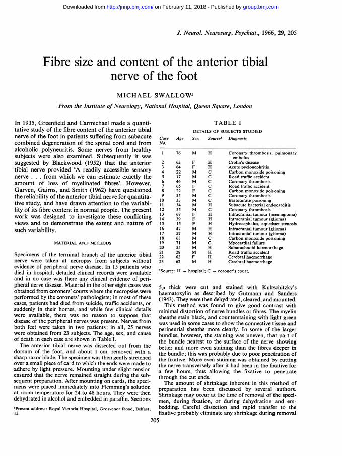
CaseNo.
TABLE IDETAILS OF SUBJECTS STUDIED
Age Sex Source' Diagnosis
1 76 M H Coronary thrombosis, pulmonaryembolus
2 62 F H Crohn's disease3 64 F H Acute pyelonephritis4 22 M C Carbon monoxide poisoning5 17 M C Road traffic accident6 46 M C Coronary thrombosis7 65 F C Road traffic accident8 22 F C Carbon monoxide poisoning9 55 M C Coronary thrombosis10 33 M C Barbiturate poisoning11 34 M H Subacute bacterial endocarditis12 55 M C Coronary thrombosis13 68 F H Intracranial tumour (meningioma)14 39 F H Intracranial tumour (glioma)15 15 F H Hydrocephalus, aqueduct stenosis16 47 M H Intracranial tumour (glioma)17 57 M H Intracranial tumour (glioma)18 63 M C Carbon monoxide poisoning19 71 M C Myocardial failure20 55 M H Subarachnoid haemorrhage21 28 M H Road traffic accident22 62 F H Cerebral haemorrhage23 62 M H Cerebral haemorrhage
'Source: H = hospital; C = coroner's court.
5 z thick were cut and stained with Kultschitzky'shaematoxylin as described by Gutmann and Sanders(1943). They were then dehydrated, cleared, and mounted.
This method was found to give good contrast withminimal distortion of nerve bundles or fibres. The myelinsheaths stain black, and counterstaining with light greenwas used in some cases to show the connective tissue andperineurial sheaths more clearly. In some of the largerbundles, however, the staining was uneven, that part ofthe bundle nearest to the surface of the nerve showingbetter and more even staining than the fibres deeper inthe bundle; this was probably due to poor penetration ofthe fixative. More even staining was obtained by cuttingthe nerve transversely after it had been in the fixative fora few hours, thus allowing the fixative to penetratethrough the cut ends.The amount of shrinkage inherent in this method of
preparation has been discussed by several authors.Shrinkage may occur at the time of removal of the speci-men, during fixation, or during dehydration and em-bedding. Careful dissection and rapid transfer to thefixative probably eliminate any shrinkage during removal
205
group.bmj.com on February 11, 2018 - Published by http://jnnp.bmj.com/Downloaded from
Michael Swallow
from the body. Duncan (1934), Rexed (1944), andSanders (1948) all state that there is no significant changein diameter during fixation in osmium-containing fluids.It is likely, however, that some shrinkage occurs duringdehydration and embedding; this was estimated bySanders (1948), and accepted by Femand and Young(1951) as causing a 6 to 8% reduction in fibre diameter.Hursh (1939) demonstrated 10% shrinkage of osmium-fixed teased fibres during dehydration. The shrinkageaffects axons and myelin sheaths to an equal extent, andfibres of all diameters are affected equally, thus pre-serving their relationship to one another with regard tosize.
All the sections were photographed at a magnificationof x 250 on a Zeiss Ultraphot II camera-microscope,using 9 x 12 cm. Ilford panchromatic plates. Enlarge-ments were made on to bromide paper and two sets ofprints were produced: (a) all sections were printed at amagnification of x 500 for estimating total fibre countsand (b) selected nerves were enlarged to x 1,000 formeasurement of fibre diameter. During the process ofenlarging, magnification was checked regularly using aplate on which a micrometer scale had beenphotographed.Myelinated fibres with an external diameter of 2 p or lesswere not always clearly delineated in enlarged photo-graphs; it was therefore essential to check the photo-graphic appearances against the original slides, using highmagnification and an oil-immersion lens when necessary.
Total fibre counts for each nerve were made, using asharpened metal pointer which penetrated the photographand made contact with a metal base-plate, thus registeringthe count on an automatic counter. When individualfascicles were circular in shape, the area of each was cal-culated from the diameter measured to the inner borderof the perineurial sheath. When fascicles were oval inshape, the mean of two diameters at right angles wastaken, any irregularities being checked with a planimeter.Fibre density was calculated as the number of fibres persq. mm. of intraperineurial area.For the construction of histograms of fibre size, the
external diameter of the myelin sheath was measured fromx 1,000 photographs by means of a perspex cursor andmechanical counter which allowed automatic registrationof fibre diameter, as described by Espir and Harding(1961). In the preparation of the histograms, fibres withan external diameter of 2 ,u or less were grouped togetherand larger fibres were subdivided into 1 ,u groups.
RESULTS
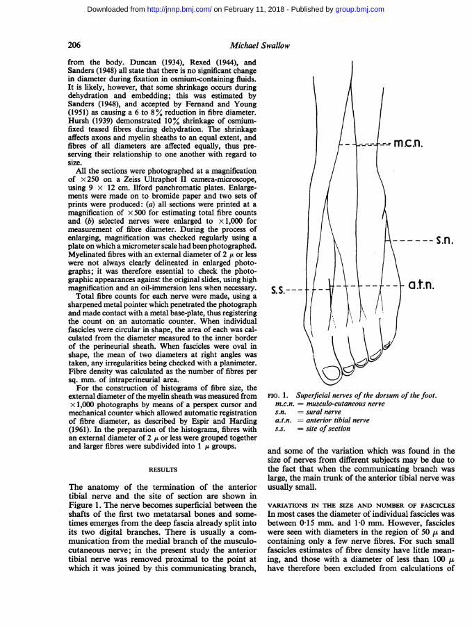
The anatomy of the termination of the anteriortibial nerve and the site of section are shown inFigure 1. The nerve becomes superficial between theshafts of the first two metatarsal bones and some-times emerges from the deep fascia already split intoits two digital branches. There is usually a com-munication from the medial branch of the musculo-cutaneous nerve; in the present study the anteriortibial nerve was removed proximal to the point atwhich it was joined by this communicating branch,
m.c.n.
a.t.n.S. S.
FIG. 1. Superficial nerves of the dorsum of the foot.m.c.n. = musculo-cutaneous nerves.n. = sural nervea.t.n. = anterior tibial nerves.s. = site of section
and some of the variation which was found in thesize of nerves from different subjects may be due tothe fact that when the communicating branch waslarge, the main trunk of the anterior tibial nerve wasusually small.
VARIATIONS IN THE SIZE AND NUMBER OF FASCICLESIn most cases the diameter of individual fascicles wasbetween 0-15 mm. and 10 mm. However, fascicleswere seen with diameters in the region of 50 ,u andcontaining only a few nerve fibres. For such smallfascicles estimates of fibre density have little mean-ing, and those with a diameter of less than 100 uhave therefore been excluded from calculations of
206
group.bmj.com on February 11, 2018 - Published by http://jnnp.bmj.com/Downloaded from

Fibre size and content of the anterior tibial nerve of the foot
:*~~~~~I
;
FIG. 2 Fibredensity (thousandfibres per sq. mm.)of individualfascicles plottedagainst the subject'sage. Values fordifferent fasciclesof the same nerveare joined by a line.
I I 1 --I20 30 40 5O 60 70 80
fascicular area and fibre density. In the 25 nerves
examined, there were in all 105 fascicles, of which 22had a diameter of less than 100 p. The distributionof the remaining 83 fascicles among the 25 nerves isshown in Fig. 2, and it can be seen that there was no
correlation between the number of fascicles and thesubject's age. Furthermore, there was no correlationbetween either the diameter of individual fascicles or
the mean fascicular diameter and the subject's age.
From the sum of the cross-sectional areas ofindividual fascicles the total fascicular area was
calculated for each subject. This varied from0-126 sq. mm. to 0-515 sq. mm. in different subjects(Table II), with a mean of 0-273 sq. mm., and showedno correlation with age.These results indicate that age has no effect on
either the number or size of the constituent fasciclesof the anterior tibial nerve, and that both may varyin a random fashion within wide limits.
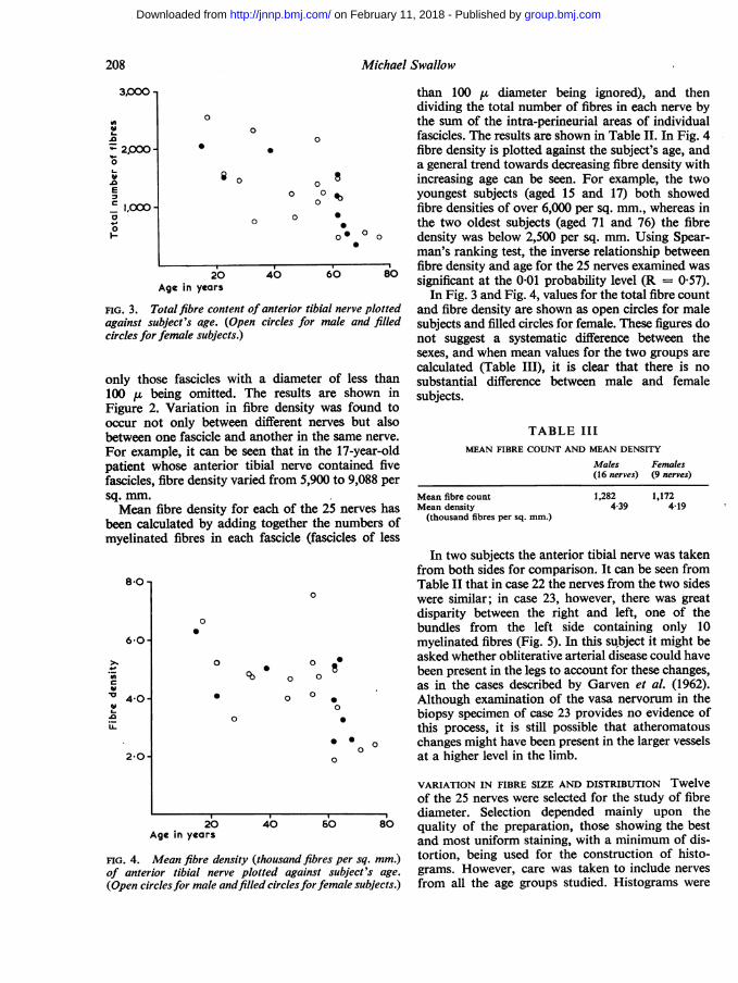
VARIATION IN TOTAL FIBRE CONTENT The totalnumber of myelinated fibres in each nerve is shownin Table II. In Fig. 3 the total fibre content is plottedagainst the subject's age. It can be seen that thelowest count (325 fibres) occurred in a 68-year-oldfemale subject and the highest (2,539 fibres) in a17-year-old male. These results indicate that thetotal fibre content of the anterior tibial nerve tendsto decrease in older subjects, although there is alsoconsiderable random variation.
In order to test the significance of the inverserelationship between total fibre count and age, thefindings in the 25 nerves were ranked according tofibre count and age, and the differences in rankingcompared by calculating Spearman's rank correla-
TABLE II
TOTAL FASCICULAR AREA FOR EACH SUBJECT
CaseNo.
234S
678910111213141516171819202122R22L23R23L
Total Fibre Content TotalFibres of Small Fascicular
Fascicles Area'(diam. <100 ,u) (sq. mm.)
462869672
1,5922,5391,220516
1,5221,066742
2,3072,131325
1,9792,033809
1,2251,121530
1,3741,4511,1631,4661,485469
- 0-19425 0 340- 0-126- 0 30510 0-382- 0-306- 0-16136 0-36814 0 200- 0-15023 0 47332 0-515- 0-127- 0-154- 0-31052 0-1628 0-258
0-308_ 0-241- 0-18289 0-414- 0-2429 0370
28 0-292- 0-259
Mean FibreDensity"(thousandfibresper sq. mm.)
2-382-485.355226-643.993-214045254.944-844072-565046-274-674-723-642-197-563-294-813.944.991-81
'Fascicles less than 100 1 in diameter omitted.
tion coefficient (R). This was found to be 0-74:Student's t test shows that this is significant at the0-001 probability level.
VARIATION IN FIBRE DENSITY Fibre density may beexpressed as the number of fibres per square milli-metre of intra-perineurial area. This has beencalculated for 83 individual fascicles in the 25 nerves,
10.0-
8 0 -
80-
e 6-0-
U- 240 -IL
2 O0
I,
10Age in years
207
i
group.bmj.com on February 11, 2018 - Published by http://jnnp.bmj.com/Downloaded from
Michael Swallow
3,000
w.04- 2,000-0
.0E
C 1,000-
0
0
0
0
0
20Age in years
FiG. 3. Total fibre content of dagainst subject's age. (Open (
circles for female subjects.)
only those fascicles with a100 ,u being omitted. The
1% W _ s _....In
than 100 ,u diameter being ignored), and thendividing the total number of fibres in each nerve bythe sum of the intra-perineurial areas of individual
0 fascicles. The results are shown in Table II. In Fig. 4fibre density is plotted against the subject's age, anda general trend towards decreasing fibre density with
8 increasing age can be seen. For example, the twoo °0 00 youngest subjects (aged 15 and 17) both showed
fibre densities of over 6,000 per sq. mm., whereas in°* the two oldest subjects (aged 71 and 76) the fibre
o * 0° o density was below 2,500 per sq. mm. Using Spear-man's ranking test, the inverse relationship between
40 60 80 fibre density and age for the 25 nerves examined wassignificant at the 0'01 probability level (R = 0 57).
In Fig. 3 and Fig. 4, values for the total fibre countanterior tibial nerve plotted and fibre density are shown as open circles for malecircles for male and filled subjects and filled circles for female. These figures do
not suggest a systematic difference between thesexes, and when mean values for the two groups arecalculated (Table III), it is clear that there is no
diameter of less than substantial difference between male and femaleD results are shown in subjects.
Figure 2. Variation in nibre density was tound tooccur not only between different nerves but alsobetween one fascicle and another in the same nerve.For example, it can be seen that in the 17-year-oldpatient whose anterior tibial nerve contained fivefascicles, fibre density varied from 5,900 to 9,088 persq. mm.Mean fibre density for each of the 25 nerves has
been calculated by adding together the numbers ofmyelinated fibres in each fascicle (fascicles of less
8-0 -
6 0
u
n 40~._
20-
0
.
0
S
0 8q 0 08
0 0
0
S0
0
* S
0
TABLE IIIMEAN FIBRE COUNT AND MEAN DENSITY
Males Females(16 nerves) (9 nerves)
Mean fibre countMean density
(thousand fibres per sq. mm.)
1,282 1,1724*39 4-19
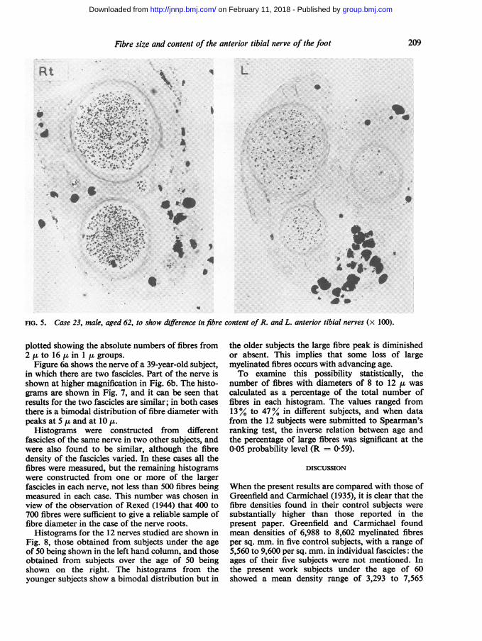
In two subjects the anterior tibial nerve was takenfrom both sides for comparison. It can be seen fromTable II that in case 22 the nerves from the two sideswere similar; in case 23, however, there was greatdisparity between the right and left, one of thebundles from the left side containing only 10myelinated fibres (Fig. 5). In this su,bject it might beasked whether obliterative arterial disease could havebeen present in the legs to account for these changes,as in the cases described by Garven et al. (1962).Although examination of the vasa nervorum in thebiopsy specimen of case 23 provides no evidence ofthis process, it is still possible that atheromatous
0 o changes might have been present in the larger vesselsat a higher level in the limb.
20Age in years
FIG. 4. Mean fibre density (tiof anterior tibial nerve plot(Open circlesfor male andfillee
VARIATION IN FIBRE SIZE AND DISTRIBUTION Twelveof the 25 nerves were selected for the study of fibrediameter. Selection depended mainly upon the
40 60 80 quality of the preparation, those showing the bestand most uniform staining, with a minimum of dis-
housand fibres per sq. mm.) tortion, being used for the construction of histo-tted against subject's age. grams. However, care was taken to include nervesd circlesforfemale subjects.) from all the age groups studied. Histograms were
208
group.bmj.com on February 11, 2018 - Published by http://jnnp.bmj.com/Downloaded from
Fibre size and content of the anterior tibial nerve of the foot
Rt
;/, .*t..;....4
_w .
14 L
*0 '#
II
*4
FIG. 5. Case 23, mak, aged 62, to show difference in fibre content of R. and L. anterior tibial nerves (x 100).
plotted showing the absolute numbers of fibres from2 IL to 16 ,u in 1 ,t groups.
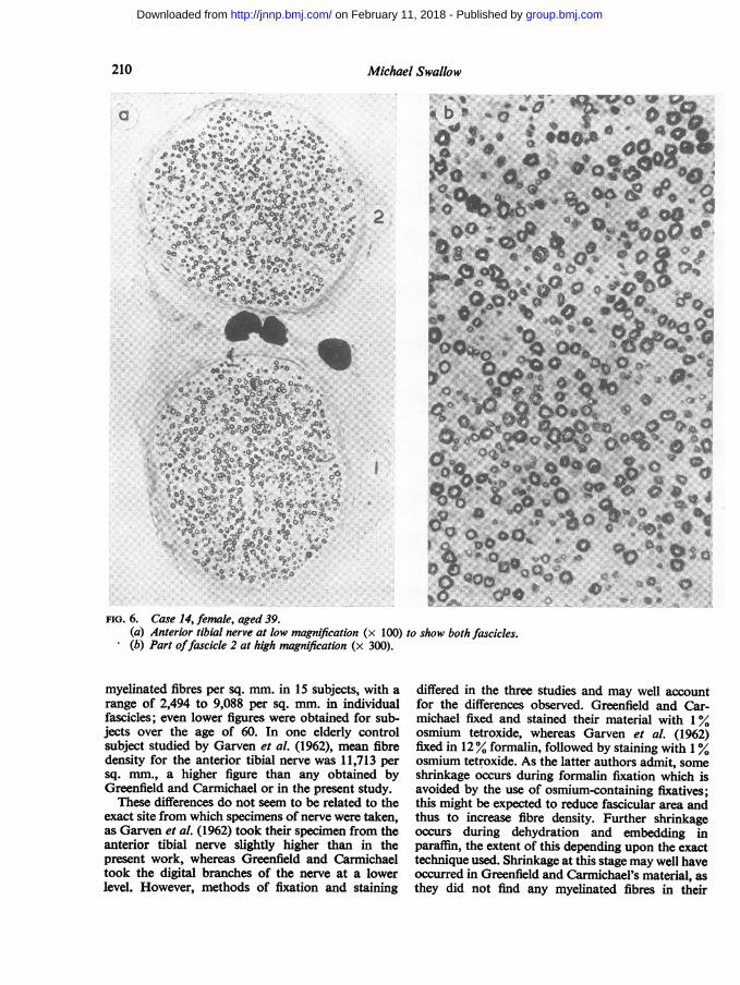
Figure 6a shows the nerve of a 39-year-old subject,in which there are two fascicles. Part of the nerve isshown at higher magnification in Fig. 6b. The histo-grams are shown in Fig. 7, and it can be seen thatresults for the two fascicles are similar; in both casesthere is a bimodal distribution of fibre diameter withpeaks at 5 , and at 10 p,.
Histograms were constructed from differentfascicles of the same nerve in two other subjects, andwere also found to be similar, although the fibredensity of the fascicles varied. In these cases all thefibres were measured, but the remaining histogramswere constructed from one or more of the largerfascicles in each nerve, not less than 500 fibres beingmeasured in each case. This number was chosen inview of the observation of Rexed (1944) that 400 to700 fibres were sufficient to give a reliable sample offibre diameter in the case of the nerve roots.
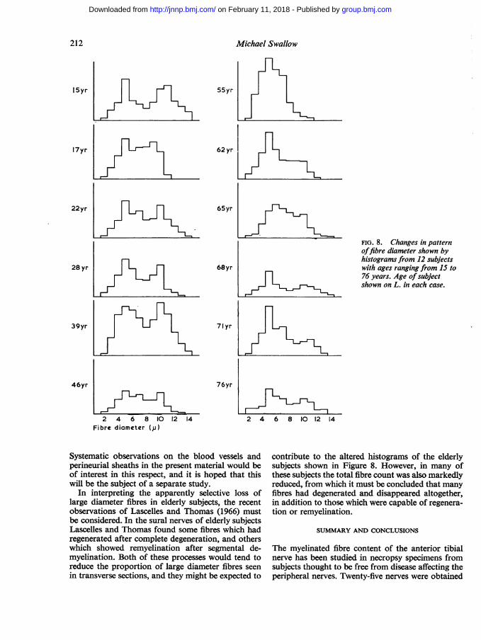
Histograms for the 12 nerves studied are shown inFig. 8, those obtained from subjects under the ageof 50 being shown in the left hand column, and thoseobtained from subjects over the age of 50 beingshown on the right. The histograms from theyounger subjects show a bimodal distribution but in
the older subjects the large fibre peak is diminishedor absent. This implies that some loss of largemyelinated fibres occurs with advancing age.To examine this possibility statistically, the
number of fibres with diameters of 8 to 12 IL wascalculated as a percentage of the total number offibres in each histogram. The values ranged from13% to 47% in different subjects, and when datafrom the 12 subjects were submitted to Spearman'sranking test, the inverse relation between age andthe percentage of large fibres was significant at the0-05 probability level (R = 059).
DISCUSSION
When the present results are compared with those ofGreenfield and Carmichael (1935), it is clear that thefibre densities found in their control subjects were
substantially higher than those reported in thepresent paper. Greenfield and Carmichael foundmean densities of 6,988 to 8,602 myelinated fibresper sq. mm. in five control subjects, with a range of5,560 to 9,600 per sq. mm. in individual fascicles: theages of their five subjects were not mentioned. Inthe present work subjects under the age of 60showed a mean density range of 3,293 to 7,565
a
.4"I.
4sa>
X P te a -Z r e ^, @ . /r . . &
* b e * o v X, ,£-- ^ . s ; _ **
t @. ' '. * *'*, t z w 0 * R
* w* w :E ^P A 4| @ -
*; *
;.s * N
# @,
a a.
x J
z s * 4e
'!a
209
k.
s. at$,
40 0
group.bmj.com on February 11, 2018 - Published by http://jnnp.bmj.com/Downloaded from
Michael Swallow
a
2
FIG. 6. Case 14, female, aged 39.(a) Anterior tibial nerve at low magnification (x 100) to show both fascicles.(b) Part offascicle 2 at high magnification (x 300).
myelinated fibres per sq. mm. in 15 subjects, with arange of 2,494 to 9,088 per sq. mm. in individualfascicles; even lower figures were obtained for sub-jects over the age of 60. In one elderly controlsubject studied by Garven et al. (1962), mean fibredensity for the anterior tibial nerve was 11,713 persq. mm., a higher figure than any obtained byGreenfield and Carmichael or in the present study.These differences do not seem to be related to the
exact site from which specimens of nerve were taken,as Garven et al. (1962) took their specimen from theanterior tibial nerve slightly higher than in thepresent work, whereas Greenfield and Carmichaeltook the digital branches of the nerve at a lowerlevel. However, methods of fixation and staining
differed in the three studies and may well accountfor the differences observed. Greenfield and Car-michael fixed and stained their material with 1 %osmium tetroxide, whereas Garven et al. (1962)fixed in 12% formalin, followed by staining with 1%osmium tetroxide. As the latter authors admit, someshrinkage occurs during formalin fixation which isavoided by the use of osmium-containing fixatives;this might be expected to reduce fascicular area andthus to increase fibre density. Further shrinkageoccurs during dehydration and embedding inparaffin, the extent of this depending upon the exacttechnique used. Shrinkage at this stage may well haveoccurred in Greenfield and Carmichael's material, asthey did not find any myelinated fibres in their
210
group.bmj.com on February 11, 2018 - Published by http://jnnp.bmj.com/Downloaded from

Fibre size and content of the anterior tibial nerve of the foot
140- for fibre density in the sural and great auricularI nerves have been published by Dyck, Beahrs, and
120- Miller (1965), who used 1 % osmium tetroxide stain-ing with cryostat sections. Their figures are only
loo - slightly higher than those given in the present paper,mean densities of less than 5,000 per sq. mm.
80- r occurring in several of their control nerves. Thisdoes not suggest that the technique used in the present
60- study has introduced serious errors due to shrinkageor failure to stain the smallest myelinated fibres.
40- By their technique, Garven et al. (1962) regard afigure of the order of 10,000 to 13,000 myelinated
20- fibres per sq. mm. as the normal density for theU, msciatic nerve and its branches; they comment that
the larger nerve trunks give more consistent resultsthan the terminal sensory branches in the foot which
o may be subject to considerable variation. Foro 40 example, one of their 'normal' digital nerves, from aE 2 young woman who had been killed in a motorEz 120 accident, had a fibre density of only 2,664 per sq.
mm., whereas the corresponding figure for theloo- n sciatic nerve was 11,953 per sq. mm.
Substantial variation in both total fibre counts80- and the fibre density has been seen in the present
series, and it seems justifiable to conclude that60- pathological changes in the anterior tibial nerve and
its branches can only be assessed correctly if sufficient40- control data are available to indicate the range ofnormal variation. While this variation may be less
for the anterior tibial nerve than for its terminal20 -
digital branches, it is still considerable, and it ismarkedly increased in elderly subjects. In spite of
2 4 6 8 10 12 114 16 this, the present study has shown an inverse relation-Fibre diameter (,) ship between fibre density and the subject's age
which was significant at the 0 01 probability level.FIG. 7. Histograms of fibre diameter for the However, the inverse relationship between the totalfascicles shown in Figure 6a. fibre count and the subject's age was significant at
Fascicle 1. 1,016 fibres; density 5,390 per sq. mm. the 0-001 probability level. This suggests that thereFascicke 2. 963 fibres; density 4,906 per sq. mm. is no advantage to be gained by expressing results in
the form of fibre density when the nerve concerned iscontrol nerves with diameters greater than 10 ,t, small enough to allow total myelinated fibre countswhereas fibres up to 14 ,u were found in the present to be made.work. Loss of myelinated fibres with increasing age hasAn alternative explanation of the low fibre been reported previously for the nerve roots by
densities in the present series might be that the tech- Corbin and Gardner (1937) and by Rexed (1944).nique used (staining with Kultschitsky's haema- Whether these changes are wholly due to arterialtoxylin followed by differentiation by Pal's method) degeneration is uncertain. Changes in the vasastains the smallest myelinated fibres less well than nervorum and in the perineurial thickness with in-1% osmium tetroxide. This was suggested by Rexed creasing age have been described by Cottrell (1940),(1944), and it raises the possibility that fewer fibres but other factors such as repeated minor traumain the 1 to 2 ,u range have been counted in the might contribute to the loss of nerve fibres. Indeedpresent work than in the two previous studies Gairns, Garven, and Smith (1960) suggest that badlymentioned, although great care was taken to check fitted shoes might damage digital nerve fibres in thephotographs against the original slides when count- foot, and thus be responsible for the low fibre den-ing, to ensure that no fibres which had been faintly sities and the degenerative changes in sensory end-stained were omitted. However, some recent figures organs which they observed in young subjects.
211
group.bmj.com on February 11, 2018 - Published by http://jnnp.bmj.com/Downloaded from
Michael Swallow
15yr
17yr
55yr
62 yr
22yr
28yr
39yr
46yr
65yr
68yr
71yr
76yr
2 4 6 8 10 12 14Fibre diameter (,u)
Systematic observations on the blood vessels andperineurial sheaths in the present material would beof interest in this respect, and it is hoped that thiswill be the subject of a separate study.
In interpreting the apparently selective loss oflarge diameter fibres in elderly subjects, the recentobservations of Lascelles and Thomas (1966) mustbe considered. In the sural nerves of elderly subjectsLascelles and Thomas found some fibres which hadregenerated after complete degeneration, and otherswhich showed remyelination after segmental de-myelination. Both of these processes would tend toreduce the proportion of large diameter fibres seenin transverse sections, and they might be expected to
FIG. 8. Changes in patternoffibre diameter shown byhistograms from 12 subjectswith ages ranging from 15 to76 years. Age ofsubjectshown on L. in each case.
2 4 6 8 10 12 14
contribute to the altered histograms of the elderlysubjects shown in Figure 8. However, in many ofthese subjects the total fibre count was also markedlyreduced, from which it must be concluded that manyfibres had degenerated and disappeared altogether,in addition to those which were capable of regenera-tion or remyelination.
SUMMARY AND CONCLUSIONS
The myelinated fibre content of the anterior tibialnerve has been studied in necropsy specimens fromsubjects thought to be free from disease affecting theperipheral nerves. Twenty-five nerves were obtained
L-L.
212
group.bmj.com on February 11, 2018 - Published by http://jnnp.bmj.com/Downloaded from
Fibre size and content of the anterior tibial nerve of the foot
from 23 subjects whose ages ranged from 16 to 82years. There was a highly significant fall in the totalfibre count and in fibre density with increasing age.The frequency distribution of fibres of different
sizes was studied in 12 cases. A significant decrease inthe proportion of large fibres was found in the oldersubjects.The wide range of variation found in young as well
as old subjects would seem to limit the usefulness ofanterior tibial nerve biopsy as a diagnostic procedurein patients with generalized neurological disease.
This work was carried out in the University Departmentof Clinical Neurology, with the aid of a generous grantfrom the Polio Research Fund. I wish to thank ProfessorR. W. Gilliatt for encouragement and help.
REFERENCES
Blackwood, W. (1952). Biopsy technique in the diagnosis of peripheralneuropathies (especially hereditary and sensory neuropathy).In Proc. Ist int. Congr. Neuropath., Rome, 1925, Vol. 3,pp. 415.424.
Corbin, K. B., and Gardner, E. D. (1937). Decrease in number ofmyelinated fibers in human spinal roots with age. Anat. Rec.,68, 63-74.
Cottrell, L. (1940). Histologic variations with age in apparentlynormal peripheral nerve trunks. Arch. Neurol. Psychiat.(Chic.), 43, 1138-1150.
Duncan, D. (1934). A relation between axon diameter and myelina-tion determined by measurement of myelinated spinal rootfibers. J. comp. Neurol., 60, 437.472.
Dyck, P. J., Beahrs, 0. H., and Miller, R. H. (1965). Peripheral nervesin hereditary neural atrophies: number and diameters ofmyelinated fibers. In Proc. 6th int. Congr. Electroenceph. clin.Neurophysiol., Vienna, pp. 673-677.
Espir, M. L. E., and Harding, D. T. C. (1961). Apparatus for measuringand counting myelinated nerve fibres. J. Neurol. Neurosurg.Psychiat., 24, 287-290.
Fernand, V. S. V., and Young, J. Z. (1951). The sizes of the nervefibres of muscle nerves. Proc. roy. Soc. B., 139, 38-58.
Gairns, F. W., Garven, H. S. D., and Smith, G. (1960). The digitalnerves and the nerve endings in progressive obliterative vasculardisease of the leg. Scot. med. J., 5, 382-391.
Garven, H. S., Gairns, F. W., and Smith, G. (1962). The nerve fibrepopulations of the nerves of the leg in chronic occlusive arterialdisease in man. Ibid., 7, 250-265.
Greenfield, J. G., and Carmichael, E. A. (1935). The peripheralnerves in cases of subacute combined degeneration of the cord.Brain, 58, 483-491.
Gutmann, E., and Sanders, F. K. (1943). Recovery of fibre numbersand diameters in the regeneration of peripheral nerves. J.Physiol. (Lond.), 101, 489-518.
Hursh, J. B. (1939). Conduction velocity and diameter of nerve fibers.Amer. J. Physiol., 127, 131-139.
Lascelles, R. G., and Thomas, P. K. (1966). Changes due to age ininternodal length in the sural nerve in man. J. Neurol. Neuro-surg. Psychiat., 29, 40-44.
Rexed, B. (1944). Contributions to the knowledge of the post-nataldevelopment of the peripheral nervous system in man. Actaphychiat. neurol. (Stockh.), suppl. 33.
Sanders, F. K. (1948). The thickness of the myelin sheaths of normaland regenerating peripheral nerve fibres. Proc. roy. Soc. B.,135, 323-357.
213
group.bmj.com on February 11, 2018 - Published by http://jnnp.bmj.com/Downloaded from

anterior tibial nerve of the foot.Fibre size and content of the
M Swallow
doi: 10.1136/jnnp.29.3.2051966 29: 205-213 J Neurol Neurosurg Psychiatry
http://jnnp.bmj.com/content/29/3/205.citationUpdated information and services can be found at:
These include:
serviceEmail alerting
online article. article. Sign up in the box at the top right corner of the Receive free email alerts when new articles cite this
Notes
http://group.bmj.com/group/rights-licensing/permissionsTo request permissions go to:
http://journals.bmj.com/cgi/reprintformTo order reprints go to:
http://group.bmj.com/subscribe/To subscribe to BMJ go to:
group.bmj.com on February 11, 2018 - Published by http://jnnp.bmj.com/Downloaded from