Embed Size (px)
Citation preview

129
J. Physiol. (1952) 117,129–151
PH. CXVII.
(Received 22 June 1951)
ACTION CURRENTS IN SINGLE AFFERENT NERVE FIBRES ELICITED BY STIMULATION OF THE SKIN
OF THE TOAD AND THE CAT
BY JURO MARUHASHI, KANJI MIZUGUCHI AND ICHIJI TASAKI*
* Present address: Central Institute for the Deaf, St Louis, Mo., U.S.A.
From the Tokugawa Biological Institute, Mejiro, Tokyo, and the Physiological Institute, Keio University, Yotsuya, Tokyo
9
INTRODUCTIONSince Adrian (1928, 1931, 1932) opened up a new method of studying the basis of sensation, the sensory impulses in single cutaneous nerve fibres have already been the object of intensive investigation by a great number of workers (Adrian & Zotterman, 1926; Adrian, Cattell & Hoagland, 1931 ; Cattell & Hoagland, 1931; Hogg, 1935; ·Zotterman, 1936, 1939, and others). The tech-nique adopted by all these workers consisted in leading off action potentials from a small group of nerve fibres, obtained either by the operative attenuation of the nerve trunk or by selecting small nerve twigs for the experiment. By this technique, Zotterman successfully estimated the :fibre-diameters of several kinds of cutaneous afferent fibres of the frog and the cat and reached important conclusions.
With this technique it is easy to record sensory discharges in single nerve fibres which develop overwhelmingly large action potentials or in fibres with especially low threshold for sensory stimuli. It is, however, extremely difficult to explore the behaviour of nerve endings which on stimulation give rise to a limited number of small action potentials, because the discharges in these fibres may be completely masked by those in other predominant fibres. This and other defects of the technique can undoubtedly be circumvented by using a single isolated fibre of the desired size for the experiments.
Since 1942 one of. us (LT.), in collaboration with several co-workers who changed from time to time, has devoted much effort to exploring the physio-logical properties of various sensory nerve endings with the single fibre technique. By means of the experimental set-up we are using now, it is possible to observe afferent impulses in a single nerve fibre, either myelinated

130 J. MARUHASHI, K. MIZUGUCHI AND I. T ASAKI or unmyelinated, of less than 2µ, diameter. Furthermore, the method of the 'bridge-insulator' (see Tasaki & Mizuguchi, 1948) enables us to apply various sorts of stimuli to the skin without causing any appreciable disturbance in the current recording device.
It is the purpose of this paper to present the results of our investigation on the properties of the 'sensory units' (Tower, 1940) in the skin. Our investiga-tion along this line is by no means complete yet. But, as the progress of our work is now very slow on account of the difficulties which confront our nation, we thought we should summarize what we have learned up to the present. Part I of this paper is concerned with experiments on the toad, and Part II on the cat.
PART I. CUTANEOUS AFFERENT FIBRES IN THE TOAD METHOD
Experiments on the toad were as a rule conducted on excised nerve-skin preparations. The skin covering the gastrocnemius muscle was dissected out together with its nerve supply, and the operation for isolating a single nerve fibre was performed at the point where the cutaneous nerve branches off from the tibial nerve. The operation was done under a low-power binocular micro-scope. The preparation was frequently brought under a high-power microscope and a single fibre of the desired size was chosen for the experiment.
The operated region of the nerve was mounted on a 'bridge-insulator' (Tasaki, 1939; Tasaki & Takeuchi, 1941). The skin was spread out on cotton-wool soaked in Ringer's solution, and a non-polarizable eleotrode in contact with this cotton-wool was earthed. Another electrode which dipped into the fluid on the opposite side of the bridge-insulator was led to the amplifier. When the conduction rate in the afferent fibre was to be determined, induction shocks were applied near the proximal stump of the sciatic nerve, and the electrode in the pool of Ringer's solution on the distal side of the bridge-insulator was led to the amplifier, the proximal pool being grounded.
Electric responses of single nerve fibres were amplified with a 4-stage resistance-capacity coupled amplifier and were recorded with an oscillograph of the Duddell type or with a cathode-ray oscillograph.
Action currents from smaller fibres were naturally smaller than those from the larger fibres. In reading our records, it should be borne in mind that the action currents from smaller fibres were amplified to a greater extent, and that consequently recorded spike heights do not give any information as to the absolute strengths of the action currents.
The amplified action current was tapped and was made to operate both a loud speaker for hearing impulses and a repetitively sweeping cathode-ray oscillograph for seeing impulses. Sensory stimuli routinely used in the experiment were air jet, light touch with a von Frey hair, pressure through the tip of a blunt glass rod, pin-prick, application of 5-10% acetic acid and light touch with a metal vessel containing hot or cold water. The details of the procedure of each particular experiment will be described in the following pages.
RESULTS
Sensory units in the toad skin were classified according to their physiological properties, the criteria of classification being (1) the diameter of the sensory nerve fibre, (2) the character of the adequate stimuli, (3) the distribution of the sensory spots in the skin, and (4) the duration of the sensory discharge under constant stimulation (i.e. the rate of adaptation). Through careful

131 ISOLATED FIBRES FROM SKIN NERVES examination of about seventy units in the toad skin, we are led to the classifica-tion shown in Table 1.
TABLE 1. Properties of various cutaneous sensory units of the toad (22-26° C)
Type Type of
discharge
Fibre size (µ.)
Conduction-rate
(m/sec) Receptive
field
No. of units
examined Tactile Phasic (tonic) 8–15 20–35 3–10spots c. 40 Pressure Tonic 4-5 5–8 1–5spots 3 Large nociceptive Phasic 6–9 10–15 10–30mm2 6 Small nociceptive Phasic or tonic 3-5 3-9 5–20mm2 19 Unmyelinated Tonic Below 2 1–0·1 ? —
The terms 'tactile' and 'pressure' in this table are used to designate the sensory fibres originating at the end-organs which are sensitive to mechanical stimuli and insensitive to acid. In most cases these fibres had a diameter above 8µ; but as we met with several cases in which the diameter was about 4µ we had to make two subdivisions in the fibres of this category. In this paper, the term 'pressure fibres' is used to represent these smaller fibres of which the endings show slow adaptation and the name 'tactile fibres' is given to the larger fibres.The term 'nociceptive fibres' is used to describe those arising at nerve endings which are sensitive both to application of acid and to pin-pricks. The details of the properties of each type of sensory unit will be presented below.
Fibres from tactile and pressure nerve endings The sensory impulses from tactile end-organs of the frog have been the
object of repeated study by members of Adrian's laboratory. In the toad, the receptive field of a single 'tactile fibre' consists of a number of clearly defined points which are distributed in an area of about 4 x 5 mm. The number of the sensitive points connecting to one afferent nerve fibre varied considerably from preparation to preparation, and was generally between 3 and 10. In female animals, where the surface of the skin is much rougher and more warty than that of male animals, it can easily be shown that the endings are located just beneath the tips of the warts; a light touch with a von Frey hair reveals that sensory impulses can be evoked most readily at definite black spots on the wart, and that in the region between these sensitive spots light stimuli are almost ineffective.
Among all the sensory nerve endings in the toad's skin, tactile endings are most sensitive to light touch and to air jet, and the fibres from these endings are the largest in the skin nerve. We have examined about forty single fibres of this type. Most of them were between 11 and 15µ in diameter. But this figure does not indicate the average diameter of the tactile fibres, because, when a single fibre of this type was desired, we selected it from among the largest
9-2

132 J. MARUHASHI, K. MIZUGUCHI AND I. TASAKI
fibres in the skin nerve. With one exception, all the tactile fibres we have isolated were above 7.5µ. At about 23° C their conduction rate, determined from the time required for conduction between the proximal stump of the sciatic nerve and the operated region of the nerve, was generally between 20 and 35 m/sec.
Discharge of impulses from this type of ending is generally phasic, i.e. under a constant pressure the discharge dies out rapidly in 0.1sec or so from the onset of the pressure stimulus (Fig. IA, top). In some preparations, however,
A B c Tactile fibre (8µ.) Pressure fibre (5µ.) Small fibres
Induction shock
lj PressingPressing . Heat
Nociceptive fibre (4·5µ.) r Air jet .. . . . . . . ......
Small fibres Tactile fibre (11 µ.)
Pricking At rest
Pricking Pressing Spontaneous '~rWr*+' Nodceptive fibre (8µ.) Nociceptive fibre (4µ.) I Pricking
Pressing Ice Acid t.J1
Pricking Heat a
Acid
50/sec 50/sec ....... .. ... Fig. 1. A and B: records of impulse discharges taken from various ' sensory unit preparations'
of the toad. C: aafferent impulses recorcied from bundles of unmyelinated fibres containing also a few fine myelinated fibres; spikes from myelinated fibres are seen in two records; time marks are 40 msec apart. All the records were taken at temperatures between 23 and 26° C. The term 'pressing' means application of a firm pressure stimulation to the receptive field of the skin through the tip of a blunt glass rod. 'Pricking' stimuli were applied with the tip of a sharp needle held by hand. 'Acid ' means application of 5–10% acetic acid. ' Induction shock' was applied to the skin with a pair ofplatinum electrodes placed on the receptive field. Records labelled 'heat' or 'cold' were taken 1–2 sec after pressing skin with a metal vessel containing water at about 50° or 0° C respectively.

133 ISOLATED FIBRES FROM SKIN NERVES the adaptation is considerably slower and the discharge may last for several seconds under constant pressure (Fig. 1, tactile fibre of 11 µ).The mode of impulse discharge gives us the impression that there are two kinds of tactile end-organs in the skin, the discharge in one being phasic and in the other 'semi-tonic'; but the fibre-size and the distribution of end-organs in the skin do not differ appreciably between the two.
In the tonic tactile endings it was observed that the rhythm of discharge was more or less irregular, due probably to collision of impulses from different end-organs innervated by the single fibre under investigation (cf. Tasaki, 1949).In the phasic endings impulses are evoked not only at the onset but also on the removal of a constant pressure.
Electrical stimulation. Although the tactile endings are not excited by the application of acid to the skin, it is possible to excite them by induction shocks or galvanic currents which traverse the skin. By application of in-duction shocks to the skin, it was found that nerve impulses are transmitted at a considerably lower rate near the end-organs; at a conduction distance of about 2 cm, the time from the stimulus to the arrival of the impulse was about 2 msec at 23° C. The average conduction rate in this peripheral region of the nerve fibre is therefore less than half the rate in the proximal region of the same fibre.
The effect of a galvanic current upon the tactile ending is of considerable interest in regard to the mechanism of production of impulses at the end-organ. When the battery circuit is so arranged that the current flows through the skin (Fig. 2, top), it is observed that a current flowing inwards throughthe skin causes an outburst of impulses at make of the current, while a current flowing in the opposite direction is apparently ineffective. The total number of impulses elicited by a galvanic current increases almost proportionately to the amount by which its strength is above the rheobase (Fig. 2).
ion Off
1·2V
5 3.6 V
6.0V 4
Fig. 2. Effect of electric current applied to one of the sensory spots of a touch fibre-skin prepara-tion of the toad. The duration of the applied current from 'on' to 'off' in the records on the left was about 0.5sec. Note a break excitation at 6 V, the cathode on the ending. 21° C.

134 J. MARUHASHI, K. MIZUGUCHI AND I. TASAKI The fact that the end-organ is excited only by a current applied with the
anode on the skin can easily be understood on the assumption that an end-organ elicits nerve impulses by transforming sensory stimuli into electric stimuli for the sensory nerve fibre (cf. Lillie, 1929). It is well known that the plasma membrane of a nerve fibre is excited by a current fl.owing outwards through this membrane (cf. Tasaki & Mizuguchi, 1948). If a sensory stimulus is to generate an outwardly directed current in the nerve fibre close to the end-organ, then the current generated by a sensory stimulus would have to be in the same direction as that generated by an anode placed on the end-organ. It therefore seems reasonable that the starting-point of the afferent fibre is excited by a current flowing inwards through the skin. Small pressure fibres. Among the fibres originating at the spots sensitive to
mechanical stimuli there are very small myelinated fibres that convey a tonic discharge of impulses on sustained pressure stimulation of the skin. We have met with such single fibres on three occasions only (see Fig. 1, pressure fibre of 5µ).They were between 4 and 5µin diameter, their conduction rate being about 5 m/sec at 24° C. The receptive field of these fibres consisted of one to five more-or-less clearly defined points. The frequency of discharge was often as high as 400/sec, and a spontaneous discharge at a low frequency (a few impulses per sec) was sometimes observed. These 'pressure endings' are less sensitive to a light touch and an air jet than the' tactile endings' described above.
Another characteristic of the pressure endings is the long latency in excita-tion by mechanical stimuli. When afferent impulses are recorded from a number of cutaneous fibres, it was often observed that a discharge in a pressure fibre in response to a constant pressure started after a short discharge in a tactile fibre had subsided.
M yelinated fibres from nociceptive endings In this category come several different types of afferent fibres. All of them
are insensitive to a light touch with a feather or to a light air jet. In response to stronger mechanical stimuli, such as tapping with a glass rod, they naturally give rise to outbursts of impulses. Large myelinated nociceptive fibres. These fibres have a diameter between
6 and 9 µ. The nerve endings of a single fibre of this type are found, when examined by light pin-pricks, to be distributed densely in an oval area of 3 x 4 mm to 5 x 6 mm. The discharge of impulses in this fibre on application of strong pressure through a blunt glass point is in general phasic; the discharge comes to an end within about 0.2sec from the onset of the stimulus. But, in preparations having relatively high sensitivity to mechanical stimuli, adapta-tion was fairly slow. The frequency of discharge is sometimes as high as 300/sec at 24° C, but impulses released by 10 % acetic acid are generally spaced by longer intervals (20 to 60 msec) at the same temperature.
135 !SOL.A.TED FIBRES FROM SKIN NERVES Small myelinated nociceptive fibres. In this category are fibres of 3 to 5µ
diameter. Their endings can be further subdivided according to the mode of impulse discharge. In one group are those which we may call tonic nociceptivefibres. They respond both to light pin-pricks and to application of acid with a relatively long train of impulses. The receptive field of a single fibre of this group varies according to the preparation from 2 x 3 mm to 4 x 6 mm, and the maximum frequency of discharge is from 50 to 130/sec at about 22° C.
Another group may be called phasic nociceptive fibres. These fibres are less sensitive to pin-pricks, and the discharge always comes to an end in O.l sec or less after the onset of the stimulus. In some preparations, only those strong pin-pricks which seem destructive to the skin are found to be effective in eliciting a small number of impulses. It is therefore probable that the nerve endings of such fibres are entirely different from those having a greater sensitivity to pin-pricks. The maximum frequency of discharge varies from 25 to 50/sec and the conduction rate from 3 to 9 m/sec at 22° C. The size of the receptive field was not determined very accurately because of the difficulty of eliciting afferent impulses by light mechanical stimuli. But, in relatively sensitive preparations, a dense distribution in an area of 2 x 2 mm to 3 x 5 mm was generally observed. On one occasion the receptive field of a single fibre seemed to consist of two separate patches of surface a few millimetres apart, each having an ordinary size.
The endings of some of the small nociceptive fibres are also sensitive to changes in temperature. Pressing the receptive field with the tip of a glass test tube filled with hot water (about 60° C) often evoked more frequent and longer discharges than pressing with a tube at room temperature; this effect was especially conspicuous when heat was applied immediately after cooling the skin with ice. It was also very often observed that not only heat but also cold increases the frequency of discharge in one and the same preparation. Since these nerve endings sensitive to temperature changes respond also to pin-pricks, we do not place them under the category of heat or cold fibres.
U nmyelinated afferent fibres It is well known that there are abundant unmyelinated fibres in the skin
nerves. They form special bundles which generally contain also some small myelinated fibres (see Fig. 3, top, left). It is possible to isolate a single unmyelinated fibre under a low-power microscope in an unstained preparation. To record action currents from such a fibre is, however, not very easy, owing to its vulnerability to injury and to desiccation.
The electric response of an unmyelinated fibre is characterized by its extremely slow development and subsidence (see Hogg, 1935) and also by its low conduction rate (Erlanger & Gasser, 1930). The size of the response was, at least in some preparations, relatively large (Fig. 3). Judging from the

136 J. MARUHASHI, K. MIZUGUCHI AND I. TASAKI
11000
son
::~-·. cm
12000
11000 1on
250/sec. 250/sec
Fig. 3. Photograph at top left shows a stage in an operation to isolate a bundle consisting mainly of unmyelinated fibres in a toad's skin nerve. The bar subtends 50µ. The bundle is marked by the cross. All the fibres, except this bundle and a large myelinated fibre, were cut across, and another photograph was taken at a magnification five times as high as in the first picture. The action current records shown were taken from this preparation. Records on the right were taken at a lower transit speed and a higher (twice) amplification. The reciprocal of the induction shock (in ohms) is given, 28° C.
all-or-none character exhibited on electrical stimulation, we may regard the action currents we recorded as being derived from single unmyelinated fibres. The strength of the action current developed by a large unmyelinated fibre is, under our experimental conditions, of the magnitude of about 10–9 A and is approximately equal to that from a small myelinated fibre. There are a number
of small unmyelinated fibres which develop weaker action currents. There are also some which seem intermediate· between a small myelinated and an unmyelinated fibre in respect to the conduction rate (1-3 m/sec).
When afferent impulses are led from a bundle consisting mainly of un-myelinated fibres, it is immediately seen that almost all kinds of sensory stimuli, such as pinching, pricking, burning with acid or applying heat or cold, give rise to an outburst of afferent impulses. In favourable cases, elec-trical responses of individual unmyelinated fibres could be distinguished from one another by their characteristic temporal configurations. An air blast or a light touch with a feather is in general ineffective in evoking impulses in

137 ISOLATED FIBRES FROM SKIN NERVES unmyelinated fibres. Impulses elicited by cold are distinctly smaller in size and less in number than those resulting from pinching, pricking or heating the skin (see Fig. 1 C).
In preparations containing only a few afferent unmyelinated fibres, we have seen that hot and cold endings are insensitive to mechanical stimuli. We also met with several preparations which are sensitive to mechanical stimuli but not to changes in temperature. In many other preparations, however, we noticed that none of the sensory stimuli, except an electric current applied to the skin, was effective in evoking an afferent impulse in some particular unmyelinated fibres. In the excitation of an unmyelinated fibre by an applica-tion of induction shocks to the skin, a long latent period and a relatively low threshold stand out clearly (Fig. 1 C, top).
The rate of conduction in individual unmyelinated fibres varies considerably from preparation to preparation; it is between 0.land 1 m/sec at about 22° C. The diphasic action currents, recorded from such fibres by the method of the 'bridge-insulator', were 10–20msec in duration, but it is possible that distortion imposed by the condenser coupling in the amplifier has had a part in curtailing the observed duration of the action current. The frequency of impulses in individual unmyelinated nerve fibres was below 50/sec. When afferent impulses were evoked by thermal stimuli, there was a latent period of about 0.5sec before an outburst of impulses started.
DISCUSSION
In our classification of the cutaneous afferent fibres, sensitivity to acid and dense distribution of the nerve endings serve to distinguish nociceptive fibres from the tactile and pressure fibres of which the endings show spot-like distribution and are insensitive to acid. Furthermore, the difference in the duration of impulse discharge under sustained stimulation served to divide nerve endings into two groups, phasic and tonic. This latter criterion seemed less reliable than the former, for change in the state of the skin could change the duration of discharge appreciably. The size of the nerve fibre, and corre-spondingly its conduction rate, are shown to differ considerably according to the function it undertakes in the body.
The contribution of the ventral and dorsal spinal roots to the sensory innervation of the skin has been investigated in a number of cases. The vertebral column of the toad was opened from behind and the dorsal roots were separated from the ventral roots. After this operation the nerve-skin preparation was made, leaving the connexion of the nerve with the spinal cord and spine uncut. The operation to isolate a single cutaneous nerve fibre was then carried out on the nerve near the skin. After the afferent discharge had been recorded in such a preparation, the position of the ground and grid electrodes in the bridge-insulator was reversed and, by applying

138 J. MARUHASHI, K. MIZUGUCHI AND I. TASAKI induction shocks to each of the roots, tests were made to determine from what root the fibre under investigation was derived. As it seemed fairly certain that in the cat (see later) some of the small myelinated afferent fibres reach the spinal cord through the ventral roots, we thought we might be able to obtain a clear-cut demonstration of afferent fibres of ventral root origin. Our attempt was not, however, successful; we could only show that the tactile fibres and most of the nociceptive fibres described above reach the spinal cord by way of the dorsal roots. Induction shocks applied to the ventral roots disclosed that, in the whole skin nerve, a number of small myelinated fibres of 3-6µin diameter and 3-8 m/sec in conduction rate (at about 25° C) are derived from the ventral roots. But we could not isolate single afferent fibres of this type.
On a few occasions we tried to record afferent impulses from ventral roots. The vertebral column of a decapitated toad was opened and lead-off electrodes were applied to one of the severed ventral roots which on cutting evoked distinct twitches in the limb muscles. We could induce afferent impulses, or add new afferent impulses, in the fibres of the ventral root by mechanical stimulation of the hind-limb. But, as we could not localize the receptive field of the afferent fibres, the experiment was discontinued.
We further examined the contribution of the sympathetic rami to the skin nerve. It was found that sympathetic rami send to the skin some small myelinated and unmyelinated fibres, 0.1–4m/sec in conduction rate; in such fibres, however, all but electric stimuli applied to the skin failed to evoke afferent impulses.
PART II. CUTANEOUS AFFERENT FIBRES IN THE CAT
In the early stage of our experiments we found it much more difficult to isolate single afferent fibres in the cat than in the toad. When, however, a simple apparatus to illuminate the nerve from beneath was devised, the operation on the cat's nerve became as easy as on the toad's or frog's nerve. The plantar nerves, the musculo-cutaneous nerves and some branches of the saphenous nerve were chosen for the operation as being best suited to the investigation, because in these skin nerves a portion of 2 cm or more could be freed from the surrounding tissue without appreciably hindering the blood supply of the skin. To increase the number of skin nerves available in one animal, the abdominal skin nerves were also frequently used.
METHODS Single fibres were isolated from skin nerves in situ. Spinal cats or cats under urethane anaesthesia were used. The animal was laid on an earthed iron table covered with cloth. The hind-limb was firmly fixed to the table by drills through both ends of the femur. The nerve was severed centrallyand was separated from the surrounding tissue for a length of about 2 cm or more. The cut end

139 ISOLATED FIBRES FROM SKIN NERVES of the nerve was brought onto a glass plate of a specially designed illuminating apparatus fixed to the table. The nerve was illuminated from below by a mechanism similar to that of a microscope. The isolation of single fibres was done in a shallow pool of Ringer's solution on the glass plate.
When the operation was over, an earthed non-pola.rizable electrode was immersed in the pool of Ringer's solution on the glass plate. The proximal, cut end of the nerve was attached to the tip of another non-polarizable electrode fixed to a movable armof a stand, and was carefully raised step by step until finally the operated region of the nerve was suspended in air. The latter electrode was led to the amplifier and afferent impulses from the end-organ were recorded. When a series of observations was over, the movable grid-electrode was lowered and the operated region of the nerve was dipped in the pool of Ringer's solution again.
Recording and stimulating arrangements were the same as in the experiments on the toad (Part I).
RESULTS
Classification of the cutaneous afferent fibres is much more complicated in the cat than in the toad, because it has been found that the types of the sensory nerve ending in the cat's skin differ according to the part of the body. The endings found in the planta are different in some respects from those found in the abdominal skin. The results of the investigations done so far have led us to the classification shown in Table 2.
TABLE 2. Classification of cat's cutaneous sensory units
Type
Fibre-diameter
(µ.) Receptive
field
No. of units
examined Touch (phasic) 8-14 1-2 spots 12 Pressure (tonic) 3–5 Spot-like 2 Large nociceptive 8-11 4–50mm2 8 Small nociceptive 3–5.5 2–4mm2 5 Cold 1.5–3 Spot-like
(below 5 mm2) 9
Wide-receptive 2-5 1500–4000mm2 19 Hair 6-12 100–500mm2 7 Subcutaneous 6–10 — 3 Scratch 8–10 Below 150 mm2 2 Unmyelinated — — —
In this table the term 'pressure' fibres is used, as in the case of the toad's skin nerve fibres, to distinguish the small myelinated fibres connected with slowly adapting mechano-receptors from the large fibres connected with quickly adapting mechano-receptors. The term 'nociceptive' is used to describe endings which are sensitive to pin-pricks.
The last column of the table gives the number of cases in which single afferent fibres of each type were successfully isolated and were subjected to thorough examination. The figures in this column give some information as to the relative number in the skin nerve of the afferent fibres of different types in each range of fibre size. Thus, it may probably be safe to conclude that, among small myelinated fibres in the skin nerve, 'wide-receptive' fibres are more numerous than 'small nociceptive' fibres.
—
140 J. MARUHASHI, K. MIZUGUOHI AND I. TASAKI
Touch fibres The nerve endings of touch fibres in our classification are present both in
the hairy abdominal skin and in the hairless plantar cushion or toe pads. They are excited by touch or pressure stimuli applied to the skin, but a very light touch with a feather is generally ineffective. Touch fibres are the largest in diameter among the fibres in the skin nerve. All of them were between 8 and 14µ with one exception in an abdominal skin nerve. On the average, touch fibres in the abdominal skin nerve are slightly smaller than those in the saphenous or plantar nerves. The receptive field of a fibre of this type is spot-like; it consists generally of one, sometimes two, touch spots. But the boundary of the spot is not very clearly defined, partly because of the stiffness of the epidermis. When a single afferent fibre is connected to two touch spots, the distance between these spots is generally less than 6 mm.
Discharge of impulses from a sensory spot of this type is generally phasic, lasting for 30–60 msec from the onset of sustained pressure stimulation (see Fig. 4). On two occasions we recorded, from single touch spots in the abdominal skin, relatively long discharges which lasted for about 0.5sec or more under constant pressure stimulation (see Fig. 5, top).
Touch spots discharge nerve impulses, as other phasic endings do, at the end as well as at the beginning of a sustained pressure stimulus. The interval between impulses in one discharge is generally longer than 3-5 msec even with very strong stimulation.
Fig. 4 gives three examples of touch fibres isolated from different skin nerves. On two occasions we made observations on touch fibres in the lingual nerve of the cat. As is expected from the results obtained by Zotterman (1936), the character of the touch fibres in the lingual nerve did not differ from those in other nerves. Their diameters were 9 and 12µ.
Pressure fibres The nerve endings of the 'pressure fibres' in our classification are character-
ized by their long, tonic discharge of impulses under sustained pressure stimulation. Their receptive field is punctiform (below 1 to 2 mm2). We have met with typical small pressure fibres on only two occasions (5 and 5.3µ). Both had their origin in the hairless area of the paw, one in the toe pad and the other in the plantar cushion. The frequency of discharge in a fibre of this type could be as high as 30 to 60/sec at the onset of very strong pressure stimulation. The discharge was maintained as long as the pressure was main-tained, although the frequency fell gradually to about 5/sec (Fig. 5, bottom). All these results are consistent with those obtained by Adrian & Zotterman (1926).

141 ISOLA.TED FIBRES FROM SKIN NERVES
...........................................................
................................ 13·5µ. ..
ns ............................................ 2.
... Fig. 4. Three examples of single 'touch fibres' with records of impulse discharges from the endings
evoked by touch stimuli. The receptive fields are marked by the black spots on the toe pad (top), on the foot joint (middle) and on the tongue (bottom). The afferent fibres were isolated in situ from the internal plantar nerve, the saphenous nerve and the lingual nerve respec-tively. Time, 10 msec .
..................... .. .......................................................... uw
t t t t t '
·················································•········ l l l l l l=
Fig. 5. Two examples of the slowly adapting afferent fibres of which the receptive field is punctiform. Above: two records taken from a single fibre isolated from one of the abdominal skin nerves. The discharge was initiated by application of pressure to the receptive fie!d. Below : two strips of a record taken from a fibre in the external plantar nerve. The first strip shows the discharge at the start of pressure stimulus applied to the skin through the tip of a blunt glass rod, and the second strip shows the discharge observed about I min later. Time, 10 msec.

142 J. MARUHASHI, K. MIZUGUCHI AND I. TASAKI
Nociceptive fibres The endings of these fibres are more susceptible to light pin-pricks than
to touch or to pressing with the tip of a blunt glass rod. Light touch with a von Frey hair was generally ineffective in eliciting impulses. The receptive field of a single nociceptive fibre arising in the plantar cushion or the toe pad
is generally 2 x 2 mm to 3 x 3 mm in area; that of a fibre arising in the hairy area is about 10 times larger. In the hairy areas, not only pin-pricks on the skin but also strong pulls at the hair were found to evoke impulses, while a light touch on the hair was ineffective (see Figs. 6 and 7).
......... ... ......... . ; ....... ........ .
......... .. ......... ... .. ................... ........... ,........... . IJ
5' j s·,, Fig. 6. Two examples of 'large nociceptive fibres', isolated from the internal plantar nerve and
from the external plantar nerve. Stimuli were pin-pricks. The dotted areas represent the receptive fields. Time, 10 msec.
f I I I I ' O 0 Ot t I IO 0 0 0 O o 1 o f 1 to t t ' 1 o ·, O O o Ito
A
-Pin-~~~I.____
B Heat
Pin I'c - U Pulling hair \~ll
! 4 µ. ··\:0
Fig. 7. Three examples of 'small nociceptive fibres' isolated from the internal plantar (A), the external plantar (B), and the musculo-cutaneous nerve (C). The diameter of the receptive field was about 2 mm in A and Band about 5 mm in C. In the record labelled 'heat' a metal vessel containing"water at 80° C was applied.
The boundary of the receptive field is relatively clearly defined; the endings

143 ISOLATED FIBRES FROM SKIN NERVES seem to be densely distributed in that area of the skin. The end-organs seemed to be located near the surface of the skin. Discharge of impulses elicited by pin-pricks and other noxious stimuli is phasic, coming to an end within about 0.2sec. On one occasion, however, we saw a fairly long discharge in a small nociceptive fibre isolated from the musculo-cutaneous nerve (Fig. 7 0).
The diameter of the nociceptive fibres was between 3 and 11µ. It was our impression that these fibres can be divided further into two groups, large and small. The larger fibres seemed to have a tendency to innervate a wider area than the smaller. As can be seen in the figure, the highest discharge frequency was much greater in the larger fibres than in the smaller.
Coldfibres The nerve endings of this group are sensitive neither to light touch nor to
a pin-prick. Application of cold through the tip of a thermo-aesthesiometer cooled with ice elicits, after a latency of about 0.5sec, an impulse discharge at a low frequency (generally below 10/sec). In some cases, not only cold but also heat evoked a number of afferent impulses in an otherwise typical cold fibre (see Fig. 8). The discharge on cooling usually lasted for at least 10sec.
o 0 t , , , , I 1 o o o 011 O 1 lo o o o o t 1 1 1 '" t o o to 0 O Io o I I 1 t" o I I"HO''• I " ' '" • ~f 1 Oto I o' 'O •I tot• o o • • o Io oo ot o'
Pressing with a cold vessel
Pressing with a wooden stick
···················································· -.o-ld-------------~1
Lukewarm
Hot
Fig. 8. Two examples of 'cold fibres'. The dots marked on the plantar and on the abdominal skin represent the receptive fields. Time, 10 msec.
Cold fibres were between l .5and 3µ, in diameter and were the smallest of the myelinated fibres. They are myelinated in the sense that nodes of Ranvier are discernible in their course. Moreover, they are less sensitive to drying than unmyelinated fibres. The receptive field appeared to be punctiform. These fibres were found both in the abdominal skin nerves and in the plantar nerves. It was relatively easy to isolate single fibres of this type arising from the toe pads or plantar cushion.
Wide-receptive fibres Afferent fibres of this type are characterized by their surprisingly broad
receptive field (see Fig. 9). In the abdominal skin area and the skin covering the hind-limb, a single fibre of this type was found to innervate an oval area

144 J. MARUHASHI, K. MIZUGUCHI AND I. TASAKI ranging from 3 x 5 cm to 5 x 9 cm (not mm!). Such a fibre isolated from the plantar nerves was found to have a receptive field covering the entire surface of one or two digits.
..................".................................."......................................................................................... 2·5"'
\ Fig. 9. Two examples of 'wide-receptive fibres'. Top: a single fibre from the saphenous nerve of
a spinal cat; the receptive field was oval in shape and was 4.5x 9 cm. Bottom: a fibre from the abdominal skin nerve of a cat under urethane; the receptive field was 5 x 9 cm. Time, 10msec.
Afferent impulses in these fibres can be evoked by extremely light touch stimuli applied to the skin surface or the hair in the receptive field. When a single fibre of this type is isolated from one of the abdominal skin nerves, it is frequently observed that the respiratory movement of the chest gives rise
to rhythmic impulse discharges in this fibre. These fibres have a diameter between 2 and 5µ. They were present abundantly in all the skin nerves examined.
It is of considerable interest that afferent fibres of this type have been demonstrated in 'deafferented' skin nerves. According to the procedure described by Eccles & Sherrington (1930), we excised aseptically the requisite dorsal root ganglia to cause degeneration in the afferent fibres in the skin and muscle nerves of the limb. In all five cats, of which five lumbar root ganglia(from 4 to 8 or from 5 to 9) had been excised 3 to 4 weeks before the experi-mentation, we could demonstrate that stimulation of any 'deafferented' skin area gives rise to outbursts of small 'afferent' impulses in the skin nerve innervating the area. We have further succeeded on two occasions in isolating single afferent fibres with broad receptive field from such deafferented skin nerves (see Fig. 10).
We have seen in fresh, unstained preparations that such a deafferented skin nerve contains a number of small myelinated fibres of below 5µ. Besides these small fibres there are also many fibres of about 10µ of which the physiological function is unknown. A histological examination (on two cats)indicated that the number of the myelinated fibres in the saphenous nerve on the operated side was about 10–30% of the number of the opposite,

145 . ISOLATED FIBRES FROM SKIN NERVES
.. ,.._.._l....UlJ...U.. .. .-..U....-JJ 1)
Fig. 10. Afferent impulses recorded from the sa.phenous nerve of a 'deafferented' cat. Before isolating a single nerve fibre, a cord wawas taken of an impulse discharge in response to stroking of the skin; note that there is no response in the 'hair fibres' which would be expected to give much higher spikes. In the last record, the discharge was evoked by a jetof air. Time, 10 msec.
unoperated side. The possibility that we might have left some of the dorsal root ganglia uncut seemed to be excluded by our failure to demonstrate other types of sensory discharge in the deafferented skin nerve (see Fig. 10, top).
In a short paper in Japanese, Takagi & Ro (1944) reported that there are in the cat's ventral roots a number of small myelinated fibres with ganglion cells in their course. The size of these fibres practically coincides with that of the afferent fibres with broad receptive field. It seems certain, therefore, that these fibres provide an exception to the Bell-Magendie law. This statement naturally
reminds us of the work of Lehmann (1924), Wartenberg (1928), Foerster & Gagel (1933) and others, who studied the cutaneous sensation in human subjects after resection of the dorsal roots. It may further be possible that Lewis's nocifensor system (1937) corresponds to the nerve plexus formed bythe afferent fibre stated in this section; the size and form of the hyperalgesicskin area, induced by faradization of the human skin, resemble to some extent those of the receptive field of this type of afferent fibre.
In connexion with the problem of afferent fibres of ventral root origin, we recorded on a few occasions afferent impulses from the dorsal and ventral roots. When the lead-off electrodes were applied to one of the severed dorsal rootlets, it was observed that there was a persistent discharge of afferent impulses and that the frequency of discharge was invariably increased bymechanical stimulation of the hind-limb. In the ventral rootlet there was no spontaneous discharge of impulses. We could induce distinct afferent impulsesin the attenuated ventral rootlet by passive movement of the homolateral hind-limb; the discharge consisted of two to six impulses following one after another at irregular intervals ranging from 4 to 400 msec. We could not locate the receptive field of these afferent fibres.
PH. CXVII. 10
146 J. MARUHASHI, K. MIZUGUCHI AND I. TASAKI
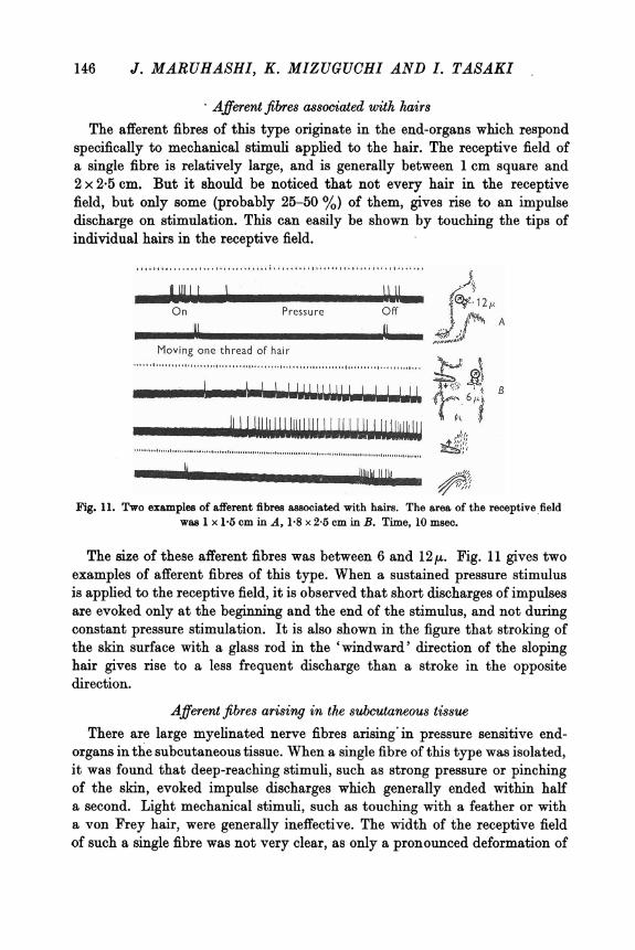
Afferent fibres associated with hairs The afferent fibres of this type originate in the end-organs which respond
specifically to mechanical stimuli applied to the hair. The receptive field of a single fibre is relatively large, and is generally between 1 cm square and 2 x 2.5 cm. But it should be noticed that not every hair in the receptive field, but only some (probably 25–50 %) of them, gives rise to an impulse discharge on stimulation. This can easily be shown by touching the tips of individual hairs in the receptive field.
The size of these afferent fibres was between 6 and 12µ. Fig. 11 gives two examples of afferent fibres of this type. When a sustained pressure stimulus is applied to the receptive field, it is observed that short discharges of impulses are evoked only at the beginning and the end of the stimulus, and not during constant pressure stimulation. It is also shown in the figure that stroking of the skin surface with a glass rod in the 'windward' direction of the sloping hair gives rise to a less frequent discharge than a stroke in the opposite direction.
,,,,,........ . .......... ,,,,,,,,,j,,,,,,, •. , •.•...•••••••.•...• , .....•.
Moving one thread of hair ................ ,.............. ,............................................,............ . I l -- ..........
...........,..............................................................................................."''''"'''"'''""''"'"''
Fig. 11. Two examples of afferent fibres associated with hairs. The area of the receptive field was l x l .5 cm in A, l .8 x 2.5 cm in B. Time, 10 msec.
Afferent fibres arising in the subcutaneous tissue There are large myelinated nerve fibres arising· in pressure sensitive end-
organs in the subcutaneous tissue. When a single fibre of this type was isolated, it was found that deep-reaching stimuli, such as strong pressure or pinching of the skin, evoked impulse discharges which generally ended within half a second. Light mechanical stimuli, such as touching with a feather or with a von Frey hair, were generally ineffective. The width of the receptive field of such a single fibre was not very clear, as only a pronounced deformation of
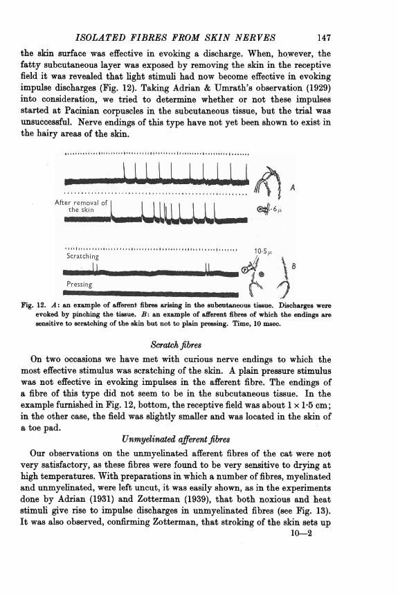
147 ISOLATED FIBRES FROM SKIN NERVES the skin surface was effective in evoking a discharge. When, ho:wever, the fatty subcutaneous layer was exposed by removing the skin in the receptive field it was revealed that light stimuli had now become effective in evoking impulse discharges (Fig. 12). Taking Adrian & Umrath's observation (1929) into consideration, we tried to determine whether or not these impulses started at Pacinian corpuscles in the subcutaneous tissue, but the trial was unsuccessful. Nerve endings of this type have not yet been shown to exist in the hairy areas of the skin.
.......................................................................,
.. .. After removal of L r.:.JJ
the sktn
Fig. 12. A: an example of afferent. fibres arising in the subcutaneous tissue. Discharges were evoked by pinching the tissue. B: an example of afferent fibres of which the endings are sensitive to scratching of the skin but not to plain pressing. Time, 10msec.
Scratchfibres On two occasions we have met with curious nerve endings to which the
most effective stimulus was scratching of the skin. A plain pressure stimulus was not effective in evoking impulses in the afferent fibre. The endings of a fibre of this type did not seem to be in the subcutaneous tissue. In the example furnished in Fig. 12, bottom, the receptive field was about 1x1.5cm; in the other case, the field was slightly smaller and was located in the skin of a toe pad.
Unmyelinat,ed afferent fibres Our observations on the unmyelinated afferent fibres of the cat were not
very satisfactory, as these fibres were found to be very sensitive to drying at high temperatures. With preparations in which a number of fibres, myelinated and unmyelinated, were left uncut, it was easily shown, as in the experiments done by Adrian (1931) and Zotterman (1939), that both noxious and heat stimuli give rise to impulse discharges in unmyelinated fibres (see Fig. 13). It was also observed, confirming Zotterman, that stroking of the skin sets up
10---2

148 J. MARUHASHI, K. MIZUGUCHI AND I. TASAKI after-discharges in the unmyelinated fibres. Action currents of these un-myelinated fibres could be easily distinguished from those of myelinated fibres by their long diphasic configuration (Adrian, Bronk & Phillips, 1932; Zotterman, 1939) on the face of the cathode-ray oscillograph, although some-times action currents of an intermediate configuration were seen.
•I I ·~I I• 11111IIII111110 I I I JI IJ 1111 I 11ItIUI11 O I I• I It I It 111II11I111 • 1 I It I I 11 •I I IJ ".'I 11 o
'~n Water at 50° C oi~
\ l1u1 I '-')IIAt rest
m i1: I Ice+ salt It® Water at 800
Fig. 13. Afferent impulses recorded from a group of small fibres including a number of unmyelinated fibres. Time, 10 msec.
On several occasions we examined the impulse discharge in afferent fibres from an area which had previously been scalded. It was observed that the spontaneous discharge was limited to small fibres, myelinated and un-myelinated, and that the after-discharge in these fibres following mechanical stimulation was remarkable in the scalded area.
DISCUSSION
In the light of the :findings stated above, it is easy to interpret the experi-mental results obtained previously by Adrian (1931), Zotterman (1939) and others. Undoubtedly a light touch on the hair gives rise to a series of impulses, in a multi-fibre preparation, in the wide-receptive and hair fibres stated above. In fact, Adrian recorded from cutaneous nerves of the cat and guinea-pig, action potentials of two different magnitudes, large and small. He further states that the afferent fibres carrying small action potentials have a much wider receptive field than those carrying large potentials. Then, a heavier touch arouses impulses further in the touch, nociceptive and unmyelinated afferent fibres described above. The action potentials called delta by Zotterman are considered to correspond to the smaller subdivision of the nociceptive fibres and also to the wide-receptive fibres in our classification.
As to the roles of these sensory units in our cutaneous sensations, it may be possible to make much speculation. But, in an attempt to correlate the functions of these afferent fibres with the sensation they may arouse in the

149 ISOLATED FIBRES FROM SKIN NERVES human subject, it should always be taken into consideration that, under normal conditions, a mechanical or noxious stimulus applied to the skin sets up impulses in more than one type of afferent fibre. It seems therefore natural to believe that our 'pain' or 'pressure' sensation is aroused by the concurrent activity of several different kinds of sensory units.
It is very important to determine the neurohistological structures corre-sponding to the endings described above. The observations made by Woollard, Weddell & Harpman (1940), Weddell (1941) and others are very important in this respect. Simultaneous use of neurohistological and electrophysiological techniques thus seems desirable.
SUMMARY
1. Impulse discharges in individual cutaneous afferent nerve fibres have been studied by isolating single fibres from various skin nerves of the toad and the cat.
2. Toad's skin afferent fibres were divided, according to the physiological property of the sensory unit, into (a) tactile fibres, (b) pressure fibres, (c) large and small nociceptive fibres and (d) unmyelinated afferent fibres.
3. (a) A tactile fibre of the toad is 8-15µ in diameter, its receptive field consists of three to ten spots and its endings adapt quickly to a constant stimulus. (b) A pressure fibre is 4–5µ.in diameter and innervates one to five spots which adapt slowly to a constant pressure. (c) Nociceptive fibres vary between 3 and 9µ,the endings being densely distributed in an area ranging from 5 to 30 mm2. (d) Unmyelinated fibres subserve perception of heat, cold and mechanical stimuli.
4. It was inferred from an experiment that sensory nerve endings transform sensory stimuli into electric currents which stimulate the starting-point of the afferent nerve fibre.
5. Cat's skin afferent fibres were classified into (a) touch, (b) pressure, (c) large and small nociceptive, (d) cold, (e) wide-receptive, {f) hair, (g) sub-cutaneous, (h) scratch, and (i) unmyelinated afferent fibres.
6. (a) A touch fibre of the cat is 8–14µ in diameter and innervates generally one, sometimes two, touch spots which adapt quickly. (b) A pressure fibre is 3-5µ in diameter, its receptive field is spot-like and adaptation is slow. (c) Nociceptive fibres vary between 3 and 11µ,the receptive fields range from 2 to 9 (sometimes 50) mm2, and the impulse discharge is generally phasic. (d) Cold fibres are 1.5–3µ in diameter, their receptive field is punctiform, and their endings are insensitive to mechanical stimuli. (e) A wide-receptive fibre is 2-5 µ,and it has a receptive field ranging from 1,500to 4,000mm2 which is sensitive to all kinds of mechanical stimuli. {f) An afferent fibre associated with hair is 6–12µ.in diameter; it innervates a large number of hairs over an area ranging from 100 to 500 mm2. (g) There are afferent fibres which arise

150 J. MA.RUH A.SHI, K. MIZUGUCHI A.ND I. TA.SA.Kl in the subcutaneous tiBBue of the cat. (h) There are fibres connected with endings which respond to scratching of the skin but not to pressing. (i) Un-myelinated fibres subserve perception of heat and mechanical stimuli.
7. It is highly probable that wide-receptive fibres reach the spinal cord through the ventral roots. ACKNOWLEDGEMENTS
We wish to express our thanks to Dr Kyoichi Oshima for the assistance given to us in the experiments on the cat and to Mrs Noburo Kamiya. for help in preparing the manuscript for publication.
The expenses of this research have been defrayed in part by a grant to I. Tasaki from the Ministry of Education.
REFERENCES
ADRIAN, E. D. (1928). The Basis o/ Sensation. London: Christophers. ADRIAN, E. �. (1931). The messages in sensory nerve fibres and their interpretation. Proc. Roy.
Soc. B, 109, 1-18. ADRIAN, E. D. (1932). The Mechanism of Nervousa Action. Philadelphia: University of Pennsyl·
vania Press. ADRIAN, E. D., BBONK, D. W. & PHILLIPS, G. (1932). Discharges in mammalian sympathetic
nerves. J. Physiol. 74, 115-133. ADRIAN, E. D., CATTELL, McK. & HOAGLAND, H. (1931). Sensory discharges in single cutaneous
nerve fibres. J. Physiol. 72, 377-391. ADRIAN, E. D. & UMRATH, K. (1929). The impulse discharge from the Pacinian corpuscle.
J. Physiol. 88, 139–154. ADRIAN, E. D. & ZOTTERMAN, Y. (1926). The impulses produced by sensory nerve endings.
Part 3. Impulses set up by touch and pressure. J. Physiol. 61, 466–483. CATTELL, McK. & HOAGLAND, H. (1931). Response of tactile receptorss to intermittent stimula-
tion. J. Physiol. 72, 392–404. ECCLES, J. C. & SHERRINGTON, C. S. (1930). Numbers and contraction-values of individual
motor-units examined in some muscles of the limb. Proc. Roy. Soc. B, 106, 326-357. EBLA.NGER, J. & GASSER, H. S. (1930). The action potential in fibres of slow conduction in spinaJ
roots and soma.tic nerves. Amer. J. Physiol. 92, 43–82. FOERSTER, O. & GAGEL, O. (1933). Ueber afferente Nervenfasern in den vorderen Wurzeln.
Z. ges. Neurol. Psychiat. 144, 313-324. HOGG, B. M. (1935). Slow impulses from the cutaneous nerves of the frog. J. Physiol. 84, 250–258. LEHMANN, W. (1924). Ueber die sensiblen Fasern der vorderen Wurzeln. Klin. Wschr. 8,
1895-1898. LEWIS, T. (1937). The nocifensor system of nerves and its reactions. Brit. med. J. i, 431–435;
491–494. LILLIE, R. S. (1929). Resemblances between the electromotor variations of rhythmically reacting
living and non-living systems. J. gene Physiol. 18, 1-11. TAKAGI, J . & Ro, U. (1944). The anatomy of the ventral spinal root (in Japanese). Igaku to
Seibutsu-gaku, 5, 35. TA.SAKI, I. (1939). The strength-duration relation of the normal, polarized and narcotized nerve
fibre. Amer. J. Physiol. 125, 367-379. TASAKI, I. (1949). Collision of two nerve impulses in the nerve fibre. Biochim. biophys. acta,
8, 494–497. TASAKI, I. & MIZUGHUCHI, K. (1948). Response of single Ranvier nodes to electrical stimuli.
J. Neurophysiol. 11, 295–303. TASAKI, I. & TAKEUCHI, T. (1941). Der am Ranvierschen Knoten entstehende Aktionsstrom und
seine Bedeutung fiir die Erregungsleitung. Pflug. Arch. ges. Physiol. 244, 696-711. TOWER, S. S. (1940). Unit for sensory reception in cornea, with notes on nerve impulses from
s.lera, iris. and lens. J. Neurophysiol. 8, 486–500. WARTENBERG, R. (1928). Klinische Studien zur Frage der Geltung des Bell-Ma.gendieschen
Gesetzes. Z. ges. Neurol. Psychiat. 118, 518–603.

151 ISOLATED FIBRES FROM SKIN NERVES WEDDELL, G. (1941). The pattern of cutaneous innervation in rela.tion to cutaneous sensibility.
J. Anat., Land., 75, 346-367. WOOLLARD, H. H., WEDDELL, G. & HARPMAN, J. A. (1940). Observations on the neurohisto-
logical basis of cutaneous pain. J. Anat., Lond., 74, ZOTTERMAN, Y. (1936). Specific action potentials in the lingual nerve of cat. Skand. Arch.
Phy.iol. 75, 105–119; ZOTTERMAN, Y. (1939). Touch, pain and tickling: an electrophysiological investigation on
cutaneous sensory nerves. J. Phy.iol. 95, 1-28.