Embed Size (px)
Citation preview
164 J Can Chiropr Assoc 2010; 54(3)
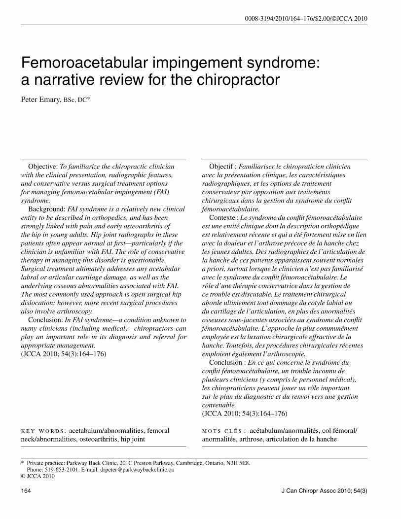
Femoroacetabular impingement syndrome: a narrative review for the chiropractorPeter Emary, BSc, DC*
* Private practice: Parkway Back Clinic, 201C Preston Parkway, Cambridge, Ontario, N3H 5E8. Phone: 519-653-2101. E-mail: [email protected]© JCCA 2010
Objectif : Familiariser le chiropraticien clinicien avec la présentation clinique, les caractéristiques radiographiques, et les options de traitement conservateur par opposition aux traitements chirurgicaux dans la gestion du syndrome du confl it fémoroacétabulaire. Contexte : Le syndrome du confl it fémoroacétabulaire est une entité clinique dont la description orthopédique est relativement récente et qui a été fortement mise en lien avec la douleur et l’arthrose précoce de la hanche chez les jeunes adultes. Des radiographies de l’articulation de la hanche de ces patients apparaissent souvent normales a priori, surtout lorsque le clinicien n’est pas familiarisé avec le syndrome du confl it fémoroacétabulaire. Le rôle d’une thérapie conservatrice dans la gestion de ce trouble est discutable. Le traitement chirurgical aborde ultimement tout dommage du cotyle labial ou du cartilage de l’articulation, en plus des anormalités osseuses sous-jacentes associées au syndrome du confl it fémoroacétabulaire. L’approche la plus communément employée est la luxation chirurgicale effractive de la hanche. Toutefois, des procédures chirurgicales récentes emploient également l’arthroscopie. Conclusion : En ce qui concerne le syndrome du confl it fémoroacétabulaire, un trouble inconnu de plusieurs cliniciens (y compris le personnel médical), les chiropraticiens peuvent jouer un rôle important sur le plan du diagnostic et du renvoi vers une gestion convenable.(JCCA 2010; 54(3):164–176)
m o t s c l é s : acétabulum/anormalités, col fémoral/anormalités, arthrose, articulation de la hanche
Objective: To familiarize the chiropractic clinician with the clinical presentation, radiographic features, and conservative versus surgical treatment options for managing femoroacetabular impingement (FAI) syndrome. Background: FAI syndrome is a relatively new clinical entity to be described in orthopedics, and has been strongly linked with pain and early osteoarthritis of the hip in young adults. Hip joint radiographs in these patients often appear normal at fi rst—particularly if the clinician is unfamiliar with FAI. The role of conservative therapy in managing this disorder is questionable. Surgical treatment ultimately addresses any acetabular labral or articular cartilage damage, as well as the underlying osseous abnormalities associated with FAI. The most commonly used approach is open surgical hip dislocation; however, more recent surgical procedures also involve arthroscopy.
Conclusion: In FAI syndrome—a condition unknown to many clinicians (including medical)—chiropractors can play an important role in its diagnosis and referral for appropriate management.(JCCA 2010; 54(3):164–176)
k e y w o r d s : acetabulum/abnormalities, femoral neck/abnormalities, osteoarthritis, hip joint
0008-3194/2010/164–176/$2.00/©JCCA 2010
J Can Chiropr Assoc 2010; 54(3) 165
P Emary
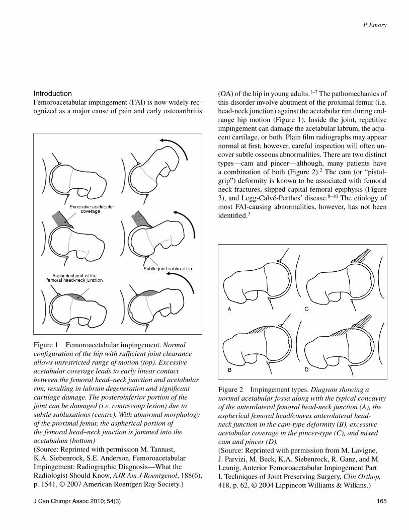
IntroductionFemoroacetabular impingement (FAI) is now widely rec-ognized as a major cause of pain and early osteoarthritis
(OA) of the hip in young adults.1–7 The pathomechanics of this disorder involve abutment of the proximal femur (i.e. head-neck junction) against the acetabular rim during end-range hip motion (Figure 1). Inside the joint, repetitive impingement can damage the acetabular labrum, the adja-cent cartilage, or both. Plain fi lm radiographs may appear normal at fi rst; however, careful inspection will often un-cover subtle osseous abnormalities. There are two distinct types—cam and pincer—although, many patients have a combination of both (Figure 2).2 The cam (or “pistol-grip”) deformity is known to be associated with femoral neck fractures, slipped capital femoral epiphysis (Figure 3), and Legg-Calvé-Perthes’ disease.8–10 The etiology of most FAI-causing abnormalities, however, has not been identifi ed.3
Figure 1 Femoroacetabular impingement. Normal confi guration of the hip with suffi cient joint clearance allows unrestricted range of motion (top). Excessive acetabular coverage leads to early linear contact between the femoral head–neck junction and acetabular rim, resulting in labrum degeneration and signifi cant cartilage damage. The posteroinferior portion of the joint can be damaged (i.e. contrecoup lesion) due to subtle subluxations (centre). With abnormal morphology of the proximal femur, the aspherical portion of the femoral head–neck junction is jammed into the acetabulum (bottom)(Source: Reprinted with permission M. Tannast, K.A. Siebenrock, S.E. Anderson, Femoroacetabular Impingement: Radiographic Diagnosis—What the Radiologist Should Know, AJR Am J Roentgenol, 188(6), p. 1541, © 2007 American Roentgen Ray Society.)
Figure 2 Impingement types. Diagram showing a normal acetabular fossa along with the typical concavity of the anterolateral femoral head-neck junction (A), the aspherical femoral head/convex anterolateral head-neck junction in the cam-type deformity (B), excessive acetabular coverage in the pincer-type (C), and mixed cam and pincer (D).(Source: Reprinted with permission from M. Lavigne, J. Parvizi, M. Beck, K.A. Siebenrock, R. Ganz, and M. Leunig, Anterior Femoroacetabular Impingement Part I. Techniques of Joint Preserving Surgery, Clin Orthop, 418, p. 62, © 2004 Lippincott Williams & Wilkins.)

166 J Can Chiropr Assoc 2010; 54(3)
Femoroacetabular impingement syndrome: a narrative review for the chiropractor
Chiropractors frequently see patients who present with hip pain that may be associated with FAI. Presented here is a review of its typical clinical and radiographic features, conservative and surgical treatment options, as well as a discussion on the role chiropractors can play in managing this disorder.
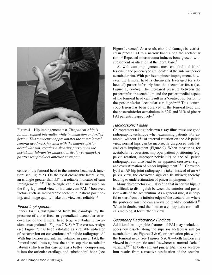
Clinical PresentationFAI syndrome presents most often in athletes of sports requiring forceful and repetitive hip fl exion, internal rota-tion, and adduction (e.g. ice hockey, soccer, martial arts, ballet).5–7 The cam-type is most common in young men between the ages of 20–30; whereas, the pincer-type is more common in middle-aged women.4 Initially, FAI symptoms are insidious and include intermittent groin pain, lateral trochanteric pain, or both.1,6 As the acetabular labrum and articular cartilage degenerate, pain frequency increases. The chief complaint is a dull ache in the anter-ior groin, especially after prolonged sitting. Occasionally, a sharp or catching pain is felt during activity, indicating a tear of the acetabular labrum.11 Examination may reveal the Trendelenburg sign (i.e. abductor weakness with full weight-bearing of the hip). Passive hip joint range of mo-tion (ROM) is limited, and often painful, in fl exion and in-ternal rotation.5 The hip impingement test elicits anterior groin pain in most patients (Figure 4).
Radiographic FeaturesRadiographic examination of FAI includes an anteropos-
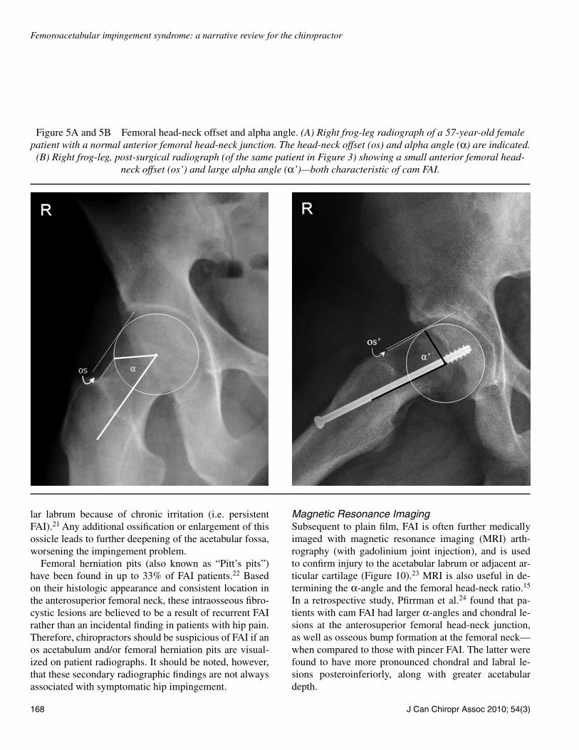
terior (AP) pelvic view, and either a frog-leg (i.e. femur externally rotated) or axial cross-table lateral view (i.e. patient supine with the hip internally rotated 15°, and the central x-ray beam is horizontally angled 45° to the supe-rior—from across the table—towards the inguinal fold).12 The AP pelvic radiograph (with the hips internally rotated 15°) provides better visualization of the contour of the lat-eral femoral head-neck junction; whereas, the frog-leg13 or axial cross-table8 lateral view allows for assessment of the anterior femoral head-neck offset (i.e. distance be-tween the widest diameter of the femoral head and most prominent part of the anterior femoral neck; Figure 5).
Cam ImpingementCam FAI is characterized on radiographs by an aspheric femoral head with morphologic rounding (i.e. lack of con-cavity) of the anterolateral head-neck junction, creating a decreased femoral head-neck offset (Figure 6; see also Figure 5b).14 Because of this abnormal morphology, hip fl exion and internal rotation force the aspheric femoral head/convex head-neck junction into the anterosuperior acetabulum (see Figure 1, bottom), inducing compression to the cartilage and shear stress between it and the labrum. As a result, the majority of chondral and labral lesions in cam impingement are located anterosuperiorly.1,2
Cam-type abnormalities can be further quantifi ed on radiographs with measurement of the alpha (α) angle (i.e. angle formed between a line drawn along the axis of the femoral neck, and a second line drawn connecting the
Figure 3A and 3B Slipped capital femoral epiphysis (SCFE) and the “pistol-grip” deformity. AP pelvic radiographs of a 16-year-old male patient showing a right-sided SCFE (A), and a cam-type (or pistol-grip) deformity (arrow) of the
same hip, along with an os acetabulum (arrowhead), after two year follow-up from in situ surgical pinning (B).
J Can Chiropr Assoc 2010; 54(3) 167
P Emary
centre of the femoral head to the anterior head-neck junc-tion; see Figure 5). On the axial cross-table lateral view, an α-angle greater than 55º is a reliable indicator of cam impingement.12,15 The α-angle can also be measured on the frog-leg lateral view to indicate cam FAI;13 however, factors such as radiographic technique, patient position-ing, and image quality make this view less reliable.16
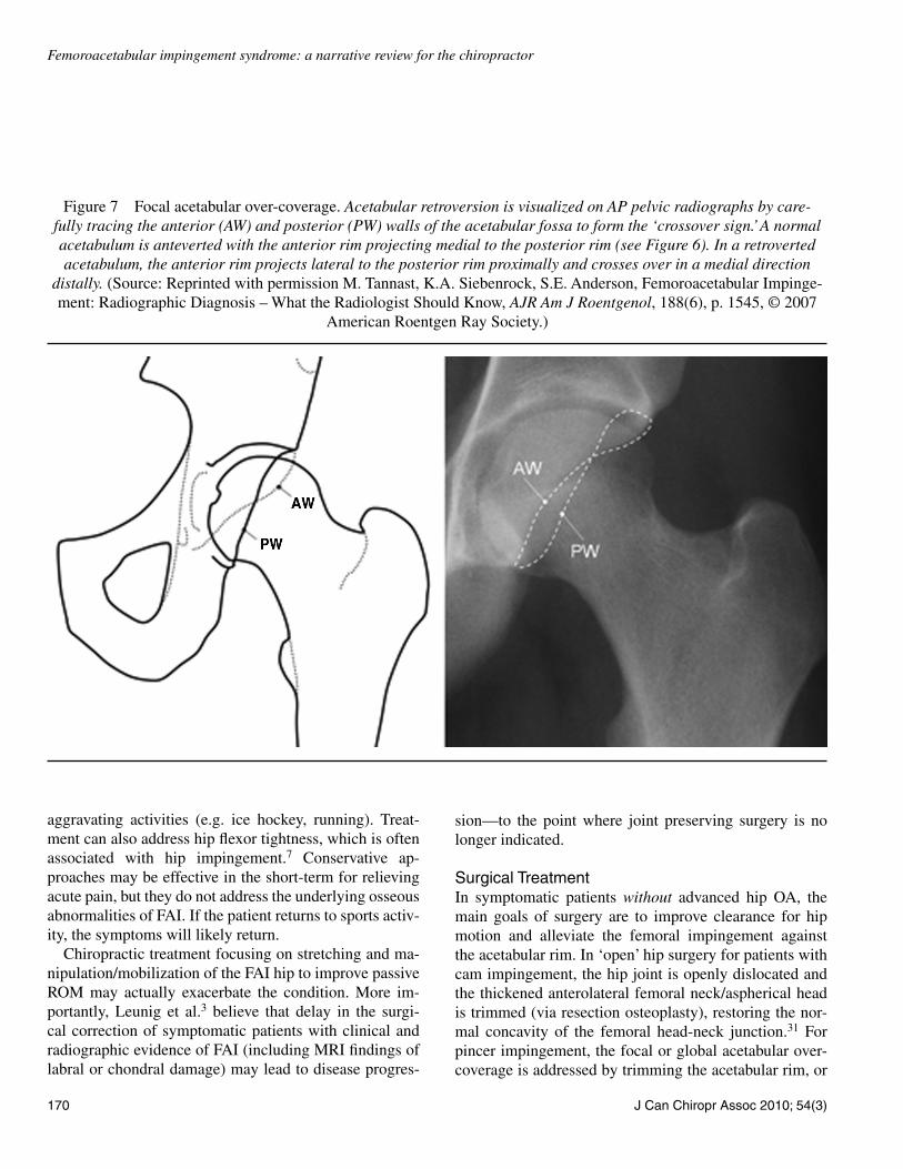
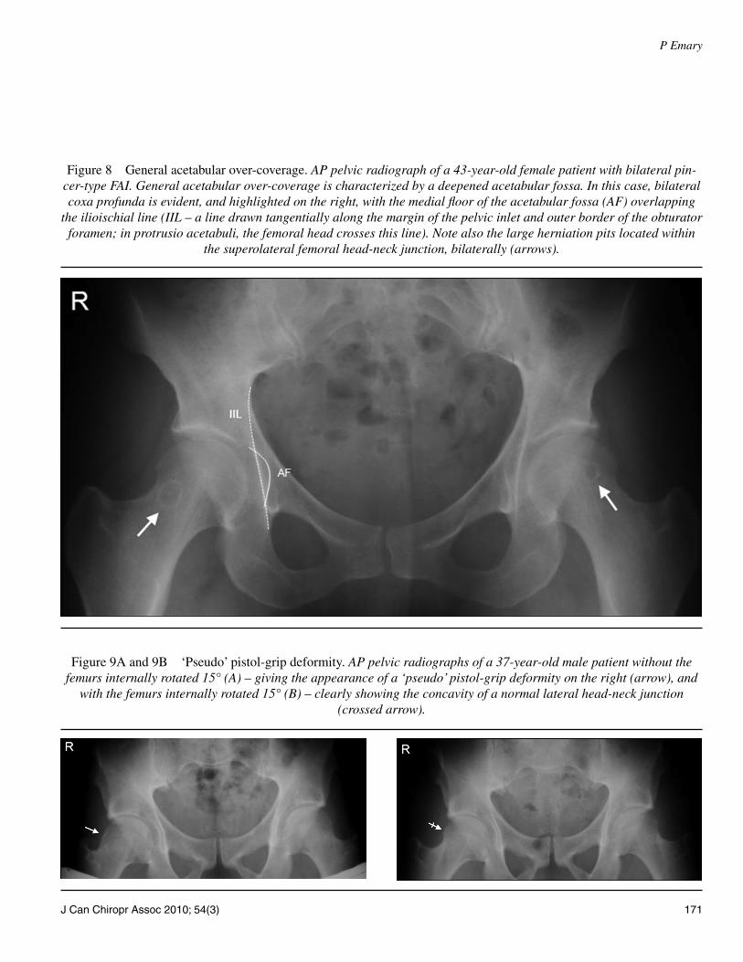
Pincer ImpingementPincer FAI is distinguished from the cam-type by the presence of either focal or generalized acetabular over-coverage of the femoral head (e.g. acetabular retrover-sion, coxa profunda; Figures 7 & 8).12 The crossover sign (see Figure 7) has been validated as a reliable indicator of retroversion on conventional AP pelvic radiographs.17 With hip fl exion and internal rotation in pincer FAI, the femoral neck abuts against the anterosuperior acetabular labrum (which in this case acts as a buffer), compressing it into the articular cartilage and subchondral bone (see
Figure 1, centre). As a result, chondral damage is restrict-ed in pincer FAI to a narrow band along the acetabular rim.1,2 Repeated microtrauma induces bone growth with subsequent ossifi cation at the labral base.2
As with cam impingement, most chondral and labral lesions in the pincer-type are located at the anterosuperior acetabular rim. With persistent pincer impingement, how-ever, the femoral head is chronically leveraged (or sub-luxated) posteroinferiorly into the acetabular fossa (see Figure 1, centre). The increased pressure between the posteroinferior acetabulum and the posteromedial aspect of the femoral head can result in a ‘contrecoup’ lesion to the posterinferior acetabular cartilage.1,2,12 This contre-coup lesion has been observed in the femoral head and the posteroinferior acetabulum in 62% and 31% of pincer FAI patients, respectively.2
Radiographic PitfallsChiropractors taking their own x-ray fi lms must use good radiographic technique when examining patients. For ex-ample, without 15° of internal rotation on the AP pelvic view, normal hips can be incorrectly diagnosed with lat-eral cam impingement (Figure 9). When measuring for acetabular retroversion, improper patient positioning (e.g. pelvic rotation, improper pelvic tilt) on the AP pelvic radiograph can also lead to an apparent crossover sign, and overestimation of pincer impingement.12,18 Converse-ly, if an AP hip joint radiograph is taken instead of an AP pelvis view, the crossover sign can be missed; thereby, leading to underestimation of pincer impingement.12
Many chiropractors will also fi nd that in certain hips, it is diffi cult to distinguish between the anterior and poste-rior walls of the acetabulum. As a general rule, it is help-ful to start from the inferior edge of the acetabulum where the posterior rim line can always be readily identifi ed.12 When in doubt, send the fi lms to a chiropractic (or medi-cal) radiologist for further review.
Secondary Radiographic FindingsAdditional radiographic features of FAI may include an accessory ossicle along the superior acetabular rim (os acetabulum; see Figures 3 & 6), or herniation pits within the femoral neck (see Figures 6 & 8)—both historically viewed in chiropractic (and elsewhere) as normal skeletal variants.19,20 In both cam and pincer FAI, the os acetabu-lum results from a reactive ossifi cation of the acetabu-
Figure 4 Hip impingement test. The patient’s hip is forcibly rotated internally, while in adduction and 90º of fl exion. This manoeuvre approximates the anterolateral femoral head-neck junction with the anterosuperior acetabular rim, creating a shearing pressure on the acetabular labrum (or adjacent articular cartilage). A positive test produces anterior groin pain.
168 J Can Chiropr Assoc 2010; 54(3)
Femoroacetabular impingement syndrome: a narrative review for the chiropractor
lar labrum because of chronic irritation (i.e. persistent FAI).21 Any additional ossifi cation or enlargement of this ossicle leads to further deepening of the acetabular fossa, worsening the impingement problem.
Femoral herniation pits (also known as “Pitt’s pits”) have been found in up to 33% of FAI patients.22 Based on their histologic appearance and consistent location in the anterosuperior femoral neck, these intraosseous fi bro-cystic lesions are believed to be a result of recurrent FAI rather than an incidental fi nding in patients with hip pain. Therefore, chiropractors should be suspicious of FAI if an os acetabulum and/or femoral herniation pits are visual-ized on patient radiographs. It should be noted, however, that these secondary radiographic fi ndings are not always associated with symptomatic hip impingement.
Magnetic Resonance ImagingSubsequent to plain fi lm, FAI is often further medically imaged with magnetic resonance imaging (MRI) arth-rography (with gadolinium joint injection), and is used to confi rm injury to the acetabular labrum or adjacent ar-ticular cartilage (Figure 10).23 MRI is also useful in de-termining the α-angle and the femoral head-neck ratio.15 In a retrospective study, Pfi rrman et al.24 found that pa-tients with cam FAI had larger α-angles and chondral le-sions at the anterosuperior femoral head-neck junction, as well as osseous bump formation at the femoral neck—when compared to those with pincer FAI. The latter were found to have more pronounced chondral and labral le-sions posteroinferiorly, along with greater acetabular depth.
Figure 5A and 5B Femoral head-neck offset and alpha angle. (A) Right frog-leg radiograph of a 57-year-old female patient with a normal anterior femoral head-neck junction. The head-neck offset (os) and alpha angle (α) are indicated.
(B) Right frog-leg, post-surgical radiograph (of the same patient in Figure 3) showing a small anterior femoral head-neck offset (os’) and large alpha angle (α’)—both characteristic of cam FAI.

J Can Chiropr Assoc 2010; 54(3) 169
P Emary
In comparison to plain fi lm, Dudda et al.25 found MR arthrography (with radial slices in the axis of the femoral head-neck junction) to be more sensitive in the assessment of the α-angle and femoral head asphericity; standard AP pelvis and lateral cross-table radiographic views resulted in underestimation of these parameters in 34.6% of pa-tients. Clohisy et al.26 also found limited reliability with many of the standard plain fi lm radiographic parameters used in diagnosing FAI; however, in order to better simu-late the clinical setting (of radiographic interpretation), x-ray parameters in this study were subjectively assessed rather than exact measurements made. For MRI, Nouh et al.27 also showed that subjective evaluation of the α-angle in cam FAI is inaccurate, and clinical experience does not
seem to help radiologists “eyeball” this angle any better. Conversely, other studies using objective rather than sub-jective impingement markers have shown good reliability for both plain fi lm13,28,29 and MRI.30 To the chiropractor, the diagnosis of FAI syndrome should not rely on radio-graphic fi ndings alone; of equal (if not greater) importance are the patient history and physical examination.
Conservative TreatmentAs with any musculoskeletal condition, FAI warrants an initial trial of conservative therapy including rest, activity modifi cation, NSAIDs, physiotherapy (or chiropractic), and if needed, corticosteroid injections.7,31 Initial treat-ment must include temporarily limiting or stopping the
Figure 6 Cam FAI. AP pelvis radiograph of a 47-year-old male patient with bilateral cam-type FAI (arrows). Note the herniation pit within the superolateral femoral neck on the right (crossed arrow), and the small ossicle (os
acetabulum) along the superior acetabular rim on the left (arrowhead). A normal anteverted acetabulum is highlighted on the right, where the anterior wall (AW) projects medial to the posterior wall (PW).
170 J Can Chiropr Assoc 2010; 54(3)
Femoroacetabular impingement syndrome: a narrative review for the chiropractor
aggravating activities (e.g. ice hockey, running). Treat-ment can also address hip fl exor tightness, which is often associated with hip impingement.7 Conservative ap-proaches may be effective in the short-term for relieving acute pain, but they do not address the underlying osseous abnormalities of FAI. If the patient returns to sports activ-ity, the symptoms will likely return.
Chiropractic treatment focusing on stretching and ma-nipulation/mobilization of the FAI hip to improve passive ROM may actually exacerbate the condition. More im-portantly, Leunig et al.3 believe that delay in the surgi-cal correction of symptomatic patients with clinical and radiographic evidence of FAI (including MRI fi ndings of labral or chondral damage) may lead to disease progres-
sion—to the point where joint preserving surgery is no longer indicated.
Surgical TreatmentIn symptomatic patients without advanced hip OA, the main goals of surgery are to improve clearance for hip motion and alleviate the femoral impingement against the acetabular rim. In ‘open’ hip surgery for patients with cam impingement, the hip joint is openly dislocated and the thickened anterolateral femoral neck/aspherical head is trimmed (via resection osteoplasty), restoring the nor-mal concavity of the femoral head-neck junction.31 For pincer impingement, the focal or global acetabular over-coverage is addressed by trimming the acetabular rim, or
Figure 7 Focal acetabular over-coverage. Acetabular retroversion is visualized on AP pelvic radiographs by care-fully tracing the anterior (AW) and posterior (PW) walls of the acetabular fossa to form the ‘crossover sign.’ A normal acetabulum is anteverted with the anterior rim projecting medial to the posterior rim (see Figure 6). In a retroverted acetabulum, the anterior rim projects lateral to the posterior rim proximally and crosses over in a medial direction
distally. (Source: Reprinted with permission M. Tannast, K.A. Siebenrock, S.E. Anderson, Femoroacetabular Impinge-ment: Radiographic Diagnosis – What the Radiologist Should Know, AJR Am J Roentgenol, 188(6), p. 1545, © 2007
American Roentgen Ray Society.)
J Can Chiropr Assoc 2010; 54(3) 171
P Emary
Figure 8 General acetabular over-coverage. AP pelvic radiograph of a 43-year-old female patient with bilateral pin-cer-type FAI. General acetabular over-coverage is characterized by a deepened acetabular fossa. In this case, bilateral coxa profunda is evident, and highlighted on the right, with the medial fl oor of the acetabular fossa (AF) overlapping
the ilioischial line (IIL – a line drawn tangentially along the margin of the pelvic inlet and outer border of the obturator foramen; in protrusio acetabuli, the femoral head crosses this line). Note also the large herniation pits located within
the superolateral femoral head-neck junction, bilaterally (arrows).
Figure 9A and 9B ‘Pseudo’ pistol-grip deformity. AP pelvic radiographs of a 37-year-old male patient without the femurs internally rotated 15° (A) – giving the appearance of a ‘pseudo’ pistol-grip deformity on the right (arrow), and
with the femurs internally rotated 15° (B) – clearly showing the concavity of a normal lateral head-neck junction (crossed arrow).

172 J Can Chiropr Assoc 2010; 54(3)
Femoroacetabular impingement syndrome: a narrative review for the chiropractor
by reorientation of a retroverted acetabulum (via periacet-abular osteotomy).31,32 Less invasive surgical approaches utilizing arthroscopy are also evolving.33 This is an at-tractive alternative to patients, particularly professional athletes, because arthroscopy involves smaller incisions, a shorter recovery time, and a lower morbidity rate.34 Sur-gery must, however, address both the labral or cartilage lesions along with the underlying osseous abnormalities causing the FAI; otherwise, the impingement problem may continue, leading to persistent pain and possible pro-gressive hip joint degeneration (Figure 11).
Early and mid-term results from the open surgical hip procedures are promising, with good to excellent clini-cal outcomes in approximately 70–80% of patients.35–37
In patients with advanced OA of the hip joint, or in those where joint preserving surgery fails, total hip replacement (i.e. arthroplasty) is the treatment of choice. Post surgical rehab of FAI typically includes the use of a continuous passive motion device (with hip fl exion limited to 70º) for the fi rst 2–3 weeks, in order to facilitate early ROM and prevention of capsular adhesions.31 Toe-touch weight-bearing while in crutches (for approximately eight weeks) is also prescribed. Albeit small, hip surgery carries risks including infection, thromboembolism, heterotopic ossi-fi cation, and neurovascular damage.5
DiscussionDespite the dramatic increase in recent literature on FAI
Figure 10A and 10B MR arthrography in FAI. (A) Oblique sagittal fat-saturated T1-weighted MR arthrographic image (600/8) of the hip in a patient with cam FAI. An abnormal anterior femoral head-neck offset (short arrow) and
anterosuperior labral tear (long arrow) are shown. (Source: Reprinted with permission A. Kassarjian, L.S. Yoon, E. Belzile, S.A. Connolly, M.B. Millis, W.E. Palmer, Triad of MR Arthrographic Findings in Patients with Cam-
Type Femoroacetabular Impingement, Radiology, 236(2), p. 592, © 2005 RSNA.) (B) Coronal spin-echo sequence T1-weighted MR image (524/14) showing ossifi cation of the acetabular labrum in a patient with pincer FAI. Bone marrow signal (arrowheads) extends into the substance of the acetabular labrum (arrow). (Source: Reprinted with
permission C.W.A. Pfi rrman, B. Mengiardi, C. Dora, F. Kalberer, M. Zanetti, J.Hodler, Femoroacetabular Impingement: Characteristic MR Arthrographic Findings in 50 Patients, Radiology, 240(3), p. 784, © 2006 RSNA.)

J Can Chiropr Assoc 2010; 54(3) 173
P Emary
Figure 11A and 11B End-stage hip degeneration in FAI. AP pelvis (A) and frog-leg right hip (B) radiographs of an 80-year-old female patient with bilateral pincer FAI, and severe right hip joint OA. Note the posteroinferior (i.e. contrecoup) joint space narrowing (arrow) and multiple osteophytes (arrowheads) on the right; coxa profunda is
also evident. Protrusio acetabuli is evident and highlighted on the left, with the femoral head (FH) overlapping the ilioischial line (IIL). Note also the linear indentation (small arrow) and reactive cortical thickening (small crossed
arrow) on the superolateral head-neck junction of the left femur.

174 J Can Chiropr Assoc 2010; 54(3)
Femoroacetabular impingement syndrome: a narrative review for the chiropractor
and its association with hip pain and OA, very little in-formation is available regarding its natural history.3 This presents a challenge to chiropractors when offering clin-
ical recommendations to patients. For instance, will every patient with radiographic evidence of FAI—regardless of symptoms—progress to end-stage hip OA, and should therefore, be referred for joint preserving surgery? In a recent study, Bardakos and Villar38 examined AP pelvic radiographs of 43 patients with a pistol-grip deformity of the femur, and mild or moderate OA of the hip. After ten-year follow-up, progression of OA was observed in 28 of the 43 patients. In other words, hip OA in one-third of these patients did not progress. Of those who did, the most important risk factors found were a lower medial proximal femoral angle (MPFA), an indication of coxa vara (Figure 12), and the presence of the ‘posterior wall’ sign (i.e. posterior acetabular wall projects medial to the femoral head centre—indicating retroversion). The auth-ors of this study suggest that a reduced MPFA in cam fe-murs may lead to hip abductor dysfunction, and that this biomechanical imbalance—especially when combined with pincer impingement—may contribute to OA pro-gression in FAI patients.
In the current literature, surgical treatment of FAI has been shown to be most successful in the absence of ad-vanced degenerative OA.3 It is still unclear, however, whether ‘preventative’ surgery should be performed in asymptomatic patients, despite radiographic evidence of FAI.4 Absence of symptoms in these cases may be due to lack of inciting activities, or FAI being at an early stage in its development. In symptomatic cases, a failure to re-solve with conservative treatment warrants referral for MRI arthrography, and in the presence of labral or chon-dral damage, orthopedic surgical consultation. Chiroprac-tors (or medical doctors) may, however, have diffi culty fi nding a surgeon with experience in treating FAI, because it is still a relatively new entity within orthopedics.6 Thus, practitioners in these situations will need to familiarize themselves with orthopedic surgeons specializing in hip joint preservation procedures, in order to make an appro-priate patient referral.
ConclusionFAI syndrome typically presents in young adults with in-sidious onset groin pain, often in association with sports activity. The hip impingement test is positive in most of these patients. Hip joint radiographs may appear normal at fi rst—particularly if the clinician is unfamiliar with FAI. Plain fi lms showing a cam (or pistol-grip) deform-
Figure 12 Medial proximal femoral angle (MPFA). AP radiograph of the left hip joint (in a 36-year-old male patient) showing the modifi ed MPFA. A line is drawn from the superior tip of the greater trochanter through to the centre of the femoral head. A second line is drawn (representing the anatomical axis of the proximal femur) from the midpoint of the most distally visible aspect of the femoral shaft, and up proximally through the piriformis fossa. The medial angle formed by these two lines is the MPFA (normal range = 80º to 89º). Note the ossifi cation of the lateral acetabular labrum (arrowhead), resulting in pincer FAI.

J Can Chiropr Assoc 2010; 54(3) 175
P Emary
ity—especially in femurs with a reduced MPFA—com-bined with pincer FAI may indicate increased risk of OA progression. Chiropractors should also be aware of the Trendelenburg sign when examining FAI patients, as hip abductor weakness/dysfunction, if present, may be an additional risk indicator for progressive hip degenera-tion. More research is necessary, however, to determine the precise etiology and natural history of FAI syndrome. Nevertheless, chiropractors can play an important role in identifying patients with possible FAI syndrome, and in facilitating the appropriate management of this disorder.
AcknowledgementThe author thanks Carolyn Simolo and the staff at the New York Chiropractic College Library for their assist-ance in retrieving reference articles for this paper.
References1 Ganz R, Parvizi J, Beck M, Leunig M, Nötzli H,
Siebenrock KA. Femoroacetabular impingement: a cause for osteoarthritis of the hip. Clin Orthop Relat Res. 2003; 417:112–120.
2 Beck M, Kalhor M, Leunig M, Ganz R. Hip morphology infl uences the pattern of damage to the acetabular cartilage: femoroacetabular impingement as a cause of early osteoarthritis of the hip. J Bone Joint Surg Br. 2005; 87(7):1012–1018.
3 Leunig M, Beaulé PE, Ganz R. The concept of femoroacetabular impingement: current status and future perspectives. Clin Orthop Relat Res. 2009; 467(3):616–622.
4 Ganz R, Leunig M, Leunig-Ganz K, Harris WH. The etiology of osteoarthritis of the hip: an integrated mechanical concept. Clin Orthop Relat Res. 2008; 466(2):264–272.
5 Zebala LP, Schoenecker PL, Clohisy JC. Anterior femoroacetabular impingement: a diverse disease with evolving treatment options. Iowa Orthop J. 2007; 27:71–81.
6 Dooley PJ. Femoroacetabular impingement syndrome: Nonarthritic hip pain in young adults. Can Fam Physician. 2008; 54(1):42–47.
7 Hart ES, Metkar US, Rebello GN, Grottkau BE. Femoroacetabular impingement in adolescents and young adults. Orthop Nurs. 2009; 28(3):117–124.
8 Eijer H, Myers SR, Ganz R. Anterior femoroacetabular impingement after femoral neck fractures. J Orthop Trauma. 2001; 15(7):475–481.
9 Goodman DA, Feighan JE, Smith AD, Latimer B, Buly RL, Cooperman DR. Subclinical slipped capital femoral epiphysis. Relationship to osteoarthrosis of the hip. J Bone Joint Surg Am. 1997; 79(10):1489–1497.
10 Snow SW, Keret D, Scarangella S, Bowen JR. Anterior impingement of the femoral head: a late phenomenon of Legg-Calvé-Perthes’ disease. J Pediatr Orthop. 1993; 13(3):286–289.
11 Burnett RS, Della Rocca GJ, Prather H, Curry M, Maloney WJ, Clohisy JC. Clinical presentation of patients with tears of the acetabular labrum. J Bone Joint Surg Am. 2006; 88(7):1448–1457.
12 Tannast M, Siebenrock KA, Anderson SE. Femoroacetabular impingement: radiographic diagnosis— what the radiologist should know. AJR Am J Roentgenol. 2007; 188(6):1540–1552.
13 Clohisy JC, Nunley RM, Otto RJ, Schoenecker PL. The frog-leg lateral radiograph accurately visualized hip cam impingement abnormalities. Clin Orthop Relat Res. 2007; 462:115–121.
14 Ito K, Minka MA 2nd, Leunig M, Werlen S, Ganz R. Femoroacetabular impingement and the cam-effect. A MRI-based quantitative anatomical study of the femoral head-neck offset. J Bone Joint Surg Br. 2001; 83(2):171–176.
15 Nötzli HP, Wyss TF, Stoecklin CH, Schmid MR, Treiber K, Hodler J. The contour of the femoral head-neck junction as a predictor for the risk of anterior impingement. J Bone Joint Surg Br. 2002; 84(4):556–560.
16 Konan S, Rayan F, Haddad FS. Is the frog lateral plain radiograph a reliable predictor of the alpha angle in femoroacetabular impingement? J Bone Joint Surg Br. 2010; 92-B(1):47–50.
17 Jamali AA, Mladenov K, Meyer DC, Martinez A, Beck M, Ganz R, Leunig M. Anteroposterior pelvic radiographs to assess acetabular retroversion: high validity of the “cross-over-sign”. J Orthop Res. 2007; 25(6):758–765.
18 Siebenrock KA, Kalbermatten DF, Ganz R. Effect of pelvic tilt on acetabular retroversion: a study of pelves from cadavers. Clin Orthop Relat Res. 2003; 407:241–248.
19 Yochum TR, Rowe LJ. Essentials of Skeletal Radiology. 3rd ed. Volume One. Congenital anomalies and normal skeletal variants. Baltimore: Williams & Wilkins, 2005:257–403.
20 Pitt MJ, Graham AR, Shipman JH, Birkby W. Herniation pit of the femoral neck. AJR Am J Roentgenol. 1982; 138(6):1115–1121.
21 Ito K, Leunig M, Ganz R. Histopathologic features of the acetabular labrum in femoroacetabular impingement. Clin Orthop Relat Res. 2004; 429:262–271.
22 Leunig M, Beck M, Kalhor M, Kim YJ, Werlen S, Ganz R. Fibrocystic changes at anterosuperior femoral neck: prevalence in hips with femoroacetabular impingement. Radiology. 2005; 236(1):237–246.
23 James SL, Ali K, Malara F, Young D, O’Donnell J, Connell DA. MRI fi ndings of femoroacetabular impingement. AJR Am J Roentgenol. 2006; 187(6):1412–1419.
24 Pfi rrman CWA, Mengiardi B, Dora C, Kalberer F,

176 J Can Chiropr Assoc 2010; 54(3)
Femoroacetabular impingement syndrome: a narrative review for the chiropractor
Zanetti M, Hodler J. Cam and pincer femoroacetabular impingement: characteristic MR arthrographic fi ndings in 50 patients. Radiology. 2006; 240(3):778–785.
25 Dudda M, Albers C, Mamisch TC, Werlen S, Beck M. Do normal radiographs exclude asphericity of the femoral head-neck junction? Clin Orthop Relat Res. 2009; 467(3):651–659.
26 Clohisy JC, Carlisle JC, Trousdale R, Kim Y-J, Beaulé PE, Morgan P, Steger-May K, Schoenecker PL, Millis M. Radiographic evaluation of the hip has limited reliability. Clin Orthop Relat Res. 2009; 467(3):666–675.
27 Nouh MR, Schweitzer ME, Rybak L, Cohen J. Femoroacetabular impingement: can the alpha angle be estimated? AJR Am J Roentgenol. 2008; 190(5):1260–1262.
28 Gosvig KK, Jacobsen S, Palm H, Sonne-Holm S, Magnusson E. A new radiological index for assessing asphericity of the femoral head in cam impingement. J Bone Joint Surg Br. 2007; 89(10):1309–1316.
29 Meyer DC, Beck M, Ellis T, Ganz R, Leunig M. Comparison of six radiographic projections to assess femoral head/neck asphericity. Clin Orthop Relat Res. 2006; 445:181–185.
30 Steppacher SD, Tannast M, Werlen S, Siebenrock KA. Femoral morphology differs between defi cient and excessive acetabular coverage. Clin Orthop Relat Res. 2008; 466(4):782–790.
31 Lavigne M, Parvizi J, Beck M, Siebenrock KA, Ganz R, Leunig M. Anterior femoroacetabular impingement: part I. Techniques of joint preserving surgery. Clin Orthop. 2004; 418:61–66.
32 Siebenrock KA, Schoeniger R, Ganz R. Anterior femoro-acetabular impingement due to acetabular retroversion. Treatment with periacetabular osteotomy. J Bone Joint Surg Am. 2003; 85-A(2):278–286.
33 Philippon MJ, Stubbs AJ, Schenker ML, Maxwell RB, Ganz R, Leunig M. Arthroscopic management of femoroacetabular impingement: osteoplasty technique and literature review. Am J Sports Med. 2007; 35(9):1571–1580.
34 Beaulé PE, Allen DJ, Clohisy JC, Schoenecker PL, Leunig M. The young adult with hip impingement: deciding on the optimal intervention. Instr Course Lect. 2009; 58:213–222.
35 Beck M, Leunig M, Parvizi J, Boutier V, Wyss D, Ganz R. Anterior femoroacetabular impingement: part II. Midterm results of surgical treatment. Clin Orthop. 2004; 418:67–73.
36 Murphy S, Tannast M, Kim YJ, Buly R, Millis MB. Debridement of the adult hip for femoroacetabular impingement: indications and preliminary clinical results. Clin Orthop Relat Res. 2004; 429:178–181.
37 Espinosa N, Rothenfl uh DA, Beck M, Ganz R, Leunig M. Treatment of femoro-acetabular impingement: preliminary results of labral refi xation. J Bone Joint Surg Am. 2006; 88(5):925–935.
38 Bardakos NV, Villar RN. Predictors of progression of osteoarthritis in femoroacetabular impingement: a radiological study with a minimum of ten years follow-up. J Bone Joint Surg Br. 2009; 91(2):162–169.


![Femoroacetabular%20 impingement[1]](https://img.dokumen.tips/doc/110x75/54559a24af7959d8748b6a78/femoroacetabular20-impingement1.jpg)