Embed Size (px)
Citation preview
FEMOROACETABULAR IMPINGEMENT
PATIENT INFORMATION
What is femoroacetabular impingement?
Femoroacetabular Impingement (FAI) is a condition in which there is abnormal contact
(impingement) between the rim of the acetabulum (hip joint socket) and femoral head-neck
junction (the bone just below the ball part of the thigh bone), on movement of the hip. The most
common movement that brings on pain is hip flexion (knee towards chest). Patients will
experience pain, usually in the groin, but sometimes further down the front of the thigh, side or
back of the hip. There may be episodes of clicking in the hip, or the sensation that it is coming out
of joint. Certain activities, particularly those which involve hip flexion (e.g football, dancing,
ballet, aerobics) will make the pain worse. Patients often find that sitting for a prolonged period
of time e.g long car journey, will bring on groin pain, and they often struggle to move into a more
comfortable position.
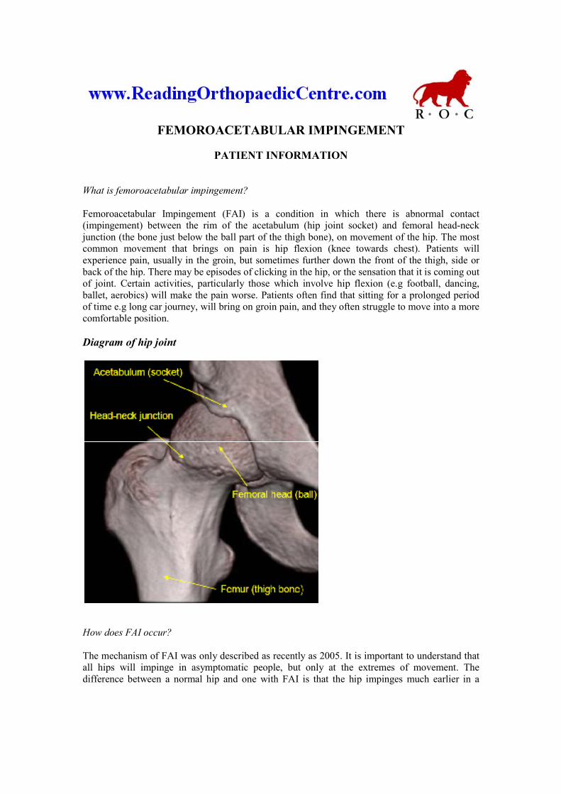
Diagram of hip joint
How does FAI occur?
The mechanism of FAI was only described as recently as 2005. It is important to understand that
all hips will impinge in asymptomatic people, but only at the extremes of movement. The
difference between a normal hip and one with FAI is that the hip impinges much earlier in a
patient with FAI. This occurs because of subtle differences in the anatomy of the hip joint.
Essentially this difference is an excess of bone, either on the edge of the acetabulum, or on the
femoral head-neck junction, or both. If the acetabulum is tilted backwards (retroverted), the same
effect occurs. The diagrams below illustrate this mechanism.
Normal hip joint, viewed from the side – no impingement at 90 degrees of hip flexion
Hip joint with impingement at 90 degrees of hip flexion, because of excess bone at the
head-neck junction. This type of impingement is termed ‘Cam’ impingement. The red
arrows indicate the shear force applied to the acetabular cartilage (right arrow) and
the displacing force applied to the acetabular labrum (left arrow)
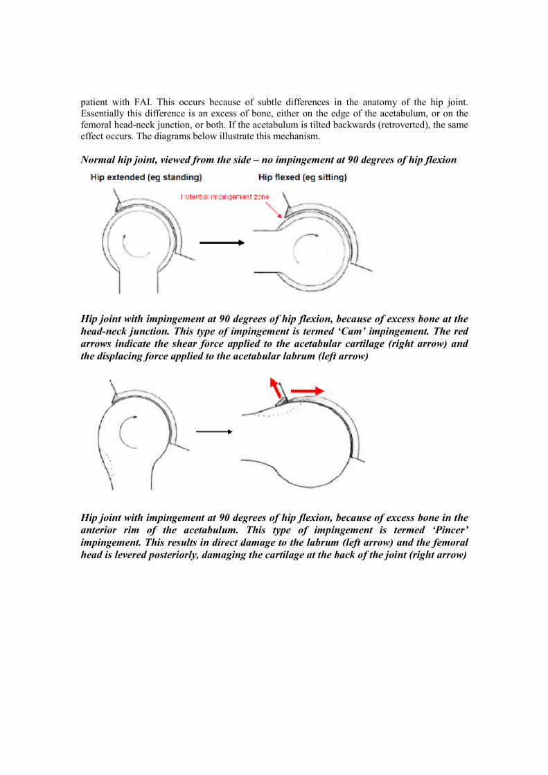
Hip joint with impingement at 90 degrees of hip flexion, because of excess bone in the
anterior rim of the acetabulum. This type of impingement is termed ‘Pincer’
impingement. This results in direct damage to the labrum (left arrow) and the femoral
head is levered posteriorly, damaging the cartilage at the back of the joint (right arrow)
Why does FAI cause hip pain?
As can be seen from the diagrams above, FAI results in damage to the cartilage (shiny shock
absorbing layer) of the hip joint, and to the acetabular labrum (gristly bumper around the edge of
the socket that helps to seal the joint). The labrum has lots of nerve endings so damage to it is
painful. Cartilage does not contain nerves, but it is likely that the increased force applied to the
bone beneath the cartilage, which does contain nerves, causes the pain.
Why is FAI important?
FAI is important for two reasons. First, it is a common cause of groin pain in young adults.
Because FAI has only recently been understood, in the past such patients may have been mis-
diagnosed as having other conditions such as a muscle strain (groin strain), hernia, or
inflammatory joint disease.
The second reason is that many surgeons believe that if untreated, FAI may result in hip arthritis,
which may ultimately require a hip replacement. It is important to understand however, that there
are not currently any studies that show that this is definitely the case, and that there are no studies
yet that show that treatment of FAI prevents progressive wearing out of the joint and arthritis.
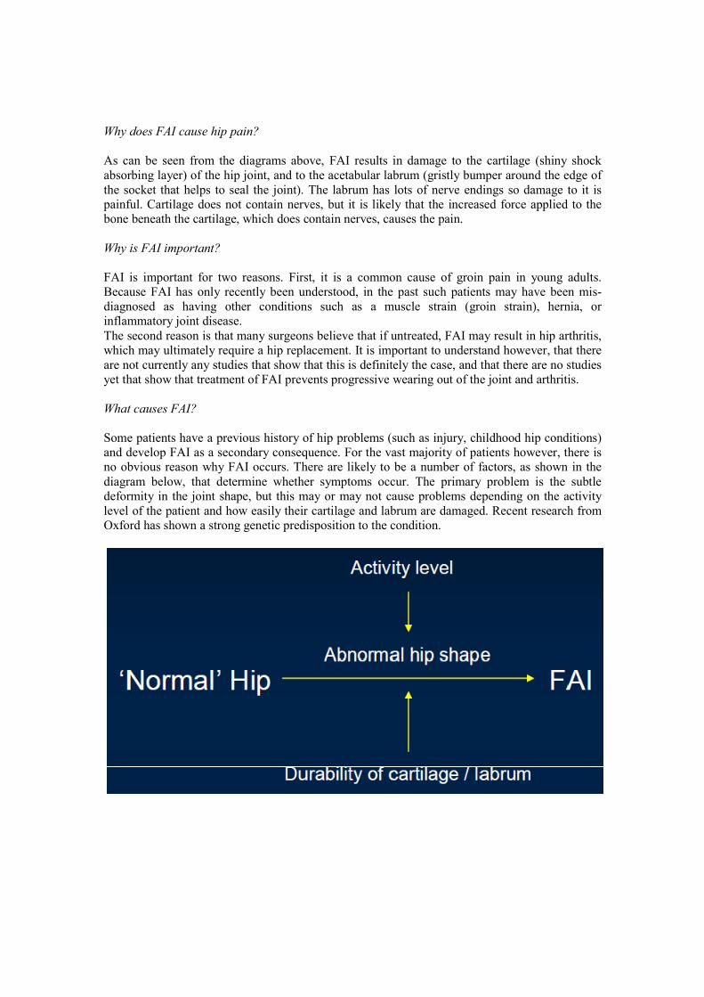
What causes FAI?
Some patients have a previous history of hip problems (such as injury, childhood hip conditions)
and develop FAI as a secondary consequence. For the vast majority of patients however, there is
no obvious reason why FAI occurs. There are likely to be a number of factors, as shown in the
diagram below, that determine whether symptoms occur. The primary problem is the subtle
deformity in the joint shape, but this may or may not cause problems depending on the activity
level of the patient and how easily their cartilage and labrum are damaged. Recent research from
Oxford has shown a strong genetic predisposition to the condition.
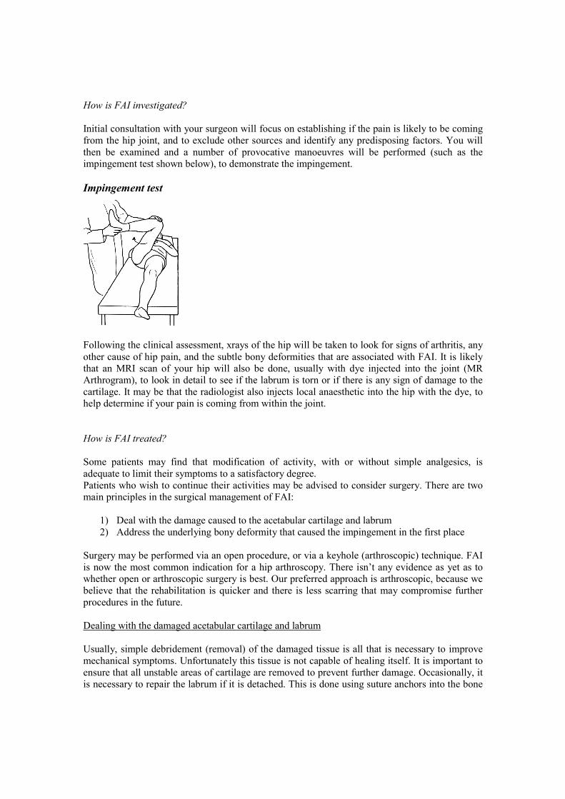
How is FAI investigated?
Initial consultation with your surgeon will focus on establishing if the pain is likely to be coming
from the hip joint, and to exclude other sources and identify any predisposing factors. You will
then be examined and a number of provocative manoeuvres will be performed (such as the
impingement test shown below), to demonstrate the impingement.
Impingement test
Following the clinical assessment, xrays of the hip will be taken to look for signs of arthritis, any
other cause of hip pain, and the subtle bony deformities that are associated with FAI. It is likely
that an MRI scan of your hip will also be done, usually with dye injected into the joint (MR
Arthrogram), to look in detail to see if the labrum is torn or if there is any sign of damage to the
cartilage. It may be that the radiologist also injects local anaesthetic into the hip with the dye, to
help determine if your pain is coming from within the joint.
How is FAI treated?
Some patients may find that modification of activity, with or without simple analgesics, is
adequate to limit their symptoms to a satisfactory degree.
Patients who wish to continue their activities may be advised to consider surgery. There are two
main principles in the surgical management of FAI:
1) Deal with the damage caused to the acetabular cartilage and labrum
2) Address the underlying bony deformity that caused the impingement in the first place
Surgery may be performed via an open procedure, or via a keyhole (arthroscopic) technique. FAI
is now the most common indication for a hip arthroscopy. There isn’t any evidence as yet as to
whether open or arthroscopic surgery is best. Our preferred approach is arthroscopic, because we
believe that the rehabilitation is quicker and there is less scarring that may compromise further
procedures in the future.
Dealing with the damaged acetabular cartilage and labrum
Usually, simple debridement (removal) of the damaged tissue is all that is necessary to improve
mechanical symptoms. Unfortunately this tissue is not capable of healing itself. It is important to
ensure that all unstable areas of cartilage are removed to prevent further damage. Occasionally, it
is necessary to repair the labrum if it is detached. This is done using suture anchors into the bone
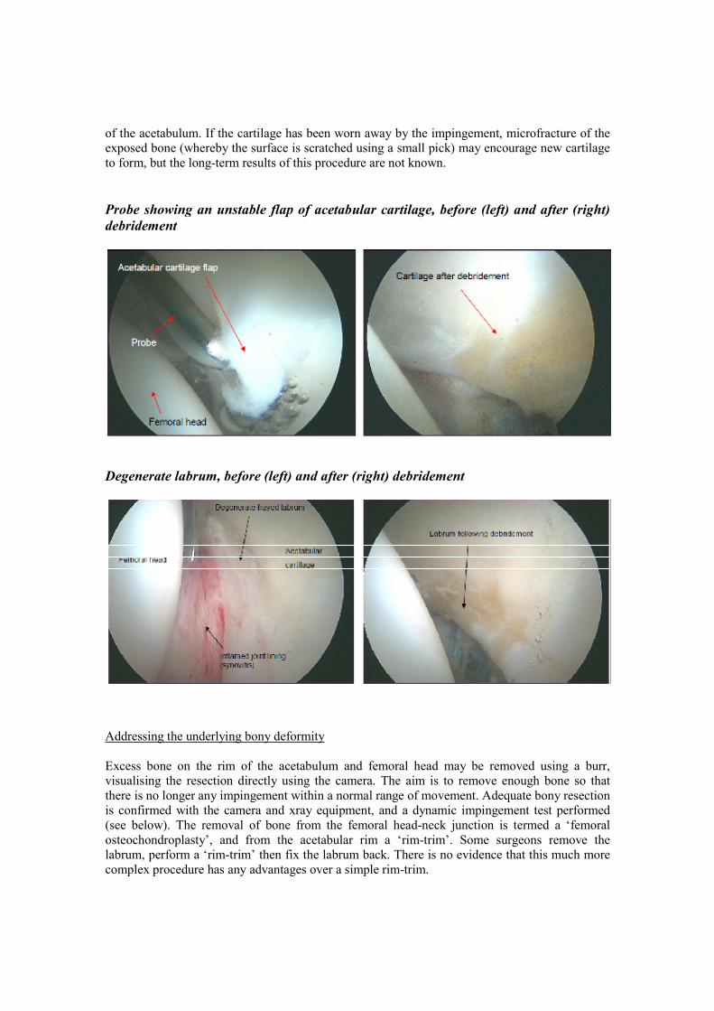
of the acetabulum. If the cartilage has been worn away by the impingement, microfracture of the
exposed bone (whereby the surface is scratched using a small pick) may encourage new cartilage
to form, but the long-term results of this procedure are not known.
Probe showing an unstable flap of acetabular cartilage, before (left) and after (right)
debridement
Degenerate labrum, before (left) and after (right) debridement
Addressing the underlying bony deformity
Excess bone on the rim of the acetabulum and femoral head may be removed using a burr,
visualising the resection directly using the camera. The aim is to remove enough bone so that
there is no longer any impingement within a normal range of movement. Adequate bony resection
is confirmed with the camera and xray equipment, and a dynamic impingement test performed
(see below). The removal of bone from the femoral head-neck junction is termed a ‘femoral
osteochondroplasty’, and from the acetabular rim a ‘rim-trim’. Some surgeons remove the
labrum, perform a ‘rim-trim’ then fix the labrum back. There is no evidence that this much more
complex procedure has any advantages over a simple rim-trim.
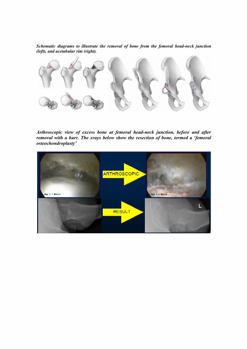
Schematic diagrams to illustrate the removal of bone from the femoral head-neck junction
(left), and acetabular rim (right).
Arthroscopic view of excess bone at femoral head-neck junction, before and after
removal with a burr. The xrays below show the resection of bone, termed a ‘femoral
osteochondroplasty’
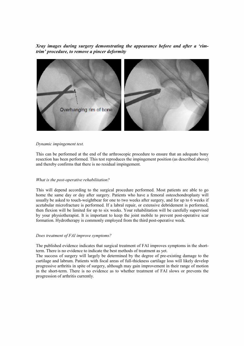
Xray images during surgery demonstrating the appearance before and after a ‘rim-
trim’ procedure, to remove a pincer deformity
Dynamic impingement test.
This can be performed at the end of the arthroscopic procedure to ensure that an adequate bony
resection has been performed. This test reproduces the impingement position (as described above)
and thereby confirms that there is no residual impingement.
What is the post-operative rehabilitation?
This will depend according to the surgical procedure performed. Most patients are able to go
home the same day or day after surgery. Patients who have a femoral osteochondroplasty will
usually be asked to touch-weightbear for one to two weeks after surgery, and for up to 6 weeks if
acetabular microfracture is performed. If a labral repair, or extensive debridement is performed,
then flexion will be limited for up to six weeks. Your rehabilitation will be carefully supervised
by your physiotherapist. It is important to keep the joint mobile to prevent post-operative scar
formation. Hydrotherapy is commonly employed from the third post-operative week.
Does treatment of FAI improve symptoms?
The published evidence indicates that surgical treatment of FAI improves symptoms in the short-
term. There is no evidence to indicate the best methods of treatment as yet.
The success of surgery will largely be determined by the degree of pre-existing damage to the
cartilage and labrum. Patients with focal areas of full-thickness cartilage loss will likely develop
progressive arthritis in spite of surgery, although may gain improvement in their range of motion
in the short-term. There is no evidence as to whether treatment of FAI slows or prevents the
progression of arthritis currently.
What are the complications of arthroscopic surgery for FAI?
The complications are the same as for a normal hip arthroscopy. In addition, femoral
osteochondroplasty may theoretically weaken the bone leading to a risk of fracture, however in
practice this is extremely rare. The main risk is that the procedure does not improve the patient’s
symptoms significantly, or that they deteriorate after initial benefit.
Research into FAI
Research is an essential part of surgical practice. FAI is a recently recognised phenomenon with
many questions unanswered. It is therefore an area of intense research activity. Furthermore,
recent NICE guidance has recommended that hip arthroscopy should only be performed by
surgeons who are collecting research data. Your surgeon will automatically collect pre- and post-
operative scores to assess the outcome of your treatment. Please do not be alarmed if you are
contacted ‘out of the blue’ regarding further research. All such studies go through a rigorous
approval process and you will only be contacted with the prior consent of your surgeon.
Participation is entirely voluntary and your care will not be influenced either way.
[Written by Tom Pollard, specialist registrar in Orthopaedic surgery, and Tony Andrade,
consultant Orthopaedic Surgeon.]
For further details, please contact:
Tony Andrade
Consultant Orthopaedic Hip and Knee Surgeon
Reading Orthopaedic Centre
and Royal Berkshire NHS Foundation Trust