Embed Size (px)
Citation preview
Feline sporotrichosis due to Sporothrix brasiliensis:an emerging animal infection in São Paulo, BrazilMontenegro et al.
Montenegro et al. BMC Veterinary Research 2014, 10:269http://www.biomedcentral.com/1746-6148/10/269

Montenegro et al. BMC Veterinary Research 2014, 10:269http://www.biomedcentral.com/1746-6148/10/269
RESEARCH ARTICLE Open Access
Feline sporotrichosis due to Sporothrix brasiliensis:an emerging animal infection in São Paulo, BrazilHildebrando Montenegro1, Anderson Messias Rodrigues2, Maria Adelaide Galvão Dias1, Elisabete Aparecida da Silva1,Fernanda Bernardi1 and Zoilo Pires de Camargo2*
Abstract
Background: Sporotrichosis is a mycotic infectious disease that is generally acquired by traumatic inoculation ofcontaminated materials especially from plant debris or through bites and scratches from diseased animals, such asdomestic cats. It affects the skin, lymphatic system, and other organs in the warm-blooded host. Etiological agents areembedded in the plant-associated order Ophiostomatales. With essential differences between possible outbreak sourcesand ecological niche, host-environment interactions are classic determinants of risk factors for disease acquisition.Sporotrichosis outbreaks with zoonotic transmission, such as those that are ongoing in southern and southeasternBrazil, have highlighted the threat of cross-species pathogen transmission. Sporothrix brasiliensis has emerged as ahuman threat owing to the intimate contact pattern between diseased cats and humans in endemic areas.
Results: We describe the recent emergence of feline sporotrichosis in the metropolitan region of São Paulo, Brazil,with an overwhelming occurrence of S. brasiliensis as the etiological agent. A phylogenetic and a haplotype approachwere used to investigate the origin of this epidemic and the impact of feline transmission on genetic diversity. Duringthe last 3-year period, 163 cases of feline sporotrichosis were reported in São Paulo with proven S. brasiliensis culture.The haplotype diversity of feline S. brasiliensis isolates revealed the expansion of a clonal population with low geneticdiversity. Haplotype analysis confirmed that isolates from São Paulo shared the haplotype originated in the long-lastingoutbreak of cat-transmitted sporotrichosis in Rio de Janeiro, which differed from the haplotype circulating in the RioGrande do Sul epidemic.
Conclusions: The fast spread of sporotrichosis in a short period of time highlights the potential for outbreaks andsuggests that the mycosis may affect an urban population with a high concentration of susceptible felines. The felinesporotrichosis epidemic shows no signs of slowing, and this epidemiological pattern may require specific public healthstrategies to control future outbreaks.
Keywords: Sporotrichosis, Feline, Sporothrix brasiliensis, Zoonosis, Emerging infectious diseases, Epidemiology, Cat,Sporothrix schenckii, Mycosis, Outbreak
BackgroundEpidemics caused by new and old fungal agents haveemerged and re-emerged over time as a threat to thehealth of vertebrate hosts [1]. The great global burdenof fungal infections in animals is specially observed as aresult of a pathogen-host shift or a recent introductionof a pathogen in a susceptible host population [2-4].Domestic animals are at risk of developing severalmycotic diseases that can be directly transmitted to
* Correspondence: [email protected] University of São Paulo (UNIFESP), Department of Microbiology,Immunology and Parasitology, Cell Biology Division, São Paulo, SP, BrazilFull list of author information is available at the end of the article
© 2014 Montenegro et al.; licensee BioMed CeCreative Commons Attribution License (http:/distribution, and reproduction in any mediumDomain Dedication waiver (http://creativecomarticle, unless otherwise stated.
humans; however, such diseases are often neglected byhealth systems. Because domestic animals have intimatecontact with their owners, they play an important role inthe emergence of human infections; this situation is alsocommon in the developing world, where environmentalconditions are juxtaposed with inadequate public healthinfrastructure. Reducing the public health risks from zoo-nosis outbreaks in urban areas requires different preventionand control strategies, as their increased frequency duringrecent decades may be related to poverty, poor sanitation,and anthropogenic changes in the environment.Sporotrichosis is a neglected disease of humans and
animals. The disease occurs worldwide in the form of
ntral Ltd. This is an Open Access article distributed under the terms of the/creativecommons.org/licenses/by/4.0), which permits unrestricted use,, provided the original work is properly credited. The Creative Commons Publicmons.org/publicdomain/zero/1.0/) applies to the data made available in this
Montenegro et al. BMC Veterinary Research 2014, 10:269 Page 2 of 10http://www.biomedcentral.com/1746-6148/10/269
sapronoses and zoonoses, mainly in tropical and subtrop-ical regions [4-7], and is the most frequent subcutaneousmycosis in Latin America [8]. Since it was first noted inthe United States in 1898, this mycosis has been describedas a disease of occupational risk, affecting farmers,gardeners, and agricultural workers. However, recentepidemics have demonstrated the potential for zoonotictransmission of the disease, and have nearly alwaysinvolved cats as the main source of infection [9,10].Around the world, the classical type of transmissionrelies on the traumatic inoculation of contaminatedplant material in the environment. In contrast, in thealternative type of transmission, bites and scratches froma diseased cat effectively disseminate the fungus. Eitherroute of infection begins by affecting the skin locally withthe development of a nodular ulcerated lesion, and even-tually spreads out from the site of trauma through thelymphatic system and causes damage to other organs ofthe warm-blooded host [11,12]. Zoonotic sporotrichosis ishighly frequent in the southern [4,5,13] and southeastern[4,5,14,15] regions of Brazil, and animals usually experi-ence the severe form of sporotrichosis.Traditionally, sporotrichosis has been attributed to the
dimorphic fungus Sporothrix schenckii sensu lato (s.l.).Multigene phylogenies have clarified species boundarieswithin cryptic isolates [16] and led to the proposal of theS. schenckii complex, which comprises a clinically import-ant clade that includes S. brasiliensis (clade I), S. schenckiisensu stricto (s. str.) (clade II), S. globosa (clade III), and S.luriei (clade VI) [17,18]. Host susceptibility, species distri-bution, and sensitivity profile to antifungal agents are alldivergent among closely related species [4,5,19,20]. A highprevalence of S. brasiliensis, the most virulent species inthe complex [20,21] and geographically restricted to Brazil[4,5,7,22], has been reported in cats [4]. Rodrigues et al.[4] suggests that the thermal resistance exhibited by S.brasiliensis may be an important mechanism of adaptationto the feline body, and may partially explain the success ofS. brasiliensis infection over the remaining species in thecomplex. Indeed, the cat-cat contact pattern during fightsand the cat-human contact pattern of scratches andbites may also support the success of horizontal diseasetransmission in a short period of time [4,5], because thefungus does not die with the feline, and can be transmittedto the next warm-blooded host. The increased proximitybetween cats and humans favors the emergence of sporotri-chosis in Brazil.Since the 1990’s, the epidemiological profile of sporo-
trichosis has changed from a low-prevalence disease to amajor health problem that affects people living inneglected urban areas [4,5]. Its prevalence may reachepidemic proportions over time. In the metropolitanarea of Rio de Janeiro, sporotrichosis is estimated toaccount for more than 3,800 feline, 4,000 human, and
120 canine cases in the period from 1998 to 2012 [23-25].Massive zoonotic transmission has also been detected inthe southern region of Brazil [5,13,26], with characteristicssimilar to the ongoing epidemic in Rio de Janeiro.In contrast to the major ongoing epidemics in other
provinces of Brazil, during the past 20 years São Paulostate has reported a basal number of sporotrichosis cases,nearly always unrelated to feline transmission types [5,27].The Zoonosis Control Center of São Paulo (ZCC-SP) hasperformed an epidemiological surveillance service amongferal cats since 2008. In December 2010, a few cases ofsporotrichosis in cats were reported to our service; sincethen, an increasing number of feline cases have been iden-tified in São Paulo and in two of its neighboring cities.Here, we report the molecular epidemiology of Sporothrixspecies as an emerging pathogen among felines in themetropolitan area of São Paulo and discuss its relevancein one of the most populous regions of the Americas.
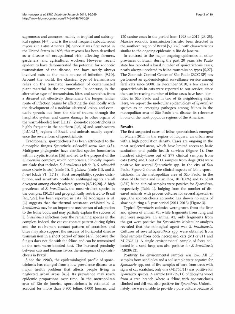
ResultsThe first suspected cases of feline sporotrichosis emergedin March 2011 in the region of Itaquera, an urban areawith a high population density. Cases are ongoing in themost neglected areas, which have limited access to basicsanitation and public health services (Figure 1). Onehundred sixty-three out of 279 clinical samples fromcats (58%) and 1 out of 11 samples from dogs (8%) werepositive for several Sporothrix spp. in the city of SãoPaulo. Figure 2 shows the clinical aspects of feline sporo-trichosis. In the metropolitan area of São Paulo, in thecities of Diadema and Guarulhos, 10 (100%) and 17 of 40(43%) feline clinical samples were positive for Sporothrix,respectively (Table 1). Judging from the number of dis-eased animals with proven cultures for several Sporothrixspp., the sporotrichosis epizootic has shown no signs ofslowing during a 3-year period (2011-2013) (Figure 3).Typical Sporothrix colonies were grown from the liver
and spleen of animal #1, while fragments from lung andgut were negative. In animal #2, only fragments fromthe gut were positive for Sporothrix. Molecular analysisrevealed that the etiological agent was S. brasiliensis.Cultures of several Sporothrix spp. were obtained fromfecal samples from both necropsied cats (M1727/11 andM1732/11). A single environmental sample of feces col-lected in a sand heap was also positive for S. brasiliensis(M039/12).Positivity for environmental samples was low. All 17
samples from sand piles and a soil sample were negative forSporothrix spp. out of five samples of bark from trees withsigns of cat scratches, only one (M1753/11) was positive forSporothrix species. A sample (M1239/11) of decaying woodfrom a tree branch where a feline with sporotrichosisclimbed and fell was also positive for Sporothrix. Unfortu-nately, we were unable to provide a pure culture because of
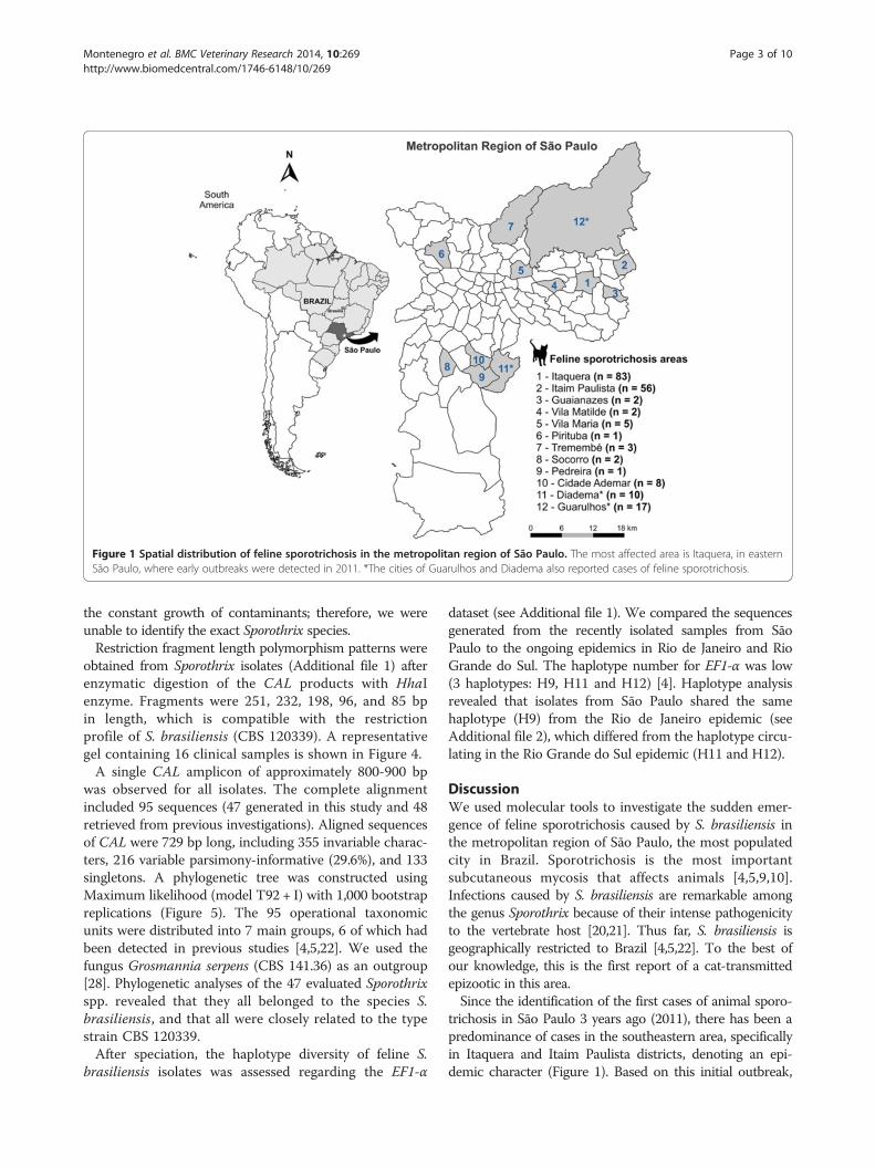
Figure 1 Spatial distribution of feline sporotrichosis in the metropolitan region of São Paulo. The most affected area is Itaquera, in easternSão Paulo, where early outbreaks were detected in 2011. *The cities of Guarulhos and Diadema also reported cases of feline sporotrichosis.
Montenegro et al. BMC Veterinary Research 2014, 10:269 Page 3 of 10http://www.biomedcentral.com/1746-6148/10/269
the constant growth of contaminants; therefore, we wereunable to identify the exact Sporothrix species.Restriction fragment length polymorphism patterns were
obtained from Sporothrix isolates (Additional file 1) afterenzymatic digestion of the CAL products with HhaIenzyme. Fragments were 251, 232, 198, 96, and 85 bpin length, which is compatible with the restrictionprofile of S. brasiliensis (CBS 120339). A representativegel containing 16 clinical samples is shown in Figure 4.A single CAL amplicon of approximately 800-900 bp
was observed for all isolates. The complete alignmentincluded 95 sequences (47 generated in this study and 48retrieved from previous investigations). Aligned sequencesof CAL were 729 bp long, including 355 invariable charac-ters, 216 variable parsimony-informative (29.6%), and 133singletons. A phylogenetic tree was constructed usingMaximum likelihood (model T92 + I) with 1,000 bootstrapreplications (Figure 5). The 95 operational taxonomicunits were distributed into 7 main groups, 6 of which hadbeen detected in previous studies [4,5,22]. We used thefungus Grosmannia serpens (CBS 141.36) as an outgroup[28]. Phylogenetic analyses of the 47 evaluated Sporothrixspp. revealed that they all belonged to the species S.brasiliensis, and that all were closely related to the typestrain CBS 120339.After speciation, the haplotype diversity of feline S.
brasiliensis isolates was assessed regarding the EF1-α
dataset (see Additional file 1). We compared the sequencesgenerated from the recently isolated samples from SãoPaulo to the ongoing epidemics in Rio de Janeiro and RioGrande do Sul. The haplotype number for EF1-α was low(3 haplotypes: H9, H11 and H12) [4]. Haplotype analysisrevealed that isolates from São Paulo shared the samehaplotype (H9) from the Rio de Janeiro epidemic (seeAdditional file 2), which differed from the haplotype circu-lating in the Rio Grande do Sul epidemic (H11 and H12).
DiscussionWe used molecular tools to investigate the sudden emer-gence of feline sporotrichosis caused by S. brasiliensis inthe metropolitan region of São Paulo, the most populatedcity in Brazil. Sporotrichosis is the most importantsubcutaneous mycosis that affects animals [4,5,9,10].Infections caused by S. brasiliensis are remarkable amongthe genus Sporothrix because of their intense pathogenicityto the vertebrate host [20,21]. Thus far, S. brasiliensis isgeographically restricted to Brazil [4,5,22]. To the best ofour knowledge, this is the first report of a cat-transmittedepizootic in this area.Since the identification of the first cases of animal sporo-
trichosis in São Paulo 3 years ago (2011), there has been apredominance of cases in the southeastern area, specificallyin Itaquera and Itaim Paulista districts, denoting an epi-demic character (Figure 1). Based on this initial outbreak,
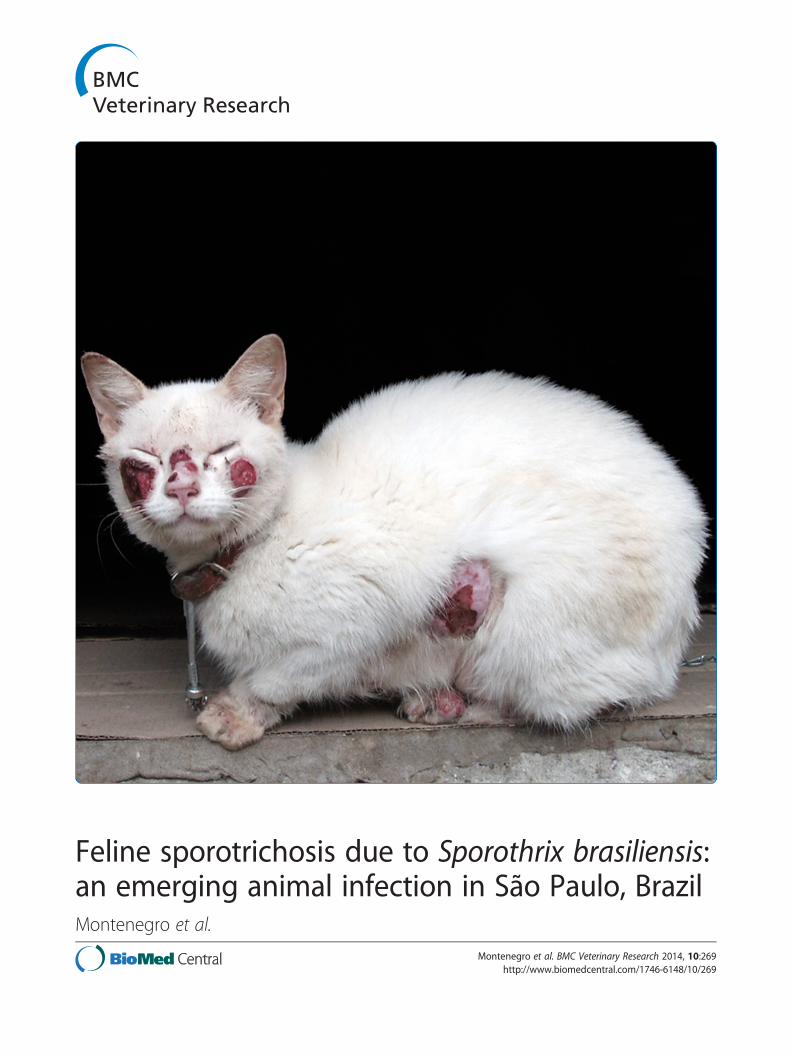
Figure 2 Clinical aspects of feline sporotrichosis. (A) Wet, ulceratedskin lesions, often particularly concentrated in the cephalic region.(B) Weight loss during the evolution of the disease.
Table 1 Sporotrichosis in feline and canine samplescollected from different cities of São Paulo State, Brazil(March 2011 to April 2014)
City District Host Positive Negative Positivity
São Paulo Cidade Ademar Feline 8 4 67%
Guaianazes Feline 2 0 100%
Itaim Paulista Canine 0 2 0%
Feline 56 23 71%
Itaquera Canine 1 9 10%
Feline 83 80 51%
Pedreira Feline 1 1 50%
Pirituba Feline 1 0 100%
Socorro Feline 2 3 40%
Tremembé Feline 3 0 100%
Vila Maria Feline 5 3 63%
Vila Matilde Feline 2 2 50%
Subtotal Canine 1 11 8%
Feline 163 116 58%
Diadema Canine 0 1 0%
Feline 10 0 100%
Guarulhos Canine 0 3 0%
Feline 17 23 43%
Total Canine 1 15 6%
Feline 190 139 58%
Figure 3 Temporal evolution of the feline sporotrichosis epidemicin the metropolitan region of São Paulo. The constant number ofpositive cats indicates the maintenance of cat-transmitted sporotrichosis.
Montenegro et al. BMC Veterinary Research 2014, 10:269 Page 4 of 10http://www.biomedcentral.com/1746-6148/10/269
the ZCC-SP conducted active searches in households,which resulted in finding other cases of feline sporotricho-sis. To date, a total of 83 feline cases and one canine casehave been identified in Itaquera, and 56 feline cases havebeen recorded in Itaim Paulista (Figure 3). As new activesearches are conducted in São Paulo, new cases aredetected, suggesting that the epidemic is unlikely to endspontaneously. It is important to note that a small numberof cats with sporotrichosis have also been found in otherdistricts of the city, as well as in other cities of the samemetropolitan area of São Paulo, such as Diadema (10feline cases) and Guarulhos (17 feline cases) (Table 1;Figure 1). These findings indicate the spread of theepidemic, and lead us to believe that the transmissionof this disease might have a silent character.Molecular data for 48 animals revealed that the out-
break is caused by S. brasiliensis. In this study, we havesuccessfully identified the isolates of feline sporotrichosisby CAL-RFLP, as suggested by Rodrigues et al. [29].The use of CAL-RFLP considerably reduced the cost ofmolecular identification in this epidemic scenario. The
overwhelming prevalence of S. brasiliensis during out-breaks is in agreement with results obtained by Rodrigueset al. [4] regarding feline-transmitted epidemics in Rio deJaneiro and Rio Grande do Sul. In addition, low geneticdiversity was observed during the early phase of thisepidemic, in agreement with previous results [4,5]. Ahaplotype network based on EF1-α suggests that thepredominant haplotype among felines in São Paulo andthe haplotype observed in Rio de Janeiro are the same.Although this information may not be conclusive becauseof the low number of markers used, it still indicates that
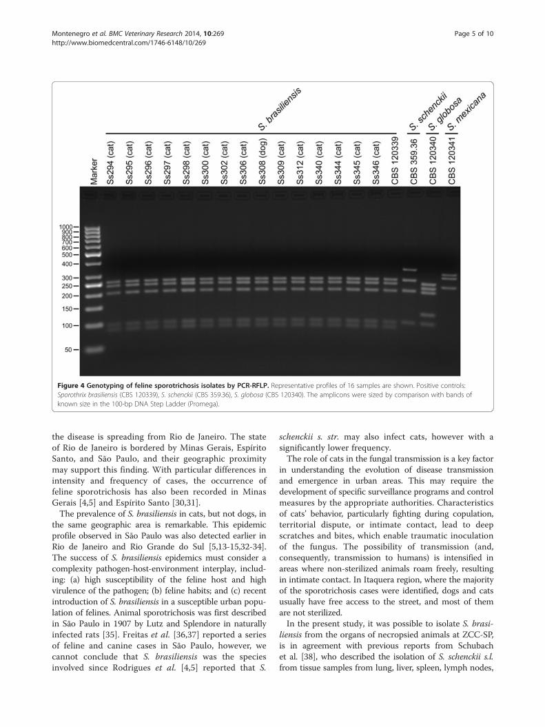
Figure 4 Genotyping of feline sporotrichosis isolates by PCR-RFLP. Representative profiles of 16 samples are shown. Positive controls:Sporothrix brasiliensis (CBS 120339), S. schenckii (CBS 359.36), S. globosa (CBS 120340). The amplicons were sized by comparison with bands ofknown size in the 100-bp DNA Step Ladder (Promega).
Montenegro et al. BMC Veterinary Research 2014, 10:269 Page 5 of 10http://www.biomedcentral.com/1746-6148/10/269
the disease is spreading from Rio de Janeiro. The stateof Rio de Janeiro is bordered by Minas Gerais, EspíritoSanto, and São Paulo, and their geographic proximitymay support this finding. With particular differences inintensity and frequency of cases, the occurrence offeline sporotrichosis has also been recorded in MinasGerais [4,5] and Espírito Santo [30,31].The prevalence of S. brasiliensis in cats, but not dogs, in
the same geographic area is remarkable. This epidemicprofile observed in São Paulo was also detected earlier inRio de Janeiro and Rio Grande do Sul [5,13-15,32-34].The success of S. brasiliensis epidemics must consider acomplexity pathogen-host-environment interplay, includ-ing: (a) high susceptibility of the feline host and highvirulence of the pathogen; (b) feline habits; and (c) recentintroduction of S. brasiliensis in a susceptible urban popu-lation of felines. Animal sporotrichosis was first describedin São Paulo in 1907 by Lutz and Splendore in naturallyinfected rats [35]. Freitas et al. [36,37] reported a seriesof feline and canine cases in São Paulo, however, wecannot conclude that S. brasiliensis was the speciesinvolved since Rodrigues et al. [4,5] reported that S.
schenckii s. str. may also infect cats, however with asignificantly lower frequency.The role of cats in the fungal transmission is a key factor
in understanding the evolution of disease transmissionand emergence in urban areas. This may require thedevelopment of specific surveillance programs and controlmeasures by the appropriate authorities. Characteristicsof cats’ behavior, particularly fighting during copulation,territorial dispute, or intimate contact, lead to deepscratches and bites, which enable traumatic inoculationof the fungus. The possibility of transmission (and,consequently, transmission to humans) is intensified inareas where non-sterilized animals roam freely, resultingin intimate contact. In Itaquera region, where the majorityof the sporotrichosis cases were identified, dogs and catsusually have free access to the street, and most of themare not sterilized.In the present study, it was possible to isolate S. brasi-
liensis from the organs of necropsied animals at ZCC-SP,is in agreement with previous reports from Schubachet al. [38], who described the isolation of S. schenckii s.l.from tissue samples from lung, liver, spleen, lymph nodes,
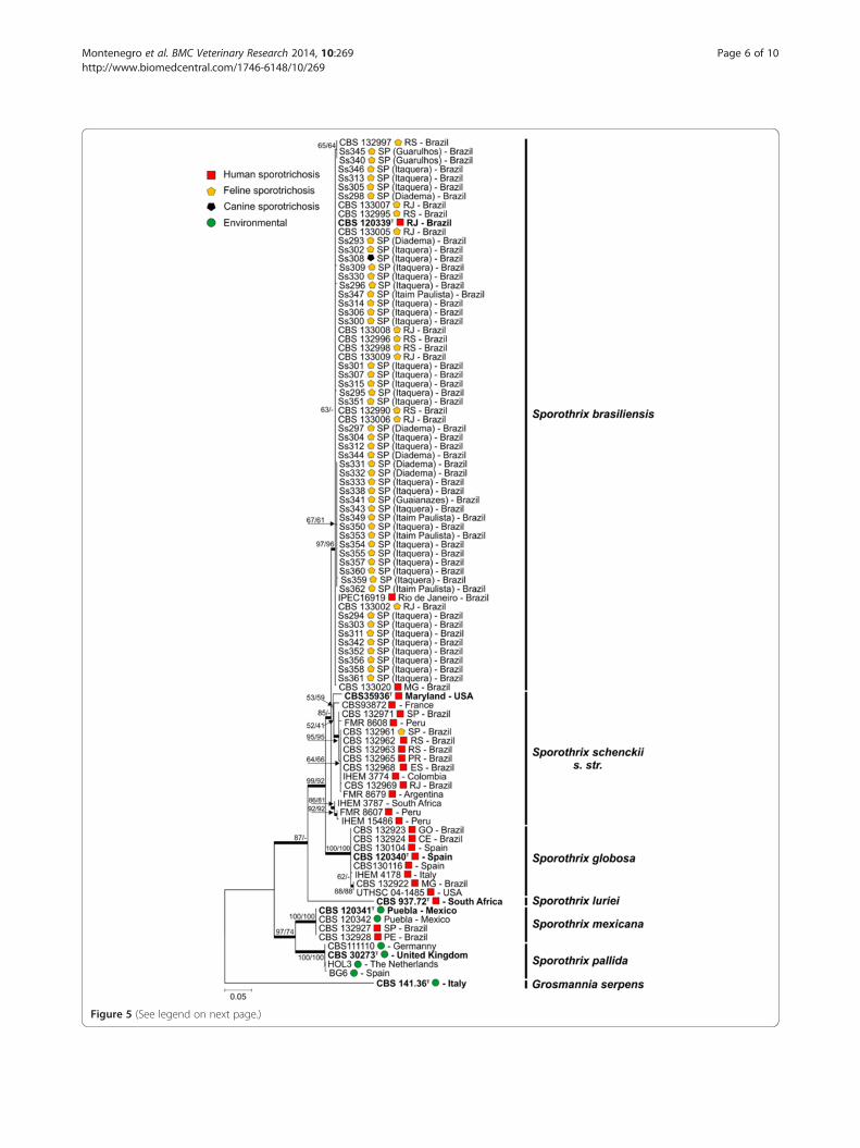
Figure 5 (See legend on next page.)
Montenegro et al. BMC Veterinary Research 2014, 10:269 Page 6 of 10http://www.biomedcentral.com/1746-6148/10/269

(See figure on previous page.)Figure 5 Phylogenetic analysis using the maximum likelihood method based on sequences from the calmodulin-encoding gene. Thepercentage of replicate trees in which the associated taxa clustered together in the bootstrap test (1000 replicates) is shown next to the branches(NJ/ML). The evolutionary distances were computed using the Tamura 3-parameter method (T92 + I). All positions containing gaps and missingdata were eliminated. Further information about isolate source and GenBank accession number can be found in the Additional file 1.
Montenegro et al. BMC Veterinary Research 2014, 10:269 Page 7 of 10http://www.biomedcentral.com/1746-6148/10/269
heart, and kidney taken postmortem from ten cats withsporotrichosis. Sporothrix brasiliensis were also isolatedfrom feces collected from the small intestine of bothnecropsied cats, as well as from feces collected from a pileof sand in Itaquera. These findings introduce new insightsregarding the ecology of S. brasiliensis. Feces fromdiseased cats may contaminate the soil, creating an envir-onmental reservoir for S. brasiliensis and becoming a newsource of contamination for animals or humans. Further-more, cats habitually bury their feces in sand or soil andsharpen their claws on tree bark, enabling the initial con-tamination of the claw by the fungus. Cats have retract-able claws and the fungus may be retained superficially inthe animal’s body [9,10]. Moreover, cats’ habit of cleaningthemselves by licking can lead to contamination of theoral mucosa, which renders biting and scratching effectivefor deep implantation of the fungus in cutaneous and sub-cutaneous sites on other animals and humans. Anotherpossibility that may render soil a source of contaminationis the inappropriate disposal of carcasses of animals thatdied with sporotrichosis, such as backyard burial, or eventhrowing the animals into the wastelands [4,33].Sporothrix was also isolated from a sample of decaying
wood and from the bark of a tree, showing that the fun-gus is present in the environment within the studiedtransmission area. However, the rate of positivity amongenvironmental samples was low, which may be relatedto the sampling strategies, seasonality, and number ofevaluated samples. Failure to isolate pathogenic Sporothrixspp. embedded in the S. schenckii complex from theoriginal source of infection in the environment is notunusual [39-41].
ConclusionsThe recent introduction of S. brasiliensis to the metro-politan area of São Paulo has resulted in the permanentinfection of felines during the 3 years since it was firstdetected. We observed striking similarities between theSão Paulo epidemic and the long-lasting outbreak of cat-transmitted sporotrichosis and those observed in Rio deJaneiro and Rio Grande do Sul. Moreover, it is unlikelythat the epidemic remains limited to cats. The threat ofcross-species pathogen transmission can lead to the riskof a massive epidemic for humans in these areas [5] andposes a significant challenge for public health systems.Strategies to control the spreading of the disease may in-clude the education of population about the main aspectsof Sporothrix transmission, animal sterilization programs,
treatment, and prophylaxis, as well as development ofcampaigns to avoid abandonment of diseased animals bytheir owners in the most affected areas.
MethodsAnimal and clinical samplesFrom March 2011 until April 2014, we studied a total of345 animals from São Paulo city and neighboring citiesthat were suspected of having sporotrichosis. Pet ownerswere informed about the risk of zoonotic transmissionof sporotrichosis, and verbal informed consent wasobtained by a professional from the ZCC-SP, before thecollection of the samples. Suspected animals had appar-ent cutaneous lesions throughout the body, especiallyin the cephalic region. The lesions were usually wet,with secretion, but were dry in rare cases. Clinical sam-ples were collected from wet lesions with sterile swabsand sent to the laboratory on the same day. However, if itwas not possible to send the samples to the laboratory onthe day of collection, they were placed in transport media(Stuart Media) and refrigerated at 4°C. Dry lesions werescraped, and the crusts were collected in sterile flasks. Aprofessional from the ZCC-SP collected all samples. Thisstudy was performed in accordance with guidelines forgood laboratory practice, and every effort was made tominimize suffering. Ethical approval was provided by theInstitutional Committee (Universidade Federal de SãoPaulo 0244/11).
Sporothrix spp. isolation and identificationClinical samples were directly inoculated on MycoselAgar slants (Becton Dickinson, Sparks, MD, USA) induplicate and incubated at 25°C for 30 days. Suspectedcolonies were subcultured on Sabouraud dextrose agarplates (Becton Dickinson, Sparks, MD, USA). Macroscopicand microscopic characteristics were applied to thedichotomous key to clinical species of the S. schenckiicomplex [18].
OrgansTwo cats that presented with terminal disseminated spo-rotrichosis were subject to gross necropsy under asepticconditions after euthanasia at ZCC-SP. The cats’ organs(liver, lung, spleen, and gut) were removed aseptically,cut into small fragments, and macerated in 2 mL of ster-ile saline solution; 0.5 mL were plated in Mycosel agarplates (in duplicate) and incubated at 25°C for 30 days.Gut samples were well rinsed with sterile saline solution
Montenegro et al. BMC Veterinary Research 2014, 10:269 Page 8 of 10http://www.biomedcentral.com/1746-6148/10/269
before sample processing. The suspected colonies wereisolated and identified as described above.
Fecal samplesFecal samples from necropsied cats were collected fromthe gut. In addition, two environmental feces sampleswere collected from a sand heap in the Itaquera region,in the backyard of a residence where diseased cats lived.Each fecal sample was diluted in sterile saline solution(1:10 dilution) with chloramphenicol (200 mg/L), vigor-ously homogenized for 5 min, and allowed to settle for15 min. Samples of the supernatant (0.5 mL) were platedon Mycosel agar plates (Becton Dickinson, Sparks, MD,USA) in duplicate and incubated at 25°C for 30 days.The suspected colonies were isolated and identified asdescribed above.
Environmental samplesEnvironmental samples (n =24) were collected in order toidentify potential reservoirs and sources of contaminationin neighboring Itaquera, where most of the cases of felinesporotrichosis were found. Sand samples (n =17) werecollected from sand piles that contained cat feces; a soilsample (n =1) was collected from a square to which theferal cats had common access at night; bark samples(n =5) were collected from trees with signs of felinescratches near a known home of diseased cats; a sampleof decaying wood (n =1) was collected for mycologicalinvestigation. Samples were processed as describedabove, inoculated on Mycosel agar plates (in duplicate),and incubated at 25°C for 30 days.
Molecular characterizationSporothrix colonies were grown on potato dextrose agarslants (Becton Dickinson, Sparks, MD, USA) for 10 daysat 25°C. DNA was extracted and purified from fungalcolonies by following the Fast DNA kit protocol (MPBiomedicals, Vista, CA, USA) [22]. The calmodulin (CAL)locus region was amplified directly from genomic DNA bypolymerase chain reaction (PCR) using the degeneratedprimers CL1 (5′-GAR TWC AAG GAG GCC TTCTC-3′) and CL2A (5′-TTT TTG CAT CAT GAG TTGGAC-3′) [42], which amplified an 800-bp ampliconcorresponding to exons 3 through 5. The CAL sequencewas used for taxonomy purposes. The translation elong-ation factor-1 alpha (EF1-α) locus region was amplifiedand sequenced using the primers EF1-F (5′-CTG AGGCTC GTT ACC AGG AG-3′) and EF1-R (5′-CGA CTTGAT GAC ACC GAC AG-3′), as described by Rodrigueset al. [4]. We used EF1-α information to compare theongoing epidemics in Rio de Janeiro, Rio Grande do Sul,and São Paulo. Only PCR products that produced singlebands were sequenced. Amplified products were gel puri-fied with the Wizard® SV Gel and PCR Clean-Up System
(Promega, Madison, WI, USA), following the manufac-turer’s instructions. PCR products were sequenced directlyin two reactions with forward and reverse primers toincrease the quality of the sequence data (Phred >30).The sequencing reactions were conducted using the
BigDye® Terminator v3.1 Cycle Sequencing Kit (AppliedBiosystems, Inc., Foster City, CA, USA) and the sequencingproducts were determined using an ABI 3730 DNAAnalyzer 48-well capillary sequencer (Applied Biosys-tems, Inc., Foster City, CA, USA). Sequences generatedin both senses were assembled into single sequences viaCAP3 implemented in BioEdit software [43]. Sequenceswere aligned with MAFFT version 7 [44], and retrievedalignments were manually edited to avoid mis-paired bases.Sequences were exported as FASTA files for BLAST searchat http://www.ncbi.nlm.nih.gov/BLAST. All sequences weredeposited online at GenBank (Additional file 1).
Phylogenetic reconstructionsRelationships among Sporothrix isolates collected duringthe São Paulo outbreak were determined by phylogeneticanalysis of CAL sequences and comparison to referencestrains (see Additional file 1) [4,5,16-18,22,45]. MaximumLikelihood and Neighbor-joining methods were employedto complete phylogenetic analyses using MEGA6 [46].Considering the Bayesian information criterion (BIC) andAkaike information criterion (AIC) [47], the Tamura 3-parameter model (T92 model) [48] was found to be thebest evolutionary model for the CAL sequence. The modelwas applied assuming that a certain fraction of sites areevolutionarily invariable. Trees were estimated using 1,000bootstrap replicates [49]; gaps and missing data were notincluded in the analysis.
Haplotype networkHaplotype analysis based on EF1-α sequences (seeAdditional file 1) were estimated using DnaSP soft-ware version 5.10 [50] in order to visualize differencesand diversity among S. brasiliensis isolates recoveredfrom zoonotic outbreaks in São Paulo, Rio de Janeiro,and Rio Grande do Sul [4]. Gaps and missing datawere excluded from the calculations. Median-joiningnetworks [51] were obtained and visualized usingNetwork 4.610 software (Fluxus Technology).
CAL restriction fragment length polymorphismMolecular characterization was also performed by polymer-ase chain reaction–restriction fragment length polymorph-ism (PCR-RFLP) as an alternative molecular approach. Thepartial CAL gene was amplified using the primers CL1 andCL2A [42] as described above and digested with HhaI asdescribed elsewhere [29]. Digested products were electro-phoresed on 2.5% (w/v) agarose gels for 90 min at 100 V inthe presence of GelRedTM (Biotium, Hayward, CA, USA).
Montenegro et al. BMC Veterinary Research 2014, 10:269 Page 9 of 10http://www.biomedcentral.com/1746-6148/10/269
We included a lane loaded with 100-bp DNA Step Ladder(Promega, Madison, WI, USA), as well as one positivecontrol from each of the following reference strains: S.brasiliensis (CBS 120339), S. schenckii (CBS 359.36), S.globosa (CBS 120340), and S. mexicana (CBS 120341). Thebands were visualized using the L-Pix Touch (LoccusBiotecnologia, São Paulo, Brazil) imaging system underUV illumination.
Availability of supporting dataAll the supporting information is included as additionalfiles.
Additional files
Additional file 1: Strains, species, origin, CAL and EF1-α, andGenBank accession numbers of Sporothrix spp. isolates used in thisstudy. All sequences were deposited online at GenBank (http://www.ncbi.nlm.nih.gov/genbank).
Additional file 2: Median-joining haplotype network of Sporothrixschenckii complex isolates, comparing all EF1-α haplotypes describedin the ongoing epidemics in Rio de Janeiro, Rio Grande do Sul, andSão Paulo. Isolates recovered in the São Paulo epidemics (2011-2013)share the same haplotype (H9) as previous outbreaks in Rio de Janeiro(1998-2012) reported by Rodrigues et al. [4]. The size of the circumference isproportional to the haplotype frequency. Isolates are coded, and theirfrequencies are represented by geographic region of isolation. Blackdots (median vectors) represent unsampled or extinct haplotypes inthe population.
Abbreviationss.l.: sensu lato; s. str.: sensu stricto; ZCC-SP: Zoonosis Control Center of São Paulo;CAL: Calmodulin; EF1-α: Translation elongation factor-1 alpha; ML: Maximumlikelihood; NJ: Neighbor-joining; BIC: Bayesian information criterion; AIC: Akaikeinformation criterion; T92: Tamura 3-parameter method; PCR-RFLP: Polymerasechain reaction–restriction fragment length polymorphism.
Competing interestsThe authors declare that they have no competing interests. The authorsalone are responsible for the content and writing of the paper.
Authors’ contributionsStudy development and design: AMR, HM, and ZPC. Epidemiologicalsurveillance, clinical observations and post-mortem examination: HM, MAGD,EAdS and FB. Molecular genetic studies and phylogenetic analysis: AMR.Drafting of the paper: AMR, HM, and ZPC. All authors read and approved thefinal manuscript.
AcknowledgmentsAMR is a fellow and acknowledges the financial support of the São PauloResearch Foundation – FAPESP (2011/07350-1). This work was supported, inpart, by grants from FAPESP (2009/54024-2), the National Council for Scientificand Technological Development – CNPq (472600/2011-7 and 472169/2012-2),and Coordenação de Aperfeiçoamento de Pessoal de Nível Superior (CAPES).
Author details1Zoonosis Control Center of São Paulo (COVISA/SMS/PMSP), São Paulo, SP,Brazil. 2Federal University of São Paulo (UNIFESP), Department of Microbiology,Immunology and Parasitology, Cell Biology Division, São Paulo, SP, Brazil.
Received: 7 July 2014 Accepted: 6 November 2014
References1. Fisher MC, Henk DA, Briggs CJ, Brownstein JS, Madoff LC, McCraw SL,
Gurr SJ: Emerging fungal threats to animal, plant and ecosystem health.Nature 2012, 484:186–194.
2. Olson DH, Aanensen DM, Ronnenberg KL, Powell CI, Walker SF, Bielby J,Garner TW, Weaver G, Fisher MC: Mapping the global emergence ofBatrachochytrium dendrobatidis, the amphibian chytrid fungus. PLoS One2013, 8:e56802.
3. Lorch JM, Meteyer CU, Behr MJ, Boyles JG, Cryan PM, Hicks AC, Ballmann AE,Coleman JT, Redell DN, Reeder DM, Blehert DS: Experimental infection ofbats with Geomyces destructans causes white-nose syndrome. Nature2011, 480:376–378.
4. Rodrigues AM, de Melo TM, de Hoog GS, Schubach TMP, Pereira SA,Fernandes GF, Bezerra LML, Felipe MS, de Camargo ZP: Phylogeneticanalysis reveals a high prevalence of Sporothrix brasiliensis in felinesporotrichosis outbreaks. PLoS Negl Trop Dis 2013, 7:e2281.
5. Rodrigues AM, de Hoog GS, Zhang Y, Camargo ZP: Emergingsporotrichosis is driven by clonal and recombinant Sporothrix species.Emerg Microbes Infect 2014, 3:e32.
6. Verma S, Verma GK, Singh G, Kanga A, Shanker V, Singh D, Gupta P, MoktaK, Sharma V: Sporotrichosis in Sub-Himalayan India. PLoS Negl Trop Dis2012, 6:e1673.
7. Zhou X, Rodrigues AM, Feng P, Hoog GS: Global ITS diversity in theSporothrix schenckii complex. Fungal Divers 2014, 66:153–165.
8. Queiroz-Telles F, Nucci M, Colombo AL, Tobón A, Restrepo A: Mycoses ofimplantation in Latin America: an overview of epidemiology, clinicalmanifestations, diagnosis and treatment. Med Mycol 2011, 49:225–236.
9. Schubach A, Barros MB, Wanke B: Epidemic sporotrichosis. Curr Opin InfectDis 2008, 21:129–133.
10. Barros MB, de Almeida PR, Schubach AO: Sporothrix schenckii andsporotrichosis. Clin Microbiol Rev 2011, 24:633–654.
11. Silva-Vergara ML, de Camargo ZP, Silva PF, Abdalla MR, Sgarbieri RN,Rodrigues AM, dos Santos KC, Barata CH, Ferreira-Paim K: DisseminatedSporothrix brasiliensis infection with endocardial and ocular involvementin an HIV-infected patient. Am J Trop Med Hyg 2012, 86:477–480.
12. Bonifaz A, Vázquez-González D: Diagnosis and treatment oflymphocutaneous sporotrichosis: What are the options? Curr Fungal Infect Rep2013, 7:252–259.
13. Madrid IM, Mattei AS, Fernandes CG, Oliveira Nobre M, Meireles MCA:Epidemiological findings and laboratory evaluation of sporotrichosis: Adescription of 103 cases in cats and dogs in Southern Brazil.Mycopathologia 2012, 173:265–273.
14. Barros MBL, Schubach AO, Schubach TMP, Wanke B, Lambert-Passos SR:An epidemic of sporotrichosis in Rio de Janeiro, Brazil: epidemiologicalaspects of a series of cases. Epidemiol Infect 2008, 136:1192–1196.
15. Schubach A, Schubach TM, Barros MB, Wanke B: Cat-transmittedsporotrichosis, Rio de Janeiro, Brazil. Emerg Infect Dis 2005, 11:1952–1954.
16. Marimon R, Gené J, Cano J, Trilles L, Dos Santos LM, Guarro J: Molecularphylogeny of Sporothrix schenckii. J Clin Microbiol 2006, 44:3251–3256.
17. Marimon R, Cano J, Gené J, Sutton DA, Kawasaki M, Guarro J: Sporothrixbrasiliensis, S. globosa, and S. mexicana, three new Sporothrix species ofclinical interest. J Clin Microbiol 2007, 45:3198–3206.
18. Marimon R, Gené J, Cano J, Guarro J: Sporothrix luriei: a rare fungus fromclinical origin. Med Mycol 2008, 46:621–625.
19. Rodrigues AM, de Hoog GS, de Cassia PD, Brihante RSN, da Costa Sidrim JJ,Gadelha MF, Colombo AL, de Camargo ZP: Genetic diversity andantifungal susceptibility profiles in causative agents of sporotrichosis.BMC Infect Dis 2014, 14:219.
20. Fernandes GF, dos Santos PO, Rodrigues AM, Sasaki AA, Burger E, deCamargo ZP: Characterization of virulence profile, protein secretion andimmunogenicity of different Sporothrix schenckii sensu stricto isolatescompared with S. globosa and S. brasiliensis species. Virulence 2013,4:241–249.
21. Arrillaga-Moncrieff I, Capilla J, Mayayo E, Marimon R, Mariné M, Gené J,Cano J, Guarro J: Different virulence levels of the species of Sporothrix ina murine model. Clin Microbiol Infect 2009, 15:651–655.
22. Rodrigues AM, de Hoog S, de Camargo ZP: Emergence of pathogenicity inthe Sporothrix schenckii complex. Med Mycol 2013, 51:405–412.
23. Barros MBL, Schubach TP, Coll JO, Gremião ID, Wanke B, Schubach A:Sporotrichosis: development and challenges of an epidemic. Rev PanamSalud Publica 2010, 27:455–460 [in Portuguese].
Montenegro et al. BMC Veterinary Research 2014, 10:269 Page 10 of 10http://www.biomedcentral.com/1746-6148/10/269
24. Pereira SA, Gremiao ID, Kitada AA, Boechat JS, Viana PG, Schubach TM: Theepidemiological scenario of feline sporotrichosis in Rio de Janeiro, Stateof Rio de Janeiro, Brazil. Rev Soc Bras Med Trop 2014, 47:392–393.
25. Silva MB, Costa MM, Torres CC, Galhardo MC, Valle AC, Magalhaes Mde A,Sabroza PC, Oliveira RM: Urban sporotrichosis: a neglected epidemic in Riode Janeiro, Brazil. Cad Saude Publica 2012, 28:1867–1880 [in Portuguese].
26. da Rosa ACM, Scroferneker ML, Vettorato R, Gervini RL, Vettorato G, Weber A:Epidemiology of sporotrichosis: a study of 304 cases in Brazil. J Am AcadDermatol 2005, 52:451–459.
27. Borges TS, Rossi CN, Fedullo JD, Taborda CP, Larsson CE: Isolation ofSporothrix schenckii from the claws of domestic cats (indoor andoutdoor) and in captivity in São Paulo (Brazil). Mycopathologia 2013,176:129–137.
28. Duong TA, de Beer ZW, Wingfield BD, Wingfield MJ: Phylogeny andtaxonomy of species in the Grosmannia serpens complex. Mycologia 2012,104:715–732.
29. Rodrigues AM, de Hoog GS, Camargo ZP: Genotyping species of theSporothrix schenckii complex by PCR-RFLP of calmodulin. Diagn MicrobiolInfect Dis 2014, 78:383–387.
30. Oliveira MM, Maifrede SB, Ribeiro MA, Zancope-Oliveira RM: Molecularidentification of Sporothrix species involved in the first familial outbreakof sporotrichosis in the state of Espirito Santo, Southeastern Brazil.Mem Inst Oswaldo Cruz 2013, 108:936–938.
31. Falqueto A, Bravim Maifrede S, Araujo Ribeiro M: Unusual clinical presentationof sporotrichosis in three members of one family. Int J Dermatol 2012,51:434–438.
32. Barros MBL, Schubach AO, Do Valle ACF, Galhardo MCG, Conceição-Silva F,Schubach TMP, Reis RS, Wanke B, Marzochi KBF, Conceição MJ: Cat-transmittedsporotrichosis epidemic in Rio de Janeiro, Brazil: Description of a series ofcases. Clin Infect Dis 2004, 38:529–535.
33. Chaves AR, de Campos MP, Barros MBL, Do Carmo CN, Gremião IDF, PereiraSA, Schubach TMP: Treatment abandonment in feline sporotrichosis – Studyof 147 cases. Zoonoses Public Health 2013, 60:149–153.
34. dos Santos IB, Schubach TMP, Leme LRP, Okamoto T, Figueiredo FB,Pereira SA, Quintella LP, Madeira MF, Coelho F, Reis RS, de O Schubach A:Sporotrichosis—The main differential diagnosis with tegumentaryleishmaniosis in dogs from Rio de Janeiro, Brazil. Vet Parasitol 2007,143:1–6.
35. Lutz A, Splendore A: Contribution to the knowledge of the so-calledsporotrichosis. Revista Medica de São Paulo 1907, 21:443–450 [in Portuguese].
36. Freitas DC, Moreno G, Saliba AM, Botino JÁ, Mós EM: Sporotrichosis in dogsand cats. Rev Fac Med Vet Univ Sao Paulo 1965, 7:381–387 [in Portuguese].
37. Freitas DC, Migliano MF, Zani Neto L: Sporotrichosis. Observation ofspontaneous case in domestic cat (Felis catus). Rev Fac Med Vet Univ SaoPaulo 1956, 5:601–604 [in Portuguese].
38. Schubach TM, Schubach Ade O, Cuzzi-Maya T, Okamoto T, Reis RS, MonteiroPC, Gutierrez-Galhardo MC, Wanke B: Pathology of sporotrichosis in 10cats in Rio de Janeiro. Vet Rec 2003, 152:172–175.
39. Dooley DP, Bostic PS, Beckius ML: Spook house sporotrichosis. A point-source outbreak of sporotrichosis associated with hay bale props in aHalloween haunted-house. Arch Intern Med 1997, 157:1885–1887.
40. Mehta KIS, Sharma NL, Kanga AK, Mahajan VK, Ranjan N: Isolation ofSporothrix schenckii from the environmental sources of cutaneoussporotrichosis patients in Himachal Pradesh, India: results of a pilotstudy. Mycoses 2007, 50:496–501.
41. Rodrigues AM, Bagagli E, de Camargo ZP, Bosco SMG: Sporothrix schenckiisensu stricto isolated from soil in an armadillo's burrow. Mycopathologia2014, 177:199–206.
42. O’Donnell K, Nirenberg H, Aoki T, Cigelnik E: A multigene phylogeny of theGibberella fujikuroi species complex: Detection of additionalphylogenetically distinct species. Mycoscience 2000, 41:61–78.
43. Hall TA: BioEdit: a user-friendly biological sequence alignment editor andanalysis program for Windows 95/98/NT. Nucleic Acids Symp Ser 1999,41:95–98.
44. Katoh K, Standley DM: MAFFT multiple sequence alignment softwareversion 7: Improvements in performance and usability. Mol Biol Evol 2013,30:772–780.
45. Romeo O, Scordino F, Criseo G: New insight into molecular phylogenyand epidemiology of Sporothrix schenckii species complex based oncalmodulin-encoding gene analysis of Italian isolates. Mycopathologia2011, 172:179–186.
46. Tamura K, Stecher G, Peterson D, Filipski A, Kumar S: MEGA6: MolecularEvolutionary Genetics Analysis Version 6.0. Mol Biol Evol 2013, 30:2725–2729.
47. Akaike H: A new look at the statistical model identification. AutomaticControl, IEEE Transactions on 1974, 19:716–723.
48. Tamura K: Estimation of the number of nucleotide substitutions when thereare strong transition-transversion and G + C-content biases. Mol Biol Evol1992, 9:678–687.
49. Felsenstein J: Evolution confidence limits on phylogenies: an approachusing the bootstrap. Evolution 1985, 39:783–791.
50. Librado P, Rozas J: DnaSP v5: a software for comprehensive analysis ofDNA polymorphism data. Bioinformatics 2009, 25:1451–1452.
51. Bandelt HJ, Forster P, Röhl A: Median-joining networks for inferringintraspecific phylogenies. Mol Biol Evol 1999, 16:37–48.
doi:10.1186/s12917-014-0269-5Cite this article as: Montenegro et al.: Feline sporotrichosis due toSporothrix brasiliensis: an emerging animal infection in São Paulo, Brazil.BMC Veterinary Research 2014 10:269.
Submit your next manuscript to BioMed Centraland take full advantage of:
• Convenient online submission
• Thorough peer review
• No space constraints or color figure charges
• Immediate publication on acceptance
• Inclusion in PubMed, CAS, Scopus and Google Scholar
• Research which is freely available for redistribution
Submit your manuscript at www.biomedcentral.com/submit


![Is Sporothrix chilensis circulating outside Chile? Sporothrix... · [1]. Is caused by the thermodimorphic fungi of the genus Sporothrix which are associated, in the environment, with](https://img.dokumen.tips/doc/110x75/5f8537b31c9c5722c234c0e4/is-sporothrix-chilensis-circulating-outside-chile-sporothrix-1-is-caused.jpg)