Embed Size (px)
Citation preview
FungiJournal of
Review
Cutaneous Disseminated and ExtracutaneousSporotrichosis: Current Status of a Complex Disease
Alexandro Bonifaz * and Andrés Tirado-Sánchez
Dermatology Service, Mycology Department, Hospital General de México “Eduardo Liceaga”, Balmis 148,Colonia Doctores, CP: 03020. Cd. de México, México; [email protected]* Correspondence: [email protected]; Tel./Fax: +52-55-5761-3923
Academic Editors: Arnaldo Lopes Colombo and Flavio Queiroz-TellesReceived: 1 December 2016; Accepted: 7 February 2017; Published: 10 February 2017
Abstract: Sporotrichosis is an implantation or inoculation mycosis caused by species of Sporothrix schenckiicomplex; its main manifestations are limited to skin; however, cutaneous-disseminated, disseminated(visceral) and extracutaneous variants of sporotrichosis can be associated with immunosuppression,including HIV-AIDS, chronic alcoholism or more virulent strains. The most common extracutaneousform of sporotrichosis includes pulmonary, osteoarticular and meningeal. The laboratory diagnosisrequires observing yeast forms and isolating the fungus; the two main causative agents areSporothrix schenckii (ss) and Sporothrix brasiliensis. Antibody levels and species recognition byPolimerase Chain Reaction using biological samples or cultures are also useful. The treatmentof choice for most cases is amphotericin B and subsequent itraconazole for maintenance therapy.
Keywords: Sporotrichosis; disseminated cutaneous sporotrichosis; AIDS; Sporothrix schenckii;Sporothrix brasiliensis; amphotericin B; itraconazole
1. Introduction
Sporotrichosis is a subacute or chronic implantation (formerly subcutaneous) fungal infection,related to dimorphic fungi included within the Sporothrix schenckii complex [1–4]. The infection iscommonly caused by inoculation of the fungus that lives in the soil, plants, and decaying material;therefore, disease is known as “rose gardener’s disease”; however, it may be transmitted by variousanimals (rodents); in Brazil, the infection is commonly transmitted by cats. Even though it is consideredas a disease of global distribution, most reports are often observed in areas with tropical temperaturesand climates [5–7] In Latin America: Brazil, Peru and Mexico reported the highest incidence [3,7–10].
Sporotrichosis is a polymorphic disease; its main clinical forms are cutaneous, the mostfrequent is cutaneous-lymphatic sporotrichosis, reported in up to 95% of cases; the second typeis cutaneous-fixed sporotrichosis, reported in up to 30% [3,4,11], and occasionally it is the predominantform [12,13]; the third form is cutaneous-disseminated sporotrichosis, only reported in up to 8% ofcases [3,4]. The extracutaneous forms are less commonly seen, and include disseminated sporotrichosis,pulmonary sporotrichosis and various osteoarticular, ocular and central nervous system disorders [4].
2. Brief Historical Background
Schenck reported the first case of sporotrichosis in 1898, while he was a medical student at JohnsHopkins Hospital (Baltimore) [14]; he described a classic case of cutaneous-lymphatic sporotrichosisand Smith isolated the fungus and identified it within the genus Sporotrichum. Later, Hektoen andPerkins [15] isolated the fungus from the exudates of skin lesions and classified it in the genusSporothrix. de Beurmann, in France, described the first case and later, Gougerot reported more than200 cases; they also described the first cases of cutaneous-disseminated sporotrichosis, in which they
J. Fungi 2017, 3, 6; doi:10.3390/jof3010006 www.mdpi.com/journal/jof

J. Fungi 2017, 3, 6 2 of 13
considered the fungus acted as an opportunist [4,16,17]. Marimon and Guarro et al., proposed that theetiology of sporotrichosis is a complex called Sporothrix schenckii, which includes five phylogeneticallydistinct species [18].
3. Etiology
For many years, Sporothrix schenckii (sensu lato) has been considered as the only etiologic agent ofsporotrichosis; however, many strains display morphological variability (micro and macroscopically).Derived from studies of molecular biology, specifically based on gene sequences: chitin synthase,β-tubulin and calmodulin, species are placed into five clades: Sporothrix brasiliensis (Clade I);Sporothrix schenckii (Clade II); Sporothrix globosa (Clade III) Sporothrix mexicana (Clade IV) andSporothrix pallida (formerly S. albicans) (Clade V) [4,18–21].
All species of the complex are dimorphic fungi; the first two exhibit the highest virulencerate. Other species result in sporadic cases, including S. pallida, which is considered a uniquephytopathogen. Other species recently described and rarely associated with sporotrichosis in humansare Sporothrix luriei and S. chilensis [4,6,22–24]. A non teleomorph state has been reported, but anassociation with Ascomycetes of the genus Ophiostoma sp has been proposed; nevertheless, in a recentinvestigation, de Beer et al. separated the two genera, those that probably have a common origin;this has provided a solution to an old problem of fungal classification [25].
4. Epidemiology
4.1. Geographical Distribution
Sporotrichosis is the most frequent implantation mycosis; it has been reported worldwide, andalthough it is not a condition of compulsory reporting, it is a subject of extensive studies motivated byits incidence [4,7]. There are several geographic areas with the highest number of cases, for example,in Transvaal (South Africa), Simson (1947) reported one of the most important epidemics (3300 casesbetween 1941 and 1943); this was related to wood from mines, which was contaminated with S. schenckii,developing multiple pulmonary and cutaneous cases [16,26]. Other endemically important areas arein China, in the Northeast, Jillin Province, in the south in Guangdong [12]; in India in the north inthe sub-Himalayan and Kangra regions and in Australia on the coast of New South Wales and theSoutheast coast [7]. In Europe, there are few reports, mostly from people who traveled to endemicareas or by immigration; France, Italy and Spain report many of those cases [3,7].
In the United States, although this was where the first cases were reported, there are some series andisolated cases; the most important case series are related to the moss sphagnum (planting Bonsai) [3,4,27];In general, the highest number of cases occurs in Latin America. A hyperendemic area has beenreported in Peru (Andean region, Abancay) [9,10,28]; however, Brazil has the largest number of cases(Rio de Janeiro, Paraná, Rio Grande do Sul, Minas Gerais and São Paulo), because of the epidemic relatedto cats (zoonoses) [4,6,8,29]; in Mexico, high endemic areas can be found in Jalisco and Puebla [3,7].Other countries with fewer but significant cases are: Colombia, Venezuela, Uruguay and Guatemala [7].
4.2. Habitat and Ecological Conditions
Sporothrix schenckii complex species usually live in warm, humid climates, with an averagetemperature of 20–25 ◦C and above 90% relative humidity; some may be thermo-resistant and othersgrow at pH ranging from 3.5 to 9.6 [3,4,12,20].
The largest number of infections occurs in the autumn and winter, where the relative humidityis higher in most endemic countries. Sporothrix schenckii (sl) lives in soil and environments high incellulose, grasses, organic matter, wood, sphagnum moss, leaves and branches [3,4,19]. It has beenisolated from different flowers and in countries like Mexico and China, has particular importance inrelation to maize, i.e., the roots and leaves [4,12,30,31].

J. Fungi 2017, 3, 6 3 of 13
The disease can also be acquired from animals acting as indirect or passive vectors, since thefungus has been isolated from hooves and teeth. Rodents, like rats, mice and squirrels, are commonvectors; cases of insects (ants, bees) and reptile’s bites, spiders and bats are also reported [3,4]. It isimportant to highlight the epidemic in Rio de Janeiro and other parts of southern Brazil, affectingdomestic and stray cats (with a value of >4000), which has affected a significant part of the population(zoonoses) [6,8,29,31].
4.3. Entrance and Incubation Period
The main route of entry is cutaneous, through injury, wounds in contact with contaminatedmaterial and to a lesser extent by respiratory route, which thus provokes primary pulmonary cases [3,4].It is believed that yeast propagation from cats is so intense that sometimes no trauma is detected.Cases of cutaneous-disseminated and disseminated sporotrichosis can be launched from a cutaneousor a pulmonary focus [4,32,33].
The incubation period depends on the size of the inoculums; for cutaneous cases, an averageincubation time of three weeks is reported. In lung cases, it is uncertain, since most of cases areasymptomatic [3,33].
4.4. Occupation, Gender and Age
Sporotrichosis has been considered an occupational disease; it is mainly present in florists, hencethe name of “Gardner’s disease” [5] or reed toxin, [12] peasants, housewives, school children (whodo field work), hunters, miners, fishermen and especially in Brazil, veterinarians and cats and dogscaregivers are often at risk [2–6].
For all types of sporotrichosis, the gender ratio is 1:1, with a slight male predominance in particulartaking into account most cutaneous-disseminated reports; however, in specific cases such as in thezoonotic epidemic in Rio de Janeiro, there is a 2:1 ratio of female: male, due to the predominancein housewives. Disseminated sporotrichosis (visceral) observed in male patients is more than 80%;a possible explanation relies in the fact that the majority of HIV-AIDS-related cases are seen in malepatients [3,4,34–36].
Most reports point out that two-thirds of the cases with sporotrichosis are young adults, mostlybetween 16 and 35 years old and only one-third occurs in children (5–15 years) [2,3,37]. For casesof cutaneous-disseminated and disseminated sporotrichosis, most cases occur in adults and areexceptional in children [4,37].
5. Predisposing Factors
Particularly, for cutaneous-disseminated and disseminated sporotrichosis, more cases arediagnosed in immunocompromised patients, mostly related to HIV-AIDS [3,34–36], chronicalcoholism [28,38–41], hematologic cancer (leukemia and lymphomas) [42–44], Diabetesmellitus [3,4,45], steroid treatment [2,3,46], transplanted patients [47,48] pregnancy [3,49] (althoughmay be considered immunocompetent), malnutrition [3,4] and exceptional cases are reported forimmunocompetent patients [49–52]. In the particular case of pregnant patients, some authors do notconsider this a predisposing factor to develop disseminated sporotrichosis; however, in our experience,the majority of cases presented as disseminated cutaneous sporotrichosis [3].
6. Pathogenesis
Primary cutaneous sporotrichosis is often initiated through trauma with contaminated material;the primary lesion occurs at the site of inoculation, in the form of a chancre (an ulcerated nodule).Two or three weeks later, an immune response involving CD4+ T-lymphocytes, macrophages, dendriticcells and neutrophils is essential for infection control and/or inoculum stabilization; this cell infiltratelater develops a granulomatous reaction [53]. INF-γ activates the Th1-type response, enhancingmacrophage functions [54,55].
J. Fungi 2017, 3, 6 4 of 13
We believe that an immunosuppressant status is essential for cutaneous-disseminated anddisseminated sporotrichosis. However, Zhang et al. [56] compared cutaneous-disseminated versuscutaneous-lymphatic and cutaneous-fixed sporotrichosis cases and showed a variation in the 10-bpdeletion genotype in the ribosomal NTS region, suggesting a more virulent strain that may producecutaneous-disseminated and disseminated sporotrichosis [4,31,57].
Pulmonary sporotrichosis is launched by primo-contact with the fungus, which needsan important inoculum; this results in an asymptomatic, limited pneumonic disease, and later,depending on the immune status, may be the focus of systemic spread [4,33].
There have been small differences in pathogenicity within S. schenckii complex species, for example,S. brasiliensis is more virulent than S. schenckii (ss), but both produce almost the same clinical picture.The main virulence factors are: fungal dimorphism, thermotolerance, melanin production (conidia),extracellular proteins (enzymes such as glycoproteins, phosphatases), epithelial adhesion and theantigenic presence of the L-rhamnose substance (part of the peptide ramnomanan) [3,4,31,56,57].
7. Clinical Features
7.1. Cutaneous Sporotrichosis
Cutaneous sporotrichosis comprises three distinct clinical forms: cutaneous-lymphaticsporotrichosis is the classic and most common presentation; it is usually located in upper limbs,lower limbs and the face; it is formed by linearly distributed, painful or pruriginous ulcerated nodulesthat in chronic stages may develop verrucous plaques [1–4,11]. The second form is cutaneous-fixedsporotrichosis, which occurs in the same inoculation site (called sporotrichoid chancre), usuallyconsisting of an asymptomatic, sole, vegetative, or slow-growing verrucous lesion, and a squamous,erythematous or violaceous halo [1–4,12,13].
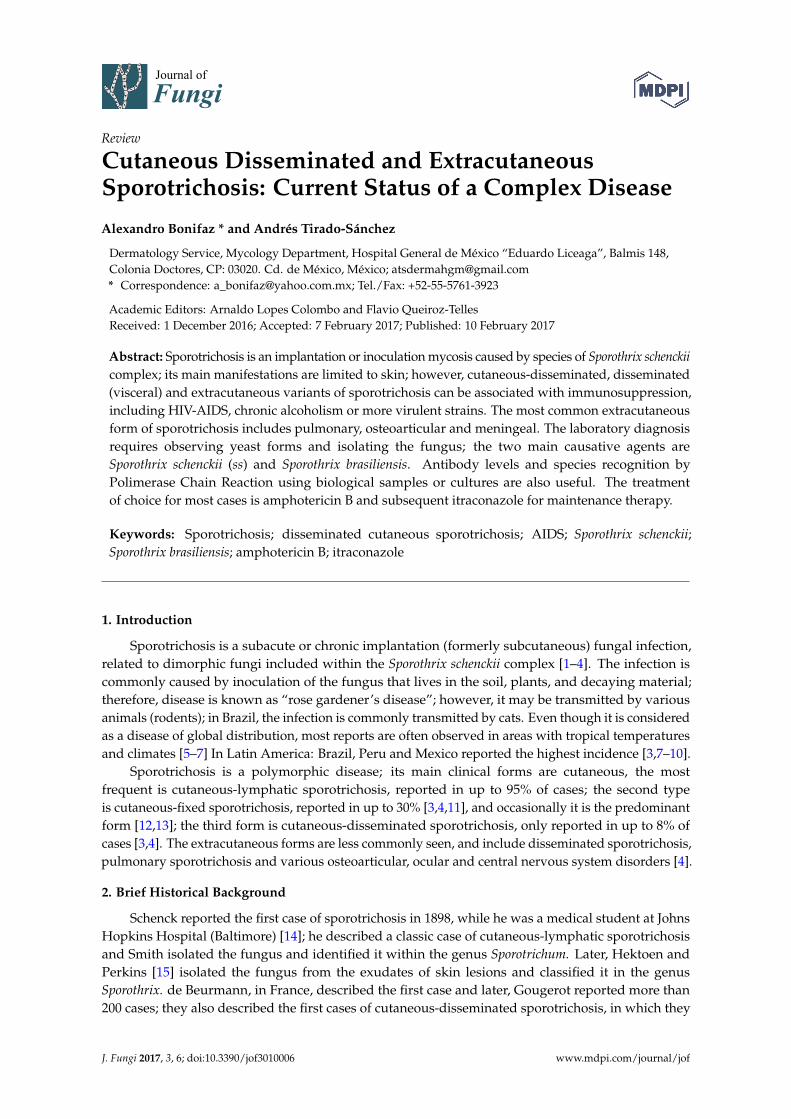
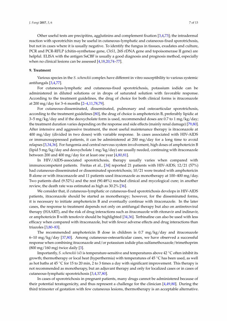
Cutaneous-disseminated sporotrichosis, also called hematogenous sporotrichosis, is a rare entity,usually seen in immunocompromised patients [3,4,46], due to the mentioned predisposing factors,where the causative agent has a role as an opportunist; there are few reports in immunocompetentpatients [49–52], pointing out that virulence is an important factor for disease development; however,this theory has not been fully verified [56,57]. Clinical manifestations of cutaneous-disseminatedsporotrichosis include ulcerated nodules and verrucous plaques (Figure 1) [58–60]; there are casesof many inoculations, this may be related to cat scratches (most cases reported in Brazil) and maydevelop in immunocompetent patients [2,4,29]. Cutaneous-disseminated sporotrichosis can be foundaffecting any part of the body surface, and even mucous membranes (mouth, pharynx, penis glans)in one third of the patients, developing ulcerations and sinus plaques [34–36,61]. It may affect bonesand joints, producing small granulomatous lesions or even extensive lytic lesions and osteomyelitis,associated with joint effusions, edema and severe pain; the most affected bones include tibia, carp andmetacarpus, ulna, knee and ankle, in that order [4,41,62–65]. Osteoarticular cases or sporotrichoidarthritis without cutaneous involvement, derived from pulmonary or hematogenous dissemination,has been reported [4,33]. Cutaneous-disseminated sporotrichosis can extend to various organs andsystems (e.g., testes, central nervous system, etc.) rapidly progressing to fungemia [3,4,34,35,38].
J. Fungi 2017, 3, 6 5 of 13
J. Fungi 2017, 3, 6 5 of 12
Figure 1. Extensive cutaneous disseminated sporotrichosis associated to chronic alcoholism.
7.2. Extracutaneous Sporotrichosis
Pulmonary sporotrichosis is a rare entity; about 100 cases have been reported so far [33,66,67], most of them are primary disease and they are usually seen in high endemic areas. It is classified into two clinical types 1. The chronic type (most common), usually asymptomatic (98%), it presents with limited cavitary zones, indistinguishable from tuberculosis; symptomatic cases manifest as pneumonia, with little cough and expectoration. The radiographs show areas of condensation, or infiltrated milliar type 2. The acute and progressive type, involves tracheobronchial lymph nodes, developing massive adenopathies, which may derive into bronchial obstruction; common symptoms include cough with abundant expectoration, dyspnea and fatigue. Chest X ray shows parahilar lymphadenopathy and, less commonly, mediastinal enlargement. Aung et al., [32] reported 86 cases diagnosed during 50 years (1960–2010), 74.4% of those were primary, while 25.6% were multifocal and most cases involved immunocompromised patients.
Central nervous system involvement is one of the deadliest complications of sporotrichosis. It has been reported in patients with severe immunosuppression often related to leukemia and post-transplanted therapy; however, the largest numbers of cases are associated with HIV-AIDS [34,36,68] and as part of zoonotic epidemics. These cases are commonly due to S. brasiliensis; invasion seems to occur in about 17% of cases; however, the exact frequency is unknown [36]. Central nervous system involvement manifests as meningoencephalitis and hydrocephalus, clinical symptoms include headache, fever, neck stiffness, mental confusion and vomiting; the main differential diagnosis is cryptococcosis [4,67]. It is also important to mention that because of the antiretroviral therapy, immune reconstitution inflammatory syndrome can be present; it is estimated that it occurs in little more than 7% and there can be diverse clinical manifestations [34,36,68,69].
Disseminated sporotrichosis can affect the skin, lungs, sinuses (sinusitis), liver, kidney, eyes (uveitis, endophthalmitis), genitalia, heart (endocarditis). Clinical features are variable and are often detected at necropsy [3,4,32,36,39,41,48].
8. Laboratory Diagnosis
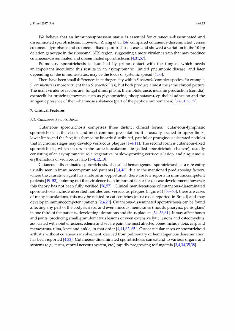
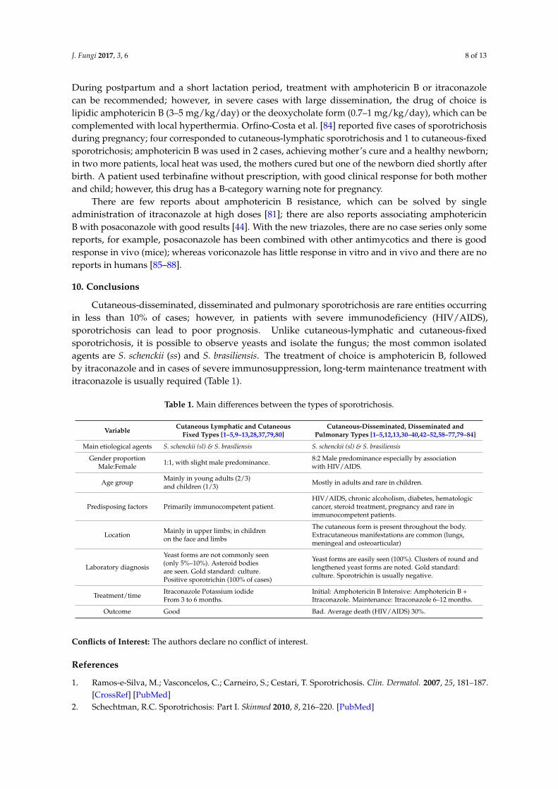
Direct examinations and staining are not useful for diagnosis of cutaneous-lymphatic and cutaneous-fixed sporotrichosis, since yeasts are observed only in a low percentage (5%–10%), whereas in cases of cutaneous-disseminated, disseminated and pulmonary sporotrichosis, Gram, Giemsa, Periodic Acid–Schiff (PAS) and Gomori-Grocott stains are useful for diagnosis as well as immunofluorescence techniques [3,4,11,70]. Yeast forms are usually round, oval or lengthened, described as “cigar-shaped” (Figure 2); in cases of immunocompromised patients, large clusters of yeast are observed, similar to feline sporotrichosis [8,29]. Differential diagnosis includes mainly histoplasmosis; in fungemia [3,4,35,36], yeasts are easily observed from blood imprints and no special staining is required [4,38,71,72].
Figure 1. Extensive cutaneous disseminated sporotrichosis associated to chronic alcoholism.
7.2. Extracutaneous Sporotrichosis
Pulmonary sporotrichosis is a rare entity; about 100 cases have been reported so far [33,66,67],most of them are primary disease and they are usually seen in high endemic areas. It is classified intotwo clinical types 1. The chronic type (most common), usually asymptomatic (98%), it presents withlimited cavitary zones, indistinguishable from tuberculosis; symptomatic cases manifest as pneumonia,with little cough and expectoration. The radiographs show areas of condensation, or infiltrated milliartype 2. The acute and progressive type, involves tracheobronchial lymph nodes, developing massiveadenopathies, which may derive into bronchial obstruction; common symptoms include cough withabundant expectoration, dyspnea and fatigue. Chest X ray shows parahilar lymphadenopathy and,less commonly, mediastinal enlargement. Aung et al., [32] reported 86 cases diagnosed during 50 years(1960–2010), 74.4% of those were primary, while 25.6% were multifocal and most cases involvedimmunocompromised patients.
Central nervous system involvement is one of the deadliest complications of sporotrichosis.It has been reported in patients with severe immunosuppression often related to leukemiaand post-transplanted therapy; however, the largest numbers of cases are associated withHIV-AIDS [34,36,68] and as part of zoonotic epidemics. These cases are commonly due to S. brasiliensis;invasion seems to occur in about 17% of cases; however, the exact frequency is unknown [36].Central nervous system involvement manifests as meningoencephalitis and hydrocephalus, clinicalsymptoms include headache, fever, neck stiffness, mental confusion and vomiting; the main differentialdiagnosis is cryptococcosis [4,67]. It is also important to mention that because of the antiretroviraltherapy, immune reconstitution inflammatory syndrome can be present; it is estimated that it occurs inlittle more than 7% and there can be diverse clinical manifestations [34,36,68,69].
Disseminated sporotrichosis can affect the skin, lungs, sinuses (sinusitis), liver, kidney,eyes (uveitis, endophthalmitis), genitalia, heart (endocarditis). Clinical features are variable andare often detected at necropsy [3,4,32,36,39,41,48].
8. Laboratory Diagnosis
Direct examinations and staining are not useful for diagnosis of cutaneous-lymphatic andcutaneous-fixed sporotrichosis, since yeasts are observed only in a low percentage (5%–10%),whereas in cases of cutaneous-disseminated, disseminated and pulmonary sporotrichosis, Gram,Giemsa, Periodic Acid–Schiff (PAS) and Gomori-Grocott stains are useful for diagnosis as well asimmunofluorescence techniques [3,4,11,70]. Yeast forms are usually round, oval or lengthened,described as “cigar-shaped” (Figure 2); in cases of immunocompromised patients, large clustersof yeast are observed, similar to feline sporotrichosis [8,29]. Differential diagnosis includes mainly
J. Fungi 2017, 3, 6 6 of 13
histoplasmosis; in fungemia [3,4,35,36], yeasts are easily observed from blood imprints and no specialstaining is required [4,38,71,72].
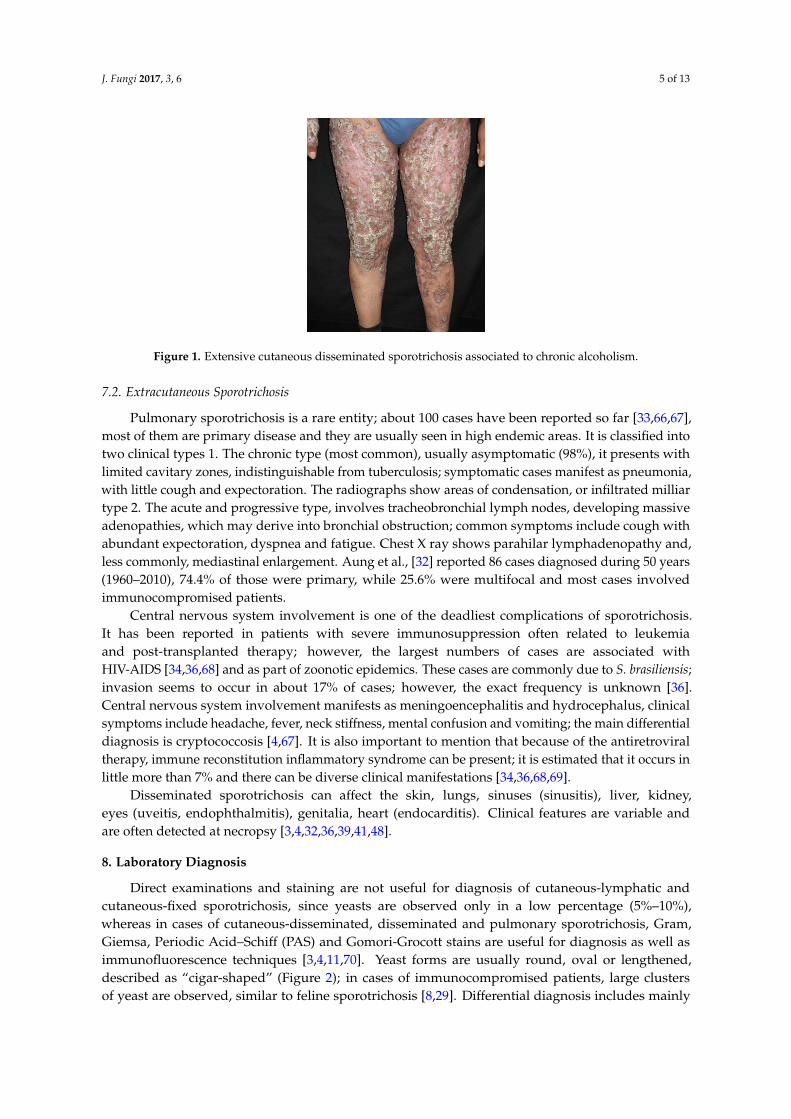
Cultures from exudative lesions, scale, tissue fragment, sputum and blood are the gold standardfor diagnosis. Sabouraud dextrose agar with and without antibiotics, incubated at 28 ◦C is often useful;the colonies may develop between 5 and 8 days; because of its dimorphic feature, Sporothrix spp.can produce yeast colonies (blood agar, chocolate agar, BHI agar) in rich media, incubated at 37 ◦C;this must be distinguished from bacterial colonies. S. schenckii (sl), presents filamentous colonieswith thin 1–3 micron septate, branched, hyaline hyphae, reproduce by ovoid, round and pyriformmicroconidia, derived from the denticle (sympudolic) form of conidiophores (10 to 30 µm in length) ordirectly from the hyphae; microscopically appear as “peach blossoms or daisies” (Figure 3) [3,4,11,24].
The histopathology offers a useful tool for cutaneous-lymphatic and cutaneous-fixedsporotrichosis diagnosis. Suppurative granulomatous pictures (84%) [70], where dispersed yeastscan rarely be seen and often with a radiated halo (asteroid bodies) are usually reported. In cases ofcutaneous-disseminated and disseminated sporotrichosis, a similar histological distribution is seenas well as the presence of yeasts, depending on the patient’s immune status; some cumulus to largeamounts of yeasts can also be noted, most of them are round or elongated; this becomes obvious withPAS and Grocott stains. It must be distinguished from histoplasmosis [3,70].
J. Fungi 2017, 3, 6 6 of 12
Cultures from exudative lesions, scale, tissue fragment, sputum and blood are the gold standard for diagnosis. Sabouraud dextrose agar with and without antibiotics, incubated at 28 °C is often useful; the colonies may develop between 5 and 8 days; because of its dimorphic feature, Sporothrix spp. can produce yeast colonies (blood agar, chocolate agar, BHI agar) in rich media, incubated at 37 °C; this must be distinguished from bacterial colonies. S. schenckii (sl), presents filamentous colonies with thin 1–3 micron septate, branched, hyaline hyphae, reproduce by ovoid, round and pyriform microconidia, derived from the denticle (sympudolic) form of conidiophores (10 to 30 μm in length) or directly from the hyphae; microscopically appear as “peach blossoms or daisies” (Figure 3) [3,4,11,24].
The histopathology offers a useful tool for cutaneous-lymphatic and cutaneous-fixed sporotrichosis diagnosis. Suppurative granulomatous pictures (84%) [70], where dispersed yeasts can rarely be seen and often with a radiated halo (asteroid bodies) are usually reported. In cases of cutaneous-disseminated and disseminated sporotrichosis, a similar histological distribution is seen as well as the presence of yeasts, depending on the patient’s immune status; some cumulus to large amounts of yeasts can also be noted, most of them are round or elongated; this becomes obvious with PAS and Grocott stains. It must be distinguished from histoplasmosis [3,70].
Figure 2. Biopsy of disseminated sporotrichosis. Renal biopsy with multiple clusters of lengthened yeast forms “cigar-shaped” (Grocott, 40×).
Figure 3. Culture of Sporothrix schenckii (Sabouraud media, 28 °C) Filamentous state with thin hyphae and denticle microconidia like “daisy flowers” (Erythrosine, 40×).
Figure 2. Biopsy of disseminated sporotrichosis. Renal biopsy with multiple clusters of lengthenedyeast forms “cigar-shaped” (Grocott, 40×).
J. Fungi 2017, 3, 6 6 of 12
Cultures from exudative lesions, scale, tissue fragment, sputum and blood are the gold standard for diagnosis. Sabouraud dextrose agar with and without antibiotics, incubated at 28 °C is often useful; the colonies may develop between 5 and 8 days; because of its dimorphic feature, Sporothrix spp. can produce yeast colonies (blood agar, chocolate agar, BHI agar) in rich media, incubated at 37 °C; this must be distinguished from bacterial colonies. S. schenckii (sl), presents filamentous colonies with thin 1–3 micron septate, branched, hyaline hyphae, reproduce by ovoid, round and pyriform microconidia, derived from the denticle (sympudolic) form of conidiophores (10 to 30 μm in length) or directly from the hyphae; microscopically appear as “peach blossoms or daisies” (Figure 3) [3,4,11,24].
The histopathology offers a useful tool for cutaneous-lymphatic and cutaneous-fixed sporotrichosis diagnosis. Suppurative granulomatous pictures (84%) [70], where dispersed yeasts can rarely be seen and often with a radiated halo (asteroid bodies) are usually reported. In cases of cutaneous-disseminated and disseminated sporotrichosis, a similar histological distribution is seen as well as the presence of yeasts, depending on the patient’s immune status; some cumulus to large amounts of yeasts can also be noted, most of them are round or elongated; this becomes obvious with PAS and Grocott stains. It must be distinguished from histoplasmosis [3,70].
Figure 2. Biopsy of disseminated sporotrichosis. Renal biopsy with multiple clusters of lengthened yeast forms “cigar-shaped” (Grocott, 40×).
Figure 3. Culture of Sporothrix schenckii (Sabouraud media, 28 °C) Filamentous state with thin hyphae and denticle microconidia like “daisy flowers” (Erythrosine, 40×).
Figure 3. Culture of Sporothrix schenckii (Sabouraud media, 28 ◦C) Filamentous state with thin hyphaeand denticle microconidia like “daisy flowers” (Erythrosine, 40×).
J. Fungi 2017, 3, 6 7 of 13
Other useful tests are precipitins, agglutinins and complement fixation [3,4,73]; the intradermalreaction with sporotrichin may be useful in cutaneous-lymphatic and cutaneous-fixed sporotrichosis,but not in cases where it is usually negative. To identify the fungus in tissues, exudates and culture,PCR and PCR-RFLP (chitin-synthetase gene, ChS1, 26S rDNA gene and topoisomerase II gene) arehelpful. ELISA with the antigen SsCBF is usually a good diagnosis and prognosis method, especiallywhen no clinical lesions can be assessed [4,18,20,74–77].
9. Treatment
Various species in the S. schenckii complex have different in vitro susceptibility to various systemicantifungals [3,4,77].
For cutaneous-lymphatic and cutaneous-fixed sporotrichosis, potassium iodide can beadministered in diluted solutions or in drops of saturated solution with favorable response.According to the treatment guidelines, the drug of choice for both clinical forms is itraconazoleat 200 mg/day for 3–6 months [2–4,11,78,79].
For cutaneous-disseminated, disseminated, pulmonary and osteoarticular sporotrichosis,according to the treatment guidelines [80], the drug of choice is amphotericin B, preferably lipidic at3–5 mg/kg/day and if the deoxycholate form is used, recommended doses are 0.7 to 1 mg/kg/day;the treatment duration varies depending on the response and side effects (mainly renal damage) [79,80].After intensive and aggressive treatment, the most useful maintenance therapy is itraconazole at400 mg/day (divided in two doses) with variable response. In cases associated with HIV-AIDSor immunosuppressed patients, it can be administered at 200 mg/day for a long time to avoidrelapses [3,34,36]. For fungemia and central nervous system involvement, high doses of amphotericin B(lipid 5 mg/kg/day and deoxycholate 1 mg/kg/day) are usually needed, continuing with itraconazolebetween 200 and 400 mg/day for at least one year [4,80,81].
In HIV/AIDS-associated sporotrichosis, therapy usually varies when compared withimmunocompetent patients. Freitas et al., [34] reported 21 patients with HIV-AIDS; 12/21 (57%)had cutaneous-disseminated or disseminated sporotrichosis; 10/21 were treated with amphotericinB alone or with itraconazole and 11 patients used itraconazole as monotherapy at 100–400 mg/day.Two patients died (9.52%) and the rest (90.48%) reached clinical and mycological cure; in anotherreview, the death rate was estimated as high as 30.2% [36].
We consider that, if cutaneous-lymphatic or cutaneous-fixed sporotrichosis develops in HIV-AIDSpatients, itraconazole should be started as monotherapy; however, for the disseminated forms,it is necessary to initiate amphotericin B and eventually continue with itraconazole. In the latercases, the response to treatment depends not only on antifungal therapy but also on antiretroviraltherapy (HAART), and the risk of drug interactions such as itraconazole with ritonavir and indinavir,or amphotericin B with tenofovir should be highlighted [34,36]. Terbinafine can also be used with lessefficacy when compared with itraconazole, but with fewer adverse effects and drug interactions thantriazoles [3,80–83].
The recommended amphotericin B dose in children is 0.7 mg/kg/day and itraconazole6–10 mg/kg/day [37,80]. Among cutaneous-osteoarticular cases, we have observed a successfulresponse when combining itraconazole and/or potassium iodide plus sulfamethoxazole/trimethoprim(800 mg/160 mg) twice daily [3].
Importantly, S. schenckii (sl) is temperature-sensitive and temperatures above 42 ◦C often inhibit itsgrowth; thermotherapy or local heat (hyperthermia) with temperatures of 45 ◦C has been used, as wellas hot baths at 45 ◦C for 15 to 20 min, 2 to 3 times a day with significant improvement. This therapy isnot recommended as monotherapy, but an adjuvant therapy and only for localized cases or in cases ofcutaneous-lymphatic sporotrichosis [3,4,37,80].
In cases of sporotrichosis in pregnant patients, many drugs cannot be administered because oftheir potential teratogenicity, and thus represent a challenge for the clinician [4,49,80]. During thethird trimester of gestation with few cutaneous lesions, thermotherapy is an acceptable alternative.
J. Fungi 2017, 3, 6 8 of 13
During postpartum and a short lactation period, treatment with amphotericin B or itraconazolecan be recommended; however, in severe cases with large dissemination, the drug of choice islipidic amphotericin B (3–5 mg/kg/day) or the deoxycholate form (0.7–1 mg/kg/day), which can becomplemented with local hyperthermia. Orfino-Costa et al. [84] reported five cases of sporotrichosisduring pregnancy; four corresponded to cutaneous-lymphatic sporotrichosis and 1 to cutaneous-fixedsporotrichosis; amphotericin B was used in 2 cases, achieving mother’s cure and a healthy newborn;in two more patients, local heat was used, the mothers cured but one of the newborn died shortly afterbirth. A patient used terbinafine without prescription, with good clinical response for both motherand child; however, this drug has a B-category warning note for pregnancy.
There are few reports about amphotericin B resistance, which can be solved by singleadministration of itraconazole at high doses [81]; there are also reports associating amphotericinB with posaconazole with good results [44]. With the new triazoles, there are no case series only somereports, for example, posaconazole has been combined with other antimycotics and there is goodresponse in vivo (mice); whereas voriconazole has little response in vitro and in vivo and there are noreports in humans [85–88].
10. Conclusions
Cutaneous-disseminated, disseminated and pulmonary sporotrichosis are rare entities occurringin less than 10% of cases; however, in patients with severe immunodeficiency (HIV/AIDS),sporotrichosis can lead to poor prognosis. Unlike cutaneous-lymphatic and cutaneous-fixedsporotrichosis, it is possible to observe yeasts and isolate the fungus; the most common isolatedagents are S. schenckii (ss) and S. brasiliensis. The treatment of choice is amphotericin B, followedby itraconazole and in cases of severe immunosuppression, long-term maintenance treatment withitraconazole is usually required (Table 1).
Table 1. Main differences between the types of sporotrichosis.
Variable Cutaneous Lymphatic and CutaneousFixed Types [1–5,9–13,28,37,79,80]
Cutaneous-Disseminated, Disseminated andPulmonary Types [1–5,12,13,30–40,42–52,58–77,79–84]
Main etiological agents S. schenckii (sl) & S. brasiliensis S. schenckii (sl) & S. brasiliensis
Gender proportionMale:Female 1:1, with slight male predominance. 8:2 Male predominance especially by association
with HIV/AIDS.
Age group Mainly in young adults (2/3)and children (1/3) Mostly in adults and rare in children.
Predisposing factors Primarily immunocompetent patient.HIV/AIDS, chronic alcoholism, diabetes, hematologiccancer, steroid treatment, pregnancy and rare inimmunocompetent patients.
Location Mainly in upper limbs; in childrenon the face and limbs
The cutaneous form is present throughout the body.Extracutaneous manifestations are common (lungs,meningeal and osteoarticular)
Laboratory diagnosis
Yeast forms are not commonly seen(only 5%–10%). Asteroid bodiesare seen. Gold standard: culture.Positive sporotrichin (100% of cases)
Yeast forms are easily seen (100%). Clusters of round andlengthened yeast forms are noted. Gold standard:culture. Sporotrichin is usually negative.
Treatment/time Itraconazole Potassium iodideFrom 3 to 6 months.
Initial: Amphotericin B Intensive: Amphotericin B +Itraconazole. Maintenance: Itraconazole 6–12 months.
Outcome Good Bad. Average death (HIV/AIDS) 30%.
Conflicts of Interest: The authors declare no conflict of interest.
References
1. Ramos-e-Silva, M.; Vasconcelos, C.; Carneiro, S.; Cestari, T. Sporotrichosis. Clin. Dermatol. 2007, 25, 181–187.[CrossRef] [PubMed]
2. Schechtman, R.C. Sporotrichosis: Part I. Skinmed 2010, 8, 216–220. [PubMed]

J. Fungi 2017, 3, 6 9 of 13
3. Bonifaz, A.; Vazquez-Gonzalez, D. Sporotrichosis: An update. G Ital. Dermatol. Venereol. 2010, 145, 659–673.[PubMed]
4. Barros, M.B.; de Almeida Paes, R.; Schubach, A.O. Sporothrix schenckii and Sporotrichosis. Clin. Microbiol. Rev.2011, 24, 633–654. [CrossRef] [PubMed]
5. Engle, J.; Desir, J.; Bernstein, J.M. A rose by any other name. Skinmed 2007, 6, 139–141. [CrossRef] [PubMed]6. Rodrigues, A.M.; de Hoog, S.; de Camargo, Z.P. Emergence of pathogenicity in the Sporothrix schenckii
complex. Med. Mycol. 2013, 51, 405–412. [CrossRef] [PubMed]7. Chakrabarti, A.; Bonifaz, A.; Gutierrez-Galhardo, M.C.; Mochizuki, T.; Li, S. Global epidemiology of
sporotrichosis. Med. Mycol. 2015, 53, 3–14. [CrossRef] [PubMed]8. Pereira, S.A.; Gremião, I.D.; Kitada, A.A.; Boechat, J.S.; Viana, P.G.; Schubach, T.M. The epidemiological
scenario of feline sporotrichosis in Rio de Janeiro, State of Rio de Janeiro, Brazil. Rev. Soc. Bras. Med. Trop.2014, 47, 392–393. [CrossRef] [PubMed]
9. Pappas, P.G.; Tellez, I.; Deep, A.E.; Nolasco, D.; Holgado, W.; Bustamante, B. Sporotrichosis in Peru:Description of an area of hyperendemicity. Clin. Infect. Dis. 2000, 30, 65–70. [CrossRef] [PubMed]
10. Bustamante, B.; Campos, P.E. Endemic sporotrichosis. Curr. Opin. Infect. Dis. 2001, 14, 145–149. [CrossRef][PubMed]
11. Bonifaz, A.; Vázquez-González, D. Diagnosis and treatment of sporotrichosis lymphocutaneous: What arethe options? Curr. Fungal Infect. Rev. 2013, 7, 252–259. [CrossRef]
12. Song, Y.; Li, S.S.; Zhong, S.X.; Liu, Y.Y.; Yao, L.; Huo, S.S. Report of 457 sporotrichosis cases from Jilinprovince, Northeast China, a serious endemic region. J. Eur. Acad. Dermatol. Venereol. 2013, 27, 313–811.[CrossRef] [PubMed]
13. Takenaka, M.; Yoshizaki, A.; Utani, A.; Nishimoto, K. A survey of 165 sporotrichosis cases examined inNagasaki prefecture from 1951 to 2012. Mycoses 2014, 57, 294–298. [CrossRef] [PubMed]
14. Schenk, B.R. On refractory subcutaneous abscesses caused by a fungus possibly related to Sporotrichia.Bull Johns Hopkins Hosp. 1898, 9, 286–290.
15. Hektoen, L.; Perkins, C.F. Refractory subcutaneous abscesses caused by Sporothrix schenckii. a new pathogenicfungus. J. Boston Soc. Med. Sci. 1900, 5, 77–89. [CrossRef]
16. Aram, H. Sporotrichosis. A historical approach. Int. J. Dermatol. 1986, 25, 203–205. [CrossRef] [PubMed]17. De Beurmann, L.H.G. Les Sporotrichoses; Felix Alcan: Paris, France, 1912.18. Marimon, R.; Cano, J.; Gene, J.; Sutton, D.A.; Kawasaki, M.; Guarro, J. Sporothrix brasiliensis, S. globosa,
and S. mexicana, three new Sporothrix species of clinical interest. J. Clin. Microbiol. 2007, 45, 3198–3206.[CrossRef] [PubMed]
19. Tellez, M.D.; Batista-Duharte, A.; Portuondo, D.; Quinello, C.; Bonne-Hernandez, R.; Carlos, I.Z.Sporothrix schenckii complex biology: Environment and fungal pathogenicity. Microbiology 2014, 160,2352–2365. [CrossRef] [PubMed]
20. Lopez-Romero, E.; Reyes-Montes Mdel, R.; Perez-Torres, A.; Ruiz-Baca, E.; Villagomez-Castro, J.C.;Mora-Montes, H.M.; Flores-Carreon, A.; Toriello, C. Sporothrix schenckii complex and sporotrichosis, anemerging health problem. Future Microbiol. 2011, 6, 85–102. [CrossRef] [PubMed]
21. Madrid, H.; Cano, J.; Gene, J.; Bonifaz, A.; Toriello, C.; Guarro, J. Sporothrix globosa, a pathogenic funguswith widespread geographical distribution. Rev. Iberoam Micol. 2009, 26, 218–222. [CrossRef] [PubMed]
22. Marimon, R.; Gené, J.; Cano, J.; Guarro, J. Sporothrix luriei: A rare fungus from clinical origin. Med. Mycol.2008, 46, 621–625. [CrossRef] [PubMed]
23. Rodrigues, A.M.; de Hoog, G.S.; de Cássia-Pires, D.; Brihante, R.S.; Sidrim, J.J.; Gadelha, M.F.; Colombo, A.L.;de Camargo, Z.P. Genetic diversity and antifungal susceptibility profiles in causative agents of sporotrichosis.BMC Infect. Dis. 2014, 23, 219. [CrossRef] [PubMed]
24. Rodrigues, A.M.; Cruz-Choappa, R.; Fernandes, G.F.; de Hoog, G.S.; de Camargo, Z.P. Sporothrix chilensissp. nov. (Ascomycota: Ophiostomatales), a soil-borne agent of human sporotrichosis with mild-pathogenicpotential to mammals. Fungal Biol. 2016, 120, 246–264. [CrossRef] [PubMed]
25. De Beer, Z.W.; Duong, T.A.; Wingfield, M.J. The divorce of Sporothrix and Ophiostoma: Solution toa problematic relationship. Stud. Mycol. 2016, 83, 165–191. [CrossRef] [PubMed]
26. Simson, F.W. Sporotrichosis infection in mines in Witwatersrand. A symposium. Proc Transv. Mine Med.Officers Assoc. 1947, 4, 51–54.

J. Fungi 2017, 3, 6 10 of 13
27. Dixon, D.M.; Salkin, I.F.; Duncan, R.A.; Hurd, N.J.; Haines, J.H.; Kemna, M.E.; Coles, F.B. Isolation andcharacterization of Sporothrix schenckii from clinical and environmental sources associated with the largestU.S. epidemic of sporotrichosis. J. Clin. Microbiol. 1991, 29, 1106–1113. [PubMed]
28. Ramirez Soto, M.C. Sporotrichosis: The story of an endemic region in Peru over 28 Years (1985 to 2012).PLoS ONE 2015, 10, e0127924. [CrossRef] [PubMed]
29. Sanchotene, K.O.; Madrid, I.M.; Klafke, G.B.; Bergamashi, M.; Della Terra, P.P.; Rodrigues, A.M.Sporothrix brasiliensis outbreaks and the rapid emergence of feline sporotrichosis. Mycoses 2015, 58, 652–658.[CrossRef] [PubMed]
30. Sanchez-Aleman, M.A.; Araiza, J.; Bonifaz, A. Aislamiento y caracterización de cepas silvestres deSporothrix schenckii e investigación de reactores a la esporotricina. Gac. Med. Mex 2004, 140, 507–512.[PubMed]
31. Zhang, Y.; Hagen, F.; Stielow, B.; Rodrigues, A.M.; Samerpitak, K.; Zhou, X.; Feng, P.; Yang, L.; Chen, M.;Deng, S.; et al. Phylogeography and evolutionary patterns in Sporothrix spanning more than 14,000 humanand animal case reports. Persoonia 2015, 35, 1–20. [CrossRef] [PubMed]
32. Aung, A.K.; Teh, B.M.; McGrath, C.; Thompson, P.J. Pulmonary sporotrichosis: Case series and systematicanalysis of literature on clinico-radiological patterns and management outcomes. Med. Mycol. 2013, 51,534–544. [CrossRef] [PubMed]
33. Aung, A.K.; Spelman, D.W.; Thompson, P.J. Pulmonary Sporotrichosis: An evolving clinical paradigm.Semin. Respir. Crit. Care Med. 2015, 36, 756–766. [CrossRef] [PubMed]
34. Freitas, D.F.; de Siqueira Hoagland, B.; do Valle, A.C.; Fraga, B.B.; de Barros, M.B.; de Oliveira Schubach, A.;de Almeida-Paes, R.; Cuzzi, T.; Rosalino, C.M.; Zancope-Oliveira, R.M.; et al. Sporotrichosis in HIV-infectedpatients: Report of 21 cases of endemic sporotrichosis in Rio de Janeiro, Brazil. Med. Mycol. 2012, 50, 170–178.[CrossRef] [PubMed]
35. Freitas, D.F.; Valle, A.C.; da Silva, M.B.; Campos, D.P.; Lyra, M.R.; de Souza, R.V.; Veloso, V.G.;Zancope-Oliveira, R.M.; Bastos, F.I.; et al. Sporotrichosis: An emerging neglected opportunistic infection inHIV-infected patients in Rio de Janeiro, Brazil. PLoS Negl. Trop. Dis. 2014, 8, e3110. [CrossRef] [PubMed]
36. Moreira, J.A.; Freitas, D.F.; Lamas, C.C. The impact of sporotrichosis in HIV-infected patients: A systematicreview. Infection 2015, 43, 267–276. [CrossRef] [PubMed]
37. Tirado-Sánchez, A.; Bonifaz, A. Sporotrichosis in Children: An Update. Curr. Fungal Infect. Rep. 2016, 10,107–116. [CrossRef]
38. Castrejon, O.V.; Robles, M.; Zubieta Arroyo, O.E. Fatal fungaemia due to Sporothrix schenckii. Mycoses 1995,38, 373–376. [CrossRef] [PubMed]
39. Espinoza-Hernandez, C.J.; Jesus-Silva, A.; Toussaint-Caire, S.; Arenas, R. Disseminated sporotrichosis withcutaneous and testicular involvement. Actas Dermosifiliogr. 2014, 105, 204–206. [CrossRef] [PubMed]
40. Nassif, P.W.; Granado, I.R.; Ferraz, J.S.; Souza, R.; Nassif, A.E. Atypical presentation of cutaneoussporotrichosis in an alcoholic patient. Dermatol. Online J. 2012, 18, 12. [PubMed]
41. Kauffman, C.A.; Pappas, P.G.; McKinsey, D.S.; Greenfield, R.A.; Perfect, J.R.; Cloud, G.A.; Thomas, C.J.;Dismukes, W.E. Treatment of lymphocutaneous and visceral sporotrichosis with fluconazole. Clin. Infect. Dis.1996, 22, 46–50. [CrossRef] [PubMed]
42. Ewing, G.E.; Bosl, G.J.; Peterson, P.K. Sporothrix schenckii meningitis in a farmer with Hodgkin’s disease.Am. J. Med. 1980, 68, 455–457. [CrossRef]
43. Kumar, S.; Kumar, D.; Gourley, W.K.; Alperin, J.B. Sporotrichosis as a presenting manifestation of hairy cellleukemia. Am. J. Hematol. 1994, 46, 134–137. [CrossRef] [PubMed]
44. Bunce, P.E.; Yang, L.; Chun, S.; Zhang, S.X.; Trinkaus, M.A.; Matukas, L.M. Disseminated sporotrichosisin a patient with hairy cell leukemia treated with amphotericin B and posaconazole. Med. Mycol. 2012, 50,197–201. [CrossRef] [PubMed]
45. Solorzano, S.; Ramirez, R.; Cabada, M.M.; Montoya, M.; Cazorla, E. Disseminated cutaneous sporotrichosiswith joint involvement in a woman with type 2 diabetes. Rev. Peru Med. Exp. Salud. Publica 2015, 32, 187–190.[CrossRef] [PubMed]
46. Severo, L.C.; Festugato, M.; Bernardi, C.; Londero, A.T. Widespread cutaneous lesions due toSporothrix schenckii in a patient under a long-term steroids therapy. Rev. Inst. Med. Trop. Sao Paulo1999, 41, 59–62. [CrossRef] [PubMed]

J. Fungi 2017, 3, 6 11 of 13
47. Gewehr, P.; Jung, B.; Aquino, V.; Manfro, R.C.; Spuldaro, F.; Rosa, R.G.; Goldani, L.Z. Sporotrichosis in renaltransplant patients. Can. J. Infect. Dis. Med. Microbiol. 2013, 24, 47–49. [CrossRef]
48. Gullberg, R.M.; Quintanilla, A.; Levin, M.L.; Williams, J.; Phair, J.P. Sporotrichosis: Recurrent cutaneous,articular, and central nervous system infection in a renal transplant recipient. Rev. Infect. Dis. 1987, 9,369–375. [CrossRef] [PubMed]
49. Ferreira, C.P.; do Valle, A.C.; Freitas, D.F.; Reis, R.; Galhardo, M.C. Pregnancy during a sporotrichosisepidemic in Rio de Janeiro, Brazil. Int. J. Gynaecol. Obstet. 2012, 117, 294–295. [CrossRef] [PubMed]
50. Yap, F.B. Disseminated cutaneous sporotrichosis in an immunocompetent individual. Int. J. Infect. Dis. 2011,15, 727–729. [CrossRef] [PubMed]
51. Romero-Cabello, R.; Bonifaz, A.; Romero-Feregrino, R.; Sanchez, C.J.; Linares, Y.; Zavala, J.T.; Romero, L.C.;Romero-Feregrino, R.; Vega, J.T. Disseminated sporotrichosis. BMJ Case Rep. 2011, 25, 2011. [CrossRef][PubMed]
52. Hassan, K.; Turker, T.; Zangeneh, T. Disseminated sporotrichosis in an immunocompetent patient. Case Rep.Plast Surg. Hand Surg. 2016, 3, 44–47. [CrossRef] [PubMed]
53. Koga, T.; Duan, H.; Furue, M. Immunohistochemical detection of interferon-γ-producing cells in granulomaformation of sporotrichosis. Med. Mycol. 2002, 40, 111–114. [CrossRef] [PubMed]
54. Uenotsuchi, T.; Takeuchi, S.; Matsuda, T.; Urabe, K.; Koga, T.; Uchi, H.; Nakahara, T.; Fukagawa, S.;Kawasaki, M.; Kajiwara, H.; et al. Differential induction of Th1-prone immunity by human dendritic cellsactivated with Sporothrix schenckii of cutaneous and visceral origins to determine their different virulence.Int. Immunol. 2006, 18, 1637–1646. [CrossRef] [PubMed]
55. Verdan, F.F.; Faleiros, J.C.; Ferreira, L.S.; Monnazzi, L.G.; Maia, D.C.; Tansine, A.; Placeres, M.C.; Carlos, I.Z.;Santos-Junior, R.R. Dendritic cell are able to differentially recognize Sporothrix schenckii antigens and promoteTh1/Th17 response in vitro. Immunobiology 2012, 217, 788–794. [CrossRef] [PubMed]
56. 56 Zhang, Z.; Liu, X.; Lv, X.; Lin, J. Variation in genotype and higher virulence of a strain of Sporothrix schenckiicausing disseminated cutaneous sporotrichosis. Mycopathologia 2011, 172, 439–446. [CrossRef] [PubMed]
57. Freitas, D.F.; Santos, S.S.; Almeida-Paes, R.; de Oliveira, M.M.; do Valle, A.C.; Gutierrez-Galhardo, M.C.;Zancope-Oliveira, R.M.; Nosanchuk, J.D. Increase in virulence of Sporothrix brasiliensis over five years ina patient with chronic disseminated sporotrichosis. Virulence 2015, 6, 112–120. [CrossRef] [PubMed]
58. Bonifaz, A.; Peniche, A.; Mercadillo, P.; Saul, A. Successful treatment of AIDS-related disseminated cutaneoussporotrichosis with itraconazole. AIDS Patient Care STDs 2001, 15, 603–606. [CrossRef] [PubMed]
59. Fujii, H.; Tanioka, M.; Yonezawa, M.; Arakawa, A.; Matsumura, Y.; Kore-eda, S.; Miyachi, Y.; Tanaka, S.;Mochizuki, T. A case of atypical sporotrichosis with multifocal cutaneous ulcers. Clin. Exp. Dermatol. 2008,33, 135–138. [CrossRef] [PubMed]
60. Stalkup, J.R.; Bell, K.; Rosen, T. Disseminated cutaneous sporotrichosis treated with itraconazole. Cutis 2002,69, 371–374. [PubMed]
61. Fontes, P.C.; Kitakawa, D.; Carvalho, Y.R.; Brandao, A.A.; Cabral, L.A.; Almeida, J.D. Sporotrichosis in anHIV-positive man with oral lesions: A case report. Acta Cytol. 2007, 51, 648–650. [CrossRef] [PubMed]
62. Anees, A.; Ali, A.; Fordham, E.W. Abnormal bone and gallium scans in a case of multifocal systemicsporotrichosis. Clin. Nucl. Med. 1986, 11, 663–664. [CrossRef] [PubMed]
63. Gordhan, A.; Ramdial, P.K.; Morar, N.; Moodley, S.D.; Aboobaker, J. Disseminated cutaneous sporotrichosis:A marker of osteoarticular sporotrichosis masquerading as gout. Int. J. Dermatol. 2001, 40, 717–719.[CrossRef] [PubMed]
64. De Carvalho Aguinaga, F.; Trope, B.M.; Fernandes, N.C.; Engel, D.C.; Ramos, E.S.M. Sporotrichosis withbone involvement: An alert to an occupational disease. Case Rep. Dermatol. 2014, 6, 114–118. [CrossRef][PubMed]
65. Lederer, H.T.; Sullivan, E.; Crum-Cianflone, N.F. Sporotrichosis as an unusual case of osteomyelitis: A casereport and review of the literature. Med. Mycol. Case Rep. 2016, 11, 31–35. [CrossRef] [PubMed]
66. Orofino-Costa, R.; Unterstell, N.; Carlos Gripp, A.; de Macedo, P.M.; Brota, A.; Dias, E.; de Melo Teixeira, M.;Felipe, M.S.; Bernardes-Engemann, A.R.; Lopes-Bezerra, L.M. Pulmonary cavitation and skin lesionsmimicking tuberculosis in a HIV negative patient caused by Sporothrix brasiliensis. Med. Mycol. Case Rep.2013, 2, 65–71. [CrossRef] [PubMed]
67. Callens, S.F.; Kitetele, F.; Lukun, P.; Lelo, P.; Van Rie, A.; Behets, F.; Colebunders, R. PulmonarySporothrix schenckii infection in a HIV positive child. J. Trop. Pediatr. 2006, 52, 144–146. [CrossRef] [PubMed]

J. Fungi 2017, 3, 6 12 of 13
68. Freitas, D.F.; Lima, M.A.; de Almeida-Paes, R.; Lamas, C.C.; do Valle, A.C.; Oliveira, M.M.;Zancope-Oliveira, R.M.; Gutierrez-Galhardo, M.C. Sporotrichosis in the central nervous system caused bySporothrix brasiliensis. Clin. Infect. Dis. 2015, 61, 663–664. [CrossRef] [PubMed]
69. Lyra, M.R.; Nascimento, M.L.; Varon, A.G.; Pimentel, M.I.; Antonio Lde, F.; Saheki, M.N.;Bedoya-Pacheco, S.J.; Valle, A.C. Immune reconstitution inflammatory syndrome in HIV and sporotrichosiscoinfection: Report of two cases and review of the literature. Rev. Soc. Bras. Med. Trop. 2014, 47, 806–809.[CrossRef] [PubMed]
70. Quintella, L.P.; Passos, S.R.; do Vale, A.C.; Galhardo, M.C.; Barros, M.B.; Cuzzi, T.; Reis Rdos, S.;de Carvalho, M.H.; Zappa, M.B.; Schubach Ade, O. Histopathology of cutaneous sporotrichosis in Riode Janeiro: A series of 119 consecutive cases. J. Cutan. Pathol. 2011, 38, 25–32. [CrossRef] [PubMed]
71. Morgan, M.A.; Cockerill, F.R., 3rd; Cortese, D.A.; Roberts, G.D. Disseminated sporotrichosis withSporothrix schenckii fungemia. Diagn. Microbiol. Infect. Dis. 1984, 2, 151–155. [CrossRef]
72. Kosinski, R.M.; Axelrod, P.; Rex, J.H.; Burday, M.; Sivaprasad, R.; Wreiole, A. Sporothrix schenckii fungemiawithout disseminated sporotrichosis. J. Clin. Microbiol. 1992, 30, 501–503. [PubMed]
73. Scott, E.N.; Kaufman, L.; Brown, A.C.; Muchmore, H.G. Serologic studies in the diagnosis and managementof meningitis due to Sporothrix schenckii. N. Engl. J. Med. 1987, 317, 935–940. [CrossRef] [PubMed]
74. Bernardes-Engemann, A.R.; Costa, R.C.; Miguens, B.R.; Penha, C.V.; Neves, E.; Pereira, B.A.; Dias, C.M.;Mattos, M.; Gutierrez, M.C.; Schubach, A.; et al. Development of an enzyme-linked immunosorbent assayfor the serodiagnosis of several clinical forms of sporotrichosis. Med. Mycol. 2005, 43, 487–493. [CrossRef][PubMed]
75. Lopes-Bezerra, L.M.; Schubach, A.; Costa, R.O. Sporothrix schenckii and sporotrichosis. An. Acad. Bras. Cienc.2006, 78, 293–308. [CrossRef] [PubMed]
76. Fernandes, G.F.; Lopes-Bezerra, L.M.; Bernardes-Engemann, A.R.; Schubach, T.M.; Dias, M.A.; Pereira, S.A.;de Camargo, Z.P. Serodiagnosis of sporotrichosis infection in cats by enzyme-linked immunosorbent assayusing a specific antigen, SsCBF, and crude exoantigens. Vet. Microbiol. 2011, 147, 445–449. [CrossRef][PubMed]
77. Rodrigues, A.M.; de Hoog, G.S.; de Camargo, Z.P. Molecular diagnosis of pathogenic Sporothrix species.PLoS Negl. Trop. Dis. 2015, 9, e0004190. [CrossRef] [PubMed]
78. Marimon, R.; Serena, C.; Gene, J.; Cano, J.; Guarro, J. In vitro antifungal susceptibilities of five species ofsporothrix. Antimicrob. Agents Chemother. 2008, 52, 732–734. [CrossRef] [PubMed]
79. Mahajan, V.K. Sporotrichosis: An overview and therapeutic options. Dermatol. Res. Pract. 2014, 2014, 272376.[CrossRef] [PubMed]
80. Kauffman, C.A.; Bustamante, B.; Chapman, S.W.; Pappas, P.G. Clinical practice guidelines for themanagement of sporotrichosis: 2007 update by the Infectious Diseases Society of America. Clin. Infect. Dis.2007, 45, 1255–1265. [CrossRef] [PubMed]
81. Bolao, F.; Podzamczer, D.; Ventin, M.; Gudiol, F. Efficacy of acute phase and maintenance therapy withitraconazole in an AIDS patient with sporotrichosis. Eur. J. Clin. Microbiol. Infect. Dis. 1994, 13, 609–612.[CrossRef] [PubMed]
82. De Lima Barros, M.B.; Schubach, A.O.; de Vasconcellos Carvalhaes de Oliveira, R.; Martins, E.B.; Teixeira, J.L.;Wanke, B. Treatment of cutaneous sporotrichosis with itraconazole—Study of 645 patients. Clin. Infect. Dis.2011, 52, e200–e206. [CrossRef] [PubMed]
83. Paixao, A.G.; Galhardo, M.C.; Almeida-Paes, R.; Nunes, E.P.; Goncalves, M.L.; Chequer, G.L.; Lamas Cda, C.The difficult management of disseminated Sporothrix brasiliensis in a patient with advanced AIDS.AIDS Res. Ther. 2015, 12, 16. [CrossRef] [PubMed]
84. Orfino-Costa, R.; Bernardes-Engemann, A.R.; Azulay-Abulafia, L.; Benvenuto, F.; Neves Mde, L.;Lopes-Bezerra, L.M. Sporotrichosis in pregnancy: Case reports of 5 patients in a zoonotic epidemic inRio de Janeiro, Brazil. An. Bras. Dermatol. 2011, 86, 995–998.
85. Baker, J.H.; Goodpasture, H.C.; Kuhns, H.R., Jr.; Rinaldi, M.G. Fungemia caused by an amphotericinB-resistant isolate of Sporothrix schenckii. Successful treatment with itraconazole. Arch. Pathol. Lab. Med. 1989,113, 1279–1281. [PubMed]
86. Fernández-Silva, F.; Capilla, J.; Mayayo, E.; Guarro, J. Efficacy of posaconazole in murine experimentalsporotrichosis. Antimicrob. Agents Chemother. 2012, 56, 2273–2277. [CrossRef] [PubMed]

J. Fungi 2017, 3, 6 13 of 13
87. Gutierrez-Galhardo, M.C.; Zancopé-Oliveira, R.M.; Monzón, A.; Rodriguez-Tudela, J.L.; Cuenca-Estrella, M.Antifungal susceptibility profile in vitro of Sporothrix schenckii in two growth phases and by two methods:Microdilution and E-test. Mycoses 2010, 53, 227–231. [CrossRef] [PubMed]
88. Fernández-Silva, F.; Capilla, J.; Mayayo, E.; Guarro, J. Modest efficacy of voriconazole against murineinfections by Sporothrix schenckii and lack of efficacy against Sporothrix brasiliensis. Mycoses 2014, 57, 121–124.[CrossRef] [PubMed]
© 2017 by the authors; licensee MDPI, Basel, Switzerland. This article is an open accessarticle distributed under the terms and conditions of the Creative Commons Attribution(CC BY) license (http://creativecommons.org/licenses/by/4.0/).