Embed Size (px)
Citation preview

9/20/21
1
1 OCTOBER 2021
Fast, Slow, Small, and TallOtherwise NOT Normal ECGs
John S. Kim, MD MSAssistant Professor of Pediatrics – Cardiology, CUSOM
Cardiologist and Intensivist, Cardiac Intensive Care Unit
Heart Institute, Children’s Hospital Colorado
1
1
2
3
4
5
Content Outline
2
Brief review of electrocardiography basics
Mostly normal things that look abnormal (PACs, PVCs, low-grade heart block)
When ECGs make the diagnosis (myocarditis, pericarditis, not-so-much MI)
HELP! (arrythmias and high-grade block)
Not so obvious things that are abnormal (long QT)
2

9/20/21
2
Cardiac Conduc+on System• Electrical impulse ini1ated at
the sinus (SA) node
• Spreads across both atria to the
AV node
• AV nodes transmits to the
ventricles via the Bundle of His
• Bundle Branches:
• Transmits impulse
throughout ventricles
• Septum à apex à walls
• Purkinje fibers – further propagate electrical ac0vity to all ventricular cells
3
3
Electrocardiogram
4
• Tracing of the electrical ac4vity of the heart
• Representa4on of vector forces that vary with 4me
(Vector = a quan0ty indicated by a magnitude and direc0on)
• P wave = atrial depolariza4on• QRS complex = ventricular
depolariza4on• T wave = ventricular
repolariza4on
4

9/20/21
3
5
ECG Leads• Electrodes are placed on:
• Each arm and leg (limb leads) – RL is grounding lead
• 6 leads across the anterolateral chest (precordial leads)
• Limb leads:• Standard limb leads (bipolar) – leads I, II, III
5
6
ECG Leads• Electrodes are placed on:
• Each arm and leg (limb leads) – RL is grounding lead
• 6 leads across the anterolateral chest (precordial leads)
• Limb leads:• Standard limb leads (bipolar) – leads I, II, III
• Augmented limb leads (unipolar) – aVR, aVL, aVF
6

9/20/21
4
7
ECG Leads• Electrodes are placed on:
• Each arm and leg (limb leads) – RL is grounding lead
• 6 leads across the anterolateral chest (precordial leads)
• Limb leads:• Standard limb leads (bipolar) – leads I, II, III
• Augmented limb leads (unipolar) – aVR, aVL, aVF
• Precordial leads:• Posi1ve unipolar leads placed on the surface of the
chest (to record the electrical ac1vity in the plane
perpendicular to the frontal plan)
• V1-V2 = anteroseptal• V3-V4 = anteroapical• V5-V6 = anterolateral
7
8
Reading and ECGStart with: Rate, Rhythm, Axis• Rate (measured in beats per minute):
– Boxes:
• Each little box = 1mm = 0.04 sec• Each big box = 5 mm = 0.20 sec• 6 big boxes = 30 mm = 1.2 sec
– Length of ECG = 10 seconds
. -------------------- PEDIATRIC ECG INTERPRETATION -------------------- . SINUS RHYTHM
1297978 06-Jun-2014 09:43:38CROSIER, MEGAN CAMILLEDOB: 01-Nov-2009 4 Years Female Race: 9999 Dept: Outpatient
OPRoom:Oper: SH
HR 90
PR 124QRSD 74QT 340QTc 416
-- AXIS --P 49QRS 57T 8
Requested By: KIM, JOHN
Enc ID: 205673075Order #: 45076759
Fellows:GOOTR LEAD:V3RINBOX:FRIDAY
Standard 12- NORMAL ECG -
Confirmed by: Schaffer, Michael 08-Jun-2014 21:37:20TCH - Main (1-01-01)
I
II
III
aVR
aVL
aVF
V1
V2
V3
V4
V5
V6
II
Chest: 10 mm/mVLimb: 10 mm/mVSpeed: 25 mm/secDevice: US50902831 F 60~ 0.15-100 Hz PH090A b LP?
300-150-100-75-60-50 rule:Rate (bpm) = 60/RR interval
• 60/0.2 = 300 bpm• 60/0.4 = 150 bpm• 60/0.6 = 100 bpm
8

9/20/21
5
9
• Regular or irregular?• Regular/equal intervals between QRS complexes
• Sinus rhythm?• P wave before every QRS• QRS a=er every P wave• Regular P-P and R-R intervals
Reading and ECGStart with: Rate, Rhythm, Axis
9
62039395Account #:
TCH (1)MAIN (01)
Requested By:
Enc ID:Order #:
. -------------------- Pediatric ECG interpretation -------------------- . Sinus tachycardia . Borderline left axis deviation
1345847 10-Sep-2021 20:24:00GARCIA VILLA, AYLINDOB: 06-Sep-2006 15 Years Female
ED (05)
ED26Room:Oper: 175123
HR 127
PR 130QRSD 65QT 276QTc 402
-- AXIS --P 19QRS -8T 5
REID VERNE WILKENING
62039395115197628
12 Lead; Standard Placement
- OTHERWISE NORMAL ECG -
Reviewed and Interpreted by: Schaffer, Michael 11-Sep-2021 07:28:16
I
II
III
aVR
aVL
aVF
V1
V2
V3
V4
V5
V6
II
Chest: 10 mm/mVLimb: 10 mm/mVSpeed: 25 mm/secDevice: USD2034093 F 60~ 0.15-100 Hz PH110C b L P?
Look
for P
wav
es in
: II,
III, a
VF(p
lus a
VR)
10

9/20/21
6
11
Irregular?... Sinus arrhythmia
TCH (1)SOUTH (03)
Reason:
. -------------------- Pediatric ECG interpretation -------------------- . Slow sinus arrhythmia
2579007 15-Sep-2021 12:34:28ABBOTT, DALTONDOB: 18-Apr-2006 15 Years Male
CSH (07)
POST OP 6Room:Oper: 160357
HR 55
PR 150QRSD 103QT 428QTc 410
-- AXIS --P 43QRS -14T 7
BRADY
12 Lead; Standard Placement
- NORMAL ECG -
Reviewed and Interpreted by: Londono Obregon, Camila 15-Sep-2021 20:36:02
I
II
III
aVR
aVL
aVF
V1
V2
V3
V4
V5
V6
II
Chest: 10 mm/mVLimb: 10 mm/mVSpeed: 25 mm/secDevice: US21931703 F 60~ 0.15-100 Hz PH100B CL P?
11
12
Can you see P waves?... I sure cannot49382461Account a:
TCH (1) MAIN (01)
Requested By:
Enc ID:Order a:
. -------------------- Pediatric ECG interpretation -------------------- * Supraventricular tachycardia . Borderline prolonged QT interval
2220576 17-Oct-2019 8:26:11KENT, JEREMYDOB: 05-Oct-2019 12 Days Male
CICU (03)
3204Room:Oper: 153898
HR 211
PR 116QRSD 76QT 249QTc 467
-- AXIS --P 113QRS 144T 10
MADELINE COQUILLETTE
4938246189958519
12 Lead; Standard Placement
- ABNORMAL ECG -
Reviewed and Interpreted by: Collins, Kathryn 18-Oct-2019 03:44:35
EditedPrevious Study:17-Oct-2019 08:08:41 - Abnormal Confirmed
I
II
III
aVR
aVL
aVF
Va
V2
V3
V4
V5
V6
II
e66caaaafaea11e94823aaa4c99caa29
Chest: 10 mm/mVLimb: 10 mm/mVSpeed: 25 mm/secDevice: US50902831 F 60~ 0.15-100 Hz PH110C b L P?
Will come back to this…
12

9/20/21
7
13
! Sum of the QRS VectorsReading and ECGStart with: Rate, Rhythm, Axis
*normal adult axis is -15 to +110 degrees
Lead I Lead aVF
13
14
Le: axis devia=on, with RVH
14

9/20/21
8
15
Extreme right axis devia=on
48395914Account a:
TCH (1) NORTH (05)
Requested By:
Enc ID:Order a:
. -------------------- Pediatric ECG interpretation -------------------- . Sinus rhythm . Right ventricular hypertrophy * Borderline T wave abnormalities
2194233 04-Sep-2019 14:30:26DEGRAW, CRUZ ROYALDOB: 17-Jul-2019 7 Weeks Male
NORTH (01)
Oper: DH
HR 144
PR 106QRSD 56QT 297QTc 458
-- AXIS --P 53QRS 242T -87
KARRIE LYNN VILLAVICENCIO
4839591488588792
12 Lead; Standard Placement
- ABNORMAL ECG -
Reviewed and Interpreted by: Yeung, Elizabeth (Liz) 05-Sep-2019 13:59:05
EditedPrevious Study:17-Jul-2019 12:29:16 - Abnormal Confirmed
I
II
III
aVR
aVL
aVF
Va
V2
V3
V4
V5
V6
II
a9da5aaacf5311e94823aaa3eb19aa29
Chest: 10 mm/mVLimb: 10 mm/mVSpeed: 25 mm/secDevice: USN1005441 F 60~ 0.15-100 Hz PH110C b L P?
15
16
Premature Atrial Contrac5ons(PACs)
• Ectopic beats arising in the atria (but not from the sinus node)• P wave morphology and PR interval is variable and different from normally generated
sinus impulse• Can be induced by s4mulants (e.g., caffeine, smoking/vaping)• Self-limi4ng, no treatment, can be reduced with correc4on of electrolyte disturbances
16

9/20/21
9
17
Premature Ventricular Contrac5ons(PVCs)
• Ectopic beats origina4ng in the ventricles resul4ng in wide complexes• Mul4ple PVCs:
• MulIple PVCs that look alike = “monomorphic”• MulIple PVCs that look different = “polymorphic”• AlternaIng with sinus beat = ventricular bigeminy
• Can be induced by s4mulants (e.g., caffeine, smoking/vaping)• Self-limi4ng, no treatment, can be reduced with correc4on of electrolyte disturbances
17
18
Low-grade AV BlockFirst Degree Heart Block
• Simply defined as PR interval greater than 200 msec (one big box)• Delay in AV conduc4on without interrup4on• Usual causes:
• Increased vagal tone• Athle<c training• Can simply be a normal varia<on
creative commons source: litfl.com
• Pathologic causes:• Myocardi<s• Electrolyte disturbances• AV node blocking drugs• Lyme disease• Myocardial ischemia #adults
18

9/20/21
10
19
. SINUS RHYTHM . FIRST DEGREE AV BLOCK . RIGHT VENTRICULAR HYPERTROPHY . ST ELEV, PROBABLE NORMAL EARLY REPOL PATTERN
747818 10-Sep-2014 13:50:42SHIOSHITA, TANNERDOB: 18-Jun-1997 17 Years Male Race: 9999 Dept: NORTH
Oper: CK
HR 61
PR 284QRSD 106QT 408QTc 411
-- AXIS --P 0QRS 31T 54
Requested By: VILLAVICENCIO, KAR>
Enc ID: 205880351Order #: 46901449
Standard 12- ABNORMAL ECG -
Confirmed by: Yeung, Elizabeth (Liz) 11-Sep-2014 09:33:31
Previous ECG:20-Jun-2000 11:09:54 - Normal Confirmed
TCH - North (1-05-01)
I
II
III
aVR
aVL
aVF
V1
V2
V3
V4
V5
V6
II
Chest: 10 mm/mVLimb: 10 mm/mVSpeed: 25 mm/secDevice: USN1005441 F 60~ 0.15-100 Hz PH090A b LP?
First Degree Heart Block
. SINUS RHYTHM . FIRST DEGREE AV BLOCK . RIGHT VENTRICULAR HYPERTROPHY . ST ELEV, PROBABLE NORMAL EARLY REPOL PATTERN
747818 10-Sep-2014 13:50:42SHIOSHITA, TANNERDOB: 18-Jun-1997 17 Years Male Race: 9999 Dept: NORTH
Oper: CK
HR 61
PR 284QRSD 106QT 408QTc 411
-- AXIS --P 0QRS 31T 54
Requested By: VILLAVICENCIO, KAR>
Enc ID: 205880351Order #: 46901449
Standard 12- ABNORMAL ECG -
Confirmed by: Yeung, Elizabeth (Liz) 11-Sep-2014 09:33:31
Previous ECG:20-Jun-2000 11:09:54 - Normal Confirmed
TCH - North (1-05-01)
I
II
III
aVR
aVL
aVF
V1
V2
V3
V4
V5
V6
II
Chest: 10 mm/mVLimb: 10 mm/mVSpeed: 25 mm/secDevice: USN1005441 F 60~ 0.15-100 Hz PH090A b LP?
19
20
Low-grade AV BlockSecond Degree, Mobitz I (Wenckebach)
• Progressive prolonga4on of PR interval… un4l non-conduc4ng P wave• Almost always benign (certainly in children)• Usual cause – increased vagal tone
• Not infrequent call to cardiology from hospital medicine service
creative commons source: litfl.com
20

9/20/21
11
21creative commons source: litfl.com
Second Degree, Mobitz I (Wenckebach)
21
22
* BORDERLINE PROLONGED QT INTERVAL although with Mobitz block QT is variable * MOBITZ I AV BLOCK (WENCKEBACH)
782195 02-Oct-2012 12:48:32SMITH, MARKIE SIERRADOB: 14-Jun-1993 19 Years Female Race: 9999 Dept: ED
ED06Room:Oper: 113410
HR 77
QRSD 90QT 440QTc 499
-- AXIS --
QRS 61T 38
Requested By: RYAN, JORDAN
Enc ID: 204443717Order #: 33934829
Fellows:Siomos
Standard 12- ABNORMAL ECG -
Not confirmedEdited
Compared to: 19-Sep-2012 20:55:26 - Normal Confirmed
TCH - Main (1-01-05)
I
II
III
aVR
aVL
aVF
V1
V2
V3
V4
V5
V6
II
Chest: 10 mm/mVLimb: 10 mm/mVSpeed: 25 mm/secDevice: USO0801601 F 60~ 0.15-100 Hz PH090AS22 b LP?
Second Degree, Mobitz I (Wenckebach)
22

9/20/21
12
23
Myocardial Infarc.on/Ischemia
Anterior MI
Inferior MI
23
Myocardial Infarc.on/Ischemia
#shamelessplug
#butidohaveshame
#butihadtomentionit
24

9/20/21
13
25
Pericardi5s • Seen with viral or inflammatory illnesses• Frequently self-limited with suppor:ve care (including NSAIDs)
25
26
. -------------------- PEDIATRIC ECG INTERPRETATION -------------------- . SINUS RHYTHM
1603257 15-Jun-2013 14:58:44BORJAS-CISNEROS, JONATHANDOB: 02-Jul-1999 13 Years Male Race: 9999 Dept: ED
ED03Room:Oper: 118600
HR 69
PR 152QRSD 86QT 380QTc 407
-- AXIS --P -36QRS 56T 49
Requested By: MENDENHALL, MARCEL>
Enc ID: 204992043Order #: 38429208
Standard 12- NORMAL ECG -
Not confirmedTCH - Main (1-01-05)
I
II
III
aVR
aVL
aVF
V1
V2
V3
V4
V5
V6
II
Chest: 10 mm/mVLimb: 10 mm/mVSpeed: 25 mm/secDevice: US90903470 F 60~ 0.15-100 Hz PH090A b LP?
Pericardi5s
26

9/20/21
14
27
60596537Account #:
TCH (1)MAIN (01)
Requested By:
Enc ID:Order #:
. Sinus rhythm . LVH by voltage . ST elevation suggests acute pericarditis
972543 11-Aug-2021 5:45:38BOHL, AREK JEREMIAHDOB: 10-Dec-2003 17 Years Male
INPATIENT (02)
3205Room:Oper: 171262
HR 71
PR 176QRSD 87QT 368QTc 401
-- AXIS --P 67QRS 51T 7
KATHRYN PLIMPTON REYNOLDS
60596537113827254
12 Lead; Standard Placement
- ABNORMAL ECG -
Reviewed and Interpreted by: von Alvensleben, Johannes 11-Aug-2021 17:35:55Previous Study:10-Aug-2021 21:18:12 - Abnormal Confirmed
I
II
III
aVR
aVL
aVF
V1
V2
V3
V4
V5
V6
II
Chest: 10 mm/mVLimb: 10 mm/mVSpeed: 25 mm/secDevice: USD2034091 F 60~ 0.15-100 Hz PH110C P?
Pericardi5s
27
28
Myocardi5s
• Inflammatory disease of the myocardium (oben myopericardi4s, in conjunc4on)• Causes:
• Numerous infec0ons (viruses, bacteria, fungal infec0ons, helminths, protozoa, spirochetes)
• Autoimmune diseases
• Hypersensi0vity reac0ons to drugs
• Toxins
• ECGs always abnormal (low QRS voltage, ST changes, conduc4on delays, PACs/PVCs)
#allthethings
28

9/20/21
15
29
Myocardi5s – diffusely low QRS voltage208293682Account #:
TCH (1)MAIN (01)
Requested By:
Enc ID:Order #:
Fellows:
. -------------------- PEDIATRIC ECG INTERPRETATION -------------------- . SINUS TACHYCARDIA * FIRST DEGREE AV DELAY WITH MOBITZ I AV BLOCK . NONSPECIFIC INTRAVENTRICULAR CONDUCTION DELAY . LOW VOLTAGE IN FRONTAL LEADS * DIFFUSE T-WAVE ABNORMALITIES * PROLONGED QT INTERVAL
1787431 31-Oct-2017 4:10:49ARYAL, PRAYASH AARONDOB: 29-Nov-2014 2 Years Male 9999
CICU (03)
3210Room:Oper: 152554
HR 179
QRSD 100QT 290QTc 501
-- AXIS --P 0QRS 97T -86
MCPHAUL, JESSICA
20829368271388719
ECG
12 Lead; Standard Placement
- ABNORMAL ECG -
Confirmed by: von Alvensleben, Johannes 31-Oct-2017 13:41:24
EditedPrevious Study:30-Oct-2017 01:23:51 - Abnormal Confirmed
I
II
III
aVR
aVL
aVF
V1
V2
V3
V4
V5
V6
II
Chest: 10 mm/mVLimb: 10 mm/mVSpeed: 25 mm/secDevice: USD1310576 F 60~ 0.15-100 Hz PH090A b L P?
29
30
Myocardi5s – diffusely low QRS voltage50919388Account #:
TCH (1)SOUTH (03)
Requested By:
Enc ID:Order #:
. -------------------- Pediatric ECG interpretation -------------------- * Narrow complex tachycardia, possibly sinus or ectopic atrial but cannot clearly identify P waves . Low voltage, precordial leads * ST segment elevations in lead V4 * nonspecific T wave flattening
2247142 10-Jan-2020 12:01:15HISER, JEREMIAHDOB: 16-Feb-2019 10 Months Male
SOSUPER (03)
17Room:Oper: 133938
HR 151
PR 276QRSD 66QT 229QTc 364
-- AXIS --P 0QRS 100T 14
TARA PAIGE LYNN NEUBRAND
5091938892969510
12 Lead; Standard Placement
- ABNORMAL ECG -
Reviewed and Interpreted by: Collins, Kathryn 10-Jan-2020 12:12:59
Edited
I
II
III
aVR
aVL
aVF
V1
V2
V3
V4
V5
V6
II
Chest: 10 mm/mVLimb: 10 mm/mVSpeed: 25 mm/secDevice: USD1310637 F 60~ 0.5-100 Hz W PH110C b L P?
30

9/20/21
16
31
High-grade AV BlockSecond Degree, Mobitz II
• More-severe form of second-degree heart block:• IntermiEent non-conducted P waves
• Without progressive prolonga0on of the PR interval, as in Wenckebach
• PR interval is constant
• P-P intervals are constant
• Caused by failure of the His-Purkinje system (Wenckebach is suppression of AV node func0on)
creative commons source: litfl.com
• Poten4al pediatric causes:• Inflammatory diseases• Autoimmune diseases• Hyperkalemia
31
32
High-grade AV BlockThird Degree – Complete Heart Block
• Complete dissocia4on of the atria and ventricles• Most likely cause for finding CHB in a child is congenital (YES! congenital!)
• Can otherwise be seen with congenital heart disease or heart surgery
creative commons source: litfl.com
. -------------------- PEDIATRIC ECG INTERPRETATION -------------------- . SINUS RHYTHM . VENTRICULAR PREMATURE COMPLEX$ . LEAD(S) I aVL V2 V4 V5 V6 WERE NOT USED FOR MORPHOLOGY ANALYSIS
1940117 22-Dec-2016 11:59:46JUNGMAN, HUNTERDOB: 09-Dec-2016 13 Days Male Race: 9999 Dept: CICU
3208Room:Oper: 143293
HR 138
QRSD 40QT 276QTc 418
-- AXIS --P 0QRS 0T 259
Requested By: MACKIE, SARA
Enc ID: 207697984Order #: 64373647
Standard 12- OTHERWISE NORMAL ECG -
Not confirmed
Previous ECG:17-Dec-2016 18:48:27 - Borderline Confirmed
TCH - MAIN (1-01-03)
I
II
III
aVR
aVL
aVF
V1
V2
V3
V4
V5
V6
II
Chest: 10 mm/mVLimb: 10 mm/mVSpeed: 25 mm/secDevice: USD1310576 Electrode: Off F 60~ 0.15-100 Hz PH090A b P?
!
32

9/20/21
17
33
Third Degree – Complete Heart Block
207531147Account #:
TCH (1)MAIN (01)
Requested By:
Enc ID:Order #:
R LEAD:INBOX:
. -------------------- PEDIATRIC ECG INTERPRETATION -------------------- . COMPLETE AV BLOCK, A-RATE 76
1290274 30-Sep-2016 13:24:07HOTH, ANYA MARIEDOB: 09-Jan-2006 10 Years Female 9999
OUTPATIENT (01)
OPRoom:Oper: CNN
HR 52
QRSD 70QT 412QTc 384
-- AXIS --P 63QRS 87T 69
PINDER, MARCO
20753114762538594
V3RFRIDAY
12 Lead; Standard Placement
- ABNORMAL ECG -
Confirmed by: Schaffer, Michael 01-Oct-2016 16:46:16Previous Study:02-May-2014 09:06:05 - Abnormal Confirmed
I
II
III
aVR
aVL
aVF
V1
V2
V3
V4
V5
V6
II
Chest: 10 mm/mVLimb: 10 mm/mVSpeed: 25 mm/secDevice: US90903477 F 60~ 0.15-100 Hz PH090A b L P?
33
34
Heart Block – Summarized• Benign:
• First Degree – simply, PR interval >200
• Second Degree, Mobitz I (aka. Wenckebach) – progressively prolonged PR, unIl dropped QRS
• Pathologic heart block (requires pacemaker):
• Second Degree, Mobitz II – constant PR, randomly dropped QRS
• Third Degree – complete AV block
* BORDERLINE PROLONGED QT INTERVAL although with Mobitz block QT is variable * MOBITZ I AV BLOCK (WENCKEBACH)
782195 02-Oct-2012 12:48:32SMITH, MARKIE SIERRADOB: 14-Jun-1993 19 Years Female Race: 9999 Dept: ED
ED06Room:Oper: 113410
HR 77
QRSD 90QT 440QTc 499
-- AXIS --
QRS 61T 38
Requested By: RYAN, JORDAN
Enc ID: 204443717Order #: 33934829
Fellows:Siomos
Standard 12- ABNORMAL ECG -
Not confirmedEdited
Compared to: 19-Sep-2012 20:55:26 - Normal Confirmed
TCH - Main (1-01-05)
I
II
III
aVR
aVL
aVF
V1
V2
V3
V4
V5
V6
II
Chest: 10 mm/mVLimb: 10 mm/mVSpeed: 25 mm/secDevice: USO0801601 F 60~ 0.15-100 Hz PH090AS22 b LP?
207531147Account #:
TCH (1)MAIN (01)
Requested By:
Enc ID:Order #:
R LEAD:INBOX:
. -------------------- PEDIATRIC ECG INTERPRETATION -------------------- . COMPLETE AV BLOCK, A-RATE 76
1290274 30-Sep-2016 13:24:07HOTH, ANYA MARIEDOB: 09-Jan-2006 10 Years Female 9999
OUTPATIENT (01)
OPRoom:Oper: CNN
HR 52
QRSD 70QT 412QTc 384
-- AXIS --P 63QRS 87T 69
PINDER, MARCO
20753114762538594
V3RFRIDAY
12 Lead; Standard Placement
- ABNORMAL ECG -
Confirmed by: Schaffer, Michael 01-Oct-2016 16:46:16Previous Study:02-May-2014 09:06:05 - Abnormal Confirmed
I
II
III
aVR
aVL
aVF
V1
V2
V3
V4
V5
V6
II
Chest: 10 mm/mVLimb: 10 mm/mVSpeed: 25 mm/secDevice: US90903477 F 60~ 0.15-100 Hz PH090A b L P?
34
9/20/21
18
35
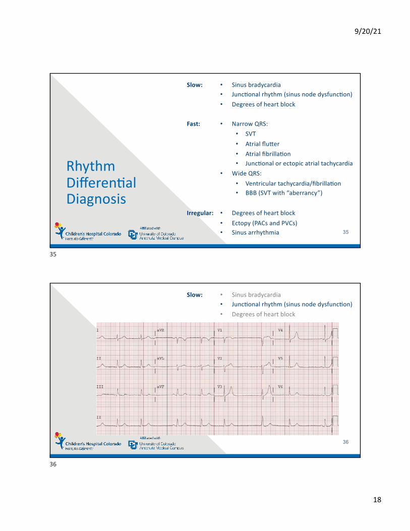
Rhythm Differen-al Diagnosis
• Sinus bradycardia• JuncIonal rhythm (sinus node dysfuncIon)• Degrees of heart block
• Narrow QRS:• SVT• Atrial fluder• Atrial fibrillaIon• JuncIonal or ectopic atrial tachycardia
• Wide QRS:• Ventricular tachycardia/fibrillaIon• BBB (SVT with “aberrancy”)
• Degrees of heart block• Ectopy (PACs and PVCs)• Sinus arrhythmia
Slow:
Fast:
Irregular:
35
36
• Sinus bradycardia• JuncIonal rhythm (sinus node dysfuncIon)• Degrees of heart block
Slow:
36

9/20/21
19
37
Rhythm Differen-al Diagnosis
• Sinus bradycardia• JuncIonal rhythm (sinus node dysfuncIon)• Degrees of heart block
• Narrow QRS:• SVT• Atrial fluder• Atrial fibrillaIon• JuncIonal or ectopic atrial tachycardia
• Wide QRS:• Ventricular tachycardia/fibrillaIon• BBB (SVT with “aberrancy”)
• Sinus arrhythmia• Ectopy (PACs and PVCs)• Degrees of heart block
Slow:
Fast:
Irregular:
TCH (1)SOUTH (03)
Reason:
. -------------------- Pediatric ECG interpretation -------------------- . Slow sinus arrhythmia
2579007 15-Sep-2021 12:34:28ABBOTT, DALTONDOB: 18-Apr-2006 15 Years Male
CSH (07)
POST OP 6Room:Oper: 160357
HR 55
PR 150QRSD 103QT 428QTc 410
-- AXIS --P 43QRS -14T 7
BRADY
12 Lead; Standard Placement
- NORMAL ECG -
Reviewed and Interpreted by: Londono Obregon, Camila 15-Sep-2021 20:36:02
I
II
III
aVR
aVL
aVF
V1
V2
V3
V4
V5
V6
II
Chest: 10 mm/mVLimb: 10 mm/mVSpeed: 25 mm/secDevice: US21931703 F 60~ 0.15-100 Hz PH100B CL P?
37
38
Rhythm Differen-al Diagnosis
• Sinus arrhythmia• Ectopy (PACs and PVCs)• Degrees of heart block
Irregular:
38

9/20/21
20
39
Rhythm Differen-al Diagnosis
• Sinus bradycardia• JuncIonal rhythm (sinus node dysfuncIon)• Degrees of heart block (e.g., third)
• Sinus arrhythmia• Ectopy (PACs and PVCs)• Degrees of heart block (e.g., second)
Slow:
Irregular:
39
40
SVT, right?
. -------------------- PEDIATRIC ECG INTERPRETATION -------------------- * A-FLUTTER W/ VARIED AV BLOCK, A-RATE 320 . RIGHT VENTRICULAR HYPERTROPHY * NONSPECIFIC REPOLARIZATION ABNORMALITIES
1209262 24-Aug-2012 09:23:50MONTOYA, ANTHONY JAMESDOB: 27-Sep-2008 3 Years Male Race: 9999 Dept: Outpatient
OPRoom:Oper: VJ
HR 159
QRSD 84QT 316QTc 515
-- AXIS --
QRS 164T -78
Requested By: NAKANO, STEPHANIE
Enc ID: 204342145Order #: 33279818
R LEAD:V3RINBOX:FRIDAY
Standard 12- ABNORMAL ECG -
Confirmed by: Schaffer, Michael 26-Aug-2012 15:45:44Edited
Previous ECG:09-Aug-2012 15:12:52 - Abnormal Confirmed
TCH - Main (1-01-01)
I
II
III
aVR
aVL
aVF
V1
V2
V3
V4
V5
V6
II
Chest: 10 mm/mVLimb: 10 mm/mVSpeed: 25 mm/secDevice: USO0801601 F 60~ 0.15-100 Hz PH090A b LP?
• SVT means just that, supraventricular tachycardia:• AV node and reentry tachycardia (accessory pathway or WPW)• Atrial fluder• Atrial fibrillaIon
40

9/20/21
21
41
• Atrial fibrilla4on:• No organized or regular atrial impulses (no normal/regular P waves)• Atrial impulses are not originaIng at the sinus node• Atrial acIvity is chaoIc (and, thus, ventricular conducIon is chaoIc)
• Atrial flujer:• “Fluder” waves rather than P waves (characterisIc “sawtooth” padern)• Regular rate of 250-320 bpm (yea, that’s right… that fast)• Only some impulses conduct through AV node (usually at regular interval)
41
42
49382461Account a:
TCH (1) MAIN (01)
Requested By:
Enc ID:Order a:
. -------------------- Pediatric ECG interpretation -------------------- * Supraventricular tachycardia . Borderline prolonged QT interval
2220576 17-Oct-2019 8:26:11KENT, JEREMYDOB: 05-Oct-2019 12 Days Male
CICU (03)
3204Room:Oper: 153898
HR 211
PR 116QRSD 76QT 249QTc 467
-- AXIS --P 113QRS 144T 10
MADELINE COQUILLETTE
4938246189958519
12 Lead; Standard Placement
- ABNORMAL ECG -
Reviewed and Interpreted by: Collins, Kathryn 18-Oct-2019 03:44:35
EditedPrevious Study:17-Oct-2019 08:08:41 - Abnormal Confirmed
I
II
III
aVR
aVL
aVF
Va
V2
V3
V4
V5
V6
II
e66caaaafaea11e94823aaa4c99caa29
Chest: 10 mm/mVLimb: 10 mm/mVSpeed: 25 mm/secDevice: US50902831 F 60~ 0.15-100 Hz PH110C b L P?
SVT, right? Yes, this +me it’s just straight up SVT
…but what exactly is usual SVT?
42

9/20/21
22
43
SVTSupra-VentricularTachycardia
• Most common rhythm disturbance in children (and young adults)• Majority of SVT occurs in structurally normal hearts (CHD is a risk factor)
• Occurs primarily by the presence of an accessory pathway:• Results in re-entry of the electrical impulse from the atrium• Creates a circuit in which the electrical impulse can cycle
repe44vely and result in rapid/regular ventricular contrac4on
• Two primary loca4ons for accessory pathways:
43
44
SVTSupra-VentricularTachycardia
• Two primary loca4ons for accessory pathways:• At the AV node – aka. Dual AV node physiology
44

9/20/21
23
45
SVTSupra-VentricularTachycardia
• Two primary loca4ons for accessory pathways:• At the AV node – aka. Dual AV node physiology• Somewhere else along the AV valve annuli
• SVT due to accessory pathways is common in neonates (1 in 250 neonates)… usually resolves by 12 mo
45
46
SVTSupra-VentricularTachycardia
• SVT occurs in 2.25 per 1000 people in the general popula4on• You probably know someone walking around who gets SVT from 4me to 4me!
• SVT can be treated with beta blockers• Some people choose to break their tachycardia with vagal maneuvers
(and live without meds!)
• SVT can be ablated via a catheter procedure• (…those crazy electricians burn the pathway from
inside the heart and… BAM! No more SVT!)
• Here, at Children’s, we do on-average 3 abla4ons for SVT every week!
46

9/20/21
24
47
49382461Account a:
TCH (1) MAIN (01)
Requested By:
Enc ID:Order a:
. -------------------- Pediatric ECG interpretation -------------------- * Supraventricular tachycardia . Borderline prolonged QT interval
2220576 17-Oct-2019 8:26:11KENT, JEREMYDOB: 05-Oct-2019 12 Days Male
CICU (03)
3204Room:Oper: 153898
HR 211
PR 116QRSD 76QT 249QTc 467
-- AXIS --P 113QRS 144T 10
MADELINE COQUILLETTE
4938246189958519
12 Lead; Standard Placement
- ABNORMAL ECG -
Reviewed and Interpreted by: Collins, Kathryn 18-Oct-2019 03:44:35
EditedPrevious Study:17-Oct-2019 08:08:41 - Abnormal Confirmed
I
II
III
aVR
aVL
aVF
Va
V2
V3
V4
V5
V6
II
e66caaaafaea11e94823aaa4c99caa29
Chest: 10 mm/mVLimb: 10 mm/mVSpeed: 25 mm/secDevice: US50902831 F 60~ 0.15-100 Hz PH110C b L P?
So… Next +me you see this…
Step 1: Is the pa+ent stable?
47
48
49382461Account a:
TCH (1) MAIN (01)
Requested By:
Enc ID:Order a:
. -------------------- Pediatric ECG interpretation -------------------- * Supraventricular tachycardia . Borderline prolonged QT interval
2220576 17-Oct-2019 8:26:11KENT, JEREMYDOB: 05-Oct-2019 12 Days Male
CICU (03)
3204Room:Oper: 153898
HR 211
PR 116QRSD 76QT 249QTc 467
-- AXIS --P 113QRS 144T 10
MADELINE COQUILLETTE
4938246189958519
12 Lead; Standard Placement
- ABNORMAL ECG -
Reviewed and Interpreted by: Collins, Kathryn 18-Oct-2019 03:44:35
EditedPrevious Study:17-Oct-2019 08:08:41 - Abnormal Confirmed
I
II
III
aVR
aVL
aVF
Va
V2
V3
V4
V5
V6
II
e66caaaafaea11e94823aaa4c99caa29
Chest: 10 mm/mVLimb: 10 mm/mVSpeed: 25 mm/secDevice: US50902831 F 60~ 0.15-100 Hz PH110C b L P?
So… Next +me you see this…
Step 1: Is the pa+ent stable?Step 2: If not-so-much
48
9/20/21
25
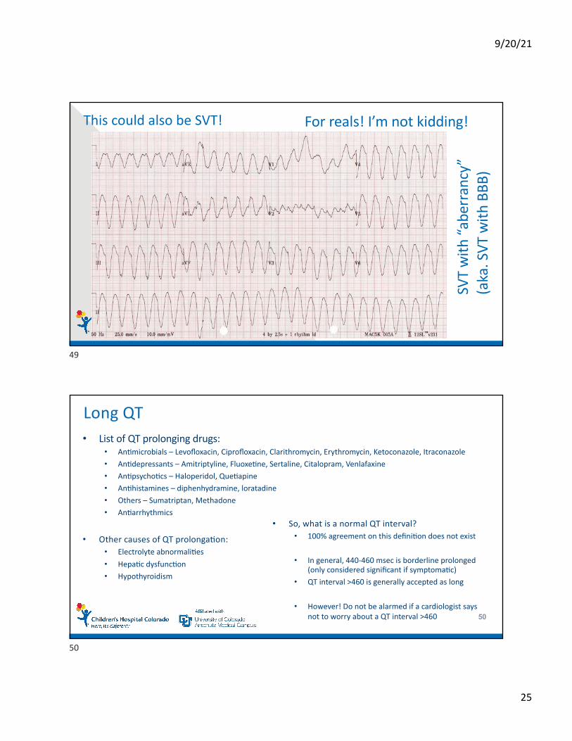
This could also be SVT! For reals! I’m not kidding!
SVT
with
“abe
rran
cy”
(aka
. SVT
with
BBB
)
49
50
Long QT• List of QT prolonging drugs:
• An0microbials – Levofloxacin, Ciprofloxacin, Clarithromycin, Erythromycin, Ketoconazole, Itraconazole
• An0depressants – Amitriptyline, Fluoxe0ne, Sertaline, Citalopram, Venlafaxine
• An0psycho0cs – Haloperidol, Que0apine
• An0histamines – diphenhydramine, loratadine
• Others – Sumatriptan, Methadone
• An0arrhythmics
• Other causes of QT prolongaIon:• Electrolyte abnormali0es
• Hepa0c dysfunc0on
• Hypothyroidism
• So, what is a normal QT interval?• 100% agreement on this defini0on does not exist
• In general, 440-460 msec is borderline prolonged (only considered significant if symptoma0c)
• QT interval >460 is generally accepted as long
• However! Do not be alarmed if a cardiologist saysnot to worry about a QT interval >460
50

9/20/21
26
51
Congenital Long QT Syndrome10 gene:cally dis:nct types of long QT syndrome (LQTS), first 3 types are most-common
TypeLQT1(KCNQ1)
LQT2(KCNH2/HERG)
LQT3(SCN5A)
QT MorphologyBroad-based, symmetrical T wave
Bifid T wave
Delayed-onset/ asymmetrical T wave
Clinical PhenotypeAdrenergic triggers (swimming, emoIon, exercise)
Commonly drug-induced,auditory sImuli
Rest/sleep
Incidence30-35%
25-30%
5-10%
51
61258191Account #:
TCH (1)MAIN (01)
Requested By:
Enc ID:Order #:
V3 LEAD:INBOX:
. -------------------- Pediatric ECG interpretation -------------------- . Sinus rhythm . LVH by voltage
2194599 15-Sep-2021 13:47:24ARNOLD, NATHANDOB: 09-Oct-2012 8 Years Male
OUTPATIENT (01)
OPRoom:Oper: GT
HR 66
PR 110QRSD 89QT 394QTc 413
-- AXIS --P 27QRS 85T 72
SAMANTHA ANN KOPS
61258191115231930
RIGHTWED
12 Lead; Standard Placement
- ABNORMAL ECG -
Reviewed and Interpreted by: Collins, Kathryn 16-Sep-2021 11:04:17Previous Study:30-Aug-2019 10:54:47 - Normal Confirmed
I
II
III
aVR
aVL
aVF
V1
V2
V3
V4
V5
V6
II
Chest: 10 mm/mVLimb: 10 mm/mVSpeed: 25 mm/secDevice: USD1723207 F 60~ 0.15-100 Hz PH110C b L P? 52
How to consider the QT interval on an ECG?Why go to the trouble of calcula=ng it?
Can you easily see the beginning and the end of the T wave?
If so, then just trust the machine read.
52
9/20/21
27
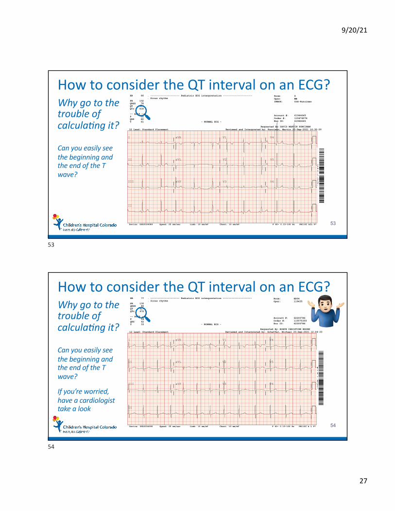
53
How to consider the QT interval on an ECG?Why go to the trouble of calcula=ng it?
Can you easily see the beginning and the end of the T wave?
61944645Account #:
TCH (1)SOUTH (03)
Requested By:
Enc ID:Order #:
INBOX:
. -------------------- Pediatric ECG interpretation -------------------- . Sinus rhythm
1984560 20-Sep-2021 13:20:50COLLIER, CAROLINE ELIZABETHDOB: 28-Jan-2015 6 Years Female
COSPRINGS (02)
6Room:Oper: MB
HR 66
PR 132QRSD 78QT 399QTc 418
-- AXIS --P 55QRS 80T 61
DAVID MARTIN RUNCIMAN
61944645115474076
COS-Runciman
12 Lead; Standard Placement
- NORMAL ECG -
Reviewed and Interpreted by: Runciman, Martin 20-Sep-2021 16:30:20
I
II
III
aVR
aVL
aVF
V1
V2
V3
V4
V5
V6
II
Chest: 10 mm/mVLimb: 10 mm/mVSpeed: 25 mm/secDevice: USD2034089 F 60~ 0.15-100 Hz PH110C bCL P?
53
54
How to consider the QT interval on an ECG?Why go to the trouble of calcula=ng it?
Can you easily see the beginning and the end of the T wave?
62200786Account #:
TCH (1)MAIN (01)
Requested By:
Enc ID:Order #:
. -------------------- Pediatric ECG interpretation -------------------- . Sinus rhythm
1247529 20-Sep-2021 9:06:00GARCIA, ITZELDOB: 21-Nov-2006 14 Years Female
ED (05)
ED04Room:Oper: 119435
HR 77
PR 130QRSD 80QT 417QTc 472
-- AXIS --P 18QRS 62T 33
ROBYN CHRISTINE MOORE
62200786115575355
12 Lead; Standard Placement
- NORMAL ECG -
Reviewed and Interpreted by: Schaffer, Michael 20-Sep-2021 12:04:23
I
II
III
aVR
aVL
aVF
V1
V2
V3
V4
V5
V6
II
Chest: 10 mm/mVLimb: 10 mm/mVSpeed: 25 mm/secDevice: USD2034090 F 60~ 0.15-100 Hz PH110C b L P?
If you’re worried, have a cardiologist take a look
!
54

9/20/21
28
1
2
3
4
5
Content Outline – Summary
55
ECG is generated by vectors formed by the leads
PACs, PVCs, and low-grade block are usually benign
Diffuse ST = pericarditis, low voltage = myocarditis
Keep calm, SVT happens!
Many things cause QT prolongation… but you can read the ECG!
55
Thank [email protected]
56