Embed Size (px)
Citation preview

V i s u a l A g n o s i a
Martha J. Farah
Vis
ua
l Ag
no
sia
S
ec
on
d
Ed
it
io
nFarah
S e c o n d E d i t i o n
,!7IA2G2-fgcade!:t;K;k;K;k
Visual Agnosia Second EditionMartha J. Farah
The cognitive neuroscience of human vision draws on two kinds of evidence: functional imaging ofnormal subjects and the study of neurological patients with visual disorders. Martha Farah’s land-mark 1990 book Visual Agnosia presented the first comprehensive analysis of disorders of visualrecognition within the framework of cognitive neuroscience and remains the authoritative work onthe subject. This long-awaited second edition provides a reorganized and updated review of thevisual agnosias, incorporating the latest research on patients with insights from the functionalneuroimaging literature.
Visual agnosia refers to a multitude of different disorders and syndromes, fascinating in theirown right and valuable for what they can tell us about normal human vision. Some patients cannotrecognize faces but can still recognize other objects, while others retain only face recognition.Some see only one object at a time; others can see multiple objects but recognize only one at atime. Some do not consciously perceive the orientation of an object but nevertheless reach for itwith perfected oriented grasp; others do not consciously recognize a face as familiar but neverthe-less respond to it autonomically. Each disorder is illustrated with a clinical vignette, followed by athorough review of the case report literature and a discussion of the theoretical implications of thedisorder for cognitive neuroscience.
The second edition extends the range of disorders covered to include disorders of topographicrecognition and both general and selective disorders of semantic memory, as well as expandedcoverage of face recognition impairments. Also included are a discussion of the complementaryroles of imaging and patient-based research in cognitive neuroscience, and a final integrativechapter presenting the “big picture” of object recognition as illuminated by agnosia research.
Martha J. Farah is Professor of Psychology and Director of the Center for Cognitive Neuroscienceat the University of Pennsylvania.
A Bradford Book
Praise for the first edition“Essential reading for all those interested in disorders of object recognition. It is the most thor-ough review available and is essential for all serious collections of books on visual perception.”—J. Davidoff, Perception
“Visual Agnosia provides a wide-ranging and provocative theoretical review of clinical disorders ofobject recognition. . . . [It is] an excellent introduction to cognitive neuroscientific theorizing and enpassant provides a helpful beginner’s guide to computational models of distributed processing. Atthe same time, the monograph is likely to inform and provoke debate amongst an audience withgreater expertise. . . . It should prove both interesting and accessible to a wide and multidiscipli-nary readership.” —Rosaleen A. McCarthy, Brain
“This is, literally, a wonderful book: filled with perceptions of things marvelous and (apparently)inexplicable. It is invigorating to see how steadfast empiricism and contemporary cognitive neuro-science can clear up parts of the mystery. Aside from the intrinsic interest of the subject matter,the book provides a case study of how cognitive neuroscientists can successfully go about theirbusiness.”—Austen Clark, Philosophical Psychology
Cover art: Juan Gris, Still Life Before an Open Window, Place Ravignan, Philadelphia Museum of Art:The Louise and Walter Arensberg Collection
The MIT PressMassachusetts Institute of TechnologyCambridge, Massachusetts 02142http://mitpress.mit.edu
0-262-56203-0
45898MIT.FARAH.VisualAgnosia 6/2/04 8:44 AM Page 1

Visual Agnosia


Visual Agnosia
second edition
By Martha J. Farah
A Bradford BookThe MIT PressCambridge, MassachusettsLondon, England

©2004 Massachusetts Institute of Technology
All rights reserved. No part of this book may be reproduced in any form by any electronicor mechanical means (including photocopying, recording, or information storage andretrieval) without permission in writing from the publisher.
This book was set in Bembo by Graphic Composition, Inc.Printed and bound in the United States of America.
Library of Congress Cataloging-in-Publication Data
Farah, Martha J.Visual agnosia / by Martha Farah.—2nd ed.
p.; cm.“A Bradford book.”ISBN 0-262-06238-0 (hc: alk. paper) —ISBN 0-262-56203-0 (pbk : alk. paper)1. Visual agnosia. 2. Visual perception. I. Title.[DNLM: 1. Agnosia—physiopathology. 2. Form Perception—physiology. 3. PatternRecognition, Visual. 4. Prosopagnosia. WL 340 F219v 2004]RC394.V57F37 2004616.8—dc22 2003059393
10 9 8 7 6 5 4 3 2 1

For Hermine Makman

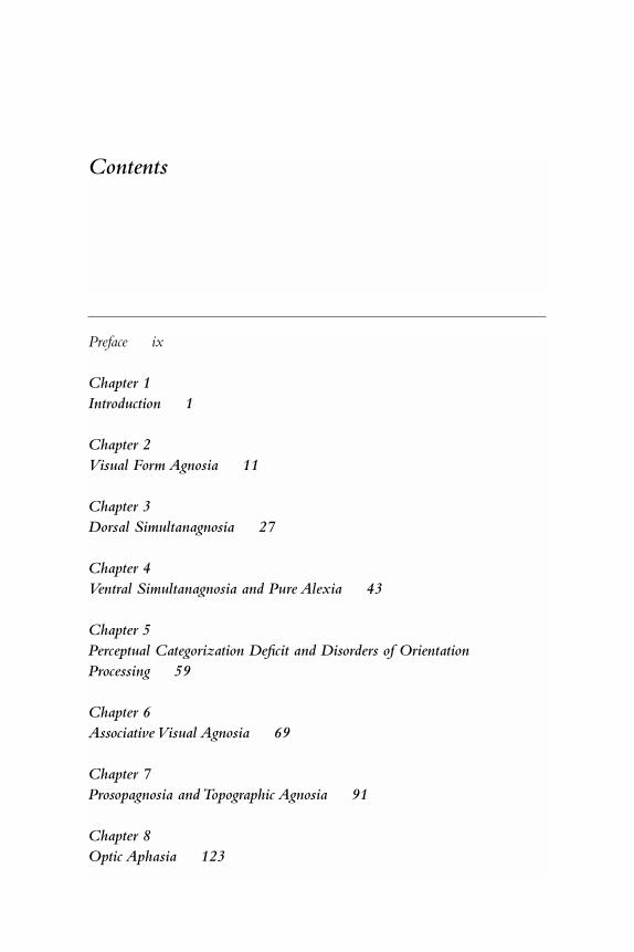
Contents
Preface ix
Chapter 1Introduction 1
Chapter 2Visual Form Agnosia 11
Chapter 3Dorsal Simultanagnosia 27
Chapter 4Ventral Simultanagnosia and Pure Alexia 43
Chapter 5Perceptual Categorization Deficit and Disorders of Orientation Processing 59
Chapter 6Associative Visual Agnosia 69
Chapter 7Prosopagnosia and Topographic Agnosia 91
Chapter 8Optic Aphasia 123

Chapter 9Semantic Knowledge Impairments 139
Chapter 10Vision When It Works 155
References 163Index 187
viii Contents

Preface
When the first edition of this book came out in 1990, I joked that mostauthors spend a number of years working on a topic and then write a bookabout it, but I had written the book first and planned to then begin work-ing on the topic. This was no exaggeration. It is a matter of record thatmy very first publication on object recognition or agnosia was the book!My backward, and some might say nervy, approach worked out surprisinglywell. The agnosia case literature was an unmined resource, and experi-mental research on agnosia to answer questions about object recognition hadbarely begun. It seemed to me that the first order of business was simplyreviewing and systematizing the case literature and posing some basicquestions that could, in principle, be answered by such cases. A book wasas good a way to do this as any. So I wrote Visual Agnosia.
Looking back at the first edition, it had an extremely high question-to-answer ratio. Many of the unanswered questions formed the basis forthe next several years of my research:Are faces “special?” Is their geometryrepresented differently from that of other objects? Are there orthography-specific brain systems? How could they develop? Do “living things” con-stitute a special category for the visual system? For the semantic system?
In the fourteen years since the first edition came out, these and manyother questions about visual object recognition have been addressed bymyself and others around the world. Where before there were just a lot ofinteresting questions, now there is consensus on some answers, healthydifferences of opinion on others, new questions, and plenty of solid sci-ence to make the second edition a very different book from the first.
My own contributions in the interim were undertaken with a verytalented and congenial group of collaborators. In particular, four of my

students played a major role in the research described here, and it is a pleas-ure to acknowledge their contributions. My former graduate studentShaun Vecera, now Associate Professor at the University of Iowa, took aset of general issues concerning attention, grouping, and early vision andtranslated them into a productive research program encompassing patient-based research, psychophysics, and computational modeling. The bestthing I did for him as an advisor was move to Penn, leaving him to relyhis own judgment and creativity. Thad Polk, a former postdoc and nowAssociate Professor at the University of Michigan, was the driving forcebehind our studies of perceptual processes in reading. In the course ofbuilding several computational models and conducting both behavioraland imaging experiments, Thad uncovered important new insights aboutthe effects of experience on pattern recognition and also learned first-hand the meaning of “going postal.” Former postdoc Jim Tanaka, nowProfessor of Psychology at the University of Victoria, took the lead in ourwork on parts and wholes in face recognition. Jim also saw the broader rel-evance of this work beyond face recognition and has made it one aspectof his multifaceted program of research on perceptual expertise. PaddyMcMullen, another former postdoc now in Canada, where she is Associ-ate Professor at Dalhousie University, was my partner in puzzlement forour initial studies of category-specific semantic impairments. She was ableto get us past that stage with her thoughtful analysis and experimentalrigor. Former postdocs Matt Kurbat, Cathy Reed, Sharon Thompson-Schill, and Lynette Tippett, graduate students Randy O’Reilly, MarcieWallace, and Kevin Wilson, and research assistants Karen Klein, KarenLevinson, Carol Rabinowitz, and Matt Stallcup all worked with me onprojects that were related in some way to the topic of this book, and theircontributions are all gratefully acknowledged.
Much of the research reported here would have been impossiblewithout the help of our agnosic subjects. These individuals worked withus in experiments that were often tedious and always difficult, designed asthey were to elicit the subjects’ agnosic impairments. I especially want toacknowledge the participation of Lincoln H., a remarkable person whohas taught me much about visual agnosia, as well as experience, adapt-ability, and hope.
Barbara Murphy of MIT Press provided advice, encouragement, andan occasional kick in the pants, without which this book would probably
x Preface

still be a manuscript. Katherine Almeida expertly guided the book throughproduction. I am grateful to them both. My colleague Russell Epstein atPenn and Tim Rogers of the MRC Cognition and Brain Unit in Cam-bridge, England read drafts of chapters and gave me their very knowl-edgeable and diplomatic advice, which I have tried to follow. Finally, myacknowledgments would not be complete without thanking three wise,generous and fun colleagues for their collaboration and tutelage in the areaof visual object recognition, Todd Feinberg, Jay McClelland, and MikeMozer.
Preface xi


Visual Agnosia


Chapter 1
Introduction
Virtually everything we know about the brain functions underlying hu-man cognition has been learned by one of two methods: studying brain-lesioned patients and functional neuroimaging. The two methods tend toyield reassuringly consistent evidence. Yet they have significantly differ-ent strengths and weaknesses, to be discussed later in this chapter, and forthis reason neither method is dispensable.
Disorders of visual object recognition following brain damage areknown as visual agnosias. There is amazing diversity to the ways in whichobject recognition can break down, from visual form agnosia in which pa-tients with normal acuity cannot recognize something as simple as a circleor a square, to topographic agnosia in which patients with normal face,object, and word recognition cannot recognize locales. In each case thepatterns of preserved and impaired abilities put useful constraints on ourtheories of how the normal visual recognition system works.
1.1 A Brief History of Agnosia
For much of its history, the study of agnosia focused on the question ofwhether there is such a thing as agnosia. Researchers began with this mostbasic of questions, and perhaps in retrospect stayed with it too long, be-cause the syndrome seemed so counterintuitive and contradictory. Howcould someone be able, in the words of Humphreys and Riddoch’s (1987b)book title, To See but Not to See? Repeatedly over the years, the concept ofvisual agnosia has met with skepticism. First Bay (1953), and then Benderand Feldman (1972), argued that visual agnosia, in the sense of a selectiveimpairment in visual recognition per se, does not exist. Bay proposed that

the appearance of a selective impairment in object recognition was in-variably the result of a combination of two more general characteristicsof agnosic patients. First, he suggested that these patients always havesubtle impairments in elementary visual functions, which may be less ap-parent under the conditions of standard tests of visual fields, acuity, andso on, than when they are being used for object recognition under natu-ral conditions. Second, he claimed that these patients suffer from a gen-eral intellectual decline. According to Bay, impairments in elementaryvision and general intelligence may occasionally conspire to produce dis-proportionate difficulties with object recognition, but there is no suchthing as an impairment in object recognition per se. Bender and Feldman(1972) supported Bay’s claims with a systematic review of a large numberof neurological patients. They searched all of the patient records from atwenty-year period at New York’s Mount Sinai Hospital and found rela-tively few cases with visual recognition difficulties. What they took to bemore damaging to the concept of agnosia was the fact that all of these casesalso had some significant elementary visual and/or general intellectualimpairments.
Bay, and Bender and Feldman won over many influential neuropsy-chologists to their point of view on agnosia (e.g., Critchley, 1964; Teu-ber, 1968), but their skepticism was not shared by everyone. Even thougha “pure” case of agnosia (a patient with impaired visual object recogni-tion and perfectly normal elementary visual and intellectual capabilities)would disprove the skeptics’ position, the absence of such a case does notprove it. Neuropsychologists know far too well that “nature’s experi-ments” are executed rather sloppily, and they would have very little tostudy if they confined themselves to pure cases of anything. With this inmind, Ettlinger (1956) made the important point that finding a “pure” ag-nosic was not the only way to settle the issue empirically. Just as effectivewould be the demonstration that agnosic patients were no more impairedin their intellectual and elementary visual capabilities than many non-agnosic patients. He demonstrated that this was true by systematically as-sessing a variety of elementary visual functions in patients already screenedfor generalized intellectual decline. Although only one of his cases had atrue agnosia, and this case did have elementary visual impairments, hefound other patients with more severe elementary visual impairments whowere not agnosic. More recently, De Haan, Heywood, Young, Edelstyn,
2 Introduction

and Newcombe (1995) carried out a more stringent test of Ettlinger’s hy-pothesis with three severe visual agnosics and a more comprehensive andsophisticated battery of visual tests. Their data supported Ettlinger’s con-clusion that whatever elementary visual impairments the agnosic patientshad, they were not the cause of the agnosia. Patients with equally impairedelementary visual function were not agnosic.
The impulse to “explain away” agnosia can be understood in termsof the theories of vision available to agnosia’s skeptics in the mid-twentiethcentury. If one views object recognition as taking place in two relativelyundifferentiated stages—(1) seeing the object and (2) associating generalknowledge with the visual percept—then the only possible way to disruptobject recognition is by disrupting vision or general knowledge. If objectrecognition difficulties seem disproportionate to difficulties of vision orgeneral knowledge (as is the case, by definition, with visual agnosia), thenthis must be due to a synergistic interaction of minor difficulties in bothvision and general knowledge. However, with the advent of single unitrecording in visual cortex (e.g., Gross, Rocha-Miranda, & Bender, 1972;Hubel & Weisel, 1962) and computational modeling of vision (e.g., Marr,1982), a different view of visual object recognition emerged. Accordingto this latter view, object recognition is accomplished by repeatedly trans-forming the retinal input into stimulus representations with increasinglygreater abstraction from the retinal array and increasingly greater corre-spondence to invariant properties of objects in the physical world (seeFarah, 2000). Within such a system, brain damage affecting just the laterstages of vision would create a “pure” visual agnosia.
Eventually, neuropsychologists looked beyond the question ofwhether or not agnosia exists, to other questions about agnosia, includ-ing the possibility of different types of agnosia and their associated lesionsites. As the field of cognitive neuropsychology blossomed in the 1980s,researchers attempted to relate aspects of agnosia to theories of visual ob-ject recognition, and in the process to test those theories with data fromagnosic patients (e.g., Farah, 1990; Humphreys & Riddoch, 1987b; Rat-cliff & Newcombe, 1982). In the pages that follow, I will delineate a dozenor so distinct visual agnosic syndromes, and bring each of them to bear asevidence on the nature of visual object recognition. Examples of the ques-tions to be addressed include:Are there different recognition modules, orsubsystems, required for recognizing different kinds of stimuli (e.g., faces,
Introduction 3

common objects, printed words)? Does visual selective attention operateprior to object recognition, subsequent to it, or in parallel with it? Arethe long-term visual memory representations underlying recognition im-plemented locally or in a distributed network?
1.2 Types of Agnosia
Taxonomizing may appear to be a rather atheoretical enterprise that wouldbe better replaced by analysis of the phenomena of agnosia using cogni-tive theories. However, we must begin with issues of taxonomy becausegrouping the phenomena correctly, in any area of science, is a prerequi-site for making useful theoretical generalizations about them. This is allthe more important—and all the more difficult—in the study of agnosiabecause the entire database is comprised of single cases, no two of whichare exactly alike. Therefore, much of the scientific work to be done in thisfield involves sorting these countless variable and unique cases into atractable number of “natural kinds.”
There is no standard taxonomy of agnosia. Everyone agrees that ag-nosic patients differ from each other in certain ways, but the question ofwhich differences are differences of degree and which are differences ofkind has not found a unanimous answer. On careful reading of patients’abilities and deficits, I find that many authors have grouped patients in un-helpful ways. Their implicit taxonomies misrepresent the basic empiricalphenomena, both by overinclusive categories that blur theoretically im-portant distinctions between different syndromes, and by overfractiona-tion of syndromes, in which differences of degree are treated as differencesof kind.
Most neuropsychologists follow Lissauer (1890) in distinguishing be-tween the “apperceptive agnosias” and the “associative agnosias.” Accord-ing to Lissauer, apperceptive agnosias are those in which recognition failsbecause of an impairment in visual perception, which is nonetheless abovethe level of an elementary sensory deficit such as a visual field defect. Pa-tients do not see objects normally, and hence cannot recognize them. Incontrast, associative agnosias are those in which perception seems adequateto allow recognition, and yet recognition cannot take place. It is said toinvolve, in the oft-quoted phrase of Teuber (1968), a “normal perceptstripped of its meaning.”
4 Introduction

In this respect, the apperceptive-associative distinction, as definedabove, includes a significant assumption about the mechanisms of agnosia:that the underlying deficit in so-called associative agnosia lies outside ofthe modality-specific perceptual processing of the stimulus. Whether ornot this is true is an important issue that will be discussed later. Never-theless, the grouping of agnosics into two categories—those with promi-nent, easily noticed perceptual deficits and those without—does seem tobe empirically valid.
Within these two broad categories there is tremendous variation. Forexample, among the patients who have been labeled “apperceptive” arethose who cannot discriminate a circle from a square, those who can rec-ognize any one object but cannot see other objects presented at the sametime, and those whose difficulty with object recognition is manifest onlywith objects presented at unusual orientations. Among the patients whohave been labeled “associative” are those whose impairment is confined tospecific categories of visual stimulus such as faces, places, or printed words,as well as those with across-the-board recognition impairments and thosewho seem impaired only when naming a visually presented object. Theorganization of this book reflects my attempt to find a happy medium be-tween lumping distinct syndromes together and splitting the phenomenainto an unmanageable and unnecessary number of separate categories.Each of the next eight chapters describes a type of agnosia, along with itsrelations to theories of normal visual function.
1.3 Patient-Based Cognitive Neuroscience in the Age of Imaging
The first edition of this book was written one methodological revolutionago, just before functional neuroimaging transformed cognitive neuro-science. At that time, everything we knew about the neural bases of high-level vision in humans came from studies of patients. It was thereforeparticularly exciting to work through the rich database of clinical studiesin search of insights about normal object recognition, knowing that suchinsights lay waiting there and, at the time, only there.
The situation is very different now. Neural systems can be visualizedas they perform their functions under experimentally controlled condi-tions in normal subjects. This capability revolutionized all areas of cogni-tive neuroscience, and greatly expanded our understanding of high-level
Introduction 5

vision in the course of just a decade of research. It therefore bears asking:Why study visual agnosia now that functional neuroimaging is available?The answer to this question involves an accounting of the strengths andweaknesses of imaging and patient-based cognitive neuroscience.
An obvious weakness of patient-based research is that naturally oc-curring lesions do not respect anatomical or functional boundaries. Suchmessiness would be less of a problem if all possible sizes and shapes of thesemessy lesions occurred, because different patients with overlapping lesionsmight permit inferences about the functions of common and distinct sub-regions, but this is not the case; strokes, head injury, and other etiologiesof brain damage have characteristic lesions, and many possible lesion con-figurations do not occur. The greatest advantage of functional neuro-imaging is its ability to compensate for this weakness. Although some areasof the brain are better visualized with current imaging techniques thanothers, imaging is hands-down the better way to probe the functions ofspecific anatomical regions.
Functional neuroimaging has the additional advantage of studyingnormal brains, which are the subject of interest. With patient-based re-search we are operating one inferential step away from this subject. Ofcourse, the behavior of a damaged system is related in systematic ways tothe function of the intact system. But “systematic”does not mean “simple”:reorganization following injury can greatly complicate our inferencesabout normal function (Farah, 1994). An additional problem with rare dis-orders, including most of the agnosias, is that patients provide no morethan an existence proof that a certain dissociation is possible, and hencethat the inferred neurocognitive organization exists. In the early days ofcognitive neuroscience this was a minor worry, because of the implicit as-sumption that all normal human brains were wired in basically the sameway. However, as our field finally begins to grapple with individual differ-ences (Thompson, Cannon, Narr, van Erp, Poutanen, Huttunen, Lonn-qvist, Standertskjold-Nordenstam, Kaprio, Khaledy, Dail, Zoumalan, &Toga, 2001;Hamer, 2002 ), we want to know whether the functional or-ganization inferred from one patient applies to all humans or is just onevariant. Does everyone use separate systems to recognize faces and non-face objects, or just a subpopulation, who will become prosopagnosic aftercertain patterns of brain damage? The ability to analyze individual sub-jects’ images allows us to address this question by finding out what pro-
6 Introduction

portion of subjects recruits measurably different brain regions for face andobject recognition.
In weighing the advantages and disadvantages of patient-based andimaging research, there is one other drawback to patient-based researchthat is often overlooked: the difficulty of interlaboratory verification.Findings from patients with rare disorders like agnosia cannot be pursuedby any scientist with an alternative hypothesis or a good idea for a follow-up study. This is unavoidable, at least to a degree. When a patient agreesto work with one researcher, he is not making himself available to any sci-entist in the field willing to travel to him at any point in the future. How-ever, the problem is often compounded by researchers who develop apossessiveness about “their” patients. This practice is at least as dehu-manizing to the patient as offering to put them in contact with otherresearchers, and it has impeded progress in our field. Imaging studies are much more replicable, in that a finding from one imaging lab can inprinciple be pursued by any other imaging lab.
These advantages of imaging over patient-based research make animpressive list. If we were to play a variant of the childhood game “wouldyou rather” (be rich or beautiful, fly like a bird or read minds . . .) withimaging and patient-based methods, I’d be inclined to take the imaging.Happily, we do not have to choose. Patient-based methods have their ownstrengths, which complement those of imaging. As a result, the combina-tion of the two approaches is more powerful than the sum of its parts.
The great advantage of studying patients is the ability to test hy-potheses about mechanism. The goal of most cognitive neuroscienceresearch to understand how intelligent behavior is accomplished. We aretrying to describe the causal chain of events that intervene between stim-ulus and response. We share this goal with a number of other disciplines,from molecular neuroscience to cognitive psychology. What distinguishesthese disciplines is the level of description within which they cast their hy-potheses about mechanism.
The mechanistic hypotheses of cognitive neuroscience concern theinformation-processing functions of macroscopic neural systems. This levelof description includes, at the more microscopic end of the range, theemergent behavior of populations of neurons. It is this population behav-ior, during learning, normal function, and after damage, that does the ex-planatory “work” in the computational models described in this book
Introduction 7

(e.g., models of the word superiority effect, covert face recognition, opticaphasia, and selective semantic memory impairments). At the more macro-scopic end of the cognitive neuroscience level of description are modelsthat delineate distinct information processing components and their in-terrelations, such as the division of labor between form perception fromstatic spatial cues and form from motion, and between face and objectrecognition.
Our methods deliver information that is useful for testing hypothesesat this level of description. Current imaging techniques reveal distin-guishable activations at about this scale, and the relatively more fine-grained dissociations among abilities after brain damage can also bedescribed at this level. However, images and lesions are very different intheir ability to answer questions about mechanism. Only the lesion methodcan reveal the causal relations among brain systems.
Imaging data are fundamentally correlational; they tell us that thisarea becomes active when that cognitive process is being performed. Theydo not tell us what causal role, if any, is played by an activation observedin this way. Not every activation is part of a causal pathway;representationsmay become active, in a given task context, either because they are causallyinvolved in performing the task or because they have become associatedwith other representations that are causally involved. Although it mayseem odd to think of the brain as activating unnecessary systems, I suspect that superfluous or only marginally useful activity is very com-mon, and perhaps the norm. Try the following low-tech demonstrationof this point: Glance at the bottom of this page and count the letters inthe last word. Notice that you read and understood the word even thoughit was not part of your assignment. Indeed, the same thing will happeneven if you try not to read the word. Phonological and semantic repre-sentations are so highly associated with orthographic representations thatthey are activated even when not necessary. This example of associatedactivity is intentionally obvious, but the issue is not trivial when the acti-vated systems are less open to introspection and less well characterizedcognitively.
To tease apart causal and merely associated systems, and characterizethe information-processing function of each of those systems, we need to reach in and tinker. Only by seeing the consequences of removing ordisabling different candidate systems can we infer their role in producing
8 Introduction

a given behavior. Of course, with human brains we do not “tinker.” In-stead, we examine the effects of naturally occurring brain damage.
How can patient-based research determine which activated systemsplay a causal role in implementing an ability, and which are merely asso-ciated? To answer this question, let us return to the example of unneces-sary but associated activity when counting the letters in a word. Imaginethat this task has been carried out in a scanner, and consistent with intro-spection, areas subserving visual-spatial attention are activated (as they arein counting tasks), and areas subserving orthography, phonology, and se-mantics are activated (as they are when words are processed). We now wantto answer the question:which of these activations play a causal role in im-plementing letter counting, and which are merely associated? We can findout by testing patients with lesions in each of these systems on the lettercounting task.
Patients with disorders of visual-spatial attention, including the dor-sal simultanagnosics of chapter 3, will have difficulty with the letter count-ing task. This is consistent with the hypothesis that counting visual stimulirequires marking them attentionally; the movement of visual-spatial at-tention from item to item is not merely an associated but unnecessary pro-cess. In contrast, patients with orthographic impairments (e.g., the purealexic patients of chapter 4), phonological impairments, or semantic im-pairments (e.g., the optic aphasics and semantic agnosics of chapters 8 and9) will be able to perform the task. This is consistent with the hypothesisthat the lexical processes that were reliably activated in the scanner are notin fact necessary for the behavior.
The information that patients provide goes beyond simply classifyingsystems as necessary or not necessary. It can also distinguish different typesof processing and delineate multiple parallel chains of processing that en-able a behavior. Patterns of activation in functionally parallel systems do nottell us which activations are part of the same or different pathways, or whatthe unique information-processing nature of each system is. By contrast,through interrupting processing at various loci we can infer just these prop-erties of the system, through a procedure akin to trouble-shooting.
The cognitive neuroscience of object recognition has already be-nefited from the interplay of patient-based and imaging methods. Initialattempts to investigate visual recognition using functional neuroimagingsuffered from a lack of specific hypotheses and were correspondingly quite
Introduction 9

variable in matching experimental and baseline conditions. Many studiesconsisted of simply scanning subjects while they viewed pictures or per-formed tasks with assorted stimuli and fixation points. No wonder that,in the aggregate, this sizable literature succeeded only in establishing thatvisual object recognition involves the posterior half of the brain (Farah &Aguirre, 1999)! However, this changed as imagers began to test specifichypotheses about visual recognition, most of which came from the patientliterature. For example, prosopagnosia and topographic agnosia suggestedspecific hypotheses concerning specialization in ventral visual areas, andalong with more specific hypotheses came more theoretically constrainedexperimental designs. Imaging in turn clarified the degree of segregationamong specialized recognition systems, which of course are never neatlydissociated by naturally occurring lesions.
It has recently become possible to combine imaging and patient-based research in a powerful new way, by imaging patients while they en-gage in the processes of interest. This approach poses many additionaltechnical challenges beyond those of imaging a normal brain (Price &Friston, 2003), but is also uniquely well suited to understanding theanatomical and mechanistic bases of cognition. Although as yet undevel-oped, the functional imaging of visual agnosics will undoubtedly play anincreasingly dominant role in the cognitive neuroscience of high-levelvision.
10 Introduction

Chapter 2
Visual Form Agnosia
The term “apperceptive agnosia” has been used to mean any failure of ob-ject recognition in which perceptual impairments seem clearly at fault,despite relatively preserved sensory functions such as acuity, brightnessdiscrimination, and color vision. It has been applied to an extremely het-erogeneous set of patients who seem unlikely to share a single underlyingimpairment. In the first edition of this book, I reserved the term “apper-ceptive agnosia” for one particular syndrome, to which this label has mostfrequently been applied, and added a parenthetical “narrow sense” to sig-nal the difference between this usage and the more general one. Clarity isprobably better served, however, by a more distinct label, and so I pro-pose to adopt the alternative term “visual form agnosia,” introduced byBenson and Greenberg (1969).
2.1 Visual Form Agnosia:A Case Description
Benson and Greenberg (1969) touch on many of the essential features ofthis syndrome in the following description of Mr. S, a young man who suf-fered accidental carbon monoxide poisoning.
Visual acuity could not be measured with a Snellen eye chart, as he could neitheridentify letters of the alphabet nor describe their configuration. He was able toindicate the orientation of a letter “E,” however, and could detect movement ofa small object at standard distance. He could identify some familiar numbers ifthey were slowly drawn in small size on a screen. He could readily maintain opticfixation during fundoscopic examination, and optokinetic nystagmus was elicitedbilaterally with fine, 1/8 inch marks on a tape. . . . Visual fields were normal to

10 mm and 3 mm white objects, and showed only minimal inferior constrictionbilaterally to 3 mm red and green objects. . . .
The patient was able to distinguish small differences in the luminance (0.1log unit) and wavelength (7–10 mu) of a test aperture subtending a visual angleof approximately 2 degrees. While he could detect these differences in luminance,wavelength, and area, and could respond to small movements of objects beforehim, he was unable to distinguish between two objects of the same luminance,wavelength, and area when the only difference between them was shape.
Recent and remote memory, spontaneous speech, comprehension of spo-ken language, and repetition were intact. He could name colors, but was unableto name objects, pictures of objects, body parts, letters, numbers, or geometricalfigures on visual confrontation. Yet he could readily identify and name objectsfrom tactile, olfactory, or auditory cues. Confabulatory responses in visual iden-tification utilized color and size cues (a safety pin was “silver and shiny like a watchor a nail clipper” and a rubber eraser was “a small ball”). He identified a photo-graph of a white typewritten letter on a blue background as “a beach scene,”pointing to the blue background as “the ocean,” the stationery as “the beach,”and the small typewriter print as “people seen on the beach from an airplane.”
He consistently failed to identify or to match block letters; occasionally he“read” straight line numbers, but never those with curved parts. He could clum-sily write only a few letters (X, L) and numbers (1, 4, 7), but often inverted orreversed these. Although he could consistently identify Os or Xs as they wereslowly drawn, or if the paper containing them was moved slowly before him, hewas unable to identify the very same letters afterwards on the motionless page. Hewas totally unable to copy letters or simple figures, and he could neither describenor trace the outline of common objects. . . .
He was unable to select his doctor or family members from a group until theyspoke and was unable to identify family members from photographs. At one timehe identified his own face in a mirror as his doctor’s face. He did identify his ownphotograph, but only by the color of his military uniform. After closely inspect-ing a scantily attired magazine “cover girl,” he surmised that she was a woman be-cause “there is no hair on her arms.” That this surmise was based on flesh coloridentification was evident when he failed to identify any body parts. For example,when asked to locate her eyes he pointed to her breasts. . . . (pp. 83–85)
In summary, the patient had seemingly adequate elementary visualfunctions and general cognitive ability, and yet he was dramatically im-paired on the simplest forms of shape discrimination. Indeed, this patientwas described as appearing blind to casual observers (Efron, 1968). Let usrelate the findings in this case to the others in the literature.
12 Visual Form Agnosia

2.2 Visual Form Agnosia: Some Generalities
Visual form agnosia is a relatively rare syndrome, although the similarityof a number of other reported patients to Mr. S suggest that it is a usefulcategory. Other visual form agnosics include the cases of HC (Adler, 1944;Sparr, Jay, Drislane, & Venna, 1991), ES (Alexander & Albert, 1983), Mr. S(Efron, 1968;Benson & Greenberg, 1969), RC (Campion & Latto, 1985;Campion, 1987), Schn. (Gelb & Goldstein, 1918; translated by Ellis,1938), and X (Landis, Graves, Benson, & Hebben, 1982), and DF (Mil-ner, Perrett, Johnston, et al., 1991).
As was true for Mr. S, visual field defects do not seem responsible forthe visual problems of the other patients in this category. Visual fields areeither normal or sufficiently preserved that visual field defects do not seeman adequate explanation of their visual difficulties. In all cases acuity is ei-ther normal or sufficient for recognition, and in most cases color vision isroughly normal. Maintaining fixation of a visual target was possible forall but one of these cases (Alexander & Albert, 1983), and was reporteddifficult for one other (Adler, 1944). In the three cases in which depth per-ception was explicitly reported, it was either intact or recovered while thepatient was still agnosic. Motion perception was intact in some cases, al-though most did not report any specific tests of movement perception.
In striking contrast to their roughly intact visual sensory functions,visual form agnosics are severely impaired at recognizing, matching, copy-ing, or discriminating simple visual stimuli. These impairments are notsubtle:Typical examples of patients’ errors on such tasks include calling thenumeral 9 “a capital A” (Adler, 1944), a circle “a lot of dots” (Campion,1987), or being unable to discriminate simple shapes such as “Xs” from“Os” (Benson & Greenberg, 1969;Milner et al., 1991). Figure 2.1 showsthe attempts of two of these patients to copy simple forms. Figure 2.2shows the stimuli used in two shape-matching tasks that Mr. S was un-able to perform. In the first task, pairs of rectangles with the same total areawere shown to the patient, and his task was to judge whether they had thesame shape or different shapes. In the second task, he was asked to matcha sample stimulus to one of four other stimuli that had the same shape.
The case reports give a few additional clues to the nature of these pa-tients’ perception of the visual world. For some patients it was mentionedthat figures made of dots were harder to recognize than figures made of
Visual Form Agnosia 13

solid lines, and curved lines were more difficult to perceive than straight.In two of the reports it was mentioned that the patients did not seem toperceive objects as solid forms or even surfaces in three dimensions:Adler(1944, p. 252) says of her patient, “At first she perceived contours only. Forexample, during the second week she called a nickel and a round silvercompact each ‘a key ring.’” Gelb and Goldstein (Ellis, 1938, p. 318) statethat “All drawings in perspective were utterly meaningless for this patient.A circle tilted away from him was invariably described as an ellipse.”
Recognition of real objects is also impaired but is somewhat betterthan recognition of “simple” stimuli. This appears to be due to the widerset of available cues to the identity of real objects, particularly color. The
14 Visual Form Agnosia
Figure 2.1Copies of a geometric form by H. C. (top) and simple shapes, numbers, and letters by Mr. S (bottom).

patients’ identifications of objects are typically inferences, made by piecingtogether color, size, texture, and reflectance clues. Mr. S’s reliance on theseproperties is apparent from Benson and Greenberg’s recounting of his at-tempts to recognize the safety pin and the picture of the typed letter. Theyalso report that he “could select similar objects from a group only if therewere strong color and size clues; after training he could name several fa-miliar objects but failed to do so if their color and size qualities were altered.Thus he failed to identify a green toothbrush that was substituted for a pre-viously named red toothbrush. He also called a red pencil “my toothbrush”(p. 84). RC was reported to use “features of objects, such as their color orwhether they were shiny or not. He could also recognize the ‘texture’ ofobjects. If encouraged, he could often make an accurate guess about thenature of objects from such cues” (Campion, 1987, p. 209). Landis et al.(1982) report similar strategies in their patient, X:“He once mentionedbeing on the 14th floor of the hospital. Asked how he knew, he replied “It’sthe only one having red exit doors.” Adler’s patient, too, was said to rec-ognize objects by “a process of adding up visual impressions,” and oftenused color to guess the identity of objects, mistaking vanilla ice cream forscrambled eggs and a piece of white soap for a piece of paper (p. 252).
Visual Form Agnosia 15
Figure 2.2The shape-matching ability of an apperceptive agnosic patient. On the left is a set of rec-tangles matched for overall area, which were presented pairwise to Mr. S to be judgedsame or different in shape. He was unable to discriminate all but the most distinctive, andmade errors even with these. On the right are a set of rows containing a target shape (right)and a set of four choices to be matched with the target shape. Mr. S’s answers are marked.

Motion of the object to be recognized appears to be helpful to someof these patients. Not surprisingly, it was helpful only to those subjectswho had large visual fields, since a moving object would quickly pass outof view for patients with very narrow visual fields. ES recognized objectsbest when they were “alone and moving (e.g. identifying birds or planesflying at a great distance . . . )” (Alexander & Albert, 1983, p. 408). Mo-tion helped Mr. S to segregate objects from their surround: “Mr. S canpoint with his finger to an object which is held before him. He can dothis efficiently only if the object is moved before his eyes. If it is station-ary he does not appear to know what object he has been asked to look at;his eyes randomly scan the entire room and he appears to be ‘searching’”(Efron, 1968, p. 156). Motion also aided Mr. S’s perception of form. Efronreports that “When I outlined a circular figure repeatedly with a pencilhe was apparently able to see the shape. For a brief instant his face lit upwith pleasure and he claimed that he saw the circle. A few minutes later,under static conditions, he was unable to identify the same object” (p. 159).Benson and Greenberg (1969) found that this patient was better able torecognize shapes that were moved slowly in front of him, and also that hecould recognize shapes while they were being drawn, either with ink onpaper (p. 83) or with a point of light on a screen (p. 85). Adler did not for-mally test the role of motion in HC’s recognition abilities, but remarkedthat, at the movies, “the accompanying voices and her observation ofmovements contribute to her understanding” (p. 253). Landis et al. (1982)tested X’s recognition of written material moved in front of the patientand reported that movement did not help. However, this patient normallyrecognized letters and words with the help of a tracing strategy, whichwould be foiled by stimulus movement. Therefore, the appropriate com-parison would have been between moving and stationary stimuli whentracing strategies were prevented. However, like Mr. S, this patient “rec-ognized words traced letter by letter in the air in front of him and did thismuch faster than any observers” (p. 522).
2.3 Inferring the Functional Locus of Impairment
Two account have been offered of the underlying nature of the impair-ment in visual form agnosia. The first was inspired by the observation thatat least some visual form agnosics have “peppery” scotomas throughout
16 Visual Form Agnosia

their visual fields. Campion and Latto (1985) suggested that the generaldegradation of vision resulting from such visual field deficits could have adisproportionate effect on global form perception. This account has theappeal of parsimony, in postulating a simple, low-level impairment whoseemergent effects include a loss of form perception. However, it is not clearby what mechanism such an effect would emerge. Why would the per-ception of simple, geometric forms, such as the rectangles shown in figure2.2, be so profoundly disrupted by a peppery mask? Why would such amask make it so hard to trace past a break in a line? And wouldn’t the effectof a given mask vary according to stimulus size, an effect that has not beennoted in the literature? If anything, the deletion of many random bits ofa geometric figure would seem to encourage greater reliance on globalshape properties such as “good continuity,” rather than inducing a slavishreliance on local bits of contour.
Vecera and Gilds (1998) carried out an experiment with normal sub-jects that was designed to test this interpretation of visual form agnosia.Using the influence of global shape on attentional allocation as a measureof form perception, they compared the effect of two different stimulus manipulations: superimposing a peppery mask on the stimulus display, and removing the most salient grouping cues from the stimulus display.They found that the former had no effect on the pattern of subjects’reaction times, whereas the latter eliminated the shape effect. They con-cluded that peppery scotomas are not sufficient to explain the impairmentsof visual form agnosics. This conclusion was later challenged by Abramsand Law’s (2002) finding that more severe degradation of the visual dis-plays by peppery masks did eliminate the effects of shape on attentionalallocation. However, they also report that their subjects were neverthe-less able to perceive the shapes accurately, suggesting that the pepperymask hypothesis may be more relevant to explaining the attentional effectsper se than the more general failure of shape perception in visual formagnosia.
The alternative hypothesis, implicit in the comparison condition ofVecera and Gilds’s (1998) simulating peppery scotomas, is that a groupingprocess, distinct from the perception of local features, has been damaged.From the Gestalt psychologists of the early twentieth century to contem-porary computational theories of vision (e.g., Sajda & Finkel, 1995), thegrouping of local features on the basis of such perceptual properties as
Visual Form Agnosia 17

proximity, similarity, and good continuity has been treated as a funda-mental stage in visual shape perception. The shifting, scintillating struc-ture apparent in figure 2.3 is the result of grouping processes activelyorganizing the local elements of the figure into alternative global struc-tures. The “grouping hypothesis” of visual form agnosia (Farah, 1990)takes this dissociation at face value, and infers that grouping is a function-ally and anatomically separate visual function, distinct from perception oflocal visual properties.
2.4 From Stuff to Things:The Emergence of Object Shape
Early vision has been characterized as representing “stuff” rather than“things” (Adelson & Bergen, 1991), meaning that the visual system ini-tially extracts information about local visual properties before computingthe larger scale structure of the image. In many ways, visual form agnosiacan be described as preserved stuff vision in the absence of thing vision.What is striking about visual form agnosia is the complex nature of thestuff that can be represented in the absence of things. The perception of
18 Visual Form Agnosia
Figure 2.3A demonstration of grouping processes at work. The shifting, scintillating patterns seenhere are the result of rivalrous grouping processes.

depth, velocity, acuity, and especially color (as opposed to wavelength),which are at least roughly intact in many visual form agnosics, requiresconsiderable cortical computation (see Farah, 2000, chap. 2 for a review).These computations yield a kind of rich but formless visual goo, which re-quires some additional and separately lesionable grouping process to rep-resent objects.
The process by which “things” emerge has been the subject of in-tense study within the vision sciences. In the 1980s and continuing in the1990s, a central issue in computational vision research was the nature ofthe shape primitives that are first computed from the image. To continuewith the terminology of the grouping hypothesis, the local shape infor-mation could initially be grouped into larger scale elements of contour,surface, or three-dimensional volumetric shape. In the early and influen-tial model of vision proposed by Marr (1982), local features are firstgrouped into contours (the “full primal sketch”), from there into surfaces(the “two-and-a-half-D sketch”), and finally into volumetric primitives(the “three-D model”). More recently, a Bayesian approach to grouping(or image segmentation, as it is typically called in the literature) has provencapable of extracting things from stuff by using a set of probabilisticallyreliable cues to objecthood (Knill & Richards, 1996). This approach op-erates according to a kind of “any which way you can” principle, com-bining all potentially diagnostic features of the image to arrive at correctgroupings, and is not concerned with whether the feature is primarily di-agnostic of continuous contour, surface, or volume.
Visual form agnosics lack the ability to group local visual elementsinto contours, surfaces, and objects. Selectively preserved contour per-ception with impaired surface and volume perception, or preserved con-tour and surface perception and selectively impaired volume perceptionhave never been reported. This is not strictly inconsistent with ap-proaches which hypothesize a hierarchy of these different shape primi-tives. Perhaps a dissociation among these abilities is simply neurologically,unlikely given the etiologies of naturally occurring brain damage, or per-haps such a dissociation will be observed in the future. In the meantime,however, existing impairments in grouping seem most consistent withthe simultaneous extraction of contour, surface, and volumetric shapeinformation, which is characteristic of the Bayesian approach to imagesegmentation.
Visual Form Agnosia 19

2.5 Form-from-Motion
The dramatic dissociation between the recognition of static and movingforms hints at another distinction between components of the normal vi-sual system. Recall how Mr. S’s face “lit up with pleasure” when he saw acircle being drawn and was able to recognize it; whereas a few minuteslater, without the motion of the pencil, he was unable to identify the samecircle. This and the many other descriptions of improved perception ofmoving shapes, and improved or even normal perception of motion pathshape in visual form agnosics, suggests a residual spared pathway to shapeperception. Specifically, it suggests that the derivation of form based onspatiotemporal factors, such as the correlations among the motions of a rigidbody’s local elements or the shape of a path traced in time, is independentof the derivation of form based on purely spatial factors such as proximity.This accords well with the results of more recent investigations of shapeand motion processing in the primate brain, using single cell recording,and of functional neuroimaging in humans.
Both sources of evidence support the existence of anatomically sep-arate pathways mediating the perception of static and moving forms. Aventral pathway, from occipital to inferotemporal cortex, is involved instatic shape perception, whereas a different and more dorsal pathway me-diates the perception of motion and of form-from-motion (Plant, Laxer,Barbaro, Schiffman, & Nakayama, 1993). The existence of two pathwaysdoes not necessarily imply two sets of shape representations, since the twopathways process different cues to shape or group local features by purelyspatial versus spatiotemporal relations. Indeed, there is evidence that thetwo pathways share the same end point: Sary, Vogel, and Orban (1993)found that the shape preferences of neurons in inferotemporal cortex wereinvariant over three different cues to shape, including both luminosity cuesand motion cues.
2.6 Visuomotor Function in Visual Form Agnosia
Movement plays another role in the perceptual abilities of some visualform agnosics, in their use of visually guided self-movement. The abilityto “follow”a contour with a hand movement seems to have been preservedin number of cases. This was the most famous and controversial aspect of
20 Visual Form Agnosia
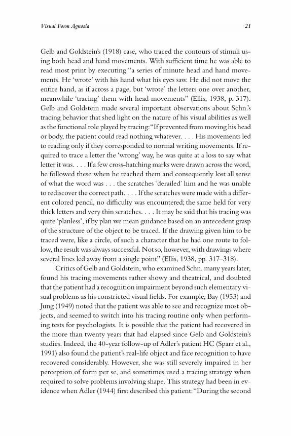
Gelb and Goldstein’s (1918) case, who traced the contours of stimuli us-ing both head and hand movements. With sufficient time he was able toread most print by executing “a series of minute head and hand move-ments. He ‘wrote’ with his hand what his eyes saw. He did not move theentire hand, as if across a page, but ‘wrote’ the letters one over another,meanwhile ‘tracing’ them with head movements” (Ellis, 1938, p. 317).Gelb and Goldstein made several important observations about Schn.’stracing behavior that shed light on the nature of his visual abilities as wellas the functional role played by tracing:“If prevented from moving his heador body, the patient could read nothing whatever. . . . His movements ledto reading only if they corresponded to normal writing movements. If re-quired to trace a letter the ‘wrong’ way, he was quite at a loss to say whatletter it was. . . . If a few cross-hatching marks were drawn across the word,he followed these when he reached them and consequently lost all senseof what the word was . . . the scratches ‘derailed’ him and he was unableto rediscover the correct path. . . . If the scratches were made with a differ-ent colored pencil, no difficulty was encountered; the same held for verythick letters and very thin scratches. . . . It may be said that his tracing wasquite ‘planless’, if by plan we mean guidance based on an antecedent graspof the structure of the object to be traced. If the drawing given him to betraced were, like a circle, of such a character that he had one route to fol-low, the result was always successful. Not so, however, with drawings whereseveral lines led away from a single point” (Ellis, 1938, pp. 317–318).
Critics of Gelb and Goldstein, who examined Schn. many years later,found his tracing movements rather showy and theatrical, and doubtedthat the patient had a recognition impairment beyond such elementary vi-sual problems as his constricted visual fields. For example, Bay (1953) andJung (1949) noted that the patient was able to see and recognize most ob-jects, and seemed to switch into his tracing routine only when perform-ing tests for psychologists. It is possible that the patient had recovered inthe more than twenty years that had elapsed since Gelb and Goldstein’sstudies. Indeed, the 40-year follow-up of Adler’s patient HC (Sparr et al.,1991) also found the patient’s real-life object and face recognition to haverecovered considerably. However, she was still severely impaired in herperception of form per se, and sometimes used a tracing strategy whenrequired to solve problems involving shape. This strategy had been in ev-idence when Adler (1944) first described this patient:“During the second
Visual Form Agnosia 21

week of her illness, the patient started to use her index finger to trace thecontour of objects” (p. 244), and that even after considerable recovery, shewould often “trace the contours of letters with her index finger in orderto enforce perception” (p. 256). The fact that other patients, with similarvisual impairments, have spontaneously adopted the same type of tracingstrategy makes it unlikely that the tracing was purely an affectation to at-tract the interest of psychologists.
Landis et al. (1982) discuss the similarity of their case X to Gelb andGoldstein’s Schn. in the spontaneous use of tracing strategies. They re-ported that “When allowed to trace, X could recognize simple geometricfigures if the point of departure for tracing was unimportant (e.g., circle,triangle). With more complex figures he was misled by unimportant lines.He would give different answers for the same drawing, dependent uponthe point of starting to trace, and often described incidental backgroundfeatures as meaningful. . . . Reading aloud was performed slowly but ac-curately. This ‘reading’ was accomplished by rapid tracing of letters, partsof letters or words with his left hand alone or with both hands. . . . [When]movement of the fingers could be prevented . . . this abolished reading.”Also, like Gelb and Goldstein’s case, X was “derailed” by slash lines, fol-lowing them off of the figure being traced. Landis et al. provide anotherdemonstration of what they call the “slavish” dependence on local conti-nuity in their patient’s tracing:when shown the stimulus in figure 2.4, thepatient consistently read it as “7415.”
Mr. S also spontaneously adopted a tracing strategy in a task in whichhe had to judge whether the orientation of two lines was the same or dif-ferent. According to Efron (1968), “He carefully followed the contoursof each by moving his head. Using this method, he frequently gave cor-rect answers. However, when prevented from making head movements hecould no longer perform the task” (p. 159). When asked to trace arounda shape, Mr. S “will often go round a simple figure many times, not know-ing that he has completed the task. . . .” In those cases in which he is askedto trace a complex object, he will almost always follow the contour of a single color area” (pp. 156–157). Finally, two of the cases were reportedto have difficulty tracing figures by hand: Case ES (Alexander & Albert,1983) had a general impairment in visually guided movements that pre-cluded tracing, and RC (Campion, 1987) was reported to have difficultytracing figures with hand movements. The latter case often resorted to
22 Visual Form Agnosia

spontaneous head movements when asked to identify an object, althoughit should be noted that Campion’s interpretation was that RC seemed tobe searching for the best view with these head movements.
Case DF displays a different form of visuomotor ability that has beenthe subject of intense study among neuropsychologists interested in per-ception and action. The earliest clue that DF had some degree of preservedvisuomotor function came from observations of her reaching behavior.Whereas she could not accurately describe or compare the sizes, shapes,and orientations of objects, her motor interactions with the world seemednormal, including shaping her hand to the proper grip size while reach-ing to grasp a doorknob or a pencil. Milner, Goodale, and colleagues (Milner, Perrett, Johnston, Benson, Jordan, Heeley, Bettucci, Mortara,Mutani, Terazzi, & Davidson, 1991;Goodale, Milner, Jakobson, & Carey,1991; Milner & Goodale, 1995) formalized this observation in a series ofingenious tests—for example, comparing DF’s hand motions when askedto put a card through a slot, with the slot at different orientations, andwhen asked to describe the angle of the slot or to turn a second slot tomatch the angle of the first. Figure 2.5 shows the difference in accuracybetween the two ways of accessing her perception of orientation:by con-scious judgment or matching, and by action. The former is variable andinaccurate; the latter, flawless.
An interesting boundary condition on this dissociation was demon-strated by Goodale, Jakobson, Milner, Perrett, Benson, and Heitanen(1994), who repeated the slot experiment with a T-shaped opening. DFwas unable to insert T-shaped blocks into the opening, suggesting that thepreserved vision for action does not extend to full-blown shape perception.
How can this dissociation between DF’s good visual motor abilitiesand poor explicit judgments of object shape and orientation be explained?Milner and Goodale suggest that the same dorsal visual pathways that have
Visual Form Agnosia 23
Figure 2.4Patient X, studied by Landis et al. (1982), consistently read this stimulus as 7415.
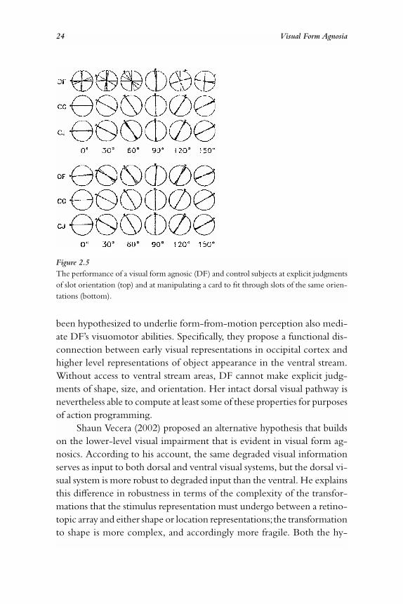
been hypothesized to underlie form-from-motion perception also medi-ate DF’s visuomotor abilities. Specifically, they propose a functional dis-connection between early visual representations in occipital cortex andhigher level representations of object appearance in the ventral stream.Without access to ventral stream areas, DF cannot make explicit judg-ments of shape, size, and orientation. Her intact dorsal visual pathway isnevertheless able to compute at least some of these properties for purposesof action programming.
Shaun Vecera (2002) proposed an alternative hypothesis that buildson the lower-level visual impairment that is evident in visual form ag-nosics. According to his account, the same degraded visual informationserves as input to both dorsal and ventral visual systems, but the dorsal vi-sual system is more robust to degraded input than the ventral. He explainsthis difference in robustness in terms of the complexity of the transfor-mations that the stimulus representation must undergo between a retino-topic array and either shape or location representations;the transformationto shape is more complex, and accordingly more fragile. Both the hy-
24 Visual Form Agnosia
Figure 2.5The performance of a visual form agnosic (DF) and control subjects at explicit judgmentsof slot orientation (top) and at manipulating a card to fit through slots of the same orien-tations (bottom).

pothesized differences in complexity and their consequences for systemrobustness were confirmed by computer simulation. In either case, thedorsal pathway’s independence of the ventral pathway in accomplishingvisuomotor control is a feature of both explanations.
2.7 Neuropathology of Visual Form Agnosia
The neuropathology in these cases of visual form agnosia shows a fairdegree of homogeneity. Five patients suffered carbon monoxide poison-ing (Adler, 1944;Alexander & Albert, 1983;Benson & Greenberg, 1969;Campion & Latto, 1985; Milner et al., 1991), one suffered mercury poi-soning (Landis et al., 1982), and one suffered a penetrating head wound(Gelb & Goldstein, 1918). Neurological signs, EEG, and structural imag-ing suggest that the brain damage in all of these patients was primarily pos-terior, affecting the occipital lobes and surrounding regions. With theexception of the penetrating head wound, the brain damage was diffuseand widespread rather than focal, and Bay (1953) suggested that the pa-tient of Gelb and Goldstein was suffering less from the focal effects of hishead wound than from increased intracranial pressure, which would alsohave diffuse and widespread effects. None of these cases has come to au-topsy, and only HC underwent MRI scanning, which disclosed occipitalatrophy. A CT scan of Campion’s patient showed subcortical white mat-ter lesions. Carbon monoxide is known to damage subcortical white mat-ter and cortex diffusely (particularly the interlaminar connections betweenneurons), and to cause patchy, disseminated lesions. Landis et al. cite re-search showing that mercury poisoning affects the white matter of the oc-cipital lobe.
Visual Form Agnosia 25


Chapter 3
Dorsal Simultanagnosia
Some authors have classified the visual form agnosics just described withanother group of patients who have a disorder known as simultanagnosia(e.g., Adler, 1944; Alexander & Albert, 1983; Bauer & Rubens, 1985;Luria, 1973). The term “simultanagnosia”was originally coined by Wolpert(1924) to describe a condition in which the patient accurately perceivesthe individual elements or details of a complex picture, but cannot appre-ciate its overall meaning. Patients with simultanagnosia resemble visualform agnosics in that they often have full visual fields, but may act as if theyare blind. Furthermore, when presented with an array of stimuli, they of-ten cannot identify stimuli indicated by an examiner. A final, qualitativesimilarity to visual form agnosics is that their perception has a “piecemeal”character to it: they may recognize some part or aspect of an object andguess the object’s identity on the basis of the perceived feature. To furthercomplicate things, simultanagnosia itself does not appear to be a homo-geneous category. I have proposed that there are two distinct disorders thathave been labeled “simultanagnosia” and often discussed interchangeably.To distinguish between them, I suggested the names “dorsal” and “ven-tral simultanagnosia,” after their characteristic lesion sites (Farah, 1990).
3.1 Dorsal Simultanagnosia:A Case Description
Luria (1959, 1963) first associated Wolpert’s rather general term with aspecific type of perceptual deficit, in which only one object, or part of anobject, can be seen at one time. This perceptual deficit is generally observedin the context of Balint’s syndrome, which consists of (1) “psychic paraly-sis of gaze,” an inability to direct voluntary eye movements to visual targets;

(2) optic ataxia, an inability to reach for or point to visual targets; and (3) avisual attentional deficit in which only one stimulus at a time is perceived,and even the attended stimulus may spontaneously slip from attention. Theelements of the syndrome occasionally occur separately from one another(Coslett & Chatterjee, 2003;Rizzo, 1993), raising the possibility that theyhave different underlying mechanisms that are associated because of neuro-anatomical proximity. It is the third element, termed simultanagnosia byLuria, that will be discussed here. Because the associated lesions are almostinvariably bilateral parieto-occipital, affecting the dorsal but not the ven-tral visual pathway, I have called this “dorsal simultanagnosia.”
A case described by Williams (1970) illustrates many of the prime fea-tures of dorsal simultanagnosia:
A sixty-eight-year-old patient studied by the author had difficulty finding his wayaround because “he couldn’t see properly.” It was found that if two objects (e.g.,pencils) were held in front of him at the same time, he could see only one of them,whether they were held side by side, one above the other, or one behind the other.Further testing showed that single stimuli representing objects or faces could beidentified correctly and even recognized when shown again, whether simple orcomplex. . . . If stimuli included more than one object, one only would be iden-tified at one time, though the other would sometimes “come into focus” as thefirst one went out. . . . If long sentences were presented, only the rightmost wordcould be read. . . . If a single word covered as large a visual area as a sentence whichcould not be read, the single word was read in its entirety. . . . If the patient wasshown a page of drawings, the contents of which overlapped (i.e., objects weredrawn on top of one another), he tended to pick out one and deny that he couldsee any others. (pp. 61–62)
3.2 General Characteristics of Dorsal Simultanagnosia
Dorsal simultanagnosia is not a common disorder, but over the decades anumber of excellent case studies have been published. The earliest datefrom World War I, when bullets and shrapnel passing through soldiers’heads could, depending on the entry and exit points, cause the relativelysymmetrical biparietal lesions of dorsal simultanagnosia (e.g., Holmes,1918; Holmes & Horrax, 1919). More recent studies include those ofBaylis, Driver, Baylis and Rafal (1994), Coslett and Saffran (1991), Gil-christ, Humphreys, and Riddoch (1996), Girotti, Milanese, Casazza,
28 Dorsal Simultanagnosia

Allegranza, Corridori, and Avanzini (1982), Godwin-Austen (1965),Hecaen and Angelergues (1954), Kase, Troncoso, Court, Tapia, and Mohr(1977), Luria (1959), Luria, Pravdina-Vinarskaya, and Yarbuss (1963),Tyler (1968), and Williams (1970). There is considerable similarity, fromcase to case, in the pattern of impaired and spared visual abilities.
The most striking feature of this syndrome is that although these pa-tients are able to recognize most objects, they generally cannot see morethan one at a time, and cannot shift rapidly from one to another. This ismanifest in several ways:As with Williams’s case, many cases can name onlyone of a set of objects. The description of complex scenes, which wasWolpert’s defining criterion for simultanagnosia, is correspondingly slowand fragmentary. For example, the Cookie Theft picture in figure 3.1,which is a frequently used to assess the language abilities needed to de-scribe a complex scene (Goodglass & Kaplan, 1978), can also be used toassess the relevant visual abilities. When viewing this picture, Coslett andSaffran’s patient “identified the boy, girl and chair, but did not know whowas standing on the chair or who was reaching for the cookie” (p. 1525).
Dorsal Simultanagnosia 29
Figure 3.1The Cookie Theft picture from the Boston Diagnostic Aphasia Examination.

When shown the scene shown in figure 3.2 for 2 seconds, Tyler’s subjectsaid she saw “a mountain.” When shown the figure for another 2 seconds,she said “a man,” and did not indicate having seen the camel, desert, orpyramids, or realize that the man was related to what she had previouslyseen. When allowed over 30 seconds to look at the picture, she eventu-ally said, “It’s a man looking at the mountains.” She said she never saw the“whole,” but only bits of it that would “fade out” (p. 161). Similar com-plaints emerge in connection with reading: patients complain that wordspop out from the page, then disappear and are replaced by other bits oftext, not necessarily adjacent to the previous bit, making reading difficultor impossible.
Counting is another task that requires seeing more than one objectat time, in order that the subject may keep track of which objects he hasalready counted and which he has yet to count. By contrasting visualcounting ability with tactile or auditory counting ability, one can deducewhether or not the visual component of the task per se is the source ofthe patient’s difficulty. Holmes (1918) describes the behavior of a typicalcase:“When asked to count a row of coins he became hopelessly confused,went from one end to the other and back again, and often passed oversome of the series;but he succeeded in enumerating them correctly whenhe was allowed to run his left fingers over them” (p. 461).
These patients are often described as acting like blind people, grop-ing for things as if in the dark, walking into furniture, and so on. For ex-
30 Dorsal Simultanagnosia
Figure 3.2Drawing described by Tyler’s (1968) patient, after prolonged scrutiny, as “a man lookingat mountains.” She never noticed the camel.

ample, Coslett and Saffran say of their patient that “On one occasion sheattempted to find her way to her bedroom by using a large lamp as a land-mark;while walking toward the lamp she fell over her dining room table”(p. 1525). They add that she could find her way at home more easily withher eyes closed than with them open. This suggests that the problem is in fact quite different from the visual impairment of a blind person. Mostauthors have described the impairment as one of visual attention, for rea-sons stated succinctly by Holmes and Horrax (1919): “The essential fea-ture was his inability to . . . take cognizance of two or more objects thatthrew their images on the seeing part of his retinae. As this occurred nomatter on what parts of his retinae the images fell, it must be attributed toa special disturbance or limitation of attention, but visual attention onlywas affected as he did not behave similarly to tactile or other impressions”(p. 390). It follows from these observations that the attention system af-fected in these patients is not simply used for enhancing the processing ofattended, relative to unattended, stimuli, but is necessary for the detec-tion of stimuli.
There are some reports that even when dorsal simultanagnosics suc-ceed in seeing an object, they will then “lose” it. This is particularly likelywhen the object is moving in a quick or unpredictable way, but can happen even with stationary objects (Godwin-Austen, 1965;Luria, 1959;Tyler, 1968). Rizzo and Hurtig (1987) studied the eye movements andsubjective visual reports of three patients with dorsal simultanagnosia andfound that in all three cases, stimuli seemed to disappear while under fix-ation. Thus, the stimulus disappearance in dorsal simultanagnosia is notsecondary to eye movement problems. On the contrary, the tendency ofthese patients to make erratic, searching eye movements may sometimesbe due to the spontaneous disappearance of the object they were viewing.This may reflect fatigue or habituation of central visual representations fol-lowing the prolonged gaze that is characteristic of dorsal simultanagnosics,once their attention has locked onto an object.
3.3 Visual Disorientation
An additional characteristic of dorsal simultanagnosia is the inability to lo-calize stimuli, even when the stimuli are seen. Because of this striking fea-ture, the term “visual disorientation” is sometimes used as an alternative
Dorsal Simultanagnosia 31

label for dorsal simultanagnosia. The ability to recognize a visible objectwithout being able to localize it is one of the more surprising dissocia-tions in neuropsychology, since it is so difficult for people without this dis-order to imagine seeing and recognizing an object without knowing in which part of the visual field, or of space, they are seeing it. Yet this dis-sociation can be readily demonstrated in dorsal simultanagnosics, either byasking patients to point to or reach for the recognized visual stimulus orto describe its location. In its purest form it is specific to the visual modal-ity, so that patients can locate auditory stimuli (Godwin-Austen, 1965;Holmes, 1918, case 3; Holmes & Horrax, 1919) or their own body partswhen named (Holmes & Horrax, 1919;Kase et al., 1977, case 1) with greatprecision. Such patients provided the basis for early theorizing about theindependence of spatial and object vision, now known to us through an-imal studies and imaging as the “two cortical visual systems” frameworkof Ungerleider and Mishkin (1982).
Are the visual disorientation, just described, and the attentional lim-itation of simultanagnosia one and the same underlying disorder, or arethey merely associated because they depend on neighboring cortex? Itseems likely that visual disorientation is secondary to, and an inevitableconsequence of, the attentional disorder in dorsal simultanagnosia. Thisis because the location of an object can be specified only relative to an-other location, be it the subject’s own body (in a pointing or reachingtask), another object (when describing the object’s location relative to an-other), or the origin of some abstract coordinate system. The inability ofdorsal simultanagnosics to attend to two separate loci would therefore beexpected to impair localization. A patient studied by Holmes (1918) sup-ports this interpretation with his own introspections. When asked to de-scribe the relative positions of two objects placed side by side, one abovethe other, or one nearer to him, the patient made many errors and spon-taneously explained his performance by saying, “I can only look at one ata time” (p. 453).
The relation between representing visual location and attending tovisual stimuli has been explored more fully explored in discussions of neg-lect (Chatterjee & Coslett, 2003). One research tradition explains neg-lect in terms of an impairment of attentional processing that affects thecontralesional side of space (e.g., Posner et al., 1984), whereas anotherpostulates an impaired internal representation of the contralesional side
32 Dorsal Simultanagnosia

of space (e.g., Bisiach, Luzzatti, & Perani, 1979). However, it has beenargued that these explanations are perfectly compatible, differing only inemphasis (Farah, 2000, chap. 8). Attention is not allocated to objects atlocations in space, but rather to internal representations of objects at lo-cations in space. Therefore, even according to an attentional account, neg-lect involves internal representations of the contralesional hemispace thatcannot be used normally. Likewise, because attention operates on repre-sentations, a representational account of neglect implies that attention can-not be deployed in the contralesional hemispace. The same argumentsapply to the bilateral impairment of location representation and attentionin dorsal simultanagnosia.
3.4 Space-Based and Object-Based Attention
Cognitive psychologists distinguish between models in which visual at-tention is allocated to locations in space and to objects per se. Under mostcircumstances, these two alternatives are difficult to tell apart becauseevery object has a location. With the appropriate experimental designs,however, it has been possible to tease the alternatives apart, and the evi-dence suggests that both forms of visual attention exist. Evidence forspatial attention includes demonstrations of that attention to one objectwill attentionally prime another object which either supersedes it in thesame location (e.g., Posner, 1980) or occurs at a nearby location (e.g.,Hoffman & Nelson, 1981). Evidence for object-based representationsconsist of demonstrations that two features can be attended to more effec-tively when they belong to the same object as opposed to different objects,even when the spatial distance between the features is held constant (Dun-can, 1984) and is insensitive to the spatial distance between them (Vecera& Farah, 1994).
What is the nature of this attentional limitation in dorsal simul-tanagnosia? Is it a limitation on the region of visual space that can be at-tended to, or on the number of objects that can be attended to? The answerappears to be “some of both.” The most salient limitation in dorsal simul-tanagnosia is on the number of objects that can be seen. Holmes andHorrax say of their patient, “It might be expected that as a result of thisaffection of visual attention he would be unable to see the whole of largeobjects presented to him at a near distance;but provided part of the image
Dorsal Simultanagnosia 33

did not fall on the blind portions of his visual fields, this was never ob-vious on examination. He recognized objects and even complicated geo-metrical figures as promptly as normal persons. . . . He explained this bysaying ‘I seem to see the whole figure in the first glance’, though occa-sionally he failed to do so if some peculiarity or prominent portion of it at once claimed his attention” (p. 390). Conversely, when attention is fo-cused on a small object, even another small object nearby may not be seen.For example, Hecaen and Angelergues (1954, case 1) remark, “In light-ing a cigarette, when the flame was offered to him an inch or two awayfrom the cigarette held between his lips, he was unable to see the flamebecause his eyes were fixed on the cigarette” (p. 374). There is never-theless some evidence that spatial factors play a role in the attentionalbottleneck. On the rare occasions when multiple objects can be perceived,they are generally small, close together, and foveal (e.g., Holmes, 1918;Tyler, 1968).
The earliest experiments on object and spatial attentional limitationsin dorsal simultanagnosia were carried out by Luria and colleagues. In onestudy (Luria et al., 1963) a patient was shown drawings of common ob-jects at different sizes in a tachistoscope. He could name the objects pre-sented one at a time, whether they subtended 6º–8º or 15º–20º. However,when two were shown simultaneously, even at the smaller size, the patientcould only name one. Luria (1959) also reports that additional visual ma-terial could be perceived if grouped into a single object by so simple aschange as connecting two forms with a line, as shown in figure 3.3. Thisobservation has since replicated and extended by Godwin-Austen (1965)and Humphreys and Riddoch (1993).
The finding that the “fracture lines” of attentional impairment fol-lowing bilateral dorsal visual system damage are influenced by the bound-aries of objects might seem puzzling, given what we know about thedivision of labor between the dorsal and ventral visual systems. As alreadymentioned, there is abundant evidence supporting the division between“what,” or object-related processing, and “where,” or space-related pro-cessing. It is supposed to be the ventral visual system that represents ob-jects, and the dorsal system that is concerned with spatial location.
The resolution of this conflict involves distinguishing between twotypes of object-based attentional effects, one of which is spatially medi-ated. This distinction is crucial to the interpretation of object-based at-
34 Dorsal Simultanagnosia

tention studies, in neglect as well as dorsal simultanagnosia, but has notalways been clearly drawn (Buxbaum, Coslett, Montgomery, & Farah,1996;Mozer, 2002). Spatially mediated object effects will emerge withinany system that includes reciprocal activation between object representa-tions and earlier, spatially formatted retinotopic representations. Considerthe simple interactive model of spatial attention and object recognitionshown in figure 3.4. The bidirectional arrows linking early retinotopicrepresentations with the higher-level systems indicate the reciprocal na-ture of the influences between these pairs of systems. Spatial attention canselect certain regions of the retinotopic array for further processing in ei-ther a “bottom-up” manner (i.e., a salient stimulus attracts attention) or a“top-down” manner (i.e., the person decides to shift attention to a cer-tain region of the array). This is the most familiar mechanism by whichattention is allocated to spatial locations. However, if the connection be-tween the array and object representations enables top-down as well asbottom-up activation, then object knowledge will also influence the dis-tribution of attentional activation of the array. Specifically, in addition topatterns in the array activating their corresponding object representations,those object representations will provide top-down support for their cor-responding activation patterns in the array.
The process of object recognition therefore results in two types ofactivated object representation. The first is the object representation
Dorsal Simultanagnosia 35
Figure 3.3Stimuli of the type used by Luria (1959) and others to demonstrate the powerful effect of“objecthood” on the perception of shapes in dorsal simultanagnosia.

proper, in the object system, which possesses various constancies such asshape and location constancy and is therefore not fundamentally a spatialrepresentation. The second is the region of the retinotopic array contain-ing the object image, activated by top-down influence from the first kindof object representation. In contrast to the distribution of attention by apurely spatial attentional “spotlight,” which would be expected to acti-vate simple ovoid clusters of array locations, the regions activated by ob-ject representations conform to the silhouette of the object in the array.Although such regions do, in a sense, represent object shape, the repre-sentation is fundamentally spatial, consisting of a set of locations or“pixels” in an array. Attention could in principle single out objects for fur-ther processing among the higher-level nonspatial representations of theobject system, as Duncan (1984) suggested, or among the fundamentallyspatial representations of the retinotopic array that have been shaped bytop-down activation from the object system. In fact, both types of atten-tion exist.
Vecera (1994) tested the predictions of the second type of accountby showing that when an object is spatially attended, the attention tends
36 Dorsal Simultanagnosia
OBJECTREPRESENTATIONS
ARRAY OFLOCATIONS
ATTENTION
Figure 3.4Model of the relations among the retinotopically formatted image representations of earlyand intermediate vision and two higher-level systems, visual object recognition and vi-sual spatial attention. Each higher-level system receives input from the image, activatingobject representations and drawing attention to activated regions of the image. Eachhigher-level system also contributes activation to the image, in the form of top-down sup-port for image features consistent with activated object representations and attentional ac-tivation in regions of the image to which attention has been drawn. The result of the visualobject recognition pathway and the spatial attention pathway interacting in a commonimage representation is both the object effects in spatial attention observed with dorsal si-multanagnosics and the spatial attentional effects on object recognition observed withventral simultanagnosics.

to be distributed over the region of the image occupied by the object.Thus, a vertically oriented rectangle will attract spatial attention to regionsabove and below the rectangle’s middle, and a horizontally oriented rec-tangle will attract spatial attention to the left and right. Using computersimulation, Mozer (2002) has shown that such a system of spatial atten-tion can account for all of the demonstrations so far reported of objecteffects in the attentional limitations of parietal-damaged neglect patients.Figure 3.5 shows one such simulation.
Within this framework it is clear how the seemingly object-basedlimitation of attention in dorsal simultanagnosia can be accounted for interms of damage to a fundamentally spatial attention system. Specifically,the impaired spatial attention system is operating on a spatial representa-tion that has been parsed into objects by a normal object recognition sys-tem, and when it is unable to shift attention between regions, attentionstays fixed on a single object’s region, regardless of its size or complexity.This also explains the observation that although the visual attention ofdorsal simultanagnosics is primarily limited to a single object, there is alsosome, relatively weaker, limitation on spatial extent.
Dorsal Simultanagnosia 37
Figure 3.5Computer simulation of the emergence of object-based attentional effects in a retino-topically formatted image representation (Mozer, 2002). Attention is initially allocated tothe right end of the barbell, and tracks this portion of the object during rotation.

3.5 Spatial Relations Within and Between Objects
Objects and their parts are hierarchical, in that one can identify a bicycleas an object and a wheel as a part, or a wheel as an object and a spoke as apart. What, then, does it mean to say that attention is limited to one ob-ject at a time in dorsal simultanagnosia? Would it be limited to a bicycle,a wheel, or a spoke? The answer is that it depends. Just as we can see ob-jects at various levels in a hierarchy, the damaged attentional system in dor-sal simultanagnosia has flexibility in what it selects as an object, and bothphysical stimulus properties and higher cognitive processes contribute tothis selection.
Luria (1959) asked a patient with dorsal simultanagnosia to describetwo different versions of a Star of David. In the first version, the star wasdrawn all in the same color of ink. In the second, one of the two compo-nent triangles was drawn in red and the other was drawn in blue. The sec-ond version provided a stimulus in which there were two distinct “objects”or visual gestalts, the red and blue triangles, occupying roughly the sameregion of space and together occupying precisely the same region of spaceas the first version. When shown the first version, the patient consistentlynamed the star. When shown the second version, the patient named ei-ther the red triangle or the blue triangle.
Dorsal simultanagnosics will sometimes appear to have an objectrecognition impairment because their visual attention will be captured bya part of the object to be recognized. For example, Tyler observed the fol-lowing of his patient:“When shown a pitcher pouring water into a glass,she first noted the handle and said ‘suitcase’. When asked to look again shespotted the glass, and remembering the handle she kept looking until sheperceived the pitcher (now with the handle on it)” (Tyler, 1968, p. 158).This is reminiscent of Holmes and Horrax’s comment that their patientsometimes failed to see a whole object if “some peculiarity or prominentportion of it at once claimed his attention.” This attentional problem caninterfere with recognizing objects. However, it is more accurate to say thatit interferes with seeing objects, or seeing them at the “correct” level ofthe hierarchy of part-whole analysis;whatever dorsal simultanagnoscis cansee, they can recognize. Furthermore, such problems are rare, since theirattention is almost always claimed by what we would consider a single,whole object.
38 Dorsal Simultanagnosia
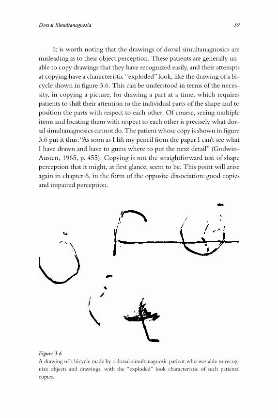
It is worth noting that the drawings of dorsal simultanagnosics aremisleading as to their object perception. These patients are generally un-able to copy drawings that they have recognized easily, and their attemptsat copying have a characteristic “exploded” look, like the drawing of a bi-cycle shown in figure 3.6. This can be understood in terms of the neces-sity, in copying a picture, for drawing a part at a time, which requirespatients to shift their attention to the individual parts of the shape and toposition the parts with respect to each other. Of course, seeing multipleitems and locating them with respect to each other is precisely what dor-sal simultanagnosics cannot do. The patient whose copy is shown in figure3.6 put it thus:“As soon as I lift my pencil from the paper I can’t see whatI have drawn and have to guess where to put the next detail” (Godwin-Austen, 1965, p. 455). Copying is not the straightforward test of shapeperception that it might, at first glance, seem to be. This point will ariseagain in chapter 6, in the form of the opposite dissociation: good copiesand impaired perception.
Dorsal Simultanagnosia 39
Figure 3.6A drawing of a bicycle made by a dorsal simultanagnosic patient who was able to recog-nize objects and drawings, with the “exploded” look characteristic of such patients’copies.

“Objecthood” is not simply a matter of size or of visual complexity,as demonstrated by Luria (1959). When he showed a rectangle made upof six dots to a patient, the patient could see the rectangle, as well as rec-ognize other geometric forms drawn with broken lines. However, as is therule in dorsal simultanagnosia, counting was impossible, even countingthe six dots of the rectangle he so easily identified. Apparently the patientcould view the stimulus as dots, and therefore see a single dot at a time,or he could view it as a rectangle, and therefore see the entire rectangle.In the latter case, the patient was still unable to count the dots, presum-ably because once he started to count the dots, he would again be seeingthem as dots and see only one at a time. In other words, the organizationof the dots into a rectangle did not increase the patient’s attentional ca-pacity to more than one object; rather, it allowed the patient to view theset of dots as a single object. An implication of this is that the attentionalsystem of dorsal simultanagnosics is sensitive to voluntary, “top-down”de-terminants of what constitutes an object.
The reading ability of dorsal simultanagnosics suggests that knowl-edge derived from previous learning also influences what our visual sys-tems take to be an object. Recall Williams’s (1970) observation that herpatient could read a single word, regardless of whether it subtended a large or small visual angle, but a sentence subtending the same large vi-sual angle could not be read. Other patients describe the experience ofsingle words jumping out at them from a page (Girotti et al., 1982;Godwin-Austen, 1965;Luria, 1959;Luria et al., 1963;Hecaen & Angel-ergues, 1954, case 2; Holmes, 1918; Holmes & Horrax, 1919). The hy-pothesis that words function as objects in attracting attention was testedby Baylis, Driver, Baylis, and Rafal (1994) with a dorsal simultanagnosicpatient. When shown letters in isolation, the patient recognized themeasily, as would be expected, given that attention is limited to one ob-ject at a time. When shown four-letter words, he read them with high ac-curacy as well. On the other hand, his ability to name nonword stringsof four letters was poor. Words and nonword strings do not differ in their objective visual complexity or wholeness, and thus provide com-pelling evidence for the role of knowledge in defining objects for theallocation of visual attention.
40 Dorsal Simultanagnosia

3.6 Neuropathology of Dorsal Simultanagnosia
The brain damage that causes dorsal simultanagnosia is bilateral and gen-erally includes the parietal and superior occipital regions. In a few casesthere has been evidence of damage confined to the occipital regions(Girotti et al., 1982;Rizzo & Hurtig, 1987), although in another only thesuperior parietal lobes were damaged (Kase et al., 1977, case 1). Etiolo-gies likely to result in this configuration of damage, with preservation ofventral visual system areas, include the penetrating head injuries originallystudied by Holmes (1918) and “watershed” infarction, in which a drop inblood pressure affects the most distal, or watershed, territory between themiddle and posterior cerebral arteries. Middle cerebral artery strokes, al-though they commonly result in parietal damage, are not often the causeof dorsal simultanagnosia because of the need for bilateral lesions.
3.7 Visual Form Agnosia and Dorsal Simultanagnosia: Similarities andDifferences
There is a “family resemblance” among several of the syndromes tradi-tionally considered to be apperceptive agnosias. In the case of visual formagnosics and dorsal simultanagnosics, both may act effectively blind. Theyare unable to negotiate visual environments of any complexity, and willmake random searching eye movements if asked to look at a particular ob-ject. Nevertheless, in both cases they may have relatively full visual fieldsand normal acuity and color perception. An additional similarity is thattheir perception appears to be piecemeal and confined to a local part orregion of the visual field. Although visual form agnosics may seem gen-erally more impaired at recognizing objects, both types of patients mayevince object recognition difficulties and, when encountering these dif-ficulties, both typically resort to guessing based on correct partial per-ceptions. Even the neuropathology fails to provide a clear basis fordistinguishing among these patients. In both groups it is bilateral and pos-terior, and spares striate cortex to at least some degree, with cases of focalparieto-occipital damage occurring in both groups.
Despite these similarities, a closer look at the abilities and impair-ments of these groups of patients leads to the conclusion that the under-lying deficits must be very different. Consider the “piecemeal” nature of
Dorsal Simultanagnosia 41

perception in each case. In visual form agnosia only very local contour isperceived. It is so local that patients cannot trace across a break in a line,trace dotted lines, or avoid “derailment” onto irrelevant slashes drawnacross a figure. This is not at all the case in dorsal simultanagnosia. Wholeshapes are perceived, even if composed of dots or broken lines. What ispiecemeal about the perception of dorsal simultanagnosics is the limita-tion of their vision to a single object or visual gestalt, without awarenessof the presence or absence of other stimuli. Furthermore, in dorsal simul-tanagnosia the nature of the “piece” is at least partially determined by con-scious attention (e.g., to the individual dots arranged in a rectangle or to the rectangle), whereas no such top-down influences have been noted toaffect the piecemeal perception of apperceptive agnosics. The guessingstrategies of visual form agnosics and dorsal simultanagnosics are likewisesimilar only on the surface. Although both make educated guesses aboutobjects’ identities based on partial perceptual information, the nature ofthe information used by each is different. Apperceptive agnosics use color,size, and texture, but do not use shape information. Dorsal simultanag-nosics do use shape information. Furthermore, the shape perception ofdorsal simultanagnosics is intact. A final difference is that whereas motiontends to facilitate shape perception by apperceptive agnosics, it interfereswith perception by dorsal simultanagnosics.
42 Dorsal Simultanagnosia

Chapter 4
Ventral Simultanagnosia and Pure Alexia
There is another group of patients who have been called simultanagnosics,and who share the following characteristics with dorsal simultanagnosics.They are generally able to recognize a single object, but do poorly withmore than one, and with complex pictures. Their introspections soundsimilar to those of dorsal simultanagnosics. For example, when viewing a complex picture, one patient said, “I can’t see it all at once. It comes tome by casting my eye around” (Kinsbourne & Warrington, 1962, p. 466).As in dorsal simultanagnosia, reading is severely impaired. Furthermore,as in dorsal simultanagnosia, the size of the objects does not matter; thelimitation is in the number of objects. It is therefore not surprising that thesame label has been applied to these patients. However, on closer inspec-tion these two groups of patients differ in important ways, implicatingdifferent underlying impairments. Ventral simultanagnosics are so calledbecause the typical lesion is in the left temporo-occipital region.
Although ventral simultanagnosics cannot recognize multiple ob-jects, they differ from dorsal simultanagnosics in that they can see multipleobjects. This is evident in their ability to count scattered dots (Kinsbourne& Warrington, 1962), as well as in their ability to manipulate objects andwalk around without bumping into obstacles. Furthermore, given suffi-
cient time, they can recognize multiple objects. The one crucial activityof daily life that ventral simultanagnosics cannot perform is reading. Thealexia of these individuals is so salient, compared to their other visual ab-normalities, that they may be referred to simply as alexics. More specifi-
cally, their reading disorder is called “pure alexia” because it occurs inisolation from other visual and language disorders.

4.1 Ventral Simultanagnosia and Pure Alexia:A Case Description
A young man described by Warrington and Shallice (1980) exemplifiesmany of the characteristics of this syndrome. The individual was a 27-year-old deputy headmaster at a comprehensive school who five years earlierhad suffered a brain hemorrhage and had undergone surgery to evacuatea left temporoparietal hematoma. His status when seen by the authors isdescribed here.
At this time his only complaint was of being able to read only very slowly and halt-ingly, and of being unable to see words, especially long words, as a whole. . . . [He]was tested on the WAIS and obtained a verbal IQ of 125 and a performance IQof 118. On both the Picture Arrangement and the Picture Completion subtestsof the WAIS (the test stimuli are complex visual scenes) he obtained an age-scaledscore of 13, which is within the high average range. . . . His perceptual skills ap-peared entirely normal; for example he scored 20/20 on a test of identifying ob-jects from unconventional views (Warrington & Taylor, 1973). . . .
On the Schonell graded word reading test he scored at a high level, hisperformance being virtually error-free (98/100); on the Nelson reading test heobtained a reading IQ of 121 (42/50); his spelling was also entirely satisfactory.However, qualitatively, his reading was markedly abnormal. He was observed tospell out most words letter-by-letter either aloud or under his breath, and then re-construct the word (correctly) from the auditory letter information. For example,he took thirty seconds to read the first fifteen words of a prose passage. Very shortwords appeared to be read relatively normally, but even these were read somewhatslowly. . . . He was able to read nonwords almost as efficiently as words. His meantime to read lists of 5 words and nonwords (the stimuli in both tests were 4 and 6letters) was 13.3 and 14.0 seconds, respectively, and only very occasional errorswere made. Numbers written in numerical form (for example 1,340,210) wereread quickly and efficiently. There was no evidence of colour agnosia.
4.2 Ventral Simultanagnosia and Pure Alexia: Some Generalities
Pure alexia is more common than the other disorders discussed in thisbook. Perhaps because the syndrome is relatively familiar, recent articlestend to offer little in the way of patient description, such as performanceon a variety of tests other than reading or real-life problems with reading,focusing instead on testing specific hypotheses. Earlier case reports, such
44 Ventral Simultanagnosia and Pure Alexia

as that of Warrington and Shallice excerpted above, are more generouswith their background information on patients (see also Patterson & Kay,1982; Warrington & Zangwill, 1957).
Of course, all reports document the letter-by-letter reading that is thehallmark of the syndrome. Even when patients do not spell words audibly,analysis of their single-word reading latencies as a function of number of letters in the word suggests a letter-by-letter strategy. Figure 4.1 showsthe monotonic, almost perfectly linear, dependence of reading latency onword length in a pure alexic patient studied by Bub, Black, and Howell(1989). The negative acceleration of this function is probably attributableto the greater possibility of guessing the word before the last letters are readwith longer words. Note the time scale on this figure: Even short wordsrequire several seconds for this patient, and each additional letter adds asecond or more to the reading time. In summarizing the characteristicsof seven pure alexic subjects from their research, Behrmann, Plaut, andNelson (1998) noted a range in the time needed per letter, from 97 milli-seconds for an extremely mild alexic to 1.4 seconds. Patients’ reading ratescan be considerably slower than that;I have seen pure alexics spend a minuteor more on a word, although in such cases misidentifications of individual
Ventral Simultanagnosia and Pure Alexia 45
Figure 4.1Single-word reading latency as a function of number of letters in the word for a pure alexicpatient studied by Bub et al. (1989).

letters are also common. For that matter, however, it has been said that allpure alexics show some degree of impaired single-letter perception (Behr-mann & Shallice, 1995).
In addition to the letter-by-letter reading strategy, the other gen-eral features of pure alexia concern visual and language ability. Patients’ability to understand spoken language and recognize objects is invariablytested and found to be grossly intact. When writing ability is tested, it alsois intact, leading to the almost paradoxical finding of patients being un-able to read what they themselves have written.
This form of alexia is called “pure”because other forms of visual pat-tern recognition appear preserved, as do spelling, writing, and other lan-guage-related abilities. However, a closer look at the visual abilities of thesepatients reveals a distinctive type of visual disorder, ventral simultanag-nosia. Kinsbourne and Warrington (1962) first described the simultanag-nosia of pure alexic patients. Their findings, and subsequent findings, arethe focus of the next section.
4.3 Parallel Shape Recognition:A Dissociable Ability?
The nature of the perceptual impairment in ventral simultanagnosia wasfirst investigated by Kinsbourne and Warrington (1962), with a series ofelegant tachistoscopic experiments in which the speed of processing singleand multiple visual shapes could be assessed. These experiments showedthat the patients’ tachistoscopic recognition thresholds for single forms(letters or simple pictures) were within normal limits, but that their thresh-olds departed dramatically from those of normal individuals when morethan one form had to be recognized. Varying spatial factors, such as thesize, position, and separation of the stimuli, had no effect; the visual pro-cessing “bottleneck” in these patients was determined solely by the num-ber of separate forms to be recognized. Given that reading words involvesthe perception of multiple letters, this account of the visual impairmentexplains the alexia of these patients, and in particular the slow, letter-by-letter manner in which they attempt to read.
Levine and Calvanio (1978) replicated and extended the findings ofKinsbourne and Warrington with three new cases of what they termed“alexia-simultanagnosia.” Among the novel results of their study werethree findings that helped to pinpoint the locus of the processing impair-
46 Ventral Simultanagnosia and Pure Alexia

ment more precisely than Kinsbourne and Warrington’s studies. First,Levine and Calvanio demonstrated that the difficulty with multiple stim-uli is present even when the task does not involve naming the stimuli, butmerely judging whether any two of the stimuli in an array are identical ornot. This implies that the limitation is truly affecting perception per se,and not the process of labeling the percept. Second, subjects made moreerrors in this matching task when the letters in the display were visuallysimilar (e.g., OCO, as opposed to OXO), again suggesting a visual locusfor the processing breakdown. Finally, Levine and Calvanio contrasted theeffects on subjects’ performance of position cues presented just before andjust after the stimulus array. If shape recognition per se is limited to justone item, then the pre-cue should improve performance because it allowsthe subject to recognize the one item that has been cued, but the post-cue should not, because it comes after the stimulus array has disappearedand thus cannot guide selective perception. In contrast, if the bottleneckis occurring after shape recognition, in some short-term memory bufferor labeling process, then the post-cues should also help. Levine and Cal-vanio found that subjects were helped by the pre-cues: if they knew inadvance which letter (indicated by the position of a pre-cue) from a multi-letter array they were to report, they could do so accurately, even with theother letters present. However, if the cue came after perceptual processinghad been completed, it did not help, again implicating visual recognitionper se as the locus of impairment.
Why might the brain recognize multiple shapes with a distinct andseparately lesionable system? Computationally, the recognition of multipleshapes poses a special problem, distinct from the problem of recognizingcomplex or unfamiliar shapes (to mention two other ways in which shapeperception can be made difficult). The special problem for multishape recog-nition is cross talk, or interference, among the representations of separateshapes, which will be more severe the more distributed the representation.
Although distributed representation has many computational be-nefits and is used in a number of brain systems, including the visual ob-ject recognition system, it is not well suited to representing a number ofitems simultaneously. This is because once two distributed representationshave been superimposed, it is difficult to know which parts of each of thetwo representations go together. This problem is illustrated in the top partof figure 4.2. The bottom part of figure 4.2 shows that one way around
Ventral Simultanagnosia and Pure Alexia 47

this problem is to develop more localist representations. A tentative in-terpretation of the perceptual impairment of ventral simultanagnosics isthat they have lost a region of cortex in which shape information is rep-resented in a relatively more local manner.
4.4 Are Ventral Simultanagnosia and Pure Alexia Equivalent?
Although I have so far written as if pure alexia results from ventral simul-tanagnosia, this is not the only explanation that has been proposed for purealexia. Indeed, in its simplest form—a general impairment in any kind ofparallel shape recognition, which is noticeable only during reading—theexplanation fails to account for certain observations, to be discussed. Inthis section the different types of explanation for pure alexia will be re-viewed, along with the evidence for and against each.
48 Ventral Simultanagnosia and Pure Alexia
Figure 4.2A demonstration of interference among multiple representations in distributed systems,and the lack of such interference in localist systems (from Farah, 2000).

The disconnection account of pure alexia was proposed by Dejerine(1892), and more recently championed by Geschwind (1965). Accordingto this account, reading consists of associating visual information in oc-cipital cortex with language representations in posterior language areas.This is done by way of the left angular gyrus, adjacent to Wernicke’s area,which is hypothesized to contain stored multimodal associations linkingthe visual and sound patterns of printed words. Thus, pure alexia resultsfrom any lesion that disconnects the visual cortex from the left angulargyrus. The neuropathology of pure alexia is generally consistent with thishypothesis (e.g., Damasio & Damasio, 1983; Greenblatt, 1983), often in-volving a left occipital lesion (causing blindness in the right visual field)and damage to the adjacent splenium (disconnecting left visual field in-formation from the left hemisphere). Despite the anatomical support forthis interpretation of pure alexia, it is not an altogether satisfying expla-nation. For one thing, although it is not in any way inconsistent with theletter-by-letter reading strategy of pure alexic patients, it also is not ex-planatory of this highly characteristic feature of the syndrome.
It is, of course, possible that disconnection may contribute to someparticularly severe cases of pure alexia. I recall one patient who was virtu-ally unable to read at all, and whose letter-by-letter reading often involvedletters that bore no relation to the word he was looking at. His oral verbalresponses seemed to be running free, independent of visual input, with atendency toward perseveration, and his picture naming had the same un-related and often perseverative qualities. The total impression was ofexactly a disconnection in the pathways that normally link visual input andverbal output. Yamadori (1980) reported two relatively severe cases of purealexia with what he called “unilateral dyscopia,” the inability to copy writ-ten characters with one hand. In both cases the right-hand copies wereworse than the left-hand, consistent with the inability of visual informa-tion to reach the left hemisphere. The more severe case, who was entirelyunable to read written characters, showed the more complete dissociationin copying between the left and right hands.
The ventral simultanagnosia account was the next to be proposed, by Kinsbourne and Warrington in 1962 and more recently advocated byMarcie Wallace and myself (Farah & Wallace, 1991). In the research alreadysummarized in section 4.3, pure alexic patients were shown to have an im-pairment in the rapid recognition of multiple shapes using tachistoscopicmethods. This impairment can also be demonstrated with more natural
Ventral Simultanagnosia and Pure Alexia 49

stimulus presentations. Wallace and I administered simple paper-and-pencil tasks that stress the rapid encoding of multiple visual shapes to a purealexic, and found a severe impairment, a finding that has since been repli-cated by Sekuler and Behrmann (1997) in three additional patients.
So far the support for the ventral simultanagnosia account has takenthe form of associations between impairments in multiple parallel shaperecognition and letter-by-letter reading. Evidence from associations issubject to a particular type of alternative explanation, specifically that thereare two separate abilities which depend on neighboring brain regions andwhich are therefore likely to be spared or impaired together. The ambi-guity of associational data provided the motivation for us to manipulatethe difficulty of visual perception and assess its effect on the reading of a pure alexic (Farah & Wallace, 1991). We used additive factors logic toidentify the stage of reading that gives rise to the abnormal word lengtheffect (i.e., the stage of reading at which the process is forced to proceedletter by letter), and specifically to test whether it is the visual shape recog-nition stage of processing. According to additive factors logic, if two ex-perimental manipulations affect the same stage of processing, their effectswill be interactive, whereas if they affect separate stages, their effects willbe additive (Sternberg, 1969).
We presented a pure alexic with words of varying length to read,printed either clearly or with spurious line segments superimposed, asshown in figure 4.3. This manipulation of visual quality would be ex-pected to affect the stage of visual shape recognition. We found that wordlength and visual quality interacted in determining reading latency. Spe-cifically, the word length effect was exacerbated by visual noise, as shownin figure 4.4. This finding is consistent with a visual shape recognition lo-cus for the word length effect in this experiment, and, in more generalterms, with the hypothesis that an impairment in the rapid perception ofmultiple shapes underlies pure alexia. Unfortunately, the most straight-
50 Ventral Simultanagnosia and Pure Alexia
Figure 4.3Examples of normal and degraded word stimuli used in a study of pure alexia.

forward version of the ventral simultanagnosia account does not explainthe observation that number recognition is preserved in some pure alexicpatients, as it was in Warrington and Shallice’s (1980) case. It also fails toexplain why single letters are often misperceived in pure alexia.
The third major approach to explaining pure alexia is as an orthography-specific impairment. There are a number of levels of orthographic represen-tation that could be impaired in pure alexia, from individual letters towhole word representations. Warrington and Shallice (1980) proposed thatpure alexia was the result of damage to relatively high-level orthographicrepresentations of words and morphemes. They called these representa-tions “word forms” and pointed out that a loss of word forms could ex-plain the characteristic letter-by-letter reading of pure alexics, since theirvisual word recognition cannot make use of word forms, and thereforemust proceed via individual letter recognition and knowledge of spelling.
The evidence presented by Warrington and Shallice in favor of theirhypothesis was of two kinds. First, they assessed various visual capabilitiesin an attempt to rule out visual perception as the locus of impairment. By
Ventral Simultanagnosia and Pure Alexia 51
Figure 4.4The interaction of visual degradation by masking and the word length in a pure alexicpatient.

a process of elimination, this would strengthen the case for a word formimpairment. However, despite having used a number of different percep-tual tasks, many of which involved displays of multiple items, none of thetasks specifically taxed the process of rapid perception of multiple shapes,which is the leading alternative hypothesis. For example, some of the taskswere not speeded, some involved foreknowledge of target locations (elim-inating the need to recognize all but the target stimuli), and so on (seeFarah & Wallace, 1991, for a detailed review of these tasks).
Second, Warrington and Shallice manipulated reading difficulty intwo ways that they believed would render subjects relatively more de-pendent on visual word forms. In one experiment they compared thereading of print to the reading of script, and found performance withscript to be worse. In a second experiment they compared reading wordsthat had been presented tachistoscopically, for half a second, to readingnontachistoscopic word presentations, and found a marked decrement inreading performance with tachistoscopic presentations. Although thesemanipulations could well increase the reader’s reliance on word forms,they would also make reading harder for a ventral simultanagnosic, andtherefore do not discriminate between the accounts.
The word form hypothesis has not fared well in the face of more re-cent demonstrations that at least some pure alexic subjects show a “wordsuperiority effect.” The word superiority effect refers to the facilitation ofletter perception when letters occur in the context of a word or pseudo-word, relative to a nonword or, in some cases, no flanking letters at all(Reicher, 1969; Wheeler, 1970). The facilitation of letter perception byword or wordlike contexts is not simply the result of a bias to guess lettersthat would make a word, because it is observed even in forced-choice taskswhen both choices make a word:for example, when the stimulus is ROADand subjects are asked whether the second character is an O or an E.
The word superiority effect might seem paradoxical at first, for oneusually thinks of letters being perceived before words, yet here words areinfluencing letter perception. The key to understanding this effect is tonote that while letters are indeed perceived before words, in the sense thattheir representations begin to be activated before word representations be-gin to be activated ( Johnston & McClelland, 1980), letter activation maynot be complete by the point at which word representations begin to beactivated. Assuming that activated words feed activation back down to
52 Ventral Simultanagnosia and Pure Alexia

their component letter representations, as well as compete with one an-other in a winner-take-all manner, then words, most likely on the basis ofmidprocess letter recognition, will reinforce perception of those letters.An early and influential connectionst model, by McClelland and Rumel-hart (1981), demonstrates the way in which such within-level compet-itive interactions and between-level reinforcing interactions togetheraccount for most of the findings concerning word superiority in letterperception. Figure 4.5 shows a schematic depiction of part of their model.
The word form hypothesis predicts an absent or at least attenuatedword superiority effect in pure alexic subjects. However, a number of purealexic patients have shown word superiority effects, in some cases com-parable to control subjects’ (see Behrmann, Plaut, & Nelson, 1998, for areview), leaving the word form hypothesis with little supporting evidenceand substantial disconfirming evidence.
A final account of pure alexia is a hybrid of the preceding two thatinvolves specialization within specialization in the brain. One specializationis for the rapid or parallel encoding of multiple shapes, which I earlier ar-gued could be accomplished by developing more localist representations.According to this account, the general manifestations of ventral simul-tanagnosia result from damage to such an area. However, a further spe-cialization within this area can be hypothesized to occur for letter and wordrepresentation because these are the stimuli most frequently processed inthis area. The development of the latter specialization has been shown toemerge naturally from basic principles of neural computation.
Thad Polk and I have explored some of the ways in which neural net-works respond to statistical regularities in our visual environment, partic-ularly orthographic regularities, using a “self-organizing system,”a networkthat learns without an external source of information conveying “right”or “wrong.” Indeed, in such systems there is no “right” or “wrong” be-cause there is no target pattern to be learned. Rather, the strength of con-nections among the neuronlike units of such a network is changed simplyas a function of the correlations among the activity levels of the units inthe network. The best-known learning rule for self-organizing systems isthe Hebbian rule: “Neurons that fire together wire together.” In otherwords, when the activity levels of two units are positively correlated, theconnection strength between them increases. This increases the likelihoodthat their activations will be correlated in the future, since activation of
Ventral Simultanagnosia and Pure Alexia 53

54 Ventral Simultanagnosia and Pure Alexia
Figure 4.5Illustration of the interactive activation model of word recognition. Letter units are ini-tially activated by an amount proportional to the input they receive from the units repre-senting their constituent features. The letter units then pass activation on to the wordswith which they are consistent. For example, if there appears to be a “t” in the first posi-tion, that will cause the words “trap,” “trip,” “take,” and “time” to gain activation. In ad-dition, word units are inhibited by the activation of units that are inconsistent with theword. For example, “able” will be inhibited by the unit representing an initial “t,” sinceactivation in these units represents incompatible hypotheses, and “able” will also be in-hibited by activation in other word units for the same reason. So far, this model wouldseem to account for word recognition, but it is not yet clear how it accounts for the wordsuperiority effect, nor is it clear how it accounts for phenomena involving pseudowords,that is, statistical approximations to real words. The word superiority effect is explainedby postulating feedback from the word level to the letter level. Switching to a set of ex-amples different from those shown in figure 4.5, if the word shown is “read,” and percep-tion of the letter in the second position is just barely adequate, so that the “e” unit is everso slightly more activated than the “o” unit, then this will give the edge to the word “read”over the word “road.” Interword inhibition will then drive the activation of the “road”unit down, and feedback from the word level to the letter level will therefore consist ofgreater “top-down” support for “e” than for “o.” Thus, people will be more accurate atdiscriminating “e” from “o” in the context of “read” than in a nonword context or alone.Why should pseudowords also give rise to superior letter perception, given that there areno units corresponding to them at the word level? Because pseudowords are sufficientlysimilar to real words that their constituent letters will at least partially activate some realword units. “Yead,” for example, will activate “year,”“bead,” read,” and so on. Once theseword units have become activated, they will inhibit other word units that are less consis-tent with the activated letter-level units, and will provide top-down support for the let-ters that do occur in the words similar to “yead,” including, for example, the “e” in thesecond position.

one will cause the other to become active by virtue of the strengthenedconnection. In this way, the network develops a repetoire of stable patternsof activation, in the sense that activation patterns which are close to thelearned pattern (e.g., contain part of it) will be transformed into the learnedpattern and tend to remain active. These stable patterns can be viewed asrepresentations of whatever inputs to the network evoke the patterns.
If it is assumed that prior to learning, neighboring neurons have ex-citatory connections among them, such that activating one neuron tendsto activate its neighbors, then it is possible to account for a number ofaspects of cortical representation by the simple mechanism of Hebbianlearning. For example, topographic maps such as the somatosensory ho-munculus arise because neighboring locations in the input to be repre-sented (locations on the body surface, in the case of the homunculus) tendto be activated at the same time. Because of the short-range excitatoryconnections, this biases neighboring units of the network to representneighboring regions of the input (e.g., Merzenich, 1987).
In the foregoing examples, statistical regularities in the environmentinteract with correlation-driven learning to give rise to organized corti-cal representations. For example, the statistical regularity of correlated ac-tivity at neighboring locations of the input space leads to topographicmapping of that space. Polk and I reasoned that orthographic statisticalregularities in the visual environment would also interact with the corre-lation-driven learning of self-organizing systems in much the same way.The most obvious aspect of the statistics of orthography is the co-occurrence among letters. If you are looking at one letter, you are prob-ably seeing many other letters, and you are unlikely to be seeing a digit.In contrast, if you are looking at a digit, there is a good chance that youare seeing other digits at the same time, rather than letters.
We found that, across a number of variations in simple Hebbian-typelearning rules and network architectures, the co-occurrence of letters withletters and digits with digits led the network to segregate its letter and digitrepresentations (Polk & Farah, 1995a). In other words, the network de-veloped specialized letter and digit areas. This is because once a unit inthe network has begun to represent one letter, spreading activation willcause its neighbors to become active when that letter is presented. Otheritems that are presented along with that letter will therefore be biased tocome to be represented by the neighbors of the original letter’s represent-
Ventral Simultanagnosia and Pure Alexia 55

ing unit, because those neighbors will be active during the presentationof the other item. The fact that the co-occurring items will usually beother letters means that other letters’ representations will cluster aroundthat first one.
If the statistics of the input are adjusted to take into account thegreater co-occurrence of letters with letters than of numbers with num-bers, the simulation then tends to organize with letter areas only. Digitsand other shapes remain intermixed. This accounts for the observationthat letter perception may be worse than digit perception in some purealexic subjects.
The idea that co-occurrence drives the organization of visual shaperepresentations toward a segregated letter area was tested with a group ofsubjects whose visual experience with letters and digits conforms to verydifferent statistics. Thad Polk and I tested postal workers who spend eighthours on alternate days sorting Canadian mail by postal code. These codesconsist of alternating letters and numbers, for example, M5M 2W9. As ameasure of the segregation of letter representations from number repre-sentations, we used the degree to which letters pop out, in a visual searchtask, against a background of numbers. If letter representations are indeedsegregated from number representations, then the presence of a letter canbe detected by the presence of activity in the letter area, without the needto individually recognize the characters in a display. Letters are indeed de-tected among numbers faster than among letters (e.g., Jonides & Gleitman,1972), consistent with the existence of segregated letter representationsin normal subjects. As predicted, Canadian mail sorters showed less of adifference between these two conditions than postal worker control sub-jects (Polk & Farah, 1995b). A variation on the same type of mechanismscan also create case-invariant letter representations, sometimes referred toas abstract letter identities (Polk & Farah, 1997). In light of this, it is in-teresting to note that pure alexic subjects are generally disproportionatelyimpaired in visual matching tasks requiring cross-case matching (Behr-mann & Shallice, 1995;Bub, Black, & Howell, 1989;Kay & Hanley, 1991;Reuter-Lorenz & Brunn, 1990).
If letter representations are segregated within the visual system, bythe type of mechanism proposed here, then our visual system does con-tain orthography-specific components and pure alexia could follow fromdamage to such a component. However, because this component segre-
56 Ventral Simultanagnosia and Pure Alexia

gates out within an area that is already specialized for representing mul-tiple shapes simultaneously, most lesions would be expected to result insome degree of general impairment in the recognition of multiple shapesas well as a more severe impairment for the recognition of orthography,and an especially severe impairment for tasks requiring the representationof abstract letter identity, such as cross-case matching.
Much of the research on pure alexia since the 1990s has focused ona binary decision: Is it a visual impairment affecting all types of stimuli, oris it orthography-specific? Reluctantly, I have come to the conclusion thatthe truth is probably more complex than either of these two alternatives.Although I was initially impressed with the evidence for a visual impair-ment that extends beyond orthography, an accumulation of clinical ob-servations suggests that visual letter perception, if not word perception,may be disproportionately impaired in some pure alexics. Both observa-tions can be accommodated by a hypothesis of “specialization within spe-cialization.” The more general visual impairments are caused by damageto a brain region specialized for rapid encoding of multiple shapes, whichmay involve a computational strategy of localist representation. Amongthe shapes most frequently processed in this area are letters, seen mainlyin the context of words and by the mechanisms proposed by Polk and my-self (Polk & Farah, 1995a;1997); these will segregate out within this mul-tiple shape-encoding area, forming an even more specialized subarea, andpossibly coming to represent abstract letter identities. Depending upon theexact location of the lesion relative to these areas, pure alexics would beexpected to have a visual impairment for rapid encoding of multiple vi-sual shapes, with varying degrees of orthography-specificity.
4.5 Dorsal and Ventral Simultanagnosia: Similarities and Differences
Ventral simultanagnosics share a number of characteristics with the pa-tients discussed in the previous chapter. Like dorsal simultanagnosics, theyperceive complex visual stimuli in a piecemeal manner, and both types ofpatients have great difficulty reading. Also like dorsal simultanagnosics, thelimitation on what they perceive is not determined by size or position butby number of objects. As a result, the two types of disorder were discussedinterchangeably for many years; indeed, they were both simply referredto as simultanagnosia.
Ventral Simultanagnosia and Pure Alexia 57

Despite these similarities, a closer look at the abilities and impairmentsof these groups of patients leads to the conclusion that the underlying defi-
cits must be very different. In dorsal simultanagnosia, perception is piece-meal in that it is limited to a single object or visual gestalt, withoutawareness of the presence or absence of other stimuli. In ventral simul-tanagnosia, recognition is piecemeal, that is, limited to one object at a time,although, in contrast to dorsal simultanagnosia, other objects are seen.
4.6 Neuropathology of Ventral Simultanagnosia and Pure Alexia
The lesions responsible for ventral simultanagnosia and pure alexia affectthe left posterior temporal or temporo-occipital cortex, and the disorderhas been considered a localizing sign for damage there (Kinsbourne &Warrington, 1963). This localization accords well with the results of func-tional neuroimaging studies, which show selective activations within thiszone (Petersen, Fox, Snyder, & Raichle, 1990;Polk & Farah, 2001;Price,Wise, Watson, Patterson, Howard, & Frackowiack, 1994). The precise lo-cation of orthography-specific activation varies somewhat from imagingstudy to imaging study, perhaps as a result of different materials and ex-perimental designs.
58 Ventral Simultanagnosia and Pure Alexia

Chapter 5
Perceptual Categorization Deficit and Disorders ofOrientation Processing
The patients in the foregoing chapters have clinically evident problems,and much of what is known about them comes from clinical case descrip-tions. There are other patients who have been termed “visual agnosic”but whose disorders rarely cause them problems in everyday life. Thesetypes of agnosia are demonstrated in experimental tasks, and may be stud-ied in groups of patients delineated by lesion site as well as in individualcases selected for their poor performance on the relevant tests.
In addition to their subtlety, the disorders of this chapter have anotherfeature in common: they are detected using misoriented stimuli and arepresumed relevant to orientation constancy in object recognition. Ori-entation constancy is our ability to perceive an object’s shape as constantacross changes in the object’s orientation relative to us. It is at the heart ofthe problem of object recognition, since the retinal image with which ob-ject recognition begins can change drastically as the object rotates. This isparticularly true of rotations in depth (i.e., out of the picture plane) be-cause of foreshortening and occlusion.
Approaches to achieving orientation constancy are of two gen-eral types: either transform the perceived image into a format that isorientation-invariant, such that two different views of a shape map to thesame orientation-invariant representation, or maintain the image formatand apply normalizing transformations to bring the different images of a shape into alignment. In the first category are the use of orientation-invariant features and object-centered representations and in the secondis mental rotation (see Farah, 2000, chap. 3, for more discussion of thesealternatives). Which approach or approaches are taken by the human vi-sual system is an open question.
5.1 Perceptual Categorization Deficit
De Renzi, Scotti, and Spinnler (1969) first described this disorder, whosemain feature is difficulty matching three-dimensional objects across shiftsof perspective. In their study, the objects being matched were faces, andthe poorest performance on this task was found in right hemisphere-damaged patients with visual field defects, implying that the critical lesionswere in the posterior right hemisphere. This initial work was followed bya series of well-known studies by Warrington and her colleagues, whosomewhat confusingly refer to the disorder as “apperceptive agnosia.”Warrington and Taylor (1973) showed that right posteriorly damaged pa-tients were no worse than normal subjects at naming objects photo-graphed from conventional views like that shown in figure 5.1a, but were,on average, quite poor at naming the same objects photographed from un-conventional views like that shown in figure 5.1b. Warrington and Tay-lor (1978) found that even when patients had recognized the conventionalview, they were sometimes unable to see that the corresponding uncon-ventional view was the same object in a matching task. The critical lesionsite for this impairment, based on superimposed reconstructions of lesionsin Warrington and Taylor’s (1973) study, appears to be the right posteriorinferior parietal lobe.
In addition to the group studies just described, in which a large num-ber of brain-damaged patients are grouped by hemisphere or quadrant ofdamage and some summary measure of the performance of these anatom-ically defined groups is compared with the performance of control sub-jects, there have also been case studies of perceptual categorization deficit.Warrington and James (1988) present three such cases. Right posteriorlydamaged patients performed within normal limits on tests of elementaryvisual function, including the Efron (1968) rectangle matching task (figure2.2), but performed extremely poorly on a series of tests of perceptual cat-egorization. They were able to recognize only six to eight out of twentyunconventional views of objects, like the photograph in figure 5.1b, al-though they named almost all of the same objects (seventeen to twenty outof twenty) when shown them later from conventional perspectives, likethe photograph in figure 5.1a. When asked to name silhouettes of objectsin an unconventional foreshortened orientation, and when shown the sil-houettes of objects being rotated from unconventional to conventional
60 Perceptual Categorization Deficit and Disorders of Orientation Processing

perspectives, they made many errors and required that the object be ro-tated to a more conventional perspective before being able to identify it.
5.2 Perceptual Categorization Deficit: Characterizing the UnderlyingImpairment
On the face of things, perceptual categorization deficit appears to corre-spond neatly to a loss of orientation constancy. For this reason patientswith an apparently selective impairment of this ability were of great in-terest. Indeed, Warrington’s research on perceptual categorization deficitwas the only neuropsychological evidence cited by David Marr in his land-mark book on vision (1982). He interpreted it as an inability to transformthe image representation to an object-centered representation of shape,from which perspective and other aspects of the viewing conditions hadbeen eliminated
There are a number of reasons to question the relevance of percep-tual categorization deficit to understanding visual object recognition, andeven orientation constancy in particular. Warrington (1985) points outthat orientation shifts are not the only manipulations of perceptual qual-ity that pose problems for these patients. She cites unpublished data ofWarrington and Ackroyd demonstrating that the matching impairmentextends to photographs of objects with uneven lighting—for example, the
Perceptual Categorization Deficit and Disorders of Orientation Processing 61
a b
Figure 5.1Usual and unusual views, from the research of Warrington and colleagues (e.g., 1985).

pair of pictures shown in figure 5.2. Warrington therefore describes theimpairment in a fairly general way, as a failure of “perceptual categoriza-tion,” rather than a failure of orientation constancy, suggesting that pa-tients can no longer categorize perceptually dissimilar images in terms ofthe distal stimulus object that they have in common. However, even thismore general interpretation meets with difficulties, as Warrington herselflater noted.
One problem is that these patients are generally not impaired ineveryday life. Their deficit is manifest only on specially designed tests. Thisis not what one would expect if so fundamental an object recognition pro-cess were impaired. A second and related problem is that these patientshave not, in fact, been demonstrated to have an impairment in matchingobjects across different views. What, you say? Isn’t this the impairment forwhich this group of patients is known? Although perceptual categoriza-tion deficit involves a problem in matching different views of objects, allthat has been demonstrated for familiar real objects is a problem match-
62 Perceptual Categorization Deficit and Disorders of Orientation Processing
Figure 5.2Usual and unusual lighting, from the research of Warrington and colleagues (e.g., 1985).

ing a usual to an unusual view. Although one could construct a test in whichdifferent usual views of objects must be matched, the tests used so far havealways included an unusual view.
The way we recognize all manner of usual object views is swift andautomatic; seeing and recognizing feel simultaneous, and metaphors of“visual inference” are just that. In contrast, when people recognize un-usual views, the process is protracted, often requiring several seconds, anddoes sometimes involve conscious inference. Data from control subjects inthese studies of unusual view recognition shows a low but nonzero errorrate (e.g., Warrington & Taylor, 1973). It is therefore likely that the recog-nition or matching of unusual views requires a kind of effortful processingabove and beyond object perception proper. Such processing might moreaptly be called visual problem-solving than visual recognition.
When the quality of visual input is degraded, as it generally is fol-lowing right parietal lesions, patients may find the problems unsolvable.Support for such an interpretation comes from Mulder, Bouma, and An-sink (1995), who administered a range of perceptual categorization tasksto patients whose visual sensory and attentional abilities had been assessed.They found a strong association between perceptual categorization, on theone hand, and sensory and attentional function on the other. Althoughtwo patients with left neglect performed within normal limits on the per-ceptual categorization tasks, the authors point out that such patients wereadministered the perceptual categorization tests in a way that minimizedthe effects of neglect.
In sum, as simple and informative as perceptual categorization defi-
cit first appeared to be, in the end it has not shed much light on visualobject recognition. Indeed, following an unfortunate pattern seen manytimes before in psychology (e.g., the lexical decision task and the P300),the effort to understand perceptual categorization deficit in all its unex-pected complexity may have superseded the effort to use it to understandmore basic questions about mind and brain.
5.3 Orientation Agnosia
If perceptual categorization deficit is not evidence for a dissociable systemof orientation-invariant object vision, are there other neuropsychologicalsyndromes that are? One possible candidate is a disorder that Turnbull andcolleagues have named “orientation agnosia,” described in a small number
Perceptual Categorization Deficit and Disorders of Orientation Processing 63
of right hemisphere-damaged patients (Turnbull, Laws, & McCarthy,1995; Turnbull, Carey, & McCarthy, 1997; Turnbull, Beschin, & DellaSala, 1997). Patients with orientation agnosia are able to recognize draw-ings of objects that have been rotated in the picture plane, but are impairedat recognizing the pictures’ orientation. For example, given a drawing ofbus, one patient correctly named it but oriented the picture upside-down.The orientation errors are manifest when patients select the orientationfor a picture, when they attempt to match pictures, and when they copypictures. Figure 5.3 shows copies of three geometric patterns made by anorientation agnosic.
The first of the two approaches to object constancy described earlier,the use of orientation-invariant shape representations, suggests a tantaliz-ing interpretation of this disorder: it results from preserved orientation-invariant representations, unaccompanied by orientation-dependentrepresentations. This interpretation would explain why patients can rec-ognize the objects (they are achieving orientation constancy by using thefirst of the two approaches described at the outset), but have lost access tothe viewer-centered (or environment-centered) image representationsthat specify the object’s orientation relative to them (or their environ-ment). Turnbull and colleagues are pursuing this account while notingsome difficulties with it: If object recognition is being achieved throughorientation-invariant features or object-centered representations, thenorientation agnosics should also be poor at discriminating the “handed-ness”of pictures—for example deciding whether two pictures are identicalor are left-right reversed. Yet at least one orientation agnosic performedthis task well (Turnbull, Beschin, & Della Salla, 1997). In addition, the ori-entation errors made by these patients are not random, but show a sys-tematic preference for vertically aligned axes of elongation, with the flatter,more baselike end on the bottom (Turnbull, Beschin, & Della Salla, 1997).While not inconsistent with the operation of an isolated orientation-invariant system of shape representation, this error pattern is not explainedby such a system either.
Finally, we do not yet know whether the preserved object represen-tations in orientation agnosia are invariant over depth rotations. Wouldpatients fail to discriminate the orientation of objects with different sidesfacing toward them? This would bear on the relevance of orientation ag-nosia to the spatial invariance of real-world object recognition. Davidoff
64 Perceptual Categorization Deficit and Disorders of Orientation Processing

and Warrington (1999) describe a patient who, like those of Turnbull andcolleagues, is poor at judging picture orientation and handedness. He isalso impaired in the recognition of the kinds of unusual views used to testperceptual categorization, as described in section 5.1. We might expectthat preserved orientation-invariant representations, if invariant overchanges in orientation along all axes, would permit such a patient to rec-ognize depth-rotated objects. However as noted earlier, the unusual viewstest orientation invariance across particularly difficult spatial transforma-tions or, when silhouetted, with reduced featural information. A more ap-propriate test for the present hypothesis would be the discrimination of realor artificial shapes rotated over a range of angular displacements. Furtherinvestigation of orientation agnosia will undoubtedly clarify these issues.
Perceptual Categorization Deficit and Disorders of Orientation Processing 65
Figure 5.3Geometric figures and copies of the figures made by an orientation agnosic studied byTurnbull, Laws, and McCarthy (1995).

5.4 Orientation Constancy and Mental Rotation
The second of the two possible approaches to orientation constancy men-tioned at the outset was mental rotation: one’s current view of an objectis mentally rotated to match a stored image. There is evidence that, at leastin some circumstances, people use mental rotation when recognizingmisoriented objects. Michael Tarr (Tarr & Pinker, 1989; Tarr, 1995) haspursued the issue with a systematic series of experiments in which nor-mal subjects are taught to recognize novel objects presented at some, butnot all, possible orientations during learning. For both two- and three-dimensional objects, subjects’ response latencies to identifying the objectsis a roughly linear function of the angular distance between the test item’sorientation and nearest orientation seen during learning, consistent witha mental rotation process bringing the two representations into alignment.With drawings of real objects that have canonical orientations, subjectsalso show rotation-like response times as a function of the misorientationof the object, although the function flattens out with repeated expos-ure to the items ( Jolicoeur, 1985). This suggests that both orientation-dependent and orientation-independent representations may play a rolein normal object recognition ( Jolicoeur, 1990). Recent imaging studiesare consistent with this conclusion (Gauthier, Hayward, Tarr, Ander-son, Skudlarski, & Gore, 2002; Vanrie, Beatse, Wagemans, Sunaert, & Van Hecke P, 2002), showing both common and distinctive patterns ofactivation for mental rotation and orientation normalization in objectrecognition.
One way to evaluate the role of mental rotation in orientation con-stancy is to investigate the effects of a loss of mental rotation ability on therecognition of misoriented objects. The study of such patients can addressthe question of whether mental rotation is a normal and essential part ofobject recognition.
Two case studies are directly relevant to this question, with con-verging support from a third. One patient, studied by Kate Hammond andmyself, had severely impaired mental rotation ability following a largestroke that damaged much of his right parietal, frontal, and temporal lobes(Farah & Hammond, 1988). On three separate tests of mental rotation,he was able to perform the 0 degree control trials, but his performance wasbarely above chance when the stimuli were rotated as little as 45 degrees.
66 Perceptual Categorization Deficit and Disorders of Orientation Processing

With real objects his visual recognition was unimpaired, and although hisrecognition of drawings was mildly impaired, he performed identicallywith inverted pictures (78 percent in both cases). He also was able to readupside down. He was even able to reinterpret letters according to theirorientation—for example, looking at a Z and naming it as a Z in that ori-entation and also a sideways N. Another patient, described by Morton andMorris (1995), became poor at mental rotation following a left parieto-occipital stroke, yet retained good object recognition ability, including theability to recognize unusual views.
The reverse dissociation has also been noted:Turnbull and McCarthy(1996) described a patient who had retained mental rotation ability despiteimpairment in the recognition of misoriented objects. Such a dissocia-tion is inconsistent with the possibility that mental rotation tasks aresimply harder tests of the same underlying ability as object constancy tasks,an alternative explanation of the previous two patients’ performance.These cases suggest that mental rotation is not essential for object con-stancy; to the extent that it is used under normal circumstances, it is anancillary or redundant process.
Perceptual Categorization Deficit and Disorders of Orientation Processing 67


Chapter 6
Associative Visual Agnosia
Like the term “apperceptive agnosia,”“associative agnosia” has been usedquite broadly to cover a heterogeneous set of conditions. It includes im-pairments in general semantic knowledge and impairments confined tothe naming (as opposed to the recognition) of visually presented objects,which are discussed in chapters 8 and 9. The more common and specificmeaning of associative agnosia is a selective impairment in the recognitionof visually presented objects, despite apparently adequate visual percep-tion of them.
There are three criteria for membership in this category. The first isdifficulty recognizing a variety of visually presented objects, as demon-strated by naming as well as such nonverbal tests of recognition such asgrouping objects together according to their semantic category or gestur-ing to indicate their normal functions. The second criterion is normalrecognition of objects through modalities other than vision—for ex-ample, by touching the object, hearing its characteristic sound, or beinggiven a verbal definition of it. The third criterion is intact visual percep-tion, or at least visual perception that seems adequate to the task of rec-ognizing the object. This last criterion is usually tested by having patientscopy objects or drawings that they cannot recognize.
6.1 Associative Visual Agnosia:A Case Description
A well-documented case of associative object agnosia was reported byRubens and Benson in 1971. Their subject was a middle-aged man whohad suffered an acute drop in blood pressure with resulting brain damage.His mental status and language abilities were normal, and his visual acuity

was 20/30, with a right homonymous hemianopia (blindness in the rightvisual hemifield). His one severe impairment was an inability to recognizemost visual stimuli.
For the first three weeks in the hospital the patient could not identify commonobjects presented visually and did not know what was on his plate until he tastedit. He identified objects immediately on touching them. When shown a stetho-scope, he described it as “a long cord with a round thing at the end,” and asked ifit could be a watch. He identified a can opener as “could be a key.” Asked to namea cigarette lighter, he said, “I don’t know” but named it after the examiner lit it.He said he was “not sure” when shown a toothbrush. Asked to identify a comb,he said, “I don’t know.” When shown a large matchbook, he said, “It could be acontainer for keys.”He correctly identifed glasses. For a pipe, he said, “Some typeof utensil, I’m not sure.” Shown a key, he said, “I don’t know what that is;perhapsa file or a tool of some sort.”
He was never able to describe or demonstrate the use of an object if he couldnot name it. If he misnamed an object his demonstration of its use would corre-spond to the mistaken identification. Identification improved very slightly whengiven the category of the object (e.g., “something to eat”) or when asked to pointto a named object instead of being required to give the name. When told the cor-rect name of an object, he usually responded with a quick nod and said, “Yes, Isee it now.” Then, often he could point out various parts of the previously un-recognized item as readily as a normal subject (e.g., the stem and bowl of a pipe,and the laces, sole, and heel of a shoe). However, if asked by the examiner “Sup-pose I told you that the last object was not really a pipe, what would you say?”He would reply, “I would take your word for it. Perhaps it’s not really a pipe.”Sim-ilar vacillation never occurred with tactilely or aurally identified objects.
After three weeks on the ward, object naming ability had improved so thathe could name many common objects, but this was variable; he might correctlyname an object one time and misname it later. Performance deteriorated severelyif any part of the object was covered by the examiner. He could match identicalobjects but not group objects by categories (clothing, food). He could draw theoutlines of objects (key, spoon, etc.) which he could not identify. He was unableto recognize members of his family, the hospital staff, or even his own face in themirror. . . . Sometimes he had difficulty distinguishing a line drawing of an ani-mal’s face from a man’s face, but he always recognized it as a face.
Ability to recognize pictures of objects was greatly impaired, and after re-peated testing he could name only one or two out of ten line drawings. He wasalways able to name geometrical forms (circle, square, triangle, cube). Remarkably,
70 Associative Visual Agnosia

he could make excellent copies of line drawings and still fail to name the sub-ject. . . . He easily matched drawings of objects that he could not identify, and hadno difficulty discriminating between complex nonrepresentational patterns di-ffering from each other only subtly. He occasionally failed in discriminating be-cause he included imperfections in the paper or in the printer’s ink. He couldnever group drawings by class unless he could first name the subject.
Reading, both aloud and for comprehension, was greatly limited. He couldread, hesitantly, most printed letters, but often misread “K” as “R” and “L” as “T”and vice versa. . . . He was able to read words slowly by spelling them aloud.(Rubens & Benson, 1971, pp. 308–309).
6.2 General Characteristics of Associative Visual Agnosia
Although associative visual agnosia is a relatively rare disorder, it is also rel-atively likely to be written up when observed, and the literature thereforecontains many detailed case reports. Representative cases that show thesame overall pattern of spared and impaired visual abilities as Rubens andBenson’s case include Albert, Reches, and Silverberg (1975);Bauer (1982);Butter and Trobe, 1994;Davidoff and Wilson (1985;also Wilson & David-off, 1993);DeRenzi and Lucchelli (1993);Hecaen and Ajuriaguerra (1956);Levine (1978); Levine and Calvanio (1989); Moscovitch, Winocur, andBehrmann (1997), R. A. McCarthy and Warrington (1986), Pillon, Sig-noret, and Lhermitte (1981);Ratcliff and Newcombe (1982);Riddoch andHumphreys (1987a);and Wapner, Judd, and Gardner (1978). These patientscannot reliably name visually presented objects, and when they hazard anincorrect guess, it is often a visually similar object, as when the patient de-scribed above called a can opener “a key” and a key as “a file or a tool ofsome sort.” Recognition is no better when tested nonverbally, for examplein a sorting task where related objects must be grouped together (e.g., abaseball mitt, and a baseball, a paring knife and an apple). Yet in some waysthe perceptual abilities of these patients are impressively good.
Tests of visual spatial processing are generally performed successfully.Indeed, it was the preservation of visual spatial processing in associative vi-sual agnosics, contrasted with the preservation of visual object recognitionbut poor spatial processing in dorsal simultanagnosics, that first suggestedthe existence of separate “what” and “where” visual processing streams(see Ungerleider & Mishkin, 1982). For example, Rubens and Benson’s
Associative Visual Agnosia 71

(1971) patient performed well on a number of clinical tests of spatial abil-ity including a visual maze. I have tested the agnosic patient LH on a num-ber of challenging tests of spatial ability, including the mental rotation ofthree-dimensional shapes and the recall of spatial relations among Amer-ican states, and can report that he does substantially better than me.
The visual-spatial prowess of agnosic patients is not that surprisinggiven that the tasks in question make minimal demands on the ability torepresent object appearance. More surprising is their copying ability. Fig-ures 6.1 and 6.2 show the impressive copies made by two associativeagnosic patients of drawings they could not recognize. This would seemto imply that their perception of these drawings is normal, or at least suffi-
cient to allow recognition. Whether this is in fact true has implicationsfor the nature of the conputations underlying visual object recognition,as discussed next.
6.3 A Perceptual or a Mnemonic Impairment?
Visual recognition requires that memory be searched for a representationthat resembles the current stimulus input. The way that recognition oc-curs in a conventional, or Von Neumann, computer is that a representa-tion of the input is compared to representations stored in memory, usingan explicit comparison process that is itself part of a stored program in thecomputer. The process is analogous to taking the title or call number of abook that you have written on a piece of paper, and searching the library
72 Associative Visual Agnosia
Figure 6.1Drawing of an anchor that an associative agnosic could not recognize, and his copy (fromRatcliff & Newcombe, 1982).

shelves to find the same title or call number written on the spine of thebook. In terms of visual object recognition, perceptual processes wouldculminate in a high-level perceptual representation of the stimulus, whichwould then be compared with stored memory representations of stimuli.When a match is found, the associated semantic knowledge of the objectis then available, just as the contents of the book become available once thetitle has been located on the shelf. An important feature of the computer,library, and object recognition scenarios is the existence of two tokens ofthe item being recognized, one that has been derived from the input andone that resides in memory.
On the assumption that human visual object recognition has thismuch in common with Von Neumann computers, a natural question toask is:Does agnosia result from a loss of high-level perceptual representa-tions, or of stored memory representations? The visual perceptual abilitiesof associative agnosic patients seem relevant to answering this question,
Associative Visual Agnosia 73
Figure 6.2Three drawings that an associative agnosic could not recognize, and his copies of thosedrawings (from Farah et al., 1988).

in that an agnosic patient who could pass all visual perceptual tests withflying colors would presumably have lost the stored visual memory repre-sentations of objects. However, despite the many impressively copieddrawings to be found in the literature, including those in figures 6.1 and6.2, close scrutiny invariably reveals significant perceptual abnormalitiesin agnosic patients.
For example, although the final products of patients’ copying maylook very good, the manner in which they are produced is strikingly ab-normal. “Slavish,” “line-by-line,” and “piecemeal” are often used in de-scribing the manner of copying in these cases (e.g., Moscovitch, Winocur,& Behrmann, 1997;Butter & Trobe, 1994;Ratcliff & Newcombe, 1982;Wapner, Judd, & Gardner, 1978; Brown, 1972). I watched as a patientdrew the pen, tea bag, and ring shown in figure 6.2, and can report thatthey were indeed executed very slowly, with many pauses to check thecorrespondence of each line of the copy and the original. Of course, onemust allow for the fact that the patient did not recognize the objects, andwas therefore in effect drawing a nonsense figure. Nevertheless, his tech-nique seemed abnormally slow and piecemeal. Although the patient ofDeRenzi and Luccelli (1993) did not strike the authors as being especiallyslow in her copying, her performance on an overlapping figures task waspoor, and also particularly informative on the role of object recognitionin the performance of “perceptual” tasks. When meaningless shapes weresubstituted for object drawings in the overlapping figures task, the gap be-tween normal and patient performance did not diminish, implying thather ability to see familiar objects was indeed impaired.
Other observations of agnosic patients are also consistent with an im-pairment in visual perception. Such patients are abnormally sensitive tothe visual quality of stimuli, performing best with real objects, next bestwith photographs, and worst with line drawings, an ordering reflectingincreasing impoverishment of the stimulus. Tachistoscopic presentation,which also reduces visual stimulus quality, also impairs associative agnosics’performance dramatically. As already mentioned, the vast majority of theirrecognition errors are visual in nature, that is, they correspond to an ob-ject of similar shape rather than a semantically related object or an objectwith a similar-sounding name. For example, on four different occasionswhen I asked an associative agnosic to name a picture of a baseball bat, hemade four different errors, each reflecting shape similarity: paddle, knife,
74 Associative Visual Agnosia

baster, thermometer. In some cases errors that are classified as semanticmight equally well have been classified as visual—for example “Pegasus”for centaur and “Taj Mahal” for pagoda (Wilson & Davidoff, 1993). If theunderlying impairment were in the visual processing of the stimulus, itseems intuitively plausible that the resulting errors would be “near misses”in visual similarity space. Although in a sufficiently interactive system, vi-sual errors can be accounted for by impaired access to semantic knowledge(Hinton & Shallice, 1991), such accounts predict accompanying seman-tic errors. Therefore, for those cases in which visual shape errors are foundin the absence of semantic errors, it is likely that visual shape perceptionis at fault.
One conclusion that could be drawn on the basis of these observa-tions is that perception is at fault in all cases of associative agnosia so farstudied. However, there is an alternative way to view the evidence on per-ception in associative agnosia, according to which the question “Percep-tion or memory?” is simply the wrong question.
Recall that the distinction between the stimulus-derived and thestored representations is a required aspect of recognition in information-processing systems based on Von Neumann computation. However, thereare other ways of implementing information processing in which recog-nition does not involve two distinct tokens of the item to be recognized.These are found in neural network-based systems.
In neural networks, representations correspond to activation of cer-tain neuronlike units, which are interconnected in a network. The extentto which the activation of one unit causes an increase or decrease in theactivation of a neighboring unit depends on the “weight” of the connec-tion between them;positive weights cause units to excite each other, andnegative weights cause units to inhibit each other. Upon presentation ofthe input pattern to the input units, all of the units connected with thoseinput units will begin to change their activation under the influence of twokinds of constraints: the activation value of the units to which they areconnected and the weights on the connections. These units might in turnconnect to others, and influence their activation levels in the same way.In recurrent, or attractor, networks the units downstream will also beginto influence the activation levels of the earlier units. Eventually, these shift-ing activation levels across the units of the network settle into a stablepattern of activation, which is the representation that corresponds to the
Associative Visual Agnosia 75
recognized object. That pattern is determined jointly by the input activa-tion (the stimulus input) and the weights of the network (the system’sknowledge of all objects).
The two ways of implementing recognition are so different that it isdifficult to compare them except at a very abstract level. For instance, onecan say that the system’s knowledge in a conventional computer’s imple-mentation of recognition consists of separate stored representations of thestimulus and the comparison procedure, whereas in a neural network itconsists just of the connection weights, which store knowledge of objectappearance and carry out the search process. In both types of system thereis a distinction between the early representations of the stimulus, closerto the input level, and the high-level object representations that underlieobject recognition. But there are two tokens of the high-level representa-tion involved in conventional systems, the “perceptual” representation de-rived from the stimulus and the stored “memory” representation, whereasthere is only one token in neural networks. Distinctions such as “struc-ture versus process” and “perception versus memory,” which seem almostlogically necessary when one is thinking in terms of Von Neumann com-putation, dissolve when one considers the neural network implementa-tion of memory search.
6.4 Object Representation in Associative Visual Agnosia
Whether the highest levels of object representation involve distinctperceptual and mnemonic tokens or a single perceptual-mnemonicrepresentation, we can ask what kinds of information are encoded inthem. By characterizing the nature of the missing information in asso-ciative agnosics’ representations, we will learn about the kinds of vis-ual information that must be extracted from the object image to enablerecognition.
Surprisingly little systematic research has addressed this question.Furthermore, what little there is must be interpreted with the awarenessthat not all results necessarily apply to all patients. It is possible that visualrepresentation can be impaired in several different ways and lead to thesame general constellation of abilities and deficits characteristic of asso-ciative visual agnosia. In the next section, I will argue that this is indeedthe case.
76 Associative Visual Agnosia

Investigations of visual representation in associative agnosia have gen-erally involved patients whose agnosia extends to faces, and have revealeddisproportionate problems with the global structure of complex objects,relative to simpler shapes or simpler parts of shapes. Ratcliff and New-combe (1982) conducted an early investigation of perceptual processing in associative agnosia and, using possible and impossible shapes of the kindshown in figure 6.3, found a loss of global or “gestalt”perception. Althoughboth possible and impossible shapes are normal at the level of local parts,the parts of an impossible shape cannot be assigned a three-dimensionalinterpretation that is consistent throughout the shape. Discriminating be-tween possible and impossible shapes therefore requires seeing the overallstructure of the shape. Ratcliff and Newcombe found that an associative vi-sual agnosic who was able to copy the two kinds of shapes was neverthe-less unable to distinguish between them. The patient of DeRenzi andLucchelli (1993) was similarly unable to distinguish possible and impossiblefigures despite good perceptual function by some criteria.
Levine and Calvanio (1989) analyzed the perception of an associativevisual agnosic patient using the factor-analyzed test kit of Ekstrom,French, and Harman (1976). They found that most of the perceptualfactors measured in the test battery were reasonably preserved in their
Associative Visual Agnosia 77
Figure 6.3Possible and impossible figures of the kind used in Ratcliff and Newcombe’s (1982) studyof object vision in associative visual agnosia.

patient. The patient’s most severe impairments were found on those testswhich measure “visual closure,” the ability to perceive the shape and iden-tity of an object that has been fragmented or degraded by visual noise.When local shape information is missing or distorted, recognition de-pends on the extraction of the overall form, as in the pictures shown infigure 6.4.
The concept of “integrative agnosia,” proposed by Riddoch andHumphreys (1987a), is based on a more explicit and specific claim aboutthe nature of the object representation impairment. They, too, observedthe difficulty of at least some agnosic patients with more global shape rep-resentation, and suggested a specific interpretation for this difficulty interms of the impaired integration of local shape parts into higher-ordershapes. This hypothesis was suggested by the observation that when ag-nosic patients guess the identity of objects or pictures they cannot recog-nize, they often base their guess on a single local feature. For example, ananimal with a long, tapered tail might engender “rat”or “mouse”as a guess.A baby carriage, with metal spokes inside the wheels, might be called a“bicycle.” This behavior invites interpretation in terms of a hierarchicalsystem of shape representation, whose lower-level part representations arerelatively intact but whose higher-level integration of the parts is damagedor unavailable. Riddoch and Humphreys (1987a) introduced the term “in-tegrative agnosia” for such cases.
In addition to the use of local parts for guessing the identity of ob-jects, Riddoch and Humphreys collected additional evidence from theirpatient, HJA, that seemed consistent with this interpretation: impairedrecognition of briefly presented stimuli (because, they argue, if parts areserially encoded, more time will be required); impaired recognition ofoverlapping drawings (because impaired part integration will be furthertaxed by the possibility of misconjoining the parts of different objects);im-paired discrimination of real objects from pseudo objects composed ofmismatched parts of real objects; and greater impairment (relative to nor-mal subjects) in recognizing more complex depictions (because these con-tain more parts). Butter and Trobe (1994) replicated many of theseobservations with another associative visual agnosic.
It is true that an impairment in integrating local shape parts intoglobal wholes is consistent with the findings just listed. However, such animpairment is not the only way to account for these findings. First con-
78 Associative Visual Agnosia

sider the basic finding that agnosics may guess the identity of objects basedon a single correctly perceived part. While consistent with an impairmentin integration of parts, it is also consistent with almost any type impair-ment in shape-processing capacity, since the shape of a part will always besimpler than the shape of a whole object. Above and beyond this, in anysystem for which there is a fixed probability of recognizing a given shape(part or whole), there will be more successes with just parts than with justwholes, simply because parts are more numerous.
The other features of integrative agnosia are similarly ambiguouswith respect to the underlying impairment in shape representation. Theslower speed of agnosic object recognition is hardly a unique prediction
Associative Visual Agnosia 79
Figure 6.4Items testing visual closure from the Ekstrom et al. (1976) battery of factor-referencedtests.

of impaired part integration. Nor is the detrimental effect of overlappingpictures, since almost any impairment of shape representation one canthink of would make it less robust to interfering contours. Similarly, object decision would be expected to be impaired whenever shape per-ception is defective in any way. The difference in performance betweensilhouettes and detailed drawings after unspecified perceptual impairmentcould take the form of better performance the more information is avail-able (hence drawings better than silhouettes) or better performance thesimpler the shape to be perceived (hence silhouttes better than drawings),but certainly the latter prediction is not unique to a specific impairmentof part integration.
The ambiguity of these data is at least in part due to the fact that withreal objects or their depictions, one cannot cleanly manipulate one typeof visual information without affecting other types. For example, silhou-etting a shape, or deleting or changing its features, also changes the gestalt.Breaking the gestalt by scrambling features introduces new, emergent fea-tures at the picture’s “seams.”
Perhaps motivated by this problem, Rentschler, Treutwein, and Lan-dis (1994) took a different approach to assessing local and global form per-ception. They operationalized global or gestalt perception by testing theperception of textures and moire patterns, and operationalized local orfeatural perception by testing the perception of individual texture ele-ments. Impaired perception was found in both of the patients they tested,but the nature of the impairment differed. The patient with face recog-nition difficulty could discriminate between texture elements such asthose shown in figure 6.5a, but not between texture displays such as thosein figure 6.5b. She was also impaired at the discrimination of strong and weak moire patterns such as those shown in figure 6.6. The other patient, who was able to recognize faces, was poor at discriminatingtexture elements, and therefore also their global organization (figure 6.5aand b), but performed normally with moire patterns, whose local elementsare dots and whose discrimination therefore depends solely on globalshape (figure 6.6). Had they merely assessed the perception of global struc-ture with the stimulus whose local elements have shape, shown in figure6.5b, the investigators would have reached conclusions very similar tothose of previous authors: a single problem with the extraction of globalstructure.
80 Associative Visual Agnosia

Associative Visual Agnosia 81
Figure 6.5Texture displays used in Rentschler, Treutwein, and Landis’ (1994) study of visual per-ception in associative agnosia.
a
b

6.5 Delineating the Subsystems of Visual Recognition
The scope of the recognition impairment in associative visual agnosia isvariable, sometimes affecting all visual stimuli, sometimes affecting justfaces, and sometimes sparing faces or printed words. This suggests thatmore than one underlying recognition system can be impaired in associa-tive visual agnosia, and raises the question: How many different systemsare there for visual recognition, and how do they differ in their represen-tation of visual stimuli?
The pairwise dissociability of both face and written word recogni-tion from object recognition more generally might seem to imply thatthere are three corresponding types of visual recognition ability. However,this three-part organization for visual recognition would follow only if allpossible triads of spared and impaired recognition of faces, objects, andwords were observed. When I tabulated the face, word, and object recog-nition of ninety-nine associative visual agnosics whose case reports were
82 Associative Visual Agnosia
Figure 6.6Moire patterns used in a study of visual perception in associative agnosia.

indexed on Medline between 1966 and 1988, I found that certain com-binations of these abilities were not observed (Farah, 1991). Specifically,there were no well-documented cases in which the recognition of com-mon objects was impaired while the recognition of both faces and wordswas spared, and a similar lack of cases in which the recognition of bothfaces and words was impaired but the recognition of objects was spared.
This pattern is of interest because it suggests that there are just two,rather than three, underlying systems of visual representation:one systemthat is necessary for printed word recognition, used for object recognition,and not used for face recognition, and another system that is necessary forface recognition, used for object recognition, and not used for printedword recognition.
Any broad and sweeping generalization in science is an invitation,to oneself and to others, to search for disconfirming instances. Three suchinstances have been put forward to challenge the “two systems” view. Mycolleagues Buxbaum, Glosser, and Coslett (1999) studied a patient whoseprosopagnosia and pure alexia ought to have been accompanied by objectagnosia, according to the hypothesis. The patient was able to recognize thereal, three-dimensional objects in his environment, which admittedlyrules out a severe object agnosia. However, I think the title of their re-port, “Impaired Face and Word Recognition Without Object Agnosia,”overstates the case, since the patient was able to name only 78 percent ofdrawings with 60 seconds viewing time for each, and his errors were allvisual in nature.
A different type of disconfirmation is offered by patients with an iso-lated object recognition impairment, in the absence of prosopagnosia oralexia. Two cases of relevance to this prediction have been reported. Inthe first, Rumiati, Humphreys, Riddoch, and Bateman (1994) described apatient who, in the course of a dementing illness, was described as neitherprosopagnosic nor alexic but performed poorly on a number of tests of ob-ject processing. However, many aspects of the case were unclear, includ-ing whether his object recognition was impaired at all! His difficulties withobject knowledge also were present with purely verbal tasks, further un-dermining the relevance of this case to the hypothesis in question. The au-thors and I subsequently discussed these issues in print (Farah, 1997a,1997b;Rumiati & Humphreys, 1997). In a more recent report this grouppresents another case of, in the words of their title, “Agnosia Without
Associative Visual Agnosia 83

Prosopagnosia or Alexia,” yet again I question its relevance, given that thepatient consistently performed below normal limits when her knowledgeabout objects was tested through reading (Humphreys & Rumiati, 1998).In both cases the patient’s impairments seem to include semantic levels ofrepresentation, which limits their usefulness in testing theories of vision.
6.6 Characterizing the Subsystems of Visual Recognition
How do the hypothesized two visual recognition systems differ, aside fromtheir involvement in face and word recognition? Evidence is accumulat-ing that the systems differ in the way shape is represented. In one system,a complex shape is broken into parts, with the result that a larger numberof relatively simpler parts must be represented. In the other system, thereis little or no part decomposition, with the result that a small number ofrelatively complex parts must be represented. Much of the relevant evi-dence comes from research on the recognition of faces, objects, andprinted words in normal subjects. This evidence will be summarizedbriefly here.
One way of measuring the relative contribution of part and wholeinformation in face recognition is based on the finding that, when a por-tion of a pattern corresponds to a part in the natural parse of the patternby the visual system, it will be better remembered. Recognition of an iso-lated portion thus provides an assay for the degree to which a portion ofa pattern is treated as a psychologically real part by the viewer. Tanaka andFarah (1993) taught subjects to identify faces and various contrastingclasses of nonface stimuli, and then assessed the degree to which the partsof these stimuli were explicitly represented in subjects’ memories. For ex-ample, in one experiment illustrated in figure 6.7, subjects learned toname a set of faces (e.g., Joe, Larry, etc.), as well as a set of houses (Bill’shouse, Tom’s house, etc.). Subjects were then given two-alternativeforced-choice tests of the identity of isolated parts (e.g., “Which is Joe’snose?”“Which is Bill’s door?”) or whole patterns in which the correct andincorrect choices differ only by a single part (e.g., “Which is Joe?” whenconfronted with Joe and a version of Joe with the alternative nose from theisolated-part test pair;“Which is Bill’s house?”when confronted with Bill’shouse and a version of Bill’s house with the alternative door from the iso-lated test pair). We found that, relative to their ability to recognize the
84 Associative Visual Agnosia

Associative Visual Agnosia 85
Figure 6.7Examples of training and test stimuli from Tanaka and Farah’s (1993) study of faceperception.

whole faces and houses, subjects were impaired at recognizing parts offaces compared to parts of houses. Could the difference be caused by thenature of the parts themselves? No, because the same pattern of results wasobtained when faces were compared to scrambled faces and inverted faces,whose parts are identical.
Tanaka and Sengco (1997) showed that these results should not be in-terpreted simply in terms of a part-based representation in which, forfaces, the configuration of parts is particularly important. If this were thecase, changes in configuration would affect overall face recognition, butso long as individual parts are explicitly represented, this manipulationshould not affect the recognition of the individual parts per se. Testing thisprediction by comparing upright faces to inverted faces and houses, theyagain found evidence of holistic coding of upright faces. The results ofthese experiments are consistent by the following hypothesis: during thelearning and subsequent recognition of the houses, scrambled faces, andinverted faces, subjects explicitly represented their parts, whereas duringthe learning and subsequent recognition of the intact upright faces theydid not, or they did so to a lesser extent.
Another way in which we have tested the holistic representation hypothesis is by seeing whether it could explain the face inversion effect(Farah, Tanaka, & Drain, 1995). If face recognition differs from otherforms of object recognition by the use of relatively undecomposed or ho-listic representations, then perhaps the face inversion effect results from theuse of holistic, or nonpart-based, representation. In the first experimentwe taught subjects to identify random dot patterns and later tested theirrecognition of the patterns either upright or inverted. Half of the patternslearned by subjects were presented in a way that encouraged parsing thepattern into parts:each portion of the pattern corresponding to a part wasa distinctive color, so that grouping by color defined parts. The other halfof the patterns learned were presented in all black, and the test stimuli forall patterns were presented in black. When subjects had been induced tosee the patterns in terms of parts during learning, their later performanceat identifying the patterns showed no effect of orientation. In contrast,when they were not induced to encode the patterns in terms of parts, theyshowed an inversion effect in later recognition.
In a second experiment on the inversion effect, we manipulated sub-jects’ encoding of faces and then tested their ability to recognize the faces
86 Associative Visual Agnosia

upright and inverted. Subjects were induced to learn half of the faces in apartwise manner, by presenting them in the “exploded” format describedearlier, whereas the other half of the faces to be learned were presented ina normal format. All faces were tested in a normal format. For the facesthat were initially encoded in terms of parts, there was no inversion effect.In contrast, faces encoded normally showed a normal inversion effect.These results suggest that what is special about face recognition, by virtueof which it is so sensitive to orientation, is that it involves representationswith relatively little or no part decomposition.
In another series of experiments we assessed the degree of part de-composition on-line during the perception of faces, using two types of ex-perimental paradigm (Farah, Wilson, Drain, & Tanaka, 1998). In the first,we measured the relative availability of part and whole representations byrequiring subjects to compare single features of simultaneously presentedpairs of faces, and observed the influence of irrelevant features on theirability to judge the similarity or difference of the probed feature. For ex-ample, they might be asked whether two faces have the same or differentnoses. To the extent that subjects have explicit representations of the sep-arate features of a face, then they should be able to compare them with oneanother. To the extent that they do not have explicit representations ofthese features, but only a holistic representation of the entire face, theyshould experience cross talk from irrelevant features when judging theprobed feature. The amount of cross talk with upright faces was signifi-
cantly more than with inverted faces, suggesting that the relative avail-ability of parts and wholes differed for the two orientations, with partsless available in upright faces.
In three additional experiments we explored the effect on face per-ception of masks composed of face parts or whole faces. As Johnston andMcClelland (1980) reasoned in their experiments on word perception, to the extent that masks contain shape elements similar to those used inrepresenting the stimulus, the mask will interfere with perception of thestimulus. The effects of part and whole masks on the perception of uprightfaces were compared to their effects on the perception of words, invertedfaces, and houses. In all cases, part masks were relatively less disruptive thanwhole masks for upright face perception, when compared to the effects ofpart and whole masks on the perception of words, inverted faces, andhouses.
Associative Visual Agnosia 87

To return to the “two systems” view of agnosia, the foregoing resultsfrom normal subjects suggest the following: If the representation of partsis only mildly impaired, then most objects will be recognized, and onlythose objects which undergo little or no decomposition into parts, andwhose parts are therefore relatively complex, will be affected. This corre-sponds to prosopagnosia. If the representation of parts is more severely im-paired, then the recognition deficit will extend to more objects, and onlyobjects with the simplest parts will be recognized. This corresponds to ob-ject agnosia with prosopagnosia but without alexia. If the ability to repre-sent parts is intact, but the ability to rapidly encode multiple parts isimpaired, then most objects will be recognized. The only objects that willbe affected will be those which undergo decomposition into many parts,and for which multiple parts must be encoded before recognition can oc-cur. This corresponds to alexia. If the impairment in this ability is severeenough that even a moderate number of parts cannot be rapidly and ac-curately encoded, then the recognition of objects other than words willbe affected as well. However, even in this case faces should not be affected,since they do not require encoding multiple separate parts. This corre-sponds to agnosia with alexia but without prosopagnosia. If both abilitiesare impaired, then the recognition of all objects will be affected. This cor-responds to agnosia with both alexia and prosopagnosia.
6.7 Neuropathology of Associative Visual Agnosia
Associative visual agnosia is most frequently seen after bilateral infarctionof the posterior cerebral arteries, but is occasionally the result of other eti-ologies, such as head injury, and is also sometimes seen with unilateral le-sions. If one considers all cases meeting the criteria for associative visualagnosia together, it is difficult to identify a critical lesion site. Most caseshave bilateral occipitotemporal lesions, but many well-documented caseshave either left or right unilateral lesions.
The variability in lesion sites might signify the existence of more thanone underlying type of associative agnosia, and indeed the neuropathol-ogy becomes more systematic when one subdivides the cases accordingto the “two systems” view described in section 6.5. When the cases arearranged in terms of their presumed impairments in parts-based and ho-listic object representation, a clear pattern emerges.
88 Associative Visual Agnosia

A selective impairment in parts-based representation is the result ofunilateral left temporo-occipital damage. Depending on severity, patientsare either pure alexics or alexic and object agnosic, but invariably showintact face recognition, since face recognition is not dependent on parts-based recognition. Both pure alexia and agnosia sparing faces are associ-ated with unilateral left temporo-occipital damage (Feinberg et al., 1994).A selective impairment in holistic representation can be the result of uni-lateral right, or bilateral, temporo-occipital damage. Depending on sever-ity, patients are either prosopagnosic or prosopagnosic and object agnosic,but invariably can read, since word recognition is not dependent on ho-listic representation. Although most cases of prosopagnosia involve bilat-eral lesions, a considerable number involve unilateral lesions of the righthemisphere, consistent with a right hemisphere dominance for this abil-ity that varies among individuals from relative to absolute. In less stronglylateralized individuals, the remaining left hemisphere capability wouldenable recognition after a right hemisphere lesion, and therefore only bi-lateral lesions will result in a noticeable impairment, whereas in morestrongly lateralized individuals a unilateral right hemisphere lesion willsuffice (DeRenzi, Perani, Carlesimo, Silveri, & Fazio, 1994). Impairmentin both parts-based and holistic shape representation would therefore beexpected only after bilateral lesions, and would be manifest as an across-the-board agnosia for faces, objects, and printed words. In such cases, thelesions are indeed bilateral (Farah, 1991).
Associative Visual Agnosia 89


Chapter 7
Prosopagnosia and Topographic Agnosia
7.1 Modules and “Special” Systems
As cognitive science emerged from the fields of psychology, computer sci-ence, and linguistics in the 1970s, it defined its main scientific business ascharacterizing the “functional architecture” of cognition (e.g., Pylyshyn,1981). “Architecture” conveyed its emphasis on the large-scale structureof the cognitive system and was suggestive of a structure with clearly de-marcated components. A general issue that arose in this context was howextensively parcelated the structure was. On one side of the issue were ad-vocates of “unified architectures,” which accomplished a wide range ofcognitive functions using a relatively small number of general-purposecognitive components (e.g., Anderson, 1983; Newell, 1990; McClelland& Rumelhart, 1981). On the other side were advocates of “modular archi-tectures,”which consisted of many special-purpose components that carryout specific functions (Barkow, Cosmides, & Tooby, 1992; Fodor, 1982;Gardner, 1983). While the unified architectures had parsimony in their fa-vor, the modular architectures found support in linguistics and biology.
Chomsky and his followers argued that language was “special,” bywhich they meant that language learning and language use did not dependon the same mental systems used for other types of learning, perception,and cognition. Fodor’s (1983) book, The Modularity of Mind, drew its ex-amples mainly from psycholinguistics. Evolutionary biopsychologistsargued that natural selection would most likely create a modular mind,because specific solutions to specific problems in our evolutionary pastwere more likely to occur by chance than were general-purpose solutions.

Neuropsychologists documented highly selective impairments in cogni-tion that were most easily explained as the loss of a specialized module.
Among the selective impairments in visual neuropsychology are facerecognition and topographic disorientation. Face recognition, in partic-ular, has been a focus of the modularity debate.
7.2 Prosopagnosia:A Case Description
Prosopagnosia is the inability to recognize faces despite intact intellectualfunctioning and even apparently intact visual recognition of most otherstimuli. Bodamer introduced the term “prosopagnosia” in 1947, in con-junction with a careful study of three cases, and many cases have been re-ported in the neurological literature since then. A particularly completedescription of a prosopagnosic patient is given by Pallis (1955):
He was of above average intelligence and his general level of awareness was ex-tremely keen. His memory was remarkable. . . . His span of digit retention was 8forward and 6 backwards. There was no hesitation in his speech and he could obeycomplex orders. He read smoothly and there was no trouble in understanding andlater describing what he had read. . . . He promptly recognized, named, and dem-onstrated the use of a wide variety of test objects. . . . The significance of linedrawings was immediately apparent to him, and he could accurately describe thecontent of various pictures he was shown.
He mixed readily with the other patients on the ward, but rarely spoke un-less spoken to first. He could not identify his medical attendants. “You must be adoctor because of your white coat, but I don’t know which one you are. I’ll knowif you speak.”He failed to identify his wife during visiting hours. She was told oneday, without his possible knowledge, to walk right past his bed, but he did notshow the least sign of recognition. Repeated attempts were made to “catch himout” but none succeeded. If the disability was a feigned one, it was a performanceof quite unbelievable virtuosity and consistency. . . . He failed to identify picturesof Mr. Churchill, Mr. Aneurin Bevan, Hitler, Stalin, Miss Marilyn Monroe, orMr. Groucho Marx. When confronted with such portraits he would proceed de-ductively, analyzing one feature after another, searching for the “critical detail”which would yield the answer. In human faces, this was rarely forthcoming. Therewas somewhat less difficulty with animal faces. A goat was eventually recognizedby its ears and beard, a giraffe by its neck, a crocodile by its dentition, and a catby its whiskers. . . .
92 Prosopagnosia and Topographic Agnosia

The patient had analysed his difficulty in identifying faces with consider-able insight. “I can see the eyes, nose, and mouth quite clearly, but they just don’tadd up. They all seem chalked in, like on a blackboard. . . . I have to tell by theclothes or by the voice whether it is a man or a woman. . . . The hair may help alot, or if there is a mustache. . . .
“At the club I saw someone strange staring at me, and asked the steward whoit was. You’ll laugh at me. I’d been looking at myself in a mirror.”
7.3 General Characteristics of Prosopagnosia
Many other cases of prosopagnosia have been reported in the literature,including Assal, Favre, and Anderes (1984);Bornstein and Kidron (1959);Cole and Perez-Cruet (1964); De Renzi (1986); De Renzi, Faglioni,Grossi, and Nichelli (1991);Farah, Levinson, and Klein (1995);M. C. Kayand Levin (1982); Lhermitte and Pillon (1975); McNeil and Warrington(1993);Nardelli, Buonanno, Coccia, Fiaschi, Terzian, and Rizzuto (1982);Sergent and Signoret (1992); Shuttleworth, Syring, and Allen (1982);Takahashi, Kawamura, Hirayama, Shiota, and Isono (1995), and Whitelyand Warrington (1977). Particularly in the more recent reports, differentpatients are contrasted with one another, and correspondingly differentforms of prosopagnosia are proposed (De Renzi et al., 1991; McNeil &Warrington, 1991; Sergent & Signoret, 1992). Although there are un-questionably differences among the patients we label “prosopagnosic,” andsome of these differences may have important implications for conclusionswe draw from the study of such patients, there are also many generalities.Therefore, at the risk of blurring some potentially important differences,I will focus on the generalities in the present discussion.
Agnosia for faces can be strikingly complete. Patients with associa-tive visual agnosia for objects are typically able to recognize at least someobjects under natural conditions in their environment, and show trulychance performance only when recognition is made more difficult by lim-iting viewing time, choosing uncommon objects, or presenting line draw-ings instead of real objects. In contrast, prosopagnosics may fail to recognizea single face reliably. Even their closest friends and family members maynot be recognized until they speak. The utter lack of face recognition is apparent in the experience of Pallis’s patient at his club, and in a verysimilar story related to me by a prosopagnosic acquaintance. This highly
Prosopagnosia and Topographic Agnosia 93

educated and intelligent man was attending a conference held at a busy ho-tel. Coming back from the men’s room, he rounded a corner and foundhimself walking toward someone. He found the man’s behavior bizarre.The man was staring at him and heading directly toward him. A few sec-onds later he realized he was facing a mirror.
As with Pallis’s patient, most prosopagnosics complain of an alter-ation in the appearance of faces. Although they have no difficulty per-ceiving that a face is a face (and do not generally mistake wives for hats),they often speak of seeing the parts individually and losing the whole orgestalt. In a survey of published cases, Shuttleworth, Syring, and Allen(1982) noted that in 85 percent of forty-four cases, patients had subjec-tive visual complaints. A common objective test of face perception de-veloped by Benton and Van Allen (1968) is the Test of Facial Recognition.This task consists of a series of photographs of unfamiliar faces, viewedfrom the front, each of which must be matched with one of a set of sixother photographs. Some of these sets of six photographs have been takenfrom a different angle or under different lighting conditions. Examplesare shown in figure 7.1.
How do prosopagnosics perform on this stringent test of face per-ception? There is a range of performance across different cases, and differ-ent authors focus on different portions of this range. For example, Damasio(1985) cites the research of Benton and Van Allen (e.g., 1972; Benton,1980) in support of the claim that these patients’ “perception of both thewhole and the parts of a facial stimulus is intact. . . . Prosopagnosic patientsare generally able to perform complex perceptual tasks (such as the Ben-ton and Van Allen test of unfamiliar facial discrimination)” (p. 263). Benton and Van Allen’s (1972) statement is somewhat less strong: “Thedisabilities underlying prosopagnosia and impairment in performance onthe visuoperceptive task of discriminating unfamiliar faces are, or at leastmay be, dissociable” (p. 170). This conclusion was based on a review ofthree cases of prosopagnosia, one of which was “markedly impaired” atdiscriminating faces and two of which were “essentially normal” (p. 168),as well a new case, who performed “on a mediocre level, but within broadnormal limits” (p. 169) on their test of face discrimination.
The interpretation of patients’ performance on this task must be fur-ther qualified by a consideration of the speed and manner of task comple-tion. Newcombe (1979) observed a prosopagnosic patient who performed
94 Prosopagnosia and Topographic Agnosia
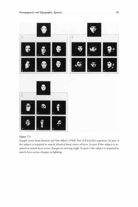
Prosopagnosia and Topographic Agnosia 95
Figure 7.1Sample items from Benton and Van Allen’s (1968) Test of Facial Recognition. In part Athe subject is required to match identical front views of faces. In part B the subject is re-quired to match faces across changes in viewing angle. In part C the subject is required tomatch faces across changes in lighting.

well on tests of face matching and discrimination, but required lengthy in-spections of the faces and, by his own report, relied on specific local fea-tures, such as the shape of the hairline. When the faces were shown to himwith oval frames blocking the hairline, his performance dropped markedly.Newcombe points out, “Some prosopagnosic patients are reported tomatch faces normally. . . . Latencies, however, are not invariably measured.Where they are, they are described as abnormally slow” (p. 319). This tellsus that the final scores of patients could be within normal limits when infact their ability to perceive faces is clearly abnormal.
A huge amount of social information comes from the half square footor so of body surface that is our face. The most obvious is individual iden-tity. This is illustrated by the old story about the two naked students sun-bathing. Their professor walks by and both rush to cover themselves withtheir hands, choosing to cover different body parts. The smart one covershis face, realizing that it is this body part and generally only this body partthat enables us to be recognized.
In addition to identity, faces are usually the most immediate and re-liable source of information about mood and emotional state. When as-sessed in prosopagnosic patients, facial expression recognition is usuallyimpaired, but some patients maintain good emotion recognition despiteimpaired recognition of identity. For example, Tranel, Damasio, and Da-masio (1988) report the emotion recognition ability of a series of patients,some of whom were prosopagnosic. Two of their subjects failed to recog-nize the identity of a single face among photographs of famous individu-als as well as of their family and friends. One of these subjects was alsoimpaired on a stringent test of facial emotion recognition, but the otherperformed comparably to control subjects. The perception of age andgender, two other personal characteristics of social relevance, followed thesame pattern as emotion in these two cases. Young, Newcombe, de Haan,Small, and Hay (1993) found evidence of the dissociability of identity andemotional expression recognition by the performance of a large group ofunilaterally brain-damaged patients.
Where a person is looking tells us what they are currently paying at-tention to, and this is another way in which the face provides socially rel-evant information. The tendency to “gaze follow,” that is, look wheresomeone else is looking, develops in infancy and can be considered aprimitive form of “theory of mind,” the ability to understand the mental
96 Prosopagnosia and Topographic Agnosia

processes of another person (Baron-Cohen, 1995). Gaze following re-quires the ability to discern, from slight variations in the position of theeye in the orbit, the distal location being fixated. This ability can also beimpaired in prosopagnosia (Campbell, Heywood, Cowey, Regard, & Lan-dis, 1990; unpublished data from patients L. H. and Adam). As with let-ter recognition in ventral simultanagnosia, the patterns of association anddissociation among face-related visual abilities suggest the existence of“specialization within specialization.” Within a broad zone of visual rep-resentation optimized for encoding faces there may be more specific re-gions necessary for specific face-related processes such as emotion and gazeperception.
A special category of prosopagnosics are those who have been pro-sopagnosic since birth. We all know individuals who are poor at facerecognition, and have seen their puzzlement or embarrassment when ap-proached by a recent or casual acquaintance. In rare cases the impairmentis so severe that it can be considered a congenital prosopagnosia. A fewparticularly severe and selective cases have been described in the litera-ture (Bentin, Deouell, & Soroker, 1999;Duchaine, 2000;Nunn, Postma,& Pearson, 2001; Temple, 1992). The individual described by Nunn andcolleagues remembers first noticing his problem at the age of seven, andreported “being unable to recognize or identify members of his own fam-ily, friends and colleagues from their faces. He also [had] difficulty recog-nizing actors in films, and would lose track of the plot if actors change theirclothing.” He was, however, normal in all other ways the authors couldthink to test, and had no evidence of brain damage on neurological examor MRI. As in other cases (Bentin et al., 1999;Duchaine, 2000) there wasa family history of congenital prosopagnosia, implying a genetic basis forthe disorder.
These cases contrast with another individual who has been prosopag-nosic since birth, due to occipitotemporal brain damage sustained as anewborn (Farah, Rabinowitz, Quinn, & Liu, 2000). This young man ismildly agnosic for objects other than faces, but is utterly unable to recog-nize his friends or family by their faces alone. The fact that functional ob-ject recognition systems could not take over face recognition in this case,despite the need for such recruitment prior to any experience with faces,represents an extreme lack of plasticity. Both the suggestion of heritabil-ity from the congenital cases without evidence of neurological damage,
Prosopagnosia and Topographic Agnosia 97

and the lack of plasticity when the relevant brain areas are damaged atbirth, indicate a genetic component of visual recognition that is specificto faces.
7.4 Are Faces “Special”?
Although many prosopagnosics have some degree of difficulty recogniz-ing objects other than faces, in many cases the deficit appears to be strik-ingly disproportionate for faces, with only rare difficulties with objects ineveryday life. This preserved ability is sometimes used to help recognizepeople—for example, by a distinctive article of clothing or hair decora-tion. The selectivity of prosopagnosia bears directly on the issue of unityversus modularity. If faces are selectively affected, then there must be someface-specific processing component that can be damaged.
To determine whether prosopagnosia is truly selective for faces, andthus implies the existence of a specialized face module, we must determinewhether faces are selectively affected merely because they are harder torecognize, or whether recognition of faces is disproportionately worsethan performance on nonface objects when the difficulty of each is takeninto account. In other words, we must assess prosopagnosic performanceon faces and nonface objects relative to the difficulty of these stimuli fornormal subjects. One technical difficulty encountered in such a project isthat normal subjects will invariably perform nearly perfectly on both faceand nonface recognition tasks. The resultant ceiling effect will mask anydifferences in difficulty that might exist between tasks, making it point-less to test normal subjects in the kinds of recognition tasks that have tra-ditionally been administered to patients. With this problem in mind,researchers have devised visual recognition tasks that test learning of novelface and nonface objects. By having subjects learn to recognize specificnew exemplars of faces and other types of objects, it is possible to titratenormal subjects’ level of recognition performance so that it falls betweenceiling and floor.
The first researchers to address this issue directly were McNeil andWarrington (1993). They studied WJ, a middle-aged professional manwho became prosopagnosic following a series of strokes. After becomingprosopagnosic, WJ made a career change and went into sheep farming. Heeventually came to recognize many of his sheep, although he remained
98 Prosopagnosia and Topographic Agnosia

unable to recognize most humans. The authors noted the potential im-plications of such a dissociation for the question of whether human facerecognition is “special,” and designed an ingenious experiment exploit-ing WJ’s newfound career. They assembled three groups of photographs—human faces, sheep faces of the same breed kept by WJ, and sheep facesof a different breed—and attempted to teach subjects names for each face.Normal subjects performed at intermediate levels between ceiling andfloor in all conditions. They performed better with the human faces thanwith sheep faces, even those who, like WJ, worked with sheep. In con-trast, WJ performed poorly with the human faces, and performednormally with the sheep faces. These data suggest that WJ’s recognitionimpairment does not affect the recognition of all groups of visually simi-lar patterns, but is selective for human faces.
My colleagues and I took a similar approach, but used common ob-jects rather than faces of another species to compare with human facerecognition (Farah, Levinson, & Klein, 1995). Our subject was LH, awell-educated professional man who has been prosopagnosic since an au-tomobile accident in college. LH is profoundly prosopagnosic, unable toreliably recognize his wife, children, or even himself in a group photo-graph. Yet he is highly intelligent, and seems to have little or no difficultyrecognizing other types of visual patterns, such as printed words or ob-jects. He has a degree of recognition impairment with drawings of objects,but this is less severe than his impairment with faces.
We employed a recognition memory paradigm, in which LH andcontrol subjects first studied a set of photographs of faces and nonface ob-jects, such as forks, chairs, and eyeglasses. Subjects were then given a largerset of photographs, and asked to make “old”/”new” judgments on them.This larger set was designed so that for each face and nonface object inthe “old” set there was a highly similar item in the “new” set. Figure 7.2shows examples of stimuli from this experiment. As a result of pilotingthe stimuli in advance, normal subjects performed equally well with thefaces and nonface objects. In contrast, LH showed a significant perfor-mance disparity, performing worse with faces than with objects.
One could still maintain that faces possess no special status in pro-sopagnosia, and instead attribute the poor performance with faces to theneed for within-category discrimination. Perhaps, in the words of Dama-sio, Damasio, and Van Hoesen (1982), “The valid dissociation is between
Prosopagnosia and Topographic Agnosia 99

100 Prosopagnosia and Topographic Agnosia
Figure 7.2Examples of stimuli from Farah, Levinson, and Klein’s (1995) experiment testing face andobject recognition in prosopagnosia.

Prosopagnosia and Topographic Agnosia 101
Figure 7.2Continued

the recognition of the generic conceptual class to which the object be-longs and the recognition of the historical context of a given object [i.e.,the individual identity of the object] vis a vis the subject” (p. 337). To testthis hypothesis directly, we carried out a second experiment in which sub-jects learned exemplars of the category “face” and an equivalent numberof highly similar exemplars all drawn from a single nonface category, eye-glass frames. As before, LH was disproportionately impaired at face recognition relative to nonface recognition, when his performance is con-sidered relative to normal subjects.
In a final experiment in this series, we compared LH’s processing offaces to a different, and in some ways ideal, nonface control stimulus: up-side-down faces (Farah, Wilson, Drain, & Tanaka, 1995). Inverted faces areare equivalent to upright faces in virtually all physical stimulus parameters,including complexity and interitem similarity. Inversion interferes with therecognition of faces more than other types of stimuli, and from this it hasbeen inferred that inverted faces do not engage (or engage to a lesser ex-tent) the hypothesized face-specific processing mechanisms (Valentine,1988). We reasoned that if LH had a general recognition impairment, thenhe would show a normal face inversion effect. In contrast, if he had suffereddamage to neural tissue implementing a specialized face recognition sys-tem, he would show an absent or attenuated face inversion effect.
LH and normal subjects were tested in a sequential matching task,in which an unfamiliar face was presented, followed by a brief interstim-ulus interval, followed by a second face, to which the subject responded“same” or “different.” The first and second faces of a trial were always inthe same orientation, and upright and inverted trials were randomly intermixed. As expected, normal subjects performed better with the up-right than with the inverted faces, replicating the usual face inversioneffect.
In contrast, LH was significantly more accurate with inverted faces—he showed an inverted inversion effect! This outcome was not among the alternatives we had considered. We drew two main conclusions from the inverted inversion effect. One concerns the “control structure” of vi-sual recognition. LH’s specialized face perception system was apparentlycontributing to his performance even though it was impaired and clearlymaladaptive. This suggests that the specialized face system operates man-datorily, reminiscent of Fodor’s (1983) characterization of special-purpose
102 Prosopagnosia and Topographic Agnosia

perceptual “modules” as engaged mandatorily by their inputs. The ideathat the face system cannot be prevented from processing faces, even whendamaged, also may explain why WJ was able to learn to recognize indi-vidual sheep after his strokes but could not learn to recognize human faces.The second implication concerns the selectivity of prosopagnosia. LH’sdisproportionate impairment on upright relative to inverted faces impliesthat an impairment of specialized processing mechanisms underlies hisprosopagnosia.
Patients who are agnosic for objects, but not for faces, provide theother half of the double dissociation supporting specialized recognitionmechanisms for faces. A number of such patients have been described inthe literature (see Feinberg, Schindler, Ochoa, Kwan, & Farah, 1994, fora case series and literature review). The most detailed study of object ag-nosia with preserved face recognition comes from Moscovitch, Winocur,and Behrmann (1997). Their patient, CK, suffered a closed head injuryresulting in severe agnosia. Despite his profound object agnosia, CK is en-tirely normal in his ability to recognize faces. In a test with 140 famousfaces, he performed well within the range of control subjects, and evenheld his own with a more challenging test of famous faces photographedat different ages (e.g., Winston Churchill as a child). In one particularlyenjoyable demonstration of the object-face dissociation, the experimentersshowed him paintings by Archimbaldo, in which collections of objectswere arranged to make faces (see figure 7.3). Whereas normal viewersquickly see both objects and faces in these pictures, CK saw only faces ini-tially, and for most of the paintings never even noticed that there werenonface objects present!
This pattern of impairment is interesting for two reasons. First, itoffers further disconfirmation of the hypothesis that prosopagnosia is justa mild disorder of a general-purpose object recognition system, with facessimply being harder to recognize than other objects. If this were true, howcould it be possible for a person to do better with faces than with otherobjects? Second, taken in conjunction with prosopagnosia, it shows thatface and object recognition are functionally independent, in that eitherone can continue to work without other. This rules out a single pathwaywith different termination points for face and nonface processing, as dia-grammed in figure 7.4a, and constrains our models of the functional ar-chitecture of visual recognition to those which include parallel pathways
Prosopagnosia and Topographic Agnosia 103

104 Prosopagnosia and Topographic Agnosia
Figure 7.3An agnosic patient with preserved face recognition studied by Moscovitch, Winocur, andBehrmann (1997) saw only a face in this picture by Archimbaldo.

for face and nonface object recognition, as diagrammed in figure 7.4b. Thisconclusion is compatible with the “two systems” hypothesis described insection 6.5.
A related double dissociation between face and object processing isinstantiated by a pair of patients with material-specific amnesias. Whereasprosopagnosics are impaired at both learning new faces and recognizingpreviously familiar faces, consistent with damage to the substrates of facerepresentation, my colleagues and I recently encountered someone withan even more selective impairment: following a head injury, CT becameamnesic for new faces (Tippett, Miller, & Farah, 2000). In contrast, hisrecognition of previously familiar faces is relatively preserved, as is hislearning of nonface visual objects. This pattern of performance is consis-tent with a disconnection between intact face representations and an intact medial temporal memory system. As such, it provides additionalevidence that the anatomical substrates of face representation are distinctfrom the representation of other objects, since they can be selectively dis-connected from the substrates of new learning.
CT’s face perception was normal, and he showed an inversion effectin face matching. His learning of verbal material and of visual materialother than faces was also normal. However, when given the face and eye-glass learning task, he performed similarly to LH. Additional evidence ofhis inability to learn faces comes from his identification of famous faces.
Prosopagnosia and Topographic Agnosia 105
FACES
OBJECTS
OBJECTS FACES
EARLYVISION
EARLYVISION
A B
Figure 7.4Two possible relations between an object recognition system and a specialized face recog-nition system.

For people who were famous prior to CT’s head injury, he performedwithin the range of age-matched control subjects on a forced-choice “famous/not famous” task, whereas for more recently famous individualshis performance was many standard deviations below normal. One in-formative exception to this is the singer Michael Jackson, who was wellknown before CT’s injury, but whose appearance changed drastically inthe years that followed. CT quickly and confidently recognized photo-graphs of Jackson taken at about the time of his injury. However, he didnot recognize a later photograph, taken the year we tested CT.
The mirror image of CT’s memory impairment was described byCarlesimo, Fadda, Turriziani, Tomaiuolo, and Caltagirone (2001). Theirpatient, a firefighter who suffered carbon monoxide poisoning, initiallyappeared to be globally amnesic. He scored extremely poorly on all stan-dard tests of memory, would repeatedly ask the same questions, and neededa list to go shopping for even a few items. Imagine the investigators’ sur-prise when, after a single encounter at the lab with a staff member, thepatient later recognized him in a different context! Subsequent testingdemonstrated an island of preserved learning ability for faces in this other-wise severely amnesic patient.
In sum, the studies of prosopagnosics, object agnosics, and amnesicsreviewed in this section all bear on the modularity of the functional ar-chitecture of vision. They show that faces are “special” in that they dependon neural systems which can be selectively damaged, spared, disconnected,or connected to other brain systems. Evidence from lesion reconstructionsand functional neuroimaging of normal subjects, discussed in section 7.7,lends further support to the hypothesis that face processing involves at leastsome neural systems not used for visual object processing more generally.
7.5 Relative and Absolute Specialization
As evidence accumulated that prosopagnosia is not simply a mild generalagnosia, but represents damage to a more specialized module, a differentissue came to the fore. The specialized system needed for face recognition,and damaged in prosopagnosia, could be used to some degree for recog-nizing other types of stimuli, or it could be dedicated exclusively to facerecognition. The evidence presented so far does not distinguish betweenthese alternatives;it merely demonstrates relatively impaired recognition of
106 Prosopagnosia and Topographic Agnosia

faces without appropriate comparisons of normal and prosopagnosic non-face object recognition.
Two lines of research suggest that cerebral specialization for facerecognition is not absolute, but is better described in terms of a gradientof specialization that encompasses certain nonface objects in certain taskcontexts. Research by De Gelder and colleagues, following up on the inverted inversion effect, provides one source of evidence (De Gelder,Bachoud-Levi, & Degos, 1998; De Gelder & Rouw, 2000). By combin-ing a challenging object perception task, involving matching subtly dif-fering shoes, with a face perception task, they showed an inversion effectfor normal subjects with both types of stimulus, and an inverted inversioneffect in prosopagnosia. They interpreted these findings in terms of theneed for “configural” representation in both tasks, a conclusion that couldas well be framed in terms of “holistic” representation as defined in theprevious chapter.
Another source of evidence comes from the work of Gauthier, Tarr,and colleagues. They have pursued the idea that what is “special” aboutface recognition is the recognition of individual patterns within the gen-eral category, when the patterns share a common spatial layout and whensuch recognition requires extensive learning or expertise (Gauthier, An-derson, Tarr, Skudlarski, & Gore, 1997;Gauthier & Tarr, 1997;Gauthier,Williams, Tarr, & Tanaka, 1998;Gauthier, Behrmann, & Tarr, 1999). Al-though face recognition might be the most commonly encountered caseof such ability, it need not be the only case. They have carried out a seriesof studies to test the hypothesis that face recognition is special only inso-far as it requires subordinate classification of similarly configured shapesand is dependent on expertise. This research involves a novel class of ob-jects called “greebles.”
As can be seen in figure 7.5, greebles share a common part structurein the same way that faces do, with two “boges,” a “quaff,” and a “dunth.”Subjects in these experiments learned names for each individual greeble.They also learned to classify the greebles by “gender” and by “family.”Theexamples shown in figure 7.6 help to clarify what these terms mean in thecontext of greebles. After many hours of training, subjects became greebleexperts, with fast, accurate greeble recognition and equivalent performancewith subordinate (individual greeble) and basic level (gender and family)recognition.
Prosopagnosia and Topographic Agnosia 107

Gauthier and Tarr (1997) sought to replicate, with greebles, the ef-fects of facial context on face part discrimination that Tanaka and I found.Recall that we taught subjects new faces, and then asked them to identifya given face’s eyes, nose, or mouth from a pair. With intact upright faces,but not other stimuli, subjects performed better when the pair was pre-sented in the context of a whole face. Although greeble parts were dis-criminated more effectively in the context of a whole greeble, the effectwas not influenced by expertise. Gauthier and Tarr did find an effect ofexpertise on the sensitivity of the context effects on configuration, anal-ogous to Tanaka and Sengco’s (1997) finding. In a later study, Gauthier et
108 Prosopagnosia and Topographic Agnosia
Figure 7.5One of Gauthier and Tarr’s (1997) “greebles,” with key parts labeled.
Figure 7.6Greebles of different families and genders (Gauthier and Tarr, 1997).

al. (1998) attempted to replicate a number of findings from the face recog-nition literature with greebles, and again obtained mixed results. The ear-lier finding of a whole greeble advantage for parts recognition was notreplicated, and no greeble inversion effect was found. However, other pur-portedly face-specific effects were found with greebles, specifically thebrightness inversion effect (reversing black and white makes faces andgreebles hard to recognize) and the composite effect (top and bottomhalves of different faces and greebles are difficult to recognize whenaligned to make a whole).
Gauthier, Behrmann, and Tarr (1999) explored the recognition abil-ities of two prosopagnosic patients for faces, real objects, and greebles. Inaddition to replicating the overall disproportionate difficulty of faces forprosopagnosics shown by others, they assessed the relative difficulty ofsubordinate, basic, and superordinate level identification for prosopag-nosics and for normal subjects. Consistent with their hypothesis thatapparently face-specific processing is actually subordinate-level processingthat requires expertise, they found that the patients’ reaction times in-creased more steeply than normal as the level of categorization becamemore specific. The question is, are the results inconsistent with the alter-native hypothesis that the system used for processing the face stimuli is not used, or is used substantially less, for processing the objects andgreebles? Given that both patients had a degree of object recognition diffi-
culty, whether resulting from the loss of the partial involvement of the sys-tem used for faces or from damage to a distinct but neighboring system,their heightened sensitivity to any sort of difficulty manipulation is notsurprising.
The contribution of greeble research may be as much conceptual asempirical, since it forces us to think precisely about what we mean whenwe claim that “faces are ‘special’.” If we mean that the hypothesized facerecognition system is never used in the recognition of nonface objects,then the greeble data have the potential to disconfirm the claim. If wemean that the system is specialized for a certain type of visual representa-tion, such as the holistic representation described in chapter 6, and thatits primary use is in face recognition but it may on occasion be recruitedfor certain other visual recognition problems, then the greeble data simplyclarify the nature of the “face recognition” system’s visual representationsand its range of possible functions.
Prosopagnosia and Topographic Agnosia 109

7.6 Covert Face Recognition in Prosopagnosia
One of the more dramatic dissociations involving prosopagnosia is thefinding of preserved “covert” face recognition, that is, the ability of somepatients to manifest knowledge of faces on indirect tests of recognition.The literature on covert recognition in prosopagnosia is large, dating backto the early 1980s and involving researchers from a number of differentlabs who were quickly drawn in by this remarkable phenomenon. Only asmall sample of the relevant findings will be summarized here. One of themost widely used methods of demonstrating preserved face recognitionin prosopagnosia is by the paired-associate face-name relearning task, inwhich patients are taught to associate the facial photographs of famouspeople (whom they cannot recognize) with the names of famous people.For some prosopagnosics, fewer learning trials are required when the pair-ing of names and faces is correct than when incorrect (e.g., RobertRedford’s face with the name “Harrison Ford”). De Haan, Young, andNewcombe (1987a) showed that this pattern of performance held evenwhen the stimulus faces were selected from among those the patient hadbeen unable to identify in a pre-experiment stimulus screening test.
Evidence of covert recognition has also come from reaction timetasks in which the familiarity or identity of faces was found to influenceprocessing time. In a visual identity match task with simultaneously pre-sented pairs of faces, De Haan, Young, and Newcombe (1987a) found thata prosopagnosic patient was faster at matching pairs of previously familiarfaces than unfamiliar faces, as is true of normal subjects. In contrast, he wasunable to name any of the previously familiar faces.
In another RT study, De Haan, Young, and Newcombe (1987b;also1987a) found evidence that photographs of faces could evoke covert se-mantic knowledge of the depicted person, despite the inability of theprosopagnosic patient to report such information about the person whentested overtly. The task was to categorize a printed name as belonging toan actor or a politician as quickly as possible. On some trials an irrelevant(i.e., to be ignored) photograph of an actor’s or politician’s face was si-multaneously presented. Normal subjects were slower to print the nameswhen the faces came from a different occupation category relative to a no-photograph baseline. Even though their prosopagnosic patient was se-verely impaired at categorizing the faces overtly as belonging to actors or
110 Prosopagnosia and Topographic Agnosia

politicians, he showed the same pattern of interference from different-category faces.
Prosopagnosic patients who manifest covert recognition appear tolack the subjective experience of recognition, at least for many of the facesfor which they show covert recognition. These patients may occasionallyrecognize a face overtly, that is, assign it the correct name and express a de-gree of confidence that they know who the person is. However, this hap-pens rarely, and the dissociation between covert recognition and awarenessof recognition holds for many faces that they fail to identify and for whichthey report no sense of familiarity.
There are several competing explanations for covert recognition inprosopagnosia. The oldest is that the face recognition system is intact inthese patients, but has been prevented from conveying information toother brain mechanisms necessary for conscious awareness. An expicitstatement of this view comes from De Haan, Bauer, and Greve (1992),who proposed the model shown in figure 7.7. According to their model,the face-specific visual and mnemonic processing of a face (carried out
Prosopagnosia and Topographic Agnosia 111
Figure 7.7Model of face recognition used by De Haan, Bauer, and Greve (1992) to explain covertface recognition in prosopagnosia in terms of a lesion at location number 1.
within the “Face processing module”) proceeds normally in covert recog-nition, but the results of this process cannot access the “Conscious aware-ness system” because of a lesion at location number 1.
Another type of explanation was put forth by Bauer (1984), who sug-gested that there may be two neural systems capable of face recognition,only one of which is associated with conscious awareness. According toBauer, the ventral visual areas damaged in prosopagnosic patients are thelocation of normal conscious face recognition. But the dorsal visual areasare hypothesized to be capable of face recognition as well, although theydo not mediate conscious recognition but, instead, affective responses tofaces. Covert recognition is explained as the isolated functioning of thedorsal face system. This account also fits into the general category of con-sciousness as a privileged property of particular brain systems. It is analo-gous to theorizing about the subcortical visual system in blindsight, andthe dorsal visual system in apperceptive agnosia, in that two systems arepostulated that carry out related but distinct visual functions, but only oneof which is endowed with conscious awareness.
Tranel and Damasio (1988) interpreted covert recognition as thenormal activation of visual face representations, which is prevented by thepatients’ lesions from activating representations in other areas of the brain,such as representations of people’s voices in auditory areas, affective va-lences in limbic areas, names in language areas, and so on. This idea wasembodied in a computer simulation of semantic priming effects, in whichcovert recognition was modeled as a partial disconnection separating in-tact visual recognition units from the rest of the system, as shown in figure7.8 (Burton, Young, Bruce, Johnston, & Ellis, 1991).
The foregoing explanations of covert recognition all include the as-sumption that visual recognition proper is intact and that the functionallesion lies outside of the visual system. A different approach to explainingcovert face recognition rejects this assumption, and posits that covertrecognition reflects the residual processing capabilities of a damaged, butnot obliterated, visual face recognition system. Randy O’Reilly, ShaunVecera, and I have argued that lower-quality visual information processingis needed to support performance in tests of covert recognition (e.g., toshow savings in relearning, and the various RT facilitation and interfer-ence effects) relative to the quality of information processing needed tosupport normal overt recognition performance (e.g., naming a face, sort-
112 Prosopagnosia and Topographic Agnosia

ing faces into those of actors and politicians; Farah, O’Reilly, & Vecera,1993; O’Reilly & Farah, 1999). Support for this view comes from a be-havioral study with normal subjects and a series of computer simulations.In the behavioral study, Wallace and Farah (1992) showed that savings in face-name relearning can be obtained with normal subjects who aretrained on a set of face-name associations and then allowed for forget theseassociations over a six-month interval. Presumably normal forgetting doesnot involve the diverting of intact information from conscious awareness,but rather the degradation of representations (albeit in a different way fromprosopagnosia). Probably the strongest evidence for this view, however, iscomputational.
Farah et al. (1993) trained a neural network, shown in figure 7.9, toassociate “face” patterns with “semantic” patterns, and to associate these,in turn, with “name” patterns. We found that, at levels of damage to theface representations which led to poor or even chance performance in
Prosopagnosia and Topographic Agnosia 113
Figure 7.8Model of face recognition used by Burton, Bruce, and Johnston (1991) to explain covertrecognition in prosopagnosia in terms of a disconnection between face recognition units(FRUs) and personal identity nodes (PINs).

overt tasks such as naming (figure 7.10a) and occupation categorization,the network showed all of the behavioral covert recognition effects re-viewed above: it relearned correct associations faster than it learned novelones (figure 7.10b), it completed the visual analysis of familiar faces fasterthan unfamiliar faces (figure 7.10c), and it showed priming and inter-ference from the faces on judgments about names (ffigure 7.10d). More re-cently, O’Reilly and Farah (1999) simulated several more covertrecognition tasks with the same basic model.
Why should a damaged neural network support performance in thisrange of covert tasks when overt recognition is poor or even at chance?The answer lies in the nature of information representation and processingin distributed, interactive networks. Representations in such networksconsist of patterns of activation over a set of units or neurons. These unitsare highly interconnected, and the extent to which the activation of oneunit causes an increase or decrease in the activation of a neighboring unitdepends on the “weight” of the connection between them. For the net-work to learn that a certain face representation goes with a certain namerepresentation, the weights among units in the network are adjusted so
114 Prosopagnosia and Topographic Agnosia
Figure 7.9Model of face recognition used by Farah, O’Reilly, and Vecera (1993) to explain covertrecognition in prosopagnosia in terms of damaged, but not obliterated, face recognition.

that presentation of either the face pattern in the face units or the namepattern in the name units causes the corresponding other pattern to be-come activated. Upon presentation of the input pattern, all of the unitsconnected with the input units will begin to change their activation inaccordance with the activation value of the units to which they are con-nected and the weights on the connections. As activation propagatesthrough the network, a stable pattern of activation eventually results, de-termined jointly by the input activation and the pattern of weights amongthe units of the network.
Our account of covert face recognition is based on the following keyidea: The set of the weights in a network that cannot correctly associatepatterns because it has never been trained (or has been trained on a dif-ferent set of patterns) is different in an important way from the set ofweights in a network that cannot correctly associate patterns because it hasbeen trained on those patterns and then damaged. The first set of weightsis random with respect to the associations in question, whereas the sec-ond is a subset of the necessary weights. Even if it is an inadequate subsetfor performing the association, it is not random; it has, “embedded” in it, some degree of knowledge of the associations. Hinton and colleagues(Hinton & Sejnowski, 1986;Hinton & Plaut, 1987) have shown that suchembedded knowledge can be demonstrated when the network relearns,suggesting that the findings of savings in relearning face-name associationsmay be explained in this way. In general, consideration of the kinds of testsused to measure covert recognition suggest that the covert measures wouldbe sensitive to this embedded knowledge. The most obvious example isthat a damaged network would be expected to relearn associations whichit previously knew faster than novel associations because of the nonran-dom starting weights as shown in figure 7.10b. Less obvious, the networkwould settle faster when given previously learned inputs than novel in-puts (figure 7.10c), because the residual weights come from a set designedto create a stable pattern from that input. Finally, to the extent that theweights continue to activate partial and subthreshold patterns over thenondamaged units in association with the input, these resultant patternscould prime (i.e., contribute toward) the activation of patterns by intactroutes (figure 7.10d).
The general implication of these ideas is that as a neural network isincreasingly damaged, there will be a window of damage in which overt
Prosopagnosia and Topographic Agnosia 115
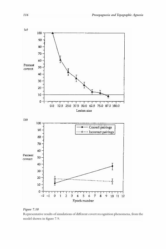
116 Prosopagnosia and Topographic Agnosia
Figure 7.10Representative results of simulations of different covert recognition phenomena, from themodel shown in figure 7.9.

Prosopagnosia and Topographic Agnosia 117

associations between patterns (e.g., faces and names) would be extremelypoor while the kinds of performance measures tapped by the covert tasksmight remain at high levels.
7.7 Neuropathology of Prosopagnosia
The lesions of prosopagnosia have received much attention. All writersagree that a right hemisphere lesion is necessary. Discussion has focusedon the intrahemispheric location of the right hemisphere lesion and onthe necessity for a second, left hemisphere lesion. In an early attempt atgeneralization, Meadows (1974) reviewed the clinical case literature onprosopagnosia and used visual field defects and autopsy reports to inferthe distribution of lesion sites. The almost invariable finding of a left su-perior quadrananopia indicated that the right inferior occipitotemporalwas critical, and the frequent finding of smaller field defects on the rightsuggested a tendency for bilateral damage. These conclusions were sup-ported by the findings in those cases that came to autopsy. Damasio,Damasio, and Van Hoesen (1982) arrived at similar conclusions, empha-sizing the need for bilateral lesions.
A revision of this view was urged by DeRenzi, Perani, Carlesimo,Silveri, and Fazio (1994), who reviewed much of the same case material,along with more recent cases and data from living patients whose braindamage was mapped using both structural MRI and PET. Their findingssupported the ventral, occipitotemporal localization of face recognition,but called for a revision of the idea that bilateral lesions are necessary. Somepatients became prosopagnosic after unilateral right hemisphere damage.The possibility of hidden left hemisphere dysfunction in these cases wasreduced by the finding of normal metabolic activity in the left hemisphereby PET scan. De Renzi et al. conclude that there is a spectrum of hemi-spheric specialization for face recognition in normal right-handed adults.Although the right hemisphere may be relatively better at face recognitionthan the left, most people have a degree of face recognition ability in bothhemispheres. Nevertheless, in a minority of cases, face recognition is sofocally represented in the right hemisphere that a unilateral lesion will leadto prosopagnosia.
The localization of face recognition has also been the focus of ex-tensive research using functional neuroimaging, with the conclusions of
118 Prosopagnosia and Topographic Agnosia

the two approaches in good agreement with one another. Early compar-isons of face recognition with other visual-spatial tasks confirmed the oc-cipitotemporal localization inferred from lesion studies (e.g., Haxby et al.,1991). As experimental designs were refined and more specific stimuluscontrasts were included to isolate face-specific regions (for example, con-trasting faces with objects, houses, body parts, and even inverted faces),the “fusiform face area” (FFA) became more focally and reliably localized(e.g., Haxby et al., 1999; Kanwisher, Tong, & Nakayama, 1998; Kan-wisher, McDermott, & Chun, 1997;Kanwisher, Stanley, & Harris, 1999;McCarthy, Puce, Gore, & Allison, 1997). Figure 7.11 shows the locationof the FFA in two subjects, on the basis of an fMRI experiment with al-ternating blocks of faces, houses and objects (Tong, Nakayama, Mosco-vitch, Weinrib, & Kanwisher, 2000). A recent report of prosopagnosiafollowing a small hematoma affecting the FFA demonstrates the conver-gence of the lesion and functional imaging approaches to the anatomy offace recognition (Wada & Yamamoto, 2001).
7.8 Topographic Agnosia:A Case Description
Like faces, scenes and landmarks are visual stimuli with great importanceto our survival. Hunting, gathering, seeking mates, and seeking refuge allrequire knowledge of one’s location within the environment, whichscenes and landmarks provide. It is probably no coincidence that half ofthe paintings in any museum gallery are either portraits or landscapes.
Recognition of scenes and landmarks is sometimes impaired in con-junction with prosopagnosia, but the two also occur in isolation. A par-ticularly pure case of topographic agnosia was described by Whitely andWarrington (1978). Following a severe head injury incurred in a motorvehicle accident, a forty-six-year-old workman recovered most of his pre-morbid abilities except for a specific problem with visual recognition.
His present complaint, four years after the accident, is of a failure to recognizebuildings, streets, and other landmarks, which incapacitates him to the extent thathe gets lost in familiar surroundings. He describes looking at a building, beingable to see and describe it clearly, yet if he looks away and looks back again, it looksdifferent as though someone had put another unfamiliar building in its place. Thestreet in which he lives seems unfamiliar, and each day he might be going along
Prosopagnosia and Topographic Agnosia 119

it as for the first time. He recognizes his own house by the number, or by his carwhen parked at the door.
He complains of considerable difficulty in getting about, and says that learn-ing new routes presents a real problem. He can, however, use a map easily and fol-low verbal instructions. He relies heavily on street names, station names, andhouse numbers.
Apart from these topographical difficulties, he reports only a minor degreeof difficulty in recognizing people’s faces, especially people he has met recently,and a minor degree of nonspecific forgetfulness. He is, nevertheless, able to reada book and follow a film or play without difficulty. He has noticed no difficultyin writing, drawing, or making scale plans.
Whitely and Warrington administered a wide range of perceptionand memory tests without finding any more general impairments thatmight have been responsible for this patient’s topographic agnosia. They
120 Prosopagnosia and Topographic Agnosia
Figure 7.11The location of the fusiform face area in two subjects, on the basis of an fMRI experi-ment with alternating blocks of faces, houses and objects (Tong et al., 2000).

assessed his ability to perceive buildings with a matching task in whichpairs of photographs of buildings, taken from different angles, were placedside by side and judged to be the same or different buildings. Althoughthe authors conclude from the patient’s perfect performance on this taskthat his perception of buildings is normal, the task does allow piecemealvisual comparison and may therefore overestimate his abilities. In contrast,he performed below the worst control subject in learning to recognizenew buildings and at the low end of the normal range in recognizingfamous city landmarks, consistent with his difficulty in learning his wayaround new locales and recognizing his own street and home.
7.9 General Characteristics of Topographic Agnosia and Related Disorders
There are a small number of similar cases in the literature. Some have pro-sopagnosia in addition to topographic agnosia, such as Pallis’s (1955) casethat was excerpted in section 7.2, and those of Cole and Perez-Cruet(1964) and Landis, Cummings, Benson and Palmer (1986, cases 2 and 4).In other cases, the topographic agnosia is an isolated visual recognitiondeficit: Incisa della Rocchetta, Cippolotti, and Warrington (1996), He-caen, Tzortzis, and Rondot (1980), and Landis et al. (1986, cases 1 and3). The impairment is appropriately grouped with disorders of visualrecognition rather than of spatial cognition, in that the patients’ spatialabilities are preserved. Indeed, it is typically reported that patients developa heavy dependence on their preserved spatial abilities to find their wayaround the environment and are able to use maps (Hecaen et al., 1980;Icisadella Rocchetta et al., 1996; Landis et al., 1986, cases 1 and 2; Whitely &Warrington, 1978). Because of the specificity of the recognition impair-ment, patients are able to recognize common objects, and also report us-ing these objects to help them orient themselves—for example, exteriorobjects such as mailboxes, water fountains, phone booths, palm trees, or acertain style of lettering, and interior objects such as the ward phonographor a certain color wall (examples from the cases of Landis et al., 1986).New learning of scenes and landmarks is dramatically impaired, alongwith recognition of personally familiar locales and, in all but Whitely andWarrington’s case, famous landmarks. Incisa della Rocchetta et al. sepa-rately tested the recognition of country landscapes, devoid of human con-structions, and city scapes, and found both severely impaired.
Prosopagnosia and Topographic Agnosia 121

Topographic agnosia should be distinguished from other disordersaffecting orientation within the large-scale environment. Aguirre andD’Esposito (1999) offer a useful taxonomy of impairments in the realmof navigation and way-finding. Of the four categories in their taxonomy,two concern purely spatial processing impairments. More likely to be con-fused are the remaining two, the disorder of topographic agnosia describedhere and a disorder of new learning that affects both visual and spatial as-pects of the environment (e.g., Epstein et al., 2001;Habib & Sirigu, 1987;Katayama, Takahashi, Ogawara, & Hattori, 1999). Patients with the lat-ter disorder can recognize premorbidly familiar locales, but have difficultylearning both the features and the layout of new environments.
7.10 Neuropathology of Topographic Agnosia
Topographic agnosia most commonly follows right or bilateral posteriorartery infarction, although Whitely and Warrington’s patient suffered ahead injury and Incisa della Rocchetta’s patient had diffuse damage dueto small vessel ischemic disease. When focal lesions can be visualized, theyare similar to those of prosopagnosia, affecting inferior medial occipito-temporal cortex, including the fusiform and lingual gyri and in some casesextending into the parahippocampal gyrus, either on the right side or bi-laterally. Functional neuroimaging has provided converging evidence fortopography-specific representations in the lingual and parahippocampalgyri (Aguirre, Zarahn, & D’Esposito, 1998;Epstein & Kanwisher, 1998),consistent with the lesions just described.
122 Prosopagnosia and Topographic Agnosia

Chapter 8
Optic Aphasia
8.1 Optic Aphasia:A Case Description
Optic aphasia is a puzzling disorder in which patients cannot name vis-ually presented objects, despite demonstrating good visual recognitionnonverbally (e.g., by pantomiming the use of a seen object or sortingsemantically related objects together) and good naming (e.g., by namingpalpated objects or objects they hear described verbally). Given that theycan get from vision to semantics (intact visual recognition) and from se-mantics to naming (intact object naming of nonvisual inputs), one wouldexpect them to be able to name visually presented objects.
One of the most thoroughly studied cases of optic aphasia was de-scribed by Lhermitte and Beauvois (1973). Their subject, Jules F, suffereda posterior left hemisphere stroke, with some amnesia, pure alexia, andmild constructional apraxia and color vision deficit. Although he maderelatively few errors in naming objects from verbal definition (5/125), orfrom their characteristic noises or their feel in his hand (1/25 and 11/120,respectively), he made a large number of errors (35/130) in naming visu-ally presented objects and pictures. Although he performed well on a taskof pointing to a visual stimulus depicting a spoken name, the authors pointout that the choice set of possible responses in this task is much smallerthan in the naming tasks just described, suggesting that performance in thistask may not be truly better than in naming visually presented stimuliwhen overall difficulty is accounted for. They also point out that when thenamed item was absent, Jules pointed to incorrect choices that were se-mantically related to the word, apparently without any awareness that thecorrect choice was missing.

Based on his naming performance in different modalities, it is clearthat Jules F. is not simply anomic, but has a specific problem naming vi-sual stimuli. However, the data so far are consistent with his being a visualagnosic, or even blind. The second critical element for classifying him asan optic aphasic is the ability to demonstrate recognition of visually pre-sented stimuli by means other than naming. A common nonverbal meansof communicating recognition, often used spontaneously by patients likeJules F, is to gesture the use of the object in pantomime. Lhermitte andBeauvois presented the patient with 100 pictures of common objects, andreport that whenever he mimed the use of an object, he did so correctly,even when he misnamed the object. For example, when shown a pictureof a boot, he mimed pulling on a boot but called it a hat. Thus, the authorsconclude that the visual naming deficit cannot be attributed to agnosia.Consistent with this is their observation that the patient had no troubleinteracting with the visual world in everyday life. They do mention oc-casional agnosialike behavior with certain complex objects: he can some-times copy or describe the appearance of a stimulus accurately withoutbeing able to identify it by any means.
The patient’s errors in naming visual stimuli seemed to be mainly se-mantic in nature, with many perseverations of responses from previous tri-als. Visual errors without semantic similarity were reported to be relativelyrare, although many errors showed both visual and semantic similarity tothe correct response. When given unlimited time to name a visual stim-ulus, Jules F would generally home in on the correct name. However, sev-eral attempts were often necessary, as is evident in the following protocol(Lhermitte and Beauvois, 1973, pp. 706–707):
A grasshopper: “a cricket, not a cricket, a grasshopper.”
A mussel: “it looks like 2 snails, 2 slugs, it is a shellfish, not an oyster, it should be mussels then.”
A bus: “a wagon . . . a public transport since there is a back door . . . a stage-coach . . . it would be . . . no . . . a city cab . . . not a cab but a city bus.”
A window-blind: “A parasol, metallic curtain rods . . . the cloth roof . . . sur-rounding sails . . . it could be a parasol . . . there are rods, but isn’t it a shelter? Awindow-blind.”
An aquarium: “A bird cage, unless it is a pot for flowers, a container, a tank, thefour aspects . . . the walls made of glass or wood . . . it could be an aquarium if itis made of glass.”
124 Optic Aphasia

A medieval crown: [a preceding picture had correctly been named “basket”] “akind of foot of metallic basket, a basket for flowers, a basket for a table . . . it cannotbe a basket for decoration on a table . . . it is not a decoration, I do not know whatit could be. . . . A basket with 4 feet, no with 4 crossed hoops . . . it cannot beworn as clothing, it would be a hat. It would be a very fancy hat to look like that.”
In order to determine whether objects were misnamed because theywere misperceived, or whether the naming errors were generated after anadequate perceptual analysis was complete, Lhermitte and Beauvois askedthe subject to draw what he had seen and what he had named on trials inwhich objects were misnamed. When asked to draw what he had seen,he invariably drew a recognizable picture of the stimulus, which was dis-tinguishable from his drawing of the named item. For example, on onetrial the stimulus was a tree and Jules F said, “it’s a house with a straw roof,”a perseveration from a previous trial in which a house was shown. Whenrequested to draw what he had seen, he drew a tree, insisting all the whilethat he was drawing a “straw roof,” and when requested to draw a house,he produced a good drawing of a house. Lhermitte and Beauvois concludethat processing within the visual system is roughly intact and cannot be thecause of the errors in visual naming. Another observation that is consis-tent with this conclusion comes from testing with tachistoscopic stimu-lus presentations. Jules F would often continue to search for and home inon the correct name for a picture long after the picture had disappeared,as if he had gotten sufficient visual information in the brief presentationand the laborious part of the visual naming process was postvisual.
8.2 General Characteristics of Optic Aphasia
A number of other cases of optic aphasia have been described in the liter-ature, including Assal and Regli (1980); Campbell and Manning (1996);Chanoine, Ferreira, Demonet, Nespoulous, and Poncet (1998); Coslettand Saffran (1989, 1992); Gil, Pluchon, Toullat, Michenau, Rogez, andLefevre (1985); Goldenberg and Karlbauer (1998); Hillis and Caramazza(1995); Larrabee et al. (1985, case 2); Poeck (1984); Riddoch and Hum-phreys (1987b); and Spreen, Benton, and Van Allen (1966). These casesshow a strong similarity to that of Lhermitte and Beauvois in several re-spects. Naming is normal or relatively unimpaired for objects presentedin the tactile modality and for sounds, as well as to spoken definition.
Optic Aphasia 125

Reading, as well as naming of visually presented objects and pictures, isinvariably poor. Like the case of Lhermitte and Beauvois, five of these pa-tients can often demonstrate their recognition by gesturing appropriatelyto a visual stimulus (Coslett & Saffran, 1992;Gil et al., 1985;Goldenberg& Karlbauer, 1998; Hillis & Caramazza, 1995; Riddoch & Humphreys,1987b). In two other cases, the patients could not be discouraged fromnaming the objects before making their gestures, and the gestures wouldthen be made for the named object rather than for the visually presentedobject (Coslett & Saffran, 1989; Larrabee et al., 1985).
The errors of some patients frequently bear no obvious relation to thetarget name, but for other patients such errors are rare. Perseverations arecommon in all cases. When naming errors can be classified in terms of theirrelation to the target word, they are predominantly semantic. For example,“vines” for a trellis (Coslett & Saffran, 1989) or “cigarette lighter” for lamp(Gil et al., 1985). Visual errors bearing no semantic relationship to the stim-ulus were relatively rare in these cases, although they were noted to occuroccasionally in many cases (Coslett & Saffran, 1989;Gil et al., 1985;Gold-enberg & Karlbauer, 1998; Hillis & Caramazza, 1995; Lhermitte & Beau-vois, 1973; Riddoch & Humpreys, 1987b). In general, visual perceptionseems intact. Hillis and Caramazza (1995) assessed object perception in anumber of ways, including the delayed copying task described in the ear-lier excerpt. Like the patient of Lhermitte and Beauvois, this patient in-variably drew the correct picture, while in each case naming it incorrectly!Also consistent with intact perception, the visual quality of the stimulus haslittle or no effect on these patients’ naming performance: When reportedseparately, the naming of objects, photographs, and drawings shows littleor no difference (Gil et al., 1985;Larrabee et al., 1985;Lhermitte & Beau-vois, 1973;Poeck, 1984;Riddoch & Humphreys, 1987b).
Although early descriptions of optic aphasia reported visual recog-nition to be intact, more recent studies have used more stringent tests ofpatients’ comprehension of visual stimuli, and obtained mixed results. Onthe one hand, several types of nonverbal task have provided evidence ofvisual recognition by optic aphasic patients: Success in pantomiming theuse of a visually presented object demonstrates access to semantics fromvision, as does normal or near-normal performance in classification tasksthat require semantically related visual stimuli to be grouped together. Forexample, given a set of pictures of furniture, vehicles, and animals, the sub-
126 Optic Aphasia

ject is to put the items in the same categories together. Optic aphasic pa-tients do well in such tasks (Assal & Regli, 1980; Chanoine et al., 1998;Coslett & Saffran, 1989; Hillis & Caramazza, 1995; Riddoch & Hum-phreys, 1987b). On the other hand, classification tasks requiring finer dis-tinctions are difficult for these patients (Chanoine et al., 1998; Gil et al.,1985;Goldenberg & Karlbauer, 1998;Hillis & Caramazza, 1995;Riddoch& Humphreys, 1987b). For example, whereas Chanoine’s patient per-formed perfectly when fairly general semantic category information wastested (putting envelope with one of the following three items: stamp, cubeand stairs), he performed at chance when subtler information is required(putting palm tree with pyramid rather than fir tree).
Why do optic aphasics fail these more difficult tests of vision-to-semantic processing? Are they really impaired in understanding the mean-ing of visually presented stimuli, and is this what underlies their visualnaming impairment? Much rides on the answers to these questions, be-cause they imply entirely different models of the optic aphasic impairmentand, by extension, normal visual naming. Although it is possible that op-tic aphasics are truly unable to derive a normal amount of semantic infor-mation from the visual appearance of objects, there are at least two reasonsto question that conclusion. First, the everyday behavior of optic aphasicpatients does not suggest impaired access to semantics from vision. In con-trast, patients with semantic memory impairments (to be discussed in thenext section) and visual agnosic patients are noticeably handicapped in reallife by their inability to understand what they see. Furthermore, even os-tensibly nonverbal tasks benefit from verbal mediation, and this seemsespecially true when irrelevant visual similarities and cross-cutting categor-ical associations are present. The unavailability of verbal codes when clas-sifying visual stimuli, or the intrusion of inappropriate verbal codes, ratherthan the unavailability of semantic information per se, may underlie thepoor performance of optic aphasic patients on stringent semantic tasks.
8.3 Neuropathology of Optic Aphasia
The neuropathology of optic aphasia shows a fair degree of uniformity. Allcases appear to have unilateral left posterior lesions; in cases with sufficientlocalizing evidence, the damage seems to include the occipital cortex andwhite matter. On the basis of their literature review, Schnider, Benson,
Optic Aphasia 127

and Scharre (1994) suggest that damage to the splenium (the most poste-rior portion of the corpus callosum) is invariably present, and distinguishesthe lesions of optic aphasia from those of visual object agnosia after uni-lateral left hemisphere damage.
8.4 Relation Between Optic Aphasia and Associative Visual Agnosia
Associative agnosics and optic aphasics share many characteristics. Theycannot name visually presented stimuli, even though their naming of tac-tile and auditory stimuli is relatively unimpaired. In addition, both typesof patients demonstrate apparently good elementary visual perception, asmeasured by their ability to copy drawings that they cannot name and de-scribe the visual appearance of objects. For this reason, the distinction be-tween them has not always been clearly drawn. For example, Ferro andSantos (1984) published a detailed report titled “Associative Visual Ag-nosia:A Case Study,” in which they reported that the patient could almostalways pantomime the use of an object if he failed to name it (p. 125), andthat his sorting of visually presented objects was reasonably good (p. 129:80 percent correct for objects) and not very different from his performanceon the analogous test with spoken words rather than visual stimuli (p. 127:90 percent correct). His naming errors were most often perseverations,and more often anomic or semantic than visual (p. 126). His ability toname pictures was insensitive to perceptual factors such as size and in-spection time (p. 126). The confusion between associative agnosia andoptic aphasia is not unique to this case. Indeed the famous case of Lissauer(1890), which prompted him to draw the apperceptive/associative dis-tinction, appears to have had a mixture of agnosia and optic aphasia, giventhe patient’s frequent semantic errors and perseverations.
Geschwind (1965) classified associative agnosia and optic aphasia asa single syndrome;Bauer and Rubens (1985) and Kertesz (1987) suggestedthat they differ only in degree rather than in kind; and Davidoff and DeBleser (1993) drew a more qualitative distinction while nevertheless high-lighting the commonality of impaired visual naming. Although there mayindeed be underlying continuities between agnosia, optic aphasia, and athird disorder, anomia (see figure 8.7), there are also multiple and inter-correlated distinctive characteristics that compel even an avid “lumper” tosplit them into separate categories.
128 Optic Aphasia

The most obvious difference is the ability of optic aphasics to deriveat least a degree of semantic information from the visual appearance of anobject, as shown by their pantomime and classification performance. Re-lated to this difference is the impact of the disorder on everyday life: as-sociative agnosics are often noted to be handicapped by an inability torecognize people, common objects, and locales, whereas no such difficul-ties are faced by optic aphasics. Associative agnosics show tremendous sen-sitivity to the visual quality of the stimulus, identifying real objects moreaccurately than photographs and photographs more accurately than draw-ings, and identifying visual stimuli better at long exposure durations thanat short ones, whereas optic aphasics are insensitive to this task dimen-sion. The nature of the errors made by these two kinds of patients differsas well: visual errors predominate in associative agnosia, whereas seman-tic errors and perseverations predominate in optic aphasia. Note that thesedifferences are not differences in degree, such that optic aphasics have thesame characteristics as agnosics but in milder form. Optic aphasics showmore perseveration and semantic errors than associative agnosics. Atroughly equivalent levels of naming peformance, associative agnosics areaffected by visual stimulus quality, whereas optic aphasics are not. In sum-mary, there are several characteristics that occur with great consistencywithin only one or the other of these groups of patients. This is inconsis-tent with a single disorder varying along a single dimension of severity. Wewill return to the relation between optic aphasia and associative visual ag-nosia in the next section, in the context of an explanation for optic aphasia.
8.5 Explaining Optic Aphasia:A Challenge for Conventional CognitiveTheory
What sorts of representations and processes underlie the normal ability toname visual stimuli, and which of these has been damaged in optic apha-sia? At first glance the answer seems perfectly obvious: optic aphasia ap-pears to be a cut-and-dried case of a disconnection syndrome, in whichintact vision centers are separated from intact naming centers. However,on closer examination of the abilities and deficits of these patients, opticaphasia poses a serious paradox that is difficult to dispel within the frame-work of conventional models of visual naming.
Optic Aphasia 129

It is generally assumed that the functional architecture underlying vi-sual naming must include three major components—vision, semantics,and naming, related to one another as shown in figure 8.1. Exactly whatthese vision, semantics, and naming mean in this context may not be en-tirely clear, but the gist of such an account must surely be correct in thefollowing sense. In order to name a visually presented object, I must see itclearly enough to be able to access some semantic information about it(i.e., to know what it is), and once I know what it is, I must then retrieveand produce its name. Surprisingly, there is no part of this model that canbe damaged to produce optic aphasia. Given that optic aphasics can ges-ture appropriately to visual stimuli they cannot name, and correctly sortor match visual stimuli according to semantic attributes, then their im-pairment cannot lie anywhere in vision, semantics, or the path betweenthe two. Given that they can supply the appropriate name to verbal defi-
nitions, sounds, and palpated objects, then their impairment cannot lieanywhere in semantics, naming operations, or the path between the two.Note that all possible loci for damage in this simple model of visual nam-ing have just been eliminated!
In order to account for optic aphasia, the model presented in figure8.1 has been revised in various ways. The earliest attempt to explain op-tic aphasia in terms of a model of normal processing was by Ratcliff andNewcombe (1982), later elaborated by Davidoff and De Bleser (1993).
130 Optic Aphasia
Figure 8.1A simplified but uncontroversial model of the systems underlying the recognition of vi-sual and nonvisual stimuli, with verbal and nonverbal responses.

They recognized the paradoxical nature of the disorder and suggested thatthe least unappealing solution is to postulate a direct route from vision tonaming. According to this model, shown in figure 8.2 in addition to se-mantically mediated object naming, there is also a nonsemantic routewhereby specific visual percepts can evoke their corresponding names di-rectly. In normal visual naming, the two routes are used in parallel and to-gether produce accurate, reliable naming. In optic aphasia, the direct routehas been interrupted, and the loss of this redundancy decreases the relia-bility of the system. Although the basic phenomena of optic aphasia areaccounted for by adding a separately lesionable route from vision to lan-guage, there is no independent support for the existence of such a route.
Another way to account for optic aphasia is to postulate multiple se-mantic systems, each associated with a different modality. Beauvois (1982)proposed that optic aphasia can be explained as a disconnection betweenvisual and verbal semantics, as shown in figure 8.3. Although she did notdefine these two terms explicitly, her discussion of them implies that vi-sual semantics consists of visual information about objects, which can beaccessed by stimuli of any modality, and verbal semantics consists of ver-bal associations and abstract properties of objects that cannot be visual-ized or represented concretely in any particular modality, also accessible bystimuli of any modality. This subdivision of semantics by modality works
Optic Aphasia 131
Figure 8.2A cognitive architecture for explaining optic aphasia, with a direct route from vision tonaming, based on the ideas of Ratcliff and Newcombe (1982) and Davidoff and De Blesser(1993).

well to explain the phenomena described by Beauvois and Saillant (1985)in a case of “optic aphasia for colors.” Although Beauvois’s idea of modal-ity-specific semantic systems does an excellent job of accounting for thebehavior of her patient with color tasks, it does not account well for thebroader range of phenomena that constitute the syndrome of optic aphasia.For example, the largely preserved ability of optic aphasics to sort visuallydissimilar objects into superordinate categories and to match visually pre-sented objects based on function (e.g., a button and a zipper) cannot beexplained in this. One could revise the modality-specific semantics ac-count to account for optic aphasic by defining the “visual semantics” offigure 8.3 as a complete store of general knowledge—including objectfunction, categorical relations with superordinate concepts, and so on—that is accessed only by visually presented stimuli. However, the notionthat we have multiple “copies” of our entire stock of semantic knowledge,one for each modality of stimulus presentation, seems just as ad hoc as thedirect route from vision to language.
Riddoch and Humphreys (1987b) suggest an interpretation of opticaphasia according to which semantics is a unitary entity, and place the pro-cessing breakdown between vision and semantics, as shown in Figure 8.4.Accordingly, they classify optic aphasia as a kind of agnosia, which theycall “semantic access agnosia,” because vision is normal but there is diffi-
132 Optic Aphasia
Figure 8.3A cognitive architecture for explaining optic aphasia with separate modality-specific se-mantic systems, based on the ideas of Beauvois (1982).

culty accessing semantic information with the products of visual percep-tion. This conception of optic aphasia coincides with the traditional con-ception of associative agnosia as “normal perception, stripped of itsmeaning.” The question is, how well does this conception of optic apha-sia account for the abilities and deficits of optic aphasics? How, for ex-ample, can it be squared with the successful gesturing of many of thesepatients in response to a visual stimulus?
Riddoch and Humphreys argue that gesturing is a misleading mea-sure of semantic access for two reasons: First, gestures are inherently lessprecise than names and are therefore a less precise means of testing com-prehension. To use an example provided by Ratcliff and Newcombe(1982), the gestures for a sock and a shoe are virtually identical, whereasthe names are highly discriminable and a patient who said “sock” whenshown a shoe would be considered to have made a semantic error. Al-though this is true, it cannot alone account for the difference in gesturingand naming performance in optic aphasia. For example, Lhermitte andBeauvois (1973) report that their patient made no gesturing errors at allfor a large set of stimuli, which were misnamed over a quarter of the time.Furthemore, this discrepancy does not seem attributable to the greater am-biguity of gesture, since the incorrect names do not generally correspondto a similar gesture (e.g., in response to a boot, a correct gesture to indi-cate a boot and the word “hat”).
Optic Aphasia 133
Figure 8.4A cognitive architecture for explaining optic aphasia as a disconnection between visionand semantics, based on the ideas of Riddoch and Humphreys (1986).
A second reason Riddoch and Humphreys give for discounting cor-rect gestures as evidence for semantic access is that an object’s physical ap-pearance alone is often sufficient to dictate what the appropriate gesturemight be. A similar point has been made by Hillis and Caramazza (1995)in their account of optic aphasia. In Gibson’s (1979) terms, objects have“affordances,” or actions that they allow or invite. Although it is true thatthe physical appearance of an object can certainly reduce the number ofpossible gestures that a subject must consider—for example, the shape ofan orange is incompatible with hand shapes for holding thin objects suchas cigarettes—it seems wrong to conclude that the appearance of an ob-ject constrains the possible gestures to the degree that gesturing abilitycould be intact when objects are not recognized (i.e., when their seman-tics are not accessed). After all, there are many examples of similar-lookingobjects that have very different gestures associated with them:a needle anda toothpick, an eraser and a piece of taffy, a bowl and a helmet, for instance.
The idea of semantic access agnosia also predicts that optic aphasicswill do as badly on semantic tasks as naming tasks. Yet as noted in section8.2, these patients perform well on many tests of their ability to derivemeaning from visual stimuli, and fail only when the tasks are so stringentas to invite verbal mediation.
A very different account of optic aphasia, shown in figure 8.5, wasproposed by Coslett and Saffran (1989, 1992), and includes a third kindof architecture for semantics. In this model, semantics is subdivided byhemisphere. The authors suggest that optic aphasia is a disconnection syn-drome whereby the products of normal visual processing cannot access theleft hemisphere’s semantic system, but can access the right hemisphere’ssemantic system. This account is explanatory to the extent that we havesome independent information about the semantic systems of the twohemispheres which match the abilities and deficits of optic aphasics. Infact, the correspondence is striking. Right-hemisphere semantics appearto be less finely differentiated than left-hemisphere semantics (Beeman &Chiarello, 1998), thus accounting for semantic errors in naming as well asthe combination of good performance on categorization tasks that testbroad superordinate categories and poor performance on the more strin-gent tests of categorization. In addition, Coslett and Saffran present de-tailed analyses of their patients’ residual reading abilities, which closelymatch the known profile of right-hemisphere reading abilities: preserved
134 Optic Aphasia

sound-to-print matching nouns but not functors or nonwords, insensi-tivity to affixes, and poor access to phonology. There is thus independentreason to believe that in this case, at least, visual input was being inter-preted by the right hemisphere.
Finally, two recent computational models of optic aphasia show howthe key characteristics of the disorder could emerge naturally from a singlesemantic system combined with some general principles of computationin neural networks. The nonlinear behavior of neural networks in re-sponse to damage formed the basis of a model I developed with MichaelMozer and Mark Sitton (Sitton, Mozer, & Farah, 2000). We showed thatmultiple lesions can have synergistic effects, resulting in impaired per-formance only when more than one lesioned component is required for a task. Specifically, we simulated visual naming and the other tasks used in studies of optic aphasia with the simple model shown in figure8.6. When a small lesion is introduced into one of the pathways, the sys-tem’s attractors are able to “clean up” the resulting noisy representations.However, two lesions’ worth of damage to the representations exceedsthe system’s cleanup abilities, and performance suffers. Given that visualnaming is the only task requiring both the vision-to-semantics pathwayand the semantics-to-naming pathway, small lesions in these two parts ofthe system result in a selective impairment in visual naming.
Optic Aphasia 135
Figure 8.5A cognitive architecture for explaining optic aphasia with separate hemisphere-specificsemantic systems, based on the ideas of Coslett and Saffran (1989).

Empirical support for the idea that superadditive impairments under-lie optic aphasia comes from an early study of anomia by Bisiach (1966).He had anomic patients name pictures of common objects, and if theycould not name them, to convey their recognition of them in some otherway, such as gesturing or circumlocuting. The pictures were full-colorpaintings, line drawings, or line drawings with stray marks superimposed.Although the subjects were not agnosic, their naming performance waspoorest for the marked-up drawings, next poorest for the normal draw-ings, and best for the full-color paintings. Their recognition performance,while also somewhat influenced by the visual quality of the stimulus, didnot account for all of the difference in naming performance. That is, thenaming ability per se of patients with anomia is influenced by the qualityof the visual input. One way of looking at Bisiach’s experiment is that heinduced optic aphasia by taking patients with damage to the naming sys-tem, and simulating the effect of damage to a second locus, visual pro-cessing, by giving them low-quality stimuli.
In addition to explaining optic aphasia, the superadditive impair-ments account locates visual agnosia, optic aphasia and anomia relative toone another in a common framework. As shown in figure 8.7, these dis-orders occupy different regions in a two-dimensional space whose axesrepresent degree of impairment in mapping from vision to semantics andfrom semantics to language. It is possible, within such a framework, for a
136 Optic Aphasia
+
+
Figure 8.6A cognitive architecture for explaining optic aphasia with superadditive effects of damageat two loci, based on the ideas of Sitton, Mozer, and Farah (2000).

patient to evolve from agnosia to optic aphasia without supposing that theone is simply a mild case of the other.
The final explanation of optic aphasia to be reviewed here is that ofPlaut (2002), which also uses general computational principles to achievea parsimonious account with a single semantic system. Plaut points outthat the learning history, by which semantic knowledge develops, involvesspecific combinations of input modalities (e.g., visual, tactile) and out-put modalities (language, gesture). Assuming that the different input and
Optic Aphasia 137
anomiaglobal
impairments
normal
visualagnosia
opticaphasia
vision-to-semantics
sem
antic
s-to
-nam
ing
Figure 8.7An abstract representation of the relations among optic aphasia, visual agnosia, and otherdisorders based on variation in two independent underlying pathways.
NAMING GESTURE
VISUAL INPUT VERBAL INPUT
SEMANTICS
Figure 8.8A cognitive architecture for explaining optic aphasia based on graded specializationwithin an initially unified semantic system, based on the ideas of Plaut (2002).

output modalities are localized and that the system is biased to assignknowledge to units and connections which minimize connection length,certain topographically defined regions will tend to participate relativelymore than others in the semantic patterns that mediate specific input-output (e.g., visual-language) mappings. This will result in a semantic sys-tem that, while not divided into hard-and-fast modules for specificmappings such as vision-to-language, will nevertheless have a degree ofregional specialization for such mappings. As represented in figure 8.8, thisgraded specialization leaves particular pairings of inputs and outputs vul-nerable to selective impairment by a small lesion restricted to one part ofthe semantic system.
138 Optic Aphasia

Chapter 9
Semantic Knowledge Impairments
Of the many different characteristics and exclusionary criteria discussedin the previous chapters, there has so far been one constant:whatever theimpairment is, it is manifest only with visual stimuli. The ability to dem-onstrate knowledge about an object when probed verbally must be estab-lished before a failure with the object presented visually can be considereda visual agnosia.
The word “agnosia” has also been applied to patients with an im-pairment of knowledge that is not limited to visual stimuli. Etymologi-cally this is fair enough, since “agnosia” means “not knowing” in Greek,but it can lead to confusion nevertheless. This is especially true when theknowledge under discussion is knowledge of the visual properties of ob-jects, which can be accessed verbally as well as visually (e.g., when we an-swer a question like “Does a duck have a pointy beak?”).
Semantic knowledge refers to our general knowledge about thepeople, places, events, and things of the world. The word “semantic” hascome to be used in this way following the memory researcher Endel Tul-ving’s (1972) distinction between “episodic memory,” which is memoryfor specific episodes in one’s life, and what he termed “semantic memory,”which is memory of a more general nature. When we remember that weate oatmeal for breakfast, or that “cockatoo”was on a list of words we wereto remember, we are retrieving episodic memories. When we rememberthat oatmeal is a breakfast food and nachos are not, or that a cockatoo is abig white bird from Australia, we are retrieving semantic memories.
The reason for including a chapter on disorders of semantic knowl-edge in a book on visual agnosia is not simply for the sake of distinguish-ing the two classes of problems. Vision and semantics are intimately related,

and the whole purpose of visual object recognition is to access semanticknowledge about seen objects. We didn’t evolve visual systems just to seethe shape, color, texture, and movement of the tiger in the bushes; weneeded to see those properties so that we could access further nonvisualknowledge, such as “One of those ate Harry last week.” Furthermore,disorders of semantic knowledge hold clues to the nature of the vision-semantics interface.
A number of different neurological conditions affect semanticknowledge. For example, patients with Alzheimer’s disease invariably de-velop semantic memory impairment (see Milberg & McGlinchy-Berroth,2003, for a review), although this is generally preceded and overshadowedby an episodic memory impairment. Herpes encephalitis, stroke, and headinjury may also affect semantic knowledge in some cases (to be discussedlater in this chapter). In rare cases, there is a progressive yet strikingly se-lective loss of semantic memory, with preservation of nonsemantic aspectsof language (syntax, phonology), episodic learning and memory, visual-spatial cognition, and some measures of nonverbal reasoning. This syn-drome has come to be called “semantic dementia.”
9.1 Semantic Dementia:A Case Report
John Hodges, Karalyn Patterson, and their colleagues are responsible formuch of what we know about semantic dementia, and their case PP illus-trates many of the defining characteristics of the syndrome.
[This sixty-eight-year-old woman] presented to us with a 2-year history of pro-gressive loss of memory for names of people, places and things, combined withimpaired comprehension of nominal terms. She also complained from the onsetof problems recognizing people from sight, voice or description. Her fund of gen-eral knowledge was radically impoverished. When asked “Have you ever beento America?” she replied “What’s America?” or asked “What’s your favoritefood?” she replied “Food, food, I wish I knew what that was.” Despite this pro-found deficit in semantic memory, her day-to-day memory remained fairly good.She could remember appointments and keep track of family events. . . .
On a wide range of fluency tasks, she was unable to generate exemplars fromany category. . . . Repetition of single words was normal, and she was even ableto repeat grammatically complex sentences. . . . She was able to do simple men-tal arithmetic . . . her visuospatial skills were remarkably preserved.
140 Semantic Knowledge Impairments

PP’s spoken language, though empty and somewhat disrupted by word-finding difficulty, has always been syntactically well-formed, as illustrated here:
PP: My mother is 98, and my mother-in-law is 95 (note both of these facts werecorrect) . . . that’s, that’s, so we have to go down there in er . . . quiet often, nearmy mother . . . near my daughter, I mean.
Examiner: Are they well?
PP: They’re all . . . they’re not too bad, both of them. My mother is 98, she liveswith my . . . sister. And we’ve been staying with them tonight, we’ve been downsouth to see the little girl (note: she is referring to her grand-daughter).
E: What are your main problems at the moment?
PP: Main problems . . . thinking about . . . thinking about . . . I’ve been wor-ried to death thinking, trying, I am going to try and think with you today.
E: That worries you quite a bit doesn’t it?
PP: Yes, because I . . . I think of everything but I . . . I can’t always talk.
E: Is the problem with thinking or talking?
PP: I don’t know . . . I don’t . . . I can’t, I think of things . . . I can’t often say . . .er . . . say what to say.
E: How is your memory?
PP: It’s very bad.
E: Do you remember seeing us before?
PP: Yes, oh yes I do. I can remember that sort of thing.
E: Have you been in this room before?
PP: Yes, we came here before (note: correct). (Hodges et al., 1992)
9.2 Semantic Dementia: General Characteristics
PP demonstrates the key features of semantic dementia observed in othercases (Basso, Capitani, & Laiacona, 1988; Hodges, Patterson, Oxbury, &Funnell, 1992;Patterson & Hodges, 1992;Poeck & Luzzatti, 1988;Snow-den, Goulding, & Neary, 1989;Tyrell, Warrington, Frackowiak, & Rosser,1990;Warrington, 1975). The condition is progressive, and affects her rec-ognition of stimuli in all modalities (e.g., neither people’s faces, voices, ordescriptions can be recognized). The loss of semantic knowledge can bedemonstrated in a variety of ways, including tests of picture naming, word-picture matching, general knowledge questions, and, as mentioned in the
Semantic Knowledge Impairments 141

excerpt above, category fluency. Category fluency is the ability to gener-ate numerous exemplars of a named category. At one point, when askedto list as many fruits as she could, PP said, “I wish I could remember whata fruit was.” In contrast to the semantic impairment, other aspects of lan-guage are relatively preserved, including syntax and phonology.
Perceptual and nonverbal problem-solving may be entirely normal,as may be the patient’s digit span. Episodic and autobiographical memoryare also relatively preserved. It might seem hard to imagine how episodicmemory could be preserved in the context of impaired semantic mem-ory, because the “episodes” would be meaningless to the patient withoutsemantic knowledge. Indeed, one must use the word “relatively” in de-scribing the preservation of their episodic memory, since it is of courselimited by their diminished understanding. However, in comparison tothe episodic memory of patients with Alzheimer’s disease, who may havemilder semantic memory impairments, the episodic memory of patientswith semantic dementia is good.
9.3 Neuropathology of Semantic Dementia
The neuropathological changes in semantic dementia are concentrated inthe temporal lobes, especially on the left. MRI in some cases has shownhighly focal atrophy of the anterior and inferolateral portions of the tempo-ral lobe. Semantic dementia is considered a form of frontotemporal lobardegeneration (Mendez, 2003). Cases that have come to autopsy often havePick’s disease. The left temporal localization for semantic memory is con-sistent with recent imaging results (see Thompson-Schill, 2003, for a re-view). For example, when Vandenberghe et al. (1996) mapped the regionsactive during different types of semantic tasks and presented via differentmodalities, regions in left temporal cortex were invariably activated.
9.4 The Role of Semantics in High-Level Vision
The question is often raised, in the course of discussions of visual agnosia,of whether the underlying functional impairment lies in vision, semantics,or visual access to semantics (e.g., sections 6.3 and 8.5). Such a discussionis of course premised on the assumption that vision and semantics are dif-ferent, but an additional assumption is often made: that there is a clear,
142 Semantic Knowledge Impairments

bright line between the two. Both assumptions seem reasonable in termsof the criteria by which the two different domains or systems of repre-sentation are defined. Semantics is generally defined as a body of storedknowledge that can be accessed through a number of input channels (e.g.,vision, audition, touch, linguistic) and used to guide a number of outputchannels (e.g., linguistic, simple motor responses such as pointing, ges-ture). In contrast, vision is restricted to visual inputs. This seems like acategorical difference. However, just as night and day are clearly differentbut there is no sharp dividing line between them, so vision and semanticsmay blend into one another with no categorical boundary between them.Evidence from semantic dementia, as well as from the selective semanticimpairments to be discussed shortly, suggest that this is the case.
If it is the case that modality-specific information about the visual ap-pearance of objects is represented independent of semantic informationabout objects, then patients with semantic dementia should perform nor-mally on tests confined to the perception of object appearance. Two suchtests have commonly been used for this purpose. One is an object match-ing task (Humphreys & Riddoch, 1984) that is a variant of the “unusualviews” task described in section 5.1. Triads of photographs are presentedto the subject: a normal view of an object and two unusual views, one ofthe same object and one of a different object. The subject’s task is to in-dicate which two photos depict the same object. In principle, this can be accomplished without semantic knowledge, on the basis of the three-dimensional form of the object and/or its characteristic features, whichcan be recovered from the images. For this reason, the task could also bedone with novel meaningless forms, and indeed Humphreys and Riddoch(1984) proposed it as a test of purely visual processing. The other test,called an “object decision task” was also developed by Riddoch andHumphreys (1987). It is intended to probe the integrity of stored visualobject representations and, again, is hypothesized to be free of semanticprocessing. In this test, the subject must discriminate drawings of real ob-jects from drawings of nonexistent chimeric objects (e.g., the blades ofscissors attached to a screwdriver handle).
Although early in the course of her disease PP performed the objectmatching task normally, her performance on this task quickly dropped tochance levels, while she still retained the visuospatial ability necessary toperform other semantics-free tasks such as Raven’s Progressive Matrices
Semantic Knowledge Impairments 143
and the Rey Complex Figure. Her performance on the object decisiontask was poor from the outset. Although Riddoch and Humphreys (1987)reasoned that performance on these tasks should be spared in a patient withsemantic impairment, this reasoning was based on the assumption thathigh-level visual representations are independent of semantics. The per-formance of PP suggests otherwise. It suggests that the representation ofobject structure in general, and familiar shapes in particular, is influencedby general semantic knowledge or, in more precise terms, by the kind ofknowledge impaired in semantic dementia. In other words, just as higher-level visual representations provide top-down support for lower-level rep-resentations, as demonstrated by phenomena such as the word superiorityeffect in letter perception (section 4.4), so these higher-level representa-tions may themselves receive top-down support from semantic represen-tations. The relation between vision and semantics will arise again inconnection with the selective impairments of semantic knowledge (dis-cussed next).
In addition to the generalized impairments of semantic memory justdescribed, particular aspects of semantic memory can be disproportionatelyimpaired. This suggests that semantic memory has a componential or-ganization, with different components localized in different and separatelylesionable brain regions. The most clearly established example of a selec-tive impairment in semantic memory is impaired knowledge of livingthings, although even this remains the subject of some controversy.
9.5 Selective Impairment of Knowledge of Living Things:A CaseDescription
Warrington and Shallice first observed a loss of knowledge of living thingsin two postencephalitic patients, and published their findings in 1984 toconsiderable skepticism. Although these patients were generally impairedat tasks such as picture naming and word definition, they were dramati-cally worse when the pictures or words represented animals and plants thanwhen they represented artifacts. Patient JBR, for example, was a twenty-three-year-old undergraduate who had recovered from herpes simplexencephalitis but was left with considerable residual damage, particularlyaffecting temporal cortex. Although his IQ test performance recovered
144 Semantic Knowledge Impairments

to the normal range, it was undoubtedly reduced from premorbid levels,and he was profoundly amnesic.
His perceptual skills were considered to be intact (e.g., on the Warrington & Tay-lor, 1978, test of matching usual and unusual view photographs [of inanimateobjects] he scored 20/20 correct and 19/20 correct on the Warrington and James,1967, fragmented letter test). His spontaneous speech was fluent although he useda somewhat limited and repetitive vocabulary;occasional word-find difficulty wasnoted and he tended to use circumlocutory expressions. Articulation, phraselength and syntax were considered to be normal. His score on the modified To-ken Test [following instructions such as “touch the blue circle with the red square”]was satisfactory (15/15) . . . . He had no difficulty in naming or identifying col-ors, shapes and letters. However, his ability to identify animate objects by sightor by touch was impaired and he appeared to have more difficulty in identifyingpictures of animals than pictures of inanimate objects and in comprehending an-imal names than object names . . . .
[To verify the difference between living and nonliving items, forty-eightpictures from each category, matched for lexical frequency, were shown to JBR,who was asked to] identify by naming or describing each picture and . . . to defineeach picture name. JBR was almost at ceiling on the visual inanimate object con-dition, yet he virtually failed to score on the visual living things condition . . . .
Some examples of the responses to inanimate object words were as follows:
Tent—temporary outhouse, living home
Briefcase—small case used by students to carry papers
Compass—tools for telling direction you are going
Torch—hand-held light
Dustbin—bin for putting rubbish in
In contrast, some examples of responses to living things were as follows:
Parrot—don’t know
Daffodil—plant
Snail—an insect animal
Eel—not well
Ostrich—unusual
When Funnell and De Mornay-Davies (1996) tested JBR sixteenyears after Warrington and Shallice conducted their investigation, they
Semantic Knowledge Impairments 145

found him still disproportionately impaired in naming living things (30percent correct) compared to nonliving things (54 percent correct).
9.6 General Characteristics of Living Things Impairment
In the years that followed, many other researchers observed the same typeof impairment, generally in patients with herpes encephalitis, closed headinjury, or, less frequently, cerebrovascular or degenerative disease affect-ing the temporal cortex. The semantic memory disorder of these patientsseems to be distinct from the impairments seen in semantic dementia andAlzheimer’s disease. Neither semantic dementia (Hodges et al., 1992) norAlzheimer’s disease (Tippett, Grossman, & Farah, 1996) routinely affectsknowledge of living things more than of nonliving, although Gonnerman,Andersen, Devlin, Kempler, and Seidenberg (1997) report that individ-ual Alzheimer’s patients may be somewhat better or worse with livingcompared to nonliving things. In addition, selective semantic impairmentsshould also be distinguished from selective anomias, which affect onlyname retrieval, as with the “fruit and vegetable” impairment observed intwo cases (Hart, Berndt, & Caramazza, 1985;Farah & Wallace, 1992). Thepatient we studied experienced a severe tip-of-the-tongue state whenattempting to name fruits and vegetables, but could indicate his recogni-tion by circumlocutory descriptions that indicated good semantic knowl-edge of the unnamed fruits and vegetables.
Among the many published cases of living things impairment arethose of Basso, Capitani, and Laiacona (1988); Caramazza and Shelton(1998); De Renzi and Lucchelli (1994); Farah, Meyer, and McMullen(1996); Farah and Rabinowitz (2003); Gainotti and Silveri (1996); Hillisand Caramazza (1991); Samson, Pillon, and De Wilde (1998); Sartori andJob (1988), and Sheridan and Humphreys (1993). What these cases havein common is a disproportionate impairment in tasks assessing knowledgeof living, relative to nonliving, things, including naming pictures, namingdescribed items, naming touched items, defining named items, classify-ing items in terms of categories (such as large versus small), and answeringgeneral knowledge questions about items, such as “Is peacock served inFrench restaurants?” or “Does a guitar have a square hole in the middle?”
Patients may differ in terms of the kinds of semantic information thatare most affected within the category of living things. Much attention has
146 Semantic Knowledge Impairments

been paid to the question of whether the knowledge impairment for liv-ing things is unique or disproportionate for visual information, or whetherit extends equally to nonvisual information. Unfortunately, a conclusiveanswer to this question requires exceedingly careful experimental design,because patient performance on tests of visual and nonvisual knowledgeis meaningful only relative to normal subjects’ performance on the sameexperimental materials. To see why this is a crucial issue, imagine a studyin which visual knowledge of animals was tested with questions like “Howmany of a parrot’s toes point frontward on each foot?”and nonvisual knowl-edge was tested with questions like “What pet bird is known for its mim-icry?” Selective visual impairments would seem common, even in thenormal population, because the visual questions are harder.
To prevent such mistaken conclusions, most studies include data fromcontrol subjects. However, most control subjects perform at or near ceil-ing, depriving us of a sensitive measure of task difficulty. Several patientsseem to show disproportionate impairment in knowledge of visual ap-pearance (e.g., Basso, Capitani, & Laiacona, 1988;De Renzi & Lucchelli,1994; Farah, Hammond, Mehta, & Ratcliff, 1989; Sartori & Job, 1988),and others seem equally impaired (Farah & Rabinowitz, 2003; Samson,Pillon, & De Wilde, 1998;Caramazza & Shelton, 1998). In two cases, thematerials were challenging enough that normal subjects performed be-tween ceiling and floor, and the patients’ performance was measured rel-ative to age- and education-matched control subjects, providing evidenceof disproportionate visual knowledge impairment in one case (Farah et al.,1989) and modality-general impairment in another (Farah & Rabinowitz,2003).
Patients with selective impairment in knowledge of living things alsodiffer from one another in visual recognition ability. Although prosopag-nosia and impaired knowledge of living things are highly associated, andit may be tempting to view faces as a kind of living thing, the two disor-ders are not invariably linked. For example, the patient of De Renzi andLucchelli (1994) with impaired knowledge of living things had no diffi-
culty with face recognition. An additional reason to view them as sepa-rate disorders is that semantic impairments are, by definition, common tomultiple modalities of input and/or output, whereas prosopagnosia affectsonly visual recognition. Prosopagnosic patients retain full knowledge ofthe people they fail to recognize.
Semantic Knowledge Impairments 147
Finally, patients differ somewhat in the precise categories of stimuliwith which they have difficulty. “Living things” is of course shorthand forthe most affected sector of semantic memory, and it is only an approxi-mation to the true scope of the impairment. We can be fairly confidentthat a patient who fails to recognize a cat would not, upon seeing the catkilled, suddenly say “I see now, it’s a dead cat!” Patients’ impairments donot conform to the literal meaning of “living things,” and the ways inwhich they depart from that literal meaning varies from case to case. Forexample, some retain knowledge of fruits, vegetables, and other foods(e.g., Hillis & Caramazza, 1991;Laiacoina, Barbarotto, & Capitani, 1993)while others do not (De Renzi & Lucchelli, 1994;Sheridan & Humphreys,1988; Silveri & Gainotti, 1988; Warrington & Shallice, 1984). Musicalinstruments cluster with living things in some cases (Silveri & Gainotti,1988; Warrington & Shallice, 1984) but may also dissociate (De Renzi &Lucchelli, 1994). Warrington and Shallice (1984) noted a loss of knowl-edge of fabrics and gemstones in their patients. Body parts, which oughtto count as living, are generally reported to be spared (e.g., Farah, Meyer,& McMullen, 1996; Caramazza & Shelton, 1998).
9.7 Selectivity of Semantic Impairments:Apparent or Real?
The idea that certain brain regions are specialized for representing knowl-edge about living things has been controversial, and prompted a search foralternative explanations of the impairment. The simplest alternative ex-planation is that the impairment is an artifact of the greater difficulty of thetests used for knowledge about living things. This is the same problem dis-cussed in the previous section, except that the comparison of concern isbetween the difficulty of living and nonliving test items, rather than vi-sual and nonvisual. In the early 1990s, several researchers attempted to ad-dress the possibility that the living/nonliving dissociation was an artifactof poorly controlled difficulty levels. Two groups took the approach ofconstructing stimulus sets whose living and nonliving items were perfectlycontrolled in terms of factors such as familiarity, name frequency, andvisual complexity (Funnell & Sheridan, 1992;Stewart, Parkin, & Hunkin1992). They found that patients who appeared to have a selective impair-ment performed equivalently on these more controlled stimulus sets,implying that the selectivity of the impairment is only apparent. A related
148 Semantic Knowledge Impairments

approach was taken by my colleagues and myself (Farah, McMullen, &Meyer, 1991), using regression to adjust for the same and other poten-tially confounding variables, with the same stimulus set used by Funnelland Sheridan, and we found a robust living/nonliving difference in twopatients.
Why the different outcomes? One possibility is that some patientshave truly selective impairments and others have impairments that onlyappear selective when tested in the conventional ways. However, an al-terative is that the small number of items which one is forced to use whenattempting to control for multiple factors robs the experiment of statisti-cal power. This seems a likely explanation, given the results of additionalexplorations of our own data set. When we replicated the Funnell andSheridan study by selecting the data from just the items used by them, wealso failed to find a significant living/nonliving difference (Farah, Meyer,& McMullen, 1996). However, a significant difference was again foundfor both patients when we continued to limit the data set to those itemsused by Funnell and Sheridan and simply included the data from five (asopposed to their one) testing sessions.
Another attempt to take difficulty into account in assessing the liv-ing/nonliving impairment was reported by Gaffan and Heywood (1993).Rather than estimate difficulty on the basis of ratings of factors that shoulddetermine difficulty, they obtained a direct measure of difficulty in a sim-ilar task: the naming of tachistoscopically presented drawings by normalsubjects. They used the same stimulus set we did, and found that accuracywas lower for living things. However, the question remains of whether theliving/nonliving dissociation in patients can be accounted for by this dif-ference in difficulty as measured by Gaffan and Heywood’s tachistoscopicdata. To find this out, I sent the data from our two cases to these investi-gators, who regressed their difficulty measure on the performance of ourtwo patients. The result of this analysis is shown in figure 9.1. Althoughthe patients were sensitive to the differences in difficulty measured by Gaf-fan and Heywood, they were also sensitive to the living/nonliving statusof the pictures above and beyond what Gaffan and Heywood measured.
Additional evidence concerning category specificity in semanticmemory comes from patients with the opposite dissociation: worse per-formance with nonliving things. The earliest report came from Warring-ton and McCarthy (1983, 1987), although their patient was so globally
Semantic Knowledge Impairments 149

aphasic that detailed investigation was impossible. Hillis and Caramazza(1991) described two patients with selective impairments of semanticknowledge, one of whom showed a striking preservation of animal-relatedknowledge. They report that his naming of land animals, water animals,and birds ranged from 77 to 100 percent correct, compared to 8–33 per-cent correct for nonanimal categories, such as food and furniture. Qual-itatively, this pattern held across tests of spoken and written naming,word-picture verification, and defining named items. A similar dissocia-tion was documented in a patient studied by Sacchett and Humphreys(1992), and Tippett, Glosser, and Farah (1996) found that a similar butmilder dissociation was generally apparent in patients with left temporallobectomies. These patients provide the other half of a double dissociationwith impaired knowledge of living things, thus adding further support tothe hypothesis that selective semantic memory impairments are not simplydue to the differential difficulty of particular categories.
9.8 Neuropathology of Selective Semantic Impairments
As with the more general loss of semantic knowledge seen in semanticdementia, the selective loss of semantic memory for living things is asso-ciated with damage to temporal cortex, particularly on the left. A large
150 Semantic Knowledge Impairments
6
5
4
3
2
1
0
0 1 2 3 4 5
MB
’s e
rror
s
Normal subjects’ errors
5
4
3
2
1
0
0 1 2 3 4 5
LH’s
err
ors
Normal subjects’ errors
Figure 9.1Naming accuracy of normal subjects with tachistoscopically presented pictures, as a func-tion of the naming accuracy of two patients with selective semantic memory impairmentsfor living things.
proportion of such patients suffered Herpes simplex viral encephalitis,which invariably attacks medial temporal regions, causing amnesia, butmay also damage lateral regions and result in semantic memory impair-ment. When knowledge of living things is lost following unilateral dam-age, as in some cases of semantic memory impairment following stroke, itaffects the left hemisphere. The left temporal region in general seems crit-ical for knowledge of both living things and nonliving things, since thoserarer cases of the opposite dissociation have unilateral left hemispheredamage due to stroke (Hillis & Caramazza, 1991;Sacchett & Humphreys,1992) or surgery (Tippett, Glosser, & Farah, 1996).
9.9 Perceptual Versus Mnemonic Impairment Revisited
The inclusion of a chapter on semantic memory impairments in this bookwas motivated by more than just the occasional (mis)labeling of semanticmemory impairments as agnosias. As remarked earlier, the goal of visualobject recognition is the retrieval of semantic information about visuallyperceived objects. As such, visual object recognition involves repeatedtransformations of an object’s representation, from its earliest visual rep-resentation through its semantic representation. The relation between vi-sual object representation and semantic memory has been regarded as keyto understanding a variety of semantic disorders, including “optic aphasia,”discussed earlier, and category-specific impairments as well. Warringtonand Shallice (1984) first proposed that the “living things” impairmentmight be, at root, an impairment of semantic knowledge about sensoryfeatures including visual appearance. They followed Allport (1985) in sug-gesting that the brain subdivides knowledge in terms of its differentperceptual input and motor output modalities rather than in terms ofsemantic memory categories per se. Warrington and Shallice proposedthat living and nonliving things may differ from one another in their re-liance on knowledge from these different modalities, with living thingsknown predominantly by their visual and other perceptual attributes, andnonliving things known predominantly by their functional attributes(which could be viewed as an abstract form of motoric representation).Impaired knowledge of living things and nonliving things could then beexplained in terms of impaired visual and functional knowledge, respec-tively. This interpretation has the advantage of parsimony, in that it invokes
Semantic Knowledge Impairments 151

a type of organization already known to exist in the brain (modality-specific organization) rather than an organization based on semantic cat-egories or attributes such as aliveness.
Unfortunately, this hypothesis fails to account for all of the data. Myown first inkling of its failure came while videotaping a teaching demon-stration with patient MB (who is described in Farah, McMullen, & Meyer,1991). After she obligingly attempted to name a few dozen Snodgrass andVanderwart (1980) drawings and revealed the impressive dissociation be-tween her recognition of living and nonliving things, I proceeded to testher nonvisual knowledge of animals, so as to demonstrate the modalityspecificity of her semantic impairment. I was brought up short when sherefused to answer questions such as “What animal gives wool?” and “Whatanimal gives milk?” Sure that she could retrieve such basic nonvisual in-formation, and perceiving an opportunity to salvage the demonstration andintroduce the topic of response thresholds with my students, I asked herto guess the answers. With the tape still running, I was again thrown off
by her incorrect guesses. The next demonstration, recognizing animalsounds, went no better. The teaching demonstration turned out to be moreeducational than I had expected!
Jay McClelland and I attempted to explain why an impairment in vi-sual semantics would affect retrieval of nonvisual knowledge about livingthings (Farah & McClelland, 1991). We showed that in distributed inter-active systems the ability to activate any one part of a representationdepends on collateral input from a certain “critical mass” of associatedknowledge. If most of the representation of living things is visual and vi-sual knowledge is damaged, then the remaining functional knowledge willlack the collateral support it needs to become active. As discussed in sec-tion 9.5, relatively few cases have been studied with the kind of experi-mental precision needed to compare the status of visual and nonvisualsemantic memory for living and nonliving things. The finding that visualknowledge often seems more affected than nonvisual knowledge is con-sistent with this model. However, not all cases can be explained in this way.In some cases the impairment is equally severe for visual and nonvisual(Farah & Rabinowitz, 2003; Samson, Pillon, & De Wilde, 1998; Shelton& Caramazza, 1998). Furthermore, the finding that visual knowledge ofnonliving things can be spared (Farah & Rabinowitz, 2003) is inconsistentwith the Farah and McClelland (1991) modality-specific model, as is
152 Semantic Knowledge Impairments

the finding that visual semantics can be impaired without creating aliving/nonliving dissociation in semantic memory (Lambon Ralph,Howard, Nightingale, & Ellis, 1998). Elaborations of this simple model,which incorporate patterns of correlation between visual and nonvisualfeatures (Devlin, Gonnerman, Anderson, & Seidenberg, 1998), are able toexplain some additional patterns of impairment, although a number of themodel’s predictions do not square with the data from patients (see Lam-bon Ralph et al., 1998, for a discussion).
If one turns to functional neuroimaging to adjudicate between theseconflicting results from patients, one simply finds the same problem:somestudies provide evidence of modality specificity in knowledge of livingthings (Thompson-Schill et al., 1999), while others find evidence of a cat-egorical segregation of living things within the visual modality-specificsemantic system (Chao & Martin, 1999). In short, the data from both pa-tients and normal subjects seem to indicate that the two types of organi-zation, modality-specific and category-specific, coexist. To see why thisconclusion may not be as ad hoc as it first sounds, we must return to theidea of the perceptual-mnemonic continuum discussed in chapter 6.
Recall that associative visual agnosia can be explained in terms of aloss of representations that are mnemonic, in the sense that they are shapedby experience, and perceptual, in the sense that they are automatically ac-tivated by a stimulus and are needed to perform perceptual tasks with noostensive memory component (e.g., copying and matching tasks). Theserepresentations are part of a chain of successive representation and re-representation that the stimulus undergoes, on the way from the retinalimage to the activation of an appropriate semantic representation.
Since the days of Marr (1982) we have become familiar with this ideain the context of purely visual processing. No one would now try to tel-escope such complex and sequentially dependent processes as edge detec-tion, grouping, and orientation constancy into a single transformation ofthe retinal image. Yet when we think about processing subsequent to ori-entation-independent object representation, we go back to thinking interms of two big boxes with labels like “vision” and “semantics” and asingle arrow in between.
In neural networks, the problem of mapping between different rep-resentational domains is best accomplished with multiple layers of neu-ronal units. Mapping between perceptual and semantic representations,
Semantic Knowledge Impairments 153
for example, is best achieved in incremental transformations of the repre-sentations through one or more intermediate layers of units, rather thanin a single step implemented in direct connections between perceptual andsemantic units. Multilayer, or “deep,”networks allow more complex map-pings than are possible with two-layer perceptron-like networks in whichinputs and outputs are associated through a single set of weighted con-nections (O’Reilly & Munakata, 2000, chapter 3). Associating knowledgeof appearance with knowledge of other semantic information is a relativelycomplex mapping. In spatial terms, it involves transforming the similar-ity space of perceptual representations, in which a ball and an orange aresimilar, to the similarity space of semantic representations, in which anorange and a banana are similar. In this case the role of the intermediatelayers is to represent partial transformations of the similarity space (e.g., in-creasing the distance between some similarly shaped objects). Thus, whena deep network learns a mapping between two representational domains,by whatever learning algorithm, the intermediate layers instantiate hybridrepresentations of the two end-layer domains, with some aspects of thesimilarity structure of each. Therefore, depending on the locus of dam-age in such a system, we should expect to find evidence of purely visual,purely semantic/categorical, and hybrid representations.
154 Semantic Knowledge Impairments

Chapter 10
Vision When It Works
The chapters of this book bear the names of different agnosias and otherdisorders of high-level vision. Much of each chapter concerns patients andwhat they can’t do. Yet I would be disappointed if the book were read onlyas a review of clinical syndromes, as fascinating as many of them are. Theultimate goal of this book is to present the insights about normal visual ob-ject recognition that come to us from the study of agnosic patients.
Although readers who hang in past the opening sections of eachchapter will find discussions of each disorder’s theoretical implications,these discussions are nevertheless fragmented, spread across eight differ-ent chapters. It might therefore be helpful to renavigate some of this ma-terial, taking a more theoretically driven path.
The path traced here is organized roughly according to the stages ofvisual object recognition revealed by agnosia. The word “roughly” is im-portant here because one of the key principles that emerges from the studyof agnosia is that vision is not a serial, unidirectional process. Objectrecognition, in particular, is full of parallelism and feedback, which un-fortunately cannot be reflected in the format of the sequential summarythat follows.
A Rich but Local Representation The retinal image has already receivedtremendous amounts of processing by the time we pick up the story (seeFarah, 2000, chapters 1 and 2 for “backstory”). In particular, local edges,depths, velocities, and surface color have already been extracted from theretinal image. The surprisingly preserved ability of many visual form ag-nosics to perceive such features, despite their inability to perceive objects,surfaces, or even continuous contours, shows that these visual qualities can

be represented in the context of an extremely local representation of form(section 2.2). Visual form agnosia validates the distinction implicit in thelabels “early” and “intermediate” vision, on the one hand, and “high-level,” “object” vision, on the other, by showing that the first set of pro-cesses can continue to function when the second set is all but obliterated.It shows us a kind of richly elaborated but formless visual “stuff,” fromwhich “things” can be derived.
From Images to Objects How “things” are derived from “stuff” is one ofthe central questions in vision research. Answers to this question can bedivided into two general categories. In the first, the process is an orderlysequence of increasingly abstract information bearing an increasinglycloser correspondence to the three-dimensional geometry of the world(e.g., Marr, 1982). Local elements are grouped into the contours to whichthey belong, sets of contours are grouped into the surfaces that they boundand define, and surfaces are likewise grouped into volumetric shapes. Inthe second category, a variety of image cues are used in a variety of ways,dictated simply by what works. This Bayesian approach is messier in someways but perhaps more elegant in others. By discovering multiple cues thathappen to correlate with the presence of a contour, surface, or volume inthe image and combining these cues, images can be parsed reliably. In sucha system there is no constraint on the order in which contour, surface, andvolumetric information can be extracted. The absence, across all mannerof visually impaired patients, of dissociations between contour perceptionand surface perception, or surface perception and volume perception (sec-tion 2.4), is consistent with the latter approach, in which image is groupedinto contours, surfaces, and volumes simultaneously.
Although the cues that enable grouping may not be segregated bygeometric class of primitive such as contour, surface, and volume, theredoes appear to be segregation of static spatial visual cues and motion-basedcues. The preservation of “form from motion” in at least some of these pa-tients (section 2.4) suggests that grouping by spatiotemporal coherence isan anatomically distinct computation from grouping by the gestalt cues thatoperate for static stimuli, such as proximity, similarity, and common fate.
Spatial Attention That attention, including spatial attention, facilitatesthe processing of visual stimuli is almost definitional. Studies in which spa-
156 Vision When It Works

tial attention is manipulated in normal subjects suggest that attention fa-cilitates perception at many levels. What these studies do not tell us is thatspatial attention, at least of the type impaired in dorsal simultanagnosia, isneeded for even the most basic perceptual processes involved in detectingand localizing visual stimuli. Only the more extreme “manipulation” ofspatial attention in dorsal simultanagnosia reveals this role for attention inperception. Recall that these patients often seem blind to unattendedstimuli, falling over objects in their path and groping the space in front ofthem as they walk (section 3.2). This is not the behavior of someone witha high “beta” or response threshold! By any reasonable criteria of “detec-tion,” these people do not detect unattended stimuli.
This same form of attention also appears necessary for localizingstimuli that are seen, since its absence causes a striking loss of location per-ception, known as “visual disorientation” (section 3.3).
Spatial Coding of Object Shape Although object shape is, in principle,nothing more than the spatial relations among the different parts of the ob-ject, in practice our visual system makes a distinction between spatial re-lations within an object, that correspond to shape, and between objects.A double dissociation indicates that these relations are computed with dif-ferent systems. In dorsal simultanagnosia, specifically visual disorientation,we see the result of damage to the between-object spatial representations(sections 3.3, 3.5). Within-object spatial representations are spared, asdemonstrated by the essentially normal object recognition of these patients(section 3.5). In contrast, visual associative agnosia shows us the oppositedissociation, with impaired representation of within-object spatial relationsbut normal representation of between-object relations (section 6.2).
Mechanisms of Spatial Invariance in Object Recognition The ability to dis-entangle between- from within-object spatial relations is essentially theability to recognize object shape as the same across multiple different per-spectives that may present very different two-dimensional images to ourvisual systems. This ability is sometimes called “spatial invariance” and isarguably the biggest unsolved problem of vision.
Is spatial invariance achieved by the use of an object-centered frameof reference, in which the within-object spatial relations are representedwith respect to one another? Or is it the result of normalization processes,
Vision When It Works 157

such as mental rotation, bringing fundamentally viewpoint-dependentrepresentations into register? At one time perceptual categorization defi-
cit seemed to offer an answer to this question, since it was interpreted asan impairment of spatial invariance. Recent evidence suggests a less specificinterpretation of the disorder, in terms of the processes required to recog-nize degraded, distorted, or misleading views of objects (section 5.2).
There are some patients, however, whose patterns of ability and defi-
cit do address the mechanisms of spatially invariant object recognition.“Orientation agnosics” behave in many ways as if their object vision is ex-clusively mediated by viewpoint-independent representations (section5.3). If this is the proper interpretation of their residual visual abilities, itimplies that such representations are available to the normal visual system,and presumably are normally used for object recognition. Consistent withthis general conclusion is the double dissociation between orientationconstancy and mental rotation (section 5.4). If spatial invariance in objectrecognition can be accomplished in the absence of mental rotation abil-ity, and if orientation invariance in object recognition can be impaireddespite intact mental rotation ability, then spatial invariance object recog-nition cannot depend on orientation normalization by mental rotation.The evidence from agnosia therefore supports either a purely viewpoint-independent system of object representation underlying object recogni-tion, or a dual-route system including both viewpoint-independentrepresentations and viewpoint-dependent representations that are nor-malized using mental rotation.
Spatial Attention and Object Representation:The Chicken and the Egg Theorder in which spatial attention and object recognition were just reviewedmight be taken to suggest that spatial attention operates on the groupedarray representation prior to object recognition. Indeed, most previoustheories have placed spatial attention between early array-format repre-sentations and object representations (e.g., Mozer, 1991). However, theopposite serial order has also been proposed, putting object recognitionbefore attention (e.g., Duncan, 1984). However, neither serial relation-ship can fully account for the data from dorsal and ventral simultanagnosia.If spatial attention selects input for further processing prior to object rep-resentation, then why do dorsal simultanagnosics perceive one object at atime, rather than arbitrary spatial chunks of the visual field? Their atten-
158 Vision When It Works

tion appears to be limited to one object at a time, with objecthood deter-mined by their stored knowledge, such that single words of varying lengthswill be perceived in their entirety (section 3.5). On the other hand, ifobject recognition occurs prior to attentional selection, then why do ven-tral simultanagnosics benefit from a spatial attentional cue in a task thattaxes object recognition (section 4.3)?
Attention-Object Representation Interactions The solution to this seemingparadox is to eliminate the assumption of serial ordering, and introduceparallelism. The grouped array is the site of reciprocal interactions withspatial attention and stored object representation. As explained in section3.4 (see also figure 3.4), spatial attention can select certain regions of thearray for preferential processing, either intentionally or by stimulussaliency. The benefit that ventral simultanagnosics derive from a spatialattentional cue is the result of this mechanism. In addition, however, thereciprocal interaction between object representations and the array willcause regions of the array that match object representations to become ac-tivated. This activation will attract attention by the saliency mechanismjust mentioned, resulting in attention selecting object-shaped regions ofthe array. The tendency of dorsal simultanagnosics to attend to entire ob-jects, regardless of size or complexity, is the result of this attentional biasfor whole objects.
Specialized Subsystems for Object Recognition Object recognition has sofar been discussed in a generic way, without specifying type of object. Theassumption of a single system for recognizing all kinds of objects makessense in the absence of reasons to hypothesize multiple systems. However,the dissociations between face recognition, place recognition, and wordrecognition suggest at least some degree of specialization within the vi-sual recognition system. Indeed, the idea of separate systems and the ini-tial delineation of faces, places, and words as relevant categories came fromclinical observations of patients along with more systematic experimen-tation (sections 4.4, 7.4, and 7.9). That these systems operate in parallel,independent of one another, is indicated by the double dissociations be-tween face and nonface, word and nonword, and place and nonplacerecognition. More recent imaging studies of normal humans have helpedto confirm and elaborate this view of parallel, specialized systems.
Vision When It Works 159

The question of how these types of specialization come to exist isharder to answer empirically than the question of whether or not they ex-ist. Yet here, too, agnosia offers some clues. Several cases of lifelong pro-sopagnosia have been documented, including a form that is apparentlyheritable prosopagnosia and a case of prosopagnosia following neonatalbrain damage. Heritable prosopagnosia is naturally evidence for geneticcontrol of face recognition systems. Early acquired prosopagnosia has aneven more specific implication for the genesis of specialization for facerecognition. The inability of functional object recognition areas to takeover face recognition in a brain-damaged newborn indicates prenatal, andthus presumably genetic, determination of brain regions dedicated to facerecognition (section 7.3). In contrast, the variable abilities of pure alexicsto recognize multiple nonalphanumeric stimuli in parallel is consistentwith an experience-driven process that creates orthographic specializationwithin a relatively more general-purpose system for rapid multishaperecognition (section 4.4).
The Goal: Semantics The point of most, if not all, of the foregoing pro-cesses is the retrieval of semantic knowledge about the objects we see.Patient-based cognitive neuroscience has taught us much of what we knowabout semantic knowledge and its relation to visual perception. Dissocia-tions among memory abilities indicate the independence of semantic andepisodic memory (section 9.2). A different set of dissociations tells us thatsemantic knowledge is not a monolithic entity, in terms of its neural in-stantiation, but rather has an internal structure (sections 8.4, 9.8, and 9.9).
Specific hypotheses about the nature of the internal divisions, in-cluding sensorimotor distinctions or truly semantic categorical divisions,have also been proposed and tested on the basis of patient data. Resultsfrom different patients are consistent with different answers, suggestingeither major individual differences among patients premorbidly or, morelikely, multiple levels of semantic representation that vary in their percep-tual and categorical content (sections 8.4 and 9.9). Finally, patient-basedcognitive neuroscience has illuminated some of the fundamental featuresof the vision-semantics interface: it appears to be a gradual transformationof information from representation in modality-specific visual terms intoamodal or multimodal semantic terms, with intermediate forms of repre-sentation along the way (section 9.9). Furthermore, in addition to the
160 Vision When It Works

bottom-up flow of information from visual to semantic representations,information encoded in higher-level semantic areas influences “purely”visual processing (sections 8.4 and 9.4).
The first edition of this book closed with an apology, for having beenable to answer so few questions, as well as with a note of optimism, thathaving clarified some of the questions we might soon be able to answerthem. This time around there is no need for apology but there is contin-ued reason for optimism! Progress in the intervening decade-plus has beenimpressive. Compare our knowledge then and now on topics such as facerecognition, orientation invariance, visual processes in reading, and theorganization of semantics. There is still room for disagreement on the finer points, but we are no longer asking “Is face recognition just like other kinds of object recognition?” or “Does the visual system includeorthography-specific representations?”
Most of what has been established in the previous decade is a broad-stroke sketch of the process of visual object recognition akin to, in thecomputer metaphor of mid-century cognitive psychology, a flow chart.We have learned about the division of labor within the visual system,including the general character of function performed by some of the“boxes.” This is a first step toward understanding how visual object recog-nition is implemented in the brain. But until we have iteratively unpackedeach of those boxes to the point where properties of individual neuronalbehavior enter the picture, we have not fully answered the “how” ques-tion for object recognition. I am confident that, if we continue to pay at-tention to single cell physiology and computational modeling, and canattract the attention of these fields to the kind of larger scale organizationdiscussed in this book, we will get there.
Vision When It Works 161


References
Abrams, R., & Law, M. (2002). Random visual noise impairs object-based attention. Ex-perimental Brain Research, 142(3), 349–353.
Adelson, E. H., & Bergen, J. (1991). The plenoptic function and the elements of early vi-sion. In M. S. Landy & J. A. Movshon (Eds.), Computational Models of Visual Processing.Cambridge, MA: MIT Press.
Adler, A. (1944). Disintegration and restoration of optic recognition in visual agnosia:Analysis of a case. Archives of Neurology and Psychiatry, 51, 243–259.
Aguirre, G. K., & D’Esposito, M. (1999). Topographical disorientation: A synthesis andtaxonomy. Brain, 122, 1613–1628.
Aguirre, G. K., Zarahn, E., & D’Esposito, M. (1998). A critique of the use of theKolmogorov-Smirnov statistic for the analysis of BOLD fMRI. Magnetic Resonance inMedicine, 39, 500–505.
Albert, M. L., Reches, A., & Silverberg, R. (1975). Associative visual agnosia withoutalexia. Neurology, 25, 322–326.
Alexander, M. P., & Albert, M. L. (1983). The anatomical basis of visual agnosia. InA. Kertesz (Ed.), Localization in Neuropsychology. New York: Academic Press.
Allport, D. A. (1985). Distributed memory, modular subsystems and dysphasia. InS. Newman & R. Epstein (Eds.), Current Perspectives in Dysphasia. Edinburgh: ChurchillLivingstone.
Anderson, J. R. (1983). The Architecture of Cognition. Cambridge, MA: Harvard Univer-sity Press.

Assal, G., Favre, C., & Anderes, J. (1984). Nonrecognition of familiar animals by a farmer:Zooagnosia or prosopagnosia for animals. Revue Neurologique, 140, 580–584.
Assal, G., & Regli, F. (1980). Syndrome de disconnection visuo-verbale et visuo-gesturelle.Revue Neurologique, 136, 365–376.
Baron-Cohen, S. (1995). Mindblindness: An Essay on Autism and Theory of Mind. Cam-bridge, MA: MIT Press.
Basso, A., Capitani E., et al. (1988). Progressive language impairment without dementia:A case with isolated category specific semantic defect. Journal of Neurology, Neurosugery andPsychiatry, 51, 1201–1207.
Bauer, R. Baron-Cohen, S. (1995). Mindblindness: An Essay on Autism and Theory of Mind.Cambridge, MA: MIT Press.
Bauer, R. M. (1982). Visual hypoemotionality as a symptom of visual-limbic disconnec-tion in man. Archives of Neurology, 39, 702–708.
Bauer, R. M. (1984). Autonomic recognition of names and faces in prosopagnosia:A neuro-psychological application of the guilty knowledge test. Neuropsychologia, 22, 457–469.
Bauer, R. M., & Rubens, A. B. (1985). Agnosia. In K. M. Heilman & E. Valenstein (Eds.),Clinical Neuropsychology (2nd ed.). New York: Oxford University Press.
Bay, E. (1953). Disturbances of visual perception and their examination. Brain, 76, 515–530.
Baylis, G., Driver, J., Baylis, L., & Rafal, R. (1994). Reading of letters and words in apatient with Balint’s syndrome. Neuropsychologia, 32, 1273–1286.
Beauvois, M. F. (1982). Optic aphasia: A process of interaction between vision and lan-guage. Philosophical Transactions of the Royal Society of London, B298, 35–47.
Beauvois, M. F., & Saillant, B. (1985). Optic aphasia for colours and colour agnosia:A dis-tinction between visual and visuo-verbal impairments in the processing of colours. Cog-nitive Neuropsychology, 2, 1–48.
Beeman, M., & Chiarello, C. (Eds.). (1998). Right Hemisphere Language Comprehension:Perspectives from Cognitive Neuroscience. Mahwah, NJ: Lawrence Erlbaum Associates.
Beeman, M., Friedman, R. B., Grafman, J., Perez, E., Diamond, S., & Lindsay, M. B.(1994). Summation priming and coarse semantic coding in the right hemisphere. Journalof Cognitive Neuroscience, 6, 26–45.
164 References

Behrmann, M., Plaut, D. C., & Nelson, J. (1998). A literature review and new data support-ing an interactive account of letter-by-letter reading. Cognitive Neuropsychology, 15, 7–51.
Behrmann. M., & Shallice T. (1995). Pure alexia: An orthographic not spatial disorder.Cognitive Neuropsychology, 12, 409–454.
Bender, M. B., & Feldman, M. (1972). The so-called “visual agnosias.”Brain, 95, 173–186.
Benson, D. F., & Greenberg, J. P. (1969). Visual form agnosia. Archives of Neurology, 20,82–89.
Bentin, S., Deouell, L. Y., & Soroker, N. (1999). Selective visual streaming in face recog-nition: Evidence from developmental prosopagnosia. NeuroReport, 10, 823–827.
Benton, A. L. (1980). The neuropsychology of face recognition. American Psychologist, 35,176–186.
Benton, A. L., & Van Allen, M. W. (1968). Impairment in facial recognition in patientswith cerebral disease. Transactions of the American Neurological Association, 93, 38–42.
Benton, A. L., & Van Allen, M. W. (1972). Prosopagnosia and facial discrimination. Jour-nal of Neurological Sciences, 15, 167–72.
Behrmann, M. & T. Shallice (1985). Pure alexia:an orthographic not spatial disorder. Cog-nitive Neuropsychology, 12, 409–454.
Bisiach, E. (1966). Perceptual factors in the pathogenesis of anomia. Cortex, 2, 90–95.
Bisiach, E., Luzzatti, C., & Perani, D. (1979). Unilateral neglect, representational schemaand consciousness. Brain, 102, 609–18.
Bodamer, J. (1947). Die Prosop-Agnosie. Archiv für Psychiatric und Zeitschrift Neurologie,179, 6–54.
Bornstein, B., & Kidron, D. P. (1959). Prosopagnosia. Journal of Neurology, Neurosurgery,and Psychiatry, 22, 124–131.
Brown, J. W. (1972). Aphasia, Apraxia and Agnosia: Clinical and Theoretical Aspects. Spring-field, IL: Charles C. Thomas.
Bruyer, R., Laterre, C., Seron, X., Feyereisne, P., Strypstein, E., Pierrard, E., & Rectem,D. (1983). A case of prosopagnosia with some preserved covert remembrance of familiarfaces. Brain and Cognition, 2, 257–284.
References 165

Bub, D. N., Black, S., & Howell, J. (1989). Word recognition and orthographic contexteffects in a letter-by-letter reader. Brain and Language, 36, 357–376.
Burton, A. M., Young, A. W., Bruce, V., Johnston, R. A., & Ellis, A. W. (1991). Under-standing covert recognition. Cognition, 39, 129–166.
Butter, C. M., & Trobe, J. D. (1994). Integrative agnosia following progressive multifocalleukoencephalopathy. Cortex, 30, 145–158.
Buxbaum, L., Glosser, G., & Coslett, H. (1999). Impaired face and word recognition with-out object agnosia. Neuropsychologia, 37(1), 41–50.
Buxbaum, L. J., Coslett, H. B., Montgomery, M., & Farah, M. J. (1996). Mental rotationmay underlie apparent object-based neglect. Neuropsychologia, 34, 113–126.
Campbell, R., & Manning, L. (1996). Optic aphasia: A case with spared action namingand associated disorders. Brain and Language, 53, 183–221.
Campbell, R., Heywood, C. A., Cowey, A., Regard, M., & Landis T. (1990). Sensitivityto eye gaze in prosopagnosic patients and monkeys with superior temporal sulcus ablation.Neuropsychologia, 28, 1123–1142.
Campion, J. (1987). Apperceptive agnosia:The specification and description of constructs.In G. W. Humphreys & M. J. Riddoch (Eds.), Visual Object Processing: A Cognitive Neuro-psychological Approach. London: Lawrence Erlbaum Associates.
Campion, J., & Latto, R. (1985). Apperceptive agnosia due to carbon monoxide poison-ing: An interpretation based on critical band masking from disseminated lesions. Behav-ioral Brain Research, 15, 227–240.
Carlesimo, G., Fadda, L., Turriziani, P., Tomaiuolo, F., & Caltagirone, C. (2001). Selec-tive sparing of face learning in a global amnesic patient. Journal of Neurology, Neurosurgery,and Psychiatry, 71(3), 340–346.
Caramazza, A., & Shelton, J. R. (1998). Domain specific knowledge systems in the brain:The animate-inanimate distinction. Journal of Cognitive Neuroscience, 10, 1–34.
Chanoine, V., Ferreira, C., Demonet, J. F., Nespoulous, J. L., & Poncet, M. (1998). Opticaphasia with pure alexia: A mild form of visual associative agnosia? A case study. Cortex,34(3), 437–448.
Chao, L. L., & Martin, A. (1999). Cortical representation of perception, naming andknowledge of color. Journal of Cognitive Neuroscience, 11, 25–35.
166 References

Chatterjee, A., & Coslett, H. B. (2003). Neglect:Cognitive neuropsychological issues. InT. E. Feinberg and J. J. Farah (Eds.), Behavioral Neurology and Neuropsychology. New York:McGraw Hill.
Chertkow, H., Bub, D., & Seidenberg, M. (1989). Priming and semantic memory loss inAlzheimer’s disease. Brain & Language, 36, 420–446.
Cole, M., & Perez-Cruet, J. (1964). Prosopagnosia. Neuropsychologia, 2, 237–246.
Coslett, H. B., & Chatterjee, A. (2003). Balint’s syndrome. In T. E. Feinberg & M. J.Farah (Eds.), Behavioral Neurology and Neuropsychology (2nd ed.). New York:McGraw-Hill.
Coslett, H. B., & Saffran, E. M. (1989). Preserved object recognition and reading com-prehension in optic aphasia. Brain, 112, 1091–1110.
Coslett, H. B., & Saffran, E. (1991). Simultanagnosia. To see but not two see. Brain, 114,1523–1545.
Coslett, H. B., & Saffran, E. M. (1992). Optic aphasia and the right hemisphere: A repli-cation and extension. Brain and Language, 43, 148–161.
Critchley, M. (1964). The problem of visual agnosia. Journal of the Neurological Sciences, 1,274–290.
Damasio, A. R. (1985). Disorders of complex visual processing: Agnosia, achromatopsia,Balint’s syndrome, and related difficulties of orientation and construction. In M. M.Mesulam (Ed.), Principles of Behavioral Neurology. Philadelphia: F. A. Davis.
Damasio, A. R., & Damasio, H. (1983). The anatomic basis of pure alexia. Neurology, 33,1573–1583.
Damasio, A. R., Damasio, H., & Van Hoesen, G. W. (1982). Prosopagnosia:Anatomic ba-sis and behavioral mechanisms. Neurology, 32, 331–341.
Davidoff, J., & De Bleser, R. (1993). Optic aphasia: A review of past studies and reap-praisal. Aphasiology, 7, 135–154.
Davidoff, J., & Warrington, E. K. (1999). The bare bones of object recognition: Implica-tions from a case of object recognition impairment. Neuropsychologia, 37, 279–292.
Davidoff, J., & Wilson, B. (1985). A case of visual agnosia showing a disorder of pre-semantic visual classification. Cortex, 21, 121–134.
References 167

De Gelder, Bachoud-Levi, A. C., & Degos, J. (1998). Inversion superiority in visualagnosia may be common to a variety of orientation polarised objects besides faces. VisionResearch, 38(18), 2855–2861.
De Gelder, B., & Rouw, R. (2000). Paradoxical configuration effects for faces and objectsin prosopagnosia. Neuropsychologia, 38(9), 1271–1279.
Dejerine, J. (1892). Contribution a l’étude anatomoclinique des différentes variétés decécité verbale. Mémoires de la Société de Biologie, 4, 61–90.
De Haan, E. H., Bauer, R. M., & Greve, K. W. (1992). Behavioral and physiological ev-idence for covert face recognition in a prosopagnosic patient. Cortex, 28, 77–95.
De Haan, E., Heywood, C., Young, A., Edelstyn, N., & Newcombe, F. (1995). Ettlingerrevisited:The relation between agnosia and sensory impairment. Journal of Neurology, Neuro-surgery, and Psychiatry, 58, 350–356.
De Haan, E. H., Young, A. W., & Newcombe, F. (1987a). Face recognition withoutawareness. Cognitive Neuropsychology, 4, 385–415.
De Haan, E. H., Young, A., & Newcombe, F. (1987b). Faces interfere with name classi-fication in a prosopagnosic patient. Cortex, 23, 309–316.
De Renzi, E. (1986). Prosopagnosia in two patients with CT scan evidence of damageconfined to the right hemisphere. Neuropsychologia, 24, 385–389.
De Renzi, E., Faglioni, P., Grossi, D., & Nichelli, P. (1991). Apperceptive and associativeforms of prosopagnosia. Cortex, 27, 213–221.
De Renzi, E., & Lucchelli, F. (1993). The fuzzy boundaries of apperceptive agnosia. Cor-tex, 29, 187–215.
De Renzi, E., & Lucchelli, F. (1994). Are semantic systems separately represented in thebrain?: The case of living category impairment. Cortex, 30, 3–25.
De Renzi, E., Perani, D., Carlesimo, G. A., Silveri, M. C., & Fazio, F. (1994). Prosopag-nosia can be associated with damage confined to the right hemisphere—An MRI andPET study and a review of the literature. Neuropsychologia, 32, 893–902.
De Renzi, E., Scotti, G., & Spinnler, H. (1969). Perceptual and associative disorders ofvisual recognition: Relationship to the side of the cerebral lesion. Neurology, 19, 634–642.
168 References

Devlin, J. T., Gonnerman, L. M., Andersen, E. S., & Seidenberg, M. S. (1998). Category-specific semantic deficits in focal and widespread brain damage:A computational account.Journal of Cognitive Neuroscience, 10, 77–94.
Duchaine, B. C. (2000). Developmental prosopagnosia with normal configural process-ing. NeuroReport, 11, 79–83.
Duncan, J. (1984). Selective attention and the organization of visual information. Journalof Experimental Psychology: General, 113, 501–517.
Efron, R. (1968). What is perception? Boston Studies in Philosophy of Science, 4, 137–173.
Ekstrom, R., French, J. W., & Harman, H. H. (1976). Manual for Kit of Factor-ReferencedCognitive Tests. Princeton, NJ: Educational Testing Service.
Ellis, W. D. (1938). A Sourcebook of Gestalt Psychology. New York: Harcourt Brace.
Epstein, R., DeYoe, E. A., Press, D. Z., Rosen, A. C., & Kanwisher, N. (2001). Neuro-psychological evidence for a topographical learning mechanism in parahippocampal cor-tex. Cognitive Neuropsychology, 18, 481–508.
Epstein, R., & Kanwisher, N. (1998). A cortical representation of the local visual envi-ronment. Nature, 392, 598–601.
Ettlinger, G. (1956). Sensory deficits in visual agnosia. Journal of Neurology, Neurosurgery,and Psychiatry, 19, 297–301.
Farah, M. J. (1990). Visual Agnosia: Disorders of Object Recognition and What They Tell UsAbout Normal Vision. Cambridge, MA: MIT Press/Bradford Books.
Farah, M. J. (1991). Patterns of co-occurrence among the associative agnosias: Implica-tions for visual object representation. Cognitive Neuropsychology, 8, 1–19.
Farah, M. J. (1994). Neuropsychological inference with an interactive brain: A critiqueof the “locality assumption.” Behavioral and Brain Sciences, 17, 43–61.
Farah, M. J. (1997a). Distinguishing perceptual and semantic impairments affecting visualobject recognition. Visual Cognition, 4, 199–206.
Farah, M. J. (1997b). Reply to Rumiati and Humphreys. Visual Cognition, 4, 219–220.
Farah, M. J. (2000). The Cognitive Neuroscience of Vision. Oxford: Blackwell.
References 169

Farah, M. J., & Aguirre, G. K. (1999). Imaging visual recognition: PET and fMRI studiesof functional anatomy of human visual recognition. Trends in Cognitive Sciences, 3, 179–186.
Farah, M. J., & Hammond, K. M. (1988). Mental rotation and orientation-invariant ob-ject recognition: Dissociable processes. Cognition, 29, 29–46.
Farah, M. J., Hammond, K. M., Levine, D. N., & Calvanio, R. (1988). Visual and spatialmental imagery:Dissociable systems of representation. Cognitive Psychology, 20, 439–462.
Farah, M. J., Hammond, K. M., Mehta, Z., & Ratcliff, G. (1989). Category-specificityand modality-specificity in semantic memory. Neuropsychologia, 27, 193–200.
Farah, M. J., Levinson, K. L., & Klein, K. L. (1995). Face perception and within-categorydiscrimination in prosopagnosia. Neuropsychologia, 33, 661–674.
Farah, M. J., & McClelland, J. L. (1991). A computational model of semantic memoryimpairment:Modality-specificity and emergent category-specificity. Journal of Experimen-tal Psychology: General, 120, 339–357.
Farah, M. J., McMullen, P. A., & Meyer, M. M. (1991). Can recognition of living thingsbe selectively impaired? Neuropsychologia, 29, 185–193.
Farah, M. J., Meyer, M. M., & McMullen, P. A. (1996). The living/non-living dissocia-tion is not an artifact: Giving an a priori implausible hypothesis a strong test. CognitiveNeuropsychology, 13, 137–154.
Farah, M. J., O’Reilly, R. C., & Vecera, S. P. (1993). Dissociated overt and covert recog-nition as an emergent property of a lesioned neural network. Psychological Review, 100,571–588.
Farah, M., & Rabinowitz, C. (2003). Genetic and environmental influences on the or-ganization of semantic memory in the brain: Is “living things” an innate category? Cogni-tive Neuropsychology, 20, 401–408.
Farah, M. J., Rabinowitz, C., Quinn, G., & Liu, G. (2000). Early commitment of theneural substrates of face recognition. Cognitive Neuropsychology, 17, 117–123.
Farah, M. J., Tanaka, J. R. & Drain, H. M. (1995). What causes the face inversion effect?Journal of Experimental Psychology: Human Perception and Performance, 21, 628–634.
Farah, M. J., & Wallace, M. A. (1991). Pure alexia as a visual impairment: A reconsider-ation. Cognitive Neuropsychology, 8, 313–334.
170 References

Farah, M. J., & Wallace, M. A. (1992). Semantically-bounded anomia: Implications forthe neural implementation of naming. Neuropsychologia, 30, 609–621.
Farah, M. J., Wallace, M. A., & Vecera, S. P. (1993). What and where in visual attention:Evidence from the neglect syndrome. In I. H. Robertson & J. C. Marshall (Eds.), Unilat-eral Neglect: Clinical and Experimental Studies. Hove, UK: Lawrence Erlbaum Associates.
Farah, M. J., Wilson, K. D., Drain, H. M., & Tanaka, J. R. (1995). The inverted faceinversion effect in prosopagnosia:Evidence for mandatory, face-specific perceptual mech-anisms. Vision Research, 35, 2089–2093.
Farah, M. J., Wilson, K. D., Drain, H. M., & Tanaka, J. R. (1998). What is “special” aboutface perception? Psychological Review, 105, 482–498.
Feinberg, T. E., Schindler, R. J., Ochoa, E., Kwan, P. C., & Farah, M. J. (1994). Asso-ciative visual agnosia and alexia without prosopagnosia. Cortex, 30, 395–411.
Ferro, J. M., & Santos, M. E. (1984). Associative visual agnosia: A case study. Cortex, 20,121–134.
Fodor, J. A. (1983). The Modularity of Mind. Cambridge, MA: MIT Press.
Funnell, E., & de Mornay Davies, J. B. R: (1996). A re-assessment of a category-specificdisorder for living things. Neurocase, 2, 461–474.
Funnell, E., & Sheridan, J. (1992). Categories of knowledge? Unfamiliar aspects of livingand non-living things. Cognitive Neuropsychology, 9, 135–154.
Gaffan, D., & Heywood, C. A. (1993). A spurious cateory-specific visual agnosia for liv-ing things in normal humans and nonhuman primates. Journal of Cognitive Neuroscience, 5,118–128.
Gainotti, G., & Silveri, M. C. (1996). Cognitive and anatomical locus of lesion in a pa-tient with a category-specific semantic impairment for living beings. Cognitive Neuropsy-chology, 13, 357–389.
Gardner, H. (1983). Frames of Mind: The Theory of Multiple Intelligences. New York:Basic Books.
Gauthier, I., Anderson, A. W., Tarr, M. J., Skudlarski, P., & Gore, J. C. (1997). Levels ofcategorization in visual recognition studies using functional magnetic resonance imaging.Current Biology, 7, 645–651.
References 171

Gauthier, I., Behrmann, M., & Tarr, J. J. (1999). Can face recognition really be dissoci-ated from object recognition? Journal of Cognitive Neuroscience, 11, 349–370.
Gauthier, I., & Tarr, M. J. (1997). Becoming a “greeble” expert: Exploring mechanismsfor face recognition. Vision Research, 37, 1673–1682.
Gauthier, I., Williams, P., Tarr, M. J., & Tanaka, J. (1998). Training “greeble” experts:A framework for studying expert object recognition processes. Vision Research, 38, 2401–2428.
Gelb, A., & Goldstein, K. (1918). Analysis of a case of figural blindness. Neurology and Psy-chology, 41, 1–143.
Geschwind, N. (1965). Disconnexion syndromes in animals and man. Part II. Brain, 88,584–644.
Gibson, J. J. (1979). The Ecological Approach to Visual Perception. Boston:Houghton Mifflin.
Gil, R., Pluchon, C., Toullat, G., Michenau, D., Rogez, R., & Lefevre, J. P. (1985). Dis-connexion visuo-verbale (aphasie optique) pour les objets, les images, les couleurs et lesvisages avec alexie “abstractive.” Neuropsychologia, 23, 333–349.
Gilchrist, I., Humphreys, G. W., & Riddoch, M. J. (1996). Grouping and extinction: evi-dence for low-level modulation of visual selection. Cognitive Neuropsychology, 13, 223–249.
Girotti, F., Milanese, C., Casazza, M., Allegranza, A., Corridori, F., & Avanzini, G. (1982).Oculomotor disturbances in Balint’s syndrome: Anatomoclinical findings and electro-oculographic analysis in a case. Cortex, 18, 603–614.
Godwin-Austen, R. B. (1965). A case of visual disorientation. Journal of Neurology, Neuro-surgery, and Psychiatry, 28, 453–458.
Goldenberg, G., & Karlbauer, F. (1998). The more you know the less you can tell:Inhibitory effects of visuo-semantic activation on modality specific visual misnaming.Cortex, 34(4), 471–491.
Gonnerman, L. M., Andersen, E. S., Devlin, J. T., Kempler, D., & Seidenberg, M. S.(1997). Double dissociation of semantic categories in Alzheimer’s disease. Brain and Lan-guage, 57, 254–279.
Goodale, M. A., Jakobson, L. S., Milner, A. D., Perrett, D. I., Benson, P. J., & Heitanen,J. K. (1994). The nature and limits of orientation and pattern processing supporting visuo-motor control in a visual form agnosic. Journal of Cognitive Neuroscience, 6, 46–56.
172 References

Goodale, M. A., Milner, A. D., Jakobson, L. S., & Carey, D. P. (1991). A neurological dis-sociation between perceiving objects and grasping them. Nature, 349, 154–156.
Goodglass, H., & Kaplan, E. (1982). The Assessment of Aphasia and Related Disorders (2nded.). Philadelphia: Lea & Febiger.
Greenblatt, S. H. (1983). Localization of lesions in alexia. In A. Kertesz (Ed.), Localizationin Neuropsychology. New York: Academic Press.
Gross, C. G., Rocha-Miranda, C. E., & Bender, D. B. (1972). Visual properties of neu-rons in inferotemporal cortex of the macaque. Journal of Neurophysiology, 35, 96–111.
Habib, M., & Sirigu, A. (1987). Pure topographical disorientation: A definition andanatomical basis. Cortex, 23, 73–85.
Hamer, D. (2002). Rethinking behavior genetics. Science, 298, 71–72.
Hart, J., Berndt, R. S., & Caramazza, A. (1985). Category-specific naming deficit fol-lowing cerebral infarction. Nature, 316,, 439–440.
Haxby, J. V., Grady, C. L., Horowitz, B., Ungerleider, L. G., Mishkin, M., Carson, R. E.,Herscovitch, P., Schapiro, M. B., & Rapoport, S. I. (1991). Dissociation of object and spa-tial visual processing pathways in human extrastriate cortex. Proceedings of the NationalAcademy of Sciences, 88: 1621–1625.
Haxby, J. V., Ungerleider, L. G., Clark, V. P., Schouten, J. L., Hoffman, E. A., & Martin,A. (1999). The effect of face inversion on activity in human neural systems for face andobject perception. Neuron, 22, 189–199.
Hecaen, H., & Angelergues, R. (1954). Balint syndrome (psychic paralysis of visual fixa-tion) and its minor forms. Brain, 77, 373–400.
Hecaen, H., & de Ajuriaguerra, J. (1956). Agnosie visuelle pour les objets inanimés parlesion unilatérale gauche. Revue Neurologique, 94, 222–233.
Hecaen, H., Tzortzis, C., & Rondot, P. (1980). Loss of topographic memory with learn-ing deficits. Cortex, 16, 525–542.
Hillis, A., & Caramazza, A. (1991). Category-specific naming and comprehension im-pairment: A double dissociation. Brain, 114, 2081–2094.
Hillis, A., & Caramazza, A. (1995). Cognitive and neural mechanisms underlying visualand semantic processing: Implications from “optic aphasia.” Journal of Cognitive Neuro-science, 7, 457–478.
References 173

Hinton, G., & Plaut, D. C. (1987). Using fast weights to deblur old memories. Proceed-ings of the 9th Annual Meeting of the Cognitive Science Society, Lawrence ErlbaumAssociates.
Hinton, G. E., & Sejnowski, T. J. (1986). Learning and relearning in Boltzmann ma-chines. In D. E. Rumelhart & J. L. McClelland (Eds.), Parallel Distributed Processing: Ex-plorations in the Microstructure of Cognition. Cambridge, MA: MIT Press.
Hinton, G. E., & Shallice, T. (1991). Lesioning an attractor network: Investigations of ac-quired dyslexia. Psychological Review, 98, 74–95.
Hodges, J. R., Patterson, K., Oxbury, S., & Funnell, E. (1992). Semantic dementia. Brain,115, 1783–1806.
Hodges, J. R., Salmon, D. P., & Butters, N. (1991). The nature of the naming deficit inAlzheimer’s and Huntington’s disease. Brain, 114, 1547–1558.
Hoffman, J. E., & Nelson, B. (1981). Spatial selectivity in visual search. Perception and Psy-chophysics, 30, 283–290.
Holmes, G. (1918). Disturbances of visual orientation. British Journal of Ophthalmology, 2,449–468, 506–518.
Holmes, G., & Horrax, G. (1919). Disturbances of spatial orientation and visual attentionwith loss of stereoscopic vision. Archives of Neurology and Psychiatry, 1, 385–407.
Hubel, D. H., & Wiesel, T. N. (1962). Receptive fields, binocular interaction, and func-tional architecture in the cat’s visual cortex. Journal of Physiology, 160, 106–154.
Humphreys, G. W., & Riddoch, M. J. (1984). Routes to object constancy: Implicationsfrom neurological impairments of object constancy. Quarterly Journal of Experimental Psy-chology, 36A, 385–415.
Humphreys, G., & Riddoch, M. (1993). Object agnosia. Baillieres Clinical Neurology, 2,339–359.
Humphreys, G. W., & Riddoch, M. J. (1987a). The fractionation of visual agnosia. InG. W. Humphreys & M. J. Riddoch (Eds.), Visual Object Processing: A Cognitive Neuropsy-chological Approach. London: Lawrence Erlbaum Associates.
Humphreys, G. W., & Riddoch, M. J. (1987b). To See but Not to See: A Case Study of Vi-sual Agnosia. Hillsdale, NJ: Lawrence Erlbaum Associates.
174 References

Humphreys, G. W., & Rumiati, R. I. (1998). Agnosia without prosopagnosia or alexia:Evidence for stored visual memories specific to objects. Cognitive Neuropsychology, 15,243–278.
Incisa della Rochetta, A., Cipolotti, L., & Warrington, E. K. (1996). Topographical dis-orientation: Selective impairment of locomotor space? Cortex, 32, 727–735.
Johnston, J. C., & McClelland, J. C. (1980). Experimental tests of a hierarchical model ofword identification. Journal of Verbal Learning and Verbal Behavior, 19, 503–524.
Jolicoeur, P. (1985). The time to name disoriented natural objects. Memory and Cognition,13, 289–303.
Jolicoeur, P. (1990). Identification of disoriented objects:A dual systems theory. Mind andLanguage, 5, 387–410.
Jonides, J., & Gleitman, H. (1972). A conceptual category effect in visual search. Percep-tion and Psychophysics, 12, 457–460.
Jung, C. (1949). Uber eine Nachuntersuchung des Falles Schn. von Goldstein und Gelb.Psychiatrie, Neurologie und Medizinische Psychologie, 1, 353–362.
Kanwisher, N., McDermott, J., & Chun, M. M. (1997). The fusiform face area: A mod-ule in human extrastriate cortex specialized for face perception. Journal of Neuroscience, 17,4302–4311.
Kanwisher, N., Stanley, D., & Harris, A. (1999). The fusiform face area is selective forfaces not animals. NeuroReport, 10, 183–187.
Kanwisher, N., Tong, F. & Nakayama, K. (1998). The effect of face inversion on the hu-man fusiform face area. Cognition, 68, B1–B11.
Kase, C. S., Troncoso, J. F., Court, J. E., Tapia, F. J., & Mohr, J. P. (1977). Global spatialdisorientation. Journal of the Neurological Sciences, 34, 267–278.
Katayama, K., Takahashi, N., Ogawara, K., & Hattori, Y. (1999). Pure topographical dis-orientation due to right posterior cingulate lesion. Cortex, 35, 279–282.
Kay, J., & Hanley, R. (1991). Simultaneous form perception and serial letter recognitionin a case of letter-by-letter reading. Cognitive Neuropsychology, 8, 249–273.
Kay, M. C., & Levin, H. S. (1982). Prosopagnosia. American Journal of Opthalmology, 94,75–80.
References 175

Kertesz, A. (1987). The clinical spectrum and localization of visual agnosia. In G. W.Humphreys & M. J. Riddoch (Eds.), Visual Object Processing: A Cognitive Neuropsychologi-cal Approach. London: Lawrence Erlbaum Associates.
Kinsbourne, M., & Warrington, E. K. (1962). A disorder of simultaneous form percep-tion. Brain, 85, 461–486.
Kinsbourne, M., & Warrington, E. K. (1963). The localizing significance of limited si-multaneous form perception. Brain, 86, 697–702.
Knill, D. C. & Richards, W. (Eds.) (1996). Perception as Bayesian Inference. Cambridge:Cambridge University Press.
Laiacona, M., Barbarotto, R., & Capitani, E. (1993). Perceptual and associative knowl-edge in category specific impairment of semantic memory: A study of two cases. Cortex,29, 727–740.
Lambon Ralph, M. A., Howard, D., Nightingale, G., & Ellis, A. W. (1998). Are living andnon-living category-specific deficits causally linked to impaired perceptual or associativeknowledge? Evidence from a category-specific double dissociation. Neurocase, 4, 311–338.
Landis, T., Cummings, J. L., Benson, D. F., & Palmer, E. P. (1986). Loss of topographicfamiliarity: An environmental agnosia. Archives of Neurology, 43, 132–136.
Landis, T., Cummings, J. L., Christen, L., Bogen, J. E., & Imhof, H. (1986). Are unilat-eral right posterior cerebral lesions sufficient to cause prosopagnosia? Clinical and radio-logical findings in six additional patients. Cortex, 22, 243–252.
Landis, T., Graves, R., Benson, F., & Hebben, N. (1982). Visual recognition through kin-aesthetic mediation. Psychological Medicine, 12, 515–531.
Larrabee, G. J., Levin, H. S., Huff, J., Kay, M. C., & Guinto, F. C. (1985). Visual agnosiacontrasted with visual-verbal disconnection. Neuropsychologia, 23, 1–12.
Levine, D., & Calvanio, R. (1989). Prosopagnosia: A defect in visual configural pro-cessing. Brain and Cognition, 10, 149–170.
Levine, D. N. (1978). Prosopagnosia and visual object agnosia:A behavioral study. Neuro-psychologia, 5, 341–365.
Levine, D. N., & Calvanio, R. (1978). A study of the visual defect in verbal alexia-simultanagonosia. Brain, 101, 65–81.
176 References

Lhermitte, F., & Beauvois, M. F. (1973). A visual-speech disconnexion syndrome. Reportof a case with optic aphasia, agnosic alexia and colour agnosia. Brain, 96, 695–714.
Lhermitte, F., & Pillon, B. (1975). La prosopagnosie. Rôle de l`hémisphère droit dans laperception visuelle. Revue Neurologique, 131, 791–812.
Lissauer, H. (1890). Ein Fall von Seelenblindheit nebst einem Beiträge zur Theorie der-selben. Archiv für Psychiatrie und Nervenkrankheiten, 21, 222–270.
Luria, A. R. (1959). Disorders of “simultaneous perception” in a case of bilateral occipito-parietal brain injury. Brain, 83, 437–449.
Luria, A. R. (1973). The Working Brain. New York: Basic Books.
Luria, A. R., Pravdina-Vinarskaya, E. N., & Yarbuss, A. L. (1963). Disorders of ocularmovement in a case of simultanagnosia. Brain, 86, 219–228.
Mack, J. L., & Boller, F. (1977). Associative visual agnosia and its related deficits:The roleof the minor hemisphere in assigning meaning to visual perceptions. Neuropsychologia, 15,345–349.
Macrae, D., & Trolle, E. (1956). The defect of function in visual agnosia. Brain, 79, 94–110.
Marr, D. (1982). Vision. San Francisco: Freeman.
Martin, A., & Fedio, P. (1983). Word production and comprehension in Alzheimer’s dis-ease: The breakdown of semantic knowledge. Brain and Language, 19, 124–141.
McCarthy, G., Puce, A., Gore, J. C., & Allison, T. (1997). Face-specific processing in hu-man fusiform gyrus. Journal of Cognitive Neuroscience, 9, 605–610.
McCarthy, R. A., & Warrington, E. K. (1986). Visual associative agnosia: A clinico-anatomical study of a single case. Journal of Neurology, Neurosurgery, and Psychiatry, 49, 1233–1240.
McClelland, J. L., & Rumelhart, D. E. (1981). An interactive activation model of contexteffects in letter perception: Part 1. An account of basic findings. Psychological Review, 88,345–407.
McNeil, J. E., & Warrington, E. K. (1993). Prosopagnosia:A face-specific disorder. Quar-terly Journal of Experimental Psychology: Human Experimental Psychology, 46A, 1–10.
Meadows, J. C. (1974). The anatomical basis of prosopagnosia. Journal of Neurology, Neuro-surgery, and Psychiatry, 37, 489–501.
References 177

Mendez, M. (2003). Frontotemporal dementia. In T. E. Feinberg and M. J. Farah (Eds.),Behavioral Neurology and Neuropsychology, 2nd ed. New York: McGraw Hill.
Merzenich, M. M. (1987). Dynamic neocortical processes and the origins of higher brainfunctions. In J. P. Changeux & M. Konishi (Eds.), Neural and Molecular Bases of Learning(pp. 337–358). Chichester, UK: John Wiley & Sons.
Milberg, W., & McGlinchy-Berroth, B. (2003). Alzheimer’s disease:Cognitive neuropsy-chological issues. In T. E. Feinberg & M. J. Farah (Eds.), Behavioral Neurology and Neuro-psychology (2nd ed.). New York: McGraw-Hill.
Milner, A. D., & Goodale, M. A. (Eds.). (1995). The Visual Brain in Action. Oxford:Oxford Science Publications.
Milner, A. D., Perrett, D. I., Johnston, R. S., Benson, P. J., Jordan, T. R., Heeley, D. W.,Bettucci, D., Mortara, F., Mutani, R., Terrazzi, E., & Davidson, D. L. W. (1991). Percep-tion and action in “visual form agnosia.” Brain, 114, 405–428.
Morton, N. & Morris, R. G. (1995). Occipito-parietal damage: Impaired mental rota-tion. Cognitive Neuropsychology, 12, 767–791.
Moscovitch, M., Winocur, G., & Behrmann, M. (1997). What is special about face recog-nition? Nineteen experiments on a person with visual object agnosia and dyslexia butnormal face recognition. Journal of Cognitive Neuroscience, 9, 555–604.
Mozer, M. (1991). The Perception of Multiple Objects: A Connectionist Approach. Cambridge,MA: MIT Press.
Mozer, M. C. (2002). Frames of reference in unilateral neglect and visual perception: Acomputational perspective. Psychological Review, 109, 156–185.
Mulder, J. L., Bouma, A., & Ansink, B. J. J. (1995). The role of visual discrimination dis-orders and neglect in perceptual categorizion deficits in right and left hemisphere dam-aged patients. Cortex, 31, 487–501.
Nardelli, E., Buonanno, F., Coccia, G., Fiaschi, H., Terzian, H., & Rizzuto, N. (1982).Prosopagnosia: Report of four cases. European Neurology, 21, 289–297.
Newcombe, F. (1979). The processing of visual information in prosopagnosia and acquireddyslexia: Functional versus physiological interpretation. In D. J. Osborne, M. M. Grune-berg, & J. R. Eiser (Eds.), Research in Psychology and Medicine. London: Academic Press.
Nunn, J., Postma, P., & Pearson, R. (2001). Developmental prosopagnosia: Should it betaken at face value? Neurocase, 7(1), 15–27.
178 References

O’Reilly, R. C., & Farah, M. J. (1999). Simulation and explanation in neuropsychologyand beyond. Cognitive Neuropsychology, 16, 1–48.
O’Reilly, R. C., & Munakata, Y. (2000). Computational Explorations in Cognitive Neuro-science. Cambridge, MA: MIT Press.
Pallis, C. A. (1955). Impaired identification of faces and places with agnosia for colors.Journal of Neurology, Neurosurgery, and Psychiatry, 18, 218–224.
Patterson, K., & Hodges, J. R. (1992). Deterioration of word meaning: implications forreading. Neuropsychologia, 30, 1025–1040.
Patterson, K. E., & Kay, J. (1982). Letter-by-letter reading: Psychological descriptions ofa neurological syndrome. Quarterly Journal of Experimental Psychology: Human ExperimentalPsychology, 34A, 411–441.
Petersen, S. E., Fox, P. T., Snyder, A. Z., & Raichle, M. E. (1990). Activation of extrastri-ate and frontal cortical areas by visual words and word-like stimuli. Science, 249, 1041–1044.
Pillon, B., Signoret, J. L., & Lhermitte, F. (1981). Agnosie visuelle associative: Rôle del’hémisphère gauche dans la perception visuelle. Revue Neurologique, 137, 831–842.
Plant, G., Laxer, K., Barbaro, N. M., Schiffman, J. S., & Nakayama, K. (1993). Impairedvisual motion perception in the contralateral hemifield following unilateral posteriorcerebral lesions in humans. Brain, 116, 1303–1335.
Plaut, D. C. (2002). Graded modality-specific specialization in semantics: A computa-tional account of optic aphasia. Cognitive Neuropsychology, 19, 603–639.
Poeck, K. (1984). Neuropsychological demonstration of splenial interhemispheric dis-connection in a case of optic anomia. Neuropsychologia, 22, 707–713.
Poeck, K., & Luzzatti, C. (1988). Slowly progressive aphasia in three patients: The prob-lem of accompanying neuropsychological deficit. Brain, 111, 151–168.
Polk, T. A., & Farah, M. J. (1995a). Brain localization for arbitrary stimulus categories: Asimple account based on Hebbian learning. Proceedings of the National Academy of Sciences,92, 12370–12373.
Polk, T. A., & Farah, M. J. (1995b). Late experience alters vision. Nature, 376, 648–649.
Polk, T. A., & Farah, M. J. (1997). A simple common contexts explanation for the devel-opment of abstract letter identities. Neural Computation, 9, 1277–1289.
References 179

Polk, T. A., & Farah, M. J. (2002). Functional MRI evidence for an abstract, not just vi-sual, word form area. Journal of Experimental Psychology: General, 131, 65–72.
Posner, M. I. (1980). Orienting of attention. Quarterly Journal of Experimental Psychology,32, 3–25.
Posner, M. I., Walker, J. A., Friedrich, F. J., & Rafal, R. D. (1984). Effects of parietal lobeinjury on covert orienting of visual attention. Journal of Neuroscience, 4, 1863–1874.
Price, C. G., & Friston, K. J. (2003). Functional neuroimaging studies of neuropsycho-logical patients. In T. E. Feinberg and M. J. Farah (Eds.), Behavioral Neurology and Neu-ropsychology, 2nd ed. New York: McGraw-Hill.
Price, C. J., Wise, R. J. F., Watson, J. D., Patterson, K., Howard, D., & Frackowiak,R. S. J. (1994). Brain activity during reading: The effects of exposure duration and task.Brain, 117, 1255–1269.
Pylyshyn, Z. W. (1981). The imagery debate: Analogue media versus tacit knowledge.Psychological Review, 88, 16–45.
Ratcliff, G., & Newcombe, F. (1982). Object recognition: Some deductions from theclinical evidence. In A. W. Ellis (Ed.), Normality and Pathology in Cognitive Functions. NewYork: Academic Press.
Reicher, G. M. (1969). Perceptual recognition as a function of meaningfulness of stimu-lus material. Journal of Experimental Psychology, 81, 275–280.
Rentschler, I., Treutwein, B. & Landis, T. (1994). Dissociation of local and global pro-cessing in visual agnosia. Vision Research, 34, 963–971.
Reuter-Lorenz, P. A., & Brunn, J. L. (1990). A prelexical basis for letter-by-letter read-ing: A case study. Cognitive Neuropsychology, 7, 1–20.
Riddoch, M. J., & Humphreys, G. W. (1986). Perceptual and action systems in unilateralneglect. In M. Jeannerod (Ed.), Neurophysiological and Neuropsychological Aspects of SpatialNeglect. North Holland: Elsevier Science Publishers.
Riddoch, M. J., & Humphreys, G. W. (1987a). A case of integrative visual agnosia. Brain,110, 1431–1462.
Riddoch, M. J., & Humphreys, G. W. (1987b). Visual object processing in optic aphasia:A case of semantic access agnosia. Cognitive Neuropsychology, 4, 131–185.
180 References

Rizzo, M. (1993). Balint’s syndrome and associated visuospatial disorders. Clinical Neurol-ogy, 2, 415–437.
Rizzo, M., & Hurtig, R. (1987). Looking but not seeing: Attention, perception, and eyemovements in simultanagnosia. Neurology, 37, 1642–1648.
Rubens, A. B., & Benson, D. F. (1971). Associative visual agnosia. Archives of Neurology,24, 305–316.
Rumiati, R. I., & Humphreys, G. (1997). Visual object agnosia without alexia or pro-sopagnosia: Arguments for separate knowledge stores. Visual Cognition, 4, 207–217.
Rumiati, R. I., Humphreys, G. W., Riddoch, M. J., & Bateman, A. (1994). Visual objectrecognition without prosopagnosia or alexia: Evidence for hierarchical theories of objectrecognition. Visual Cognition, 1, 181–225.
Sachett, C., & Humphreys, G. W. (1992). Calling a squirrel a squirrel but a canoe a wig-wam. Cognitive Neuropsychology, 9, 73–86.
Sajda, P., & Finkel, L. H. (1995). Intermediate-level visual representations and the con-struction of surface perception. Journal of Cognitive Neuroscience, 7, 267–291.
Samson, D., Pillon, A., & De Wilde, V. (1998). Impaired knowledge of visual and non-visual attributes in a patient with a semantic impairment for living entities:A case of a truecategory-specific deficit. Neurocase, 4, 273–290.
Sartori, G., & Job, R. (1988). The oyster with four legs: A neuropsychological study onthe interaction of visual and semantic information. Cognitive Neuropsychology, 5, 105–132.
Sary, G., Vogel, R., & Orban, G. (1993). Cue-invariant shape selectivity of macaque in-ferior temporal neurons. Science, 260, 995–997.
Schnider, A., Benson, D. F., & Scharre, D. W. (1994). Visual agnosia and optic aphasia: arethey anatomically distinct? Cortex, 30, 445–447.
Sekuler, E. B., & Behrmann, M. (1996). Perceptual cues in pure alexia. Cognitive Neuro-psychology, 13, 941–974.
Sergent, J., & Signoret, J. -L. (1992). Varieties of functional deficits in prosopagnosia.Cerebral Cortex, 2, 375–388.
Sheridan, J., & Humphreys, G. W. (1993). A verbal-semantic category-specific recogni-tion impairment. Cognitive Neuropsychology, 10, 143–184.
References 181

Shuttleworth, E. C., Syring, V., & Allen, N. (1982). Further observations on the natureof prosopagnosia. Brain and Cognition, 1, 307–322.
Silveri, M. C., & Gainotti, G. (1988). Interaction between vision and language in category-specific semantic impairment. M. Coltheart, G. Sartori and R. Job (Eds.), The CognitiveNeuropsychology of Language. London: Lawrence Erlbaum Associates.
Sitton, M., Mozer, M. C., & Farah, M. J. (2000). Superadditive effects of multiple lesionsin a connectionist architecture: Implications for the neuropsychology of optic aphasia.Psychological Review, 107, 709–734.
Snodgrass, J. G., & Vanderwart, M. (1980). A standardized set of 260 pictures:Norms forname agreement, image agreement, familiarity, and visual complexity. Journal of Experi-mental Psychology: Human Learning and Memory, 6, 174–215.
Snowden, J. S., Goulding, P. J., & Neary, D. (1989). Semantic dementia: A form of cir-cumscribed cerebral atrophy. Behavioral Neurology, 2, 167–182.
Sparr, S. A., Jay, M., Drislane, F. W., & Venna, N. (1991). A historic case of visual agnosiarevisited after 40 years. Brain, 114, 789–800.
Spreen, O., Benton, A. L., & Van Allen, M. W. (1966). Dissociation of visual and tactilenaming in amnesic aphasia. Neurology, 16, 807–814.
Sternberg, S. (1969). The discovery of processing stages:Extensions of Donders’ method.Acta Psychologica, 30, 276–315.
Stewart, F., Parkin, A. J., & Hunkin, N. M. (1992). Naming impairments following re-covery from herpes simplex encephalitis: Category specific? Quarterly Journal of Experi-mental Psychology: Human Experimental Psychology, 44A, 261–284.
Takahashi, N., Kawamura, M., Hirayama, K., Shiota, J., & Isono, O. (1995). Prosopag-nosia: A clinical and anatomical study of four patients. Cortex, 31, 317–329.
Tanaka, J. W., & Farah, M. J. (1993). Parts and wholes in face recognition. Quarterly Jour-nal of Experimental Psychology: Human Experimental Psychology, 46A, 225–245.
Tanaka, J. W., & Sengco, J. A. (1997). Features and their configuration in face recogni-tion. Memory & Cognition, 25, 583–592.
Tarr, M. J. (1995). Rotating objects to recognize them:A case study on the role of view-point dependency in the recognition of three-dimensional shapes. Psychonomic Bulletin Re-view, 2, 55–82.
182 References

Tarr, M. J., & Pinker, S. (1989). Mental rotation and orientation dependence in shaperecognition. Cognitive Psychology, 21, 233–282.
Temple, C. M. (1992). Developmental memory impairments: Faces and patterns. In R.Campbell (Ed.), Mental Lives: Case Studies in Cognition. Oxford: Blackwell.
Teuber, H. L. (1968). Alteration of perception and memory in man. In L. Weiskrantz(Ed.), Analysis of Behavioral Change. New York: Harper & Row.
Thompson, P. M., Cannon, T. D., Narr, K. L., van Erp, T., Poutanen, V., Huttunen, M.Lonnqvist, J., Standerskjold-Nordenstam, C., Kaprio, J., Khaledy, M., Dail, R.,Zoumalan, C. I, & Toga, A. W. (2001). Genetic influences on brain structure. Nature Neuro-science, 4, 1253–1258.
Thompson-Schill, S. L. (2003). Neuroimaging studies of semantic memory: Inferring“how” from “where.” Neuropsychologia, 41, 280–292.
Thompson-Schill, S. L., Aguirre, G. K., D’Esposito, M., & Farah, M. J. (1999). A neuralbasis for category and modality specificity of semantic knowledge. Neuropsychologia, 37,671–676.
Tippett, L. J., Glosser, G., & Farah, M. J. (1996). A category-specific naming deficit aftertemporal lobectomy. Neuropsychologia, 34, 139–146.
Tippett, L. J., Grossman, M., & Farah, M. J. (1996). The semantic memory impairmentof Alzheimer’s disease: Category specific? Cortex, 32, 143–153.
Tippett, L. J., Miller, L. A., & Farah, M. J. (2000). Prosopamnesia:A selective impairmentin face learning. Cognitive Neuropsychology, 17, 241–255.
Tong, F., Nakayama, K., Moscovitch, M., Weinrib, O., & Kanwisher, N. (2000). Re-sponse properties of the human fusiform face area. Cognitive Neuropsychology, 17, 257–279.
Tranel, D., & Damasio, A. R. (1988). Non-conscious face recognition in patients withface agnosia. Behavioral Brain Research, 30, 235–249.
Tranel, D., Damasio, A. R., & Damasio, H. (1988). Intact recognition of facial expression,gender, and age in patients with impaired recognition of face identity. Neurology, 28, 690–696.
Tulving, E. (1972). Episodic and semantic memory. E. Tulving and W. Donaldson (Eds.),Organization of Memory. New York: Academic Press.
References 183

Turnbull, O., & McCarthy, R. (1996). When is a view unusual? A single case study oforientation-dependent visual agnosia. Brain Research Bulletin, 40(5–6), 497–503.
Turnbull, O. H., Beschin, N., & Della Sala, S. (1997). Agnosia for object orientation: Im-plications for theories of object recognition. Neuropsychologia, 35, 153–163.
Turnbull, O. H., Carey, D. P., & McCarthy, R. A. (1997). The neuropsychology of ob-ject constancy. Journal of the International Neuropsychological Society, 3, 288–298.
Turnbull, O. H., Laws, K. R., & McCarthy, R. A. (1995). Object recognition withoutknowledge of object orientation. Cortex, 31, 387–395.
Tyler, H. R. (1968). Abnormalities of perception with defective eye movements (Balint’ssyndrome). Cortex, 3, 154–171.
Tyrrell, P. H., Warrington, E. K., Frackowiak, R. S. J., & M. N. Rossor (1990). Hetero-geneity in progressive aphasia due to focal cortical atrophy: A clinical and PET study.Brain, 113, 1321–1336.
Ungerleider, L. G., & Mishkin, M. (1982). Two cortical visual systems. In D. J. Ingle,M. A. Goodale, & R. J. W. Mansfield (Eds.), Analysis of Visual Behavior. Cambridge, MA:MIT Press.
Valentine, T. (1988). Upside-down faces: A review of the effect of inversion upon facerecognition. British Journal of Psychology, 79, 471–491.
Vandenberghe, R., Price, C., Wise, R., Josephs, O., & Frackowiak, R. S. J. (1996). Func-tional anatomy of a common semantic system for words and pictures. Nature, 383, 254–256.
Vecera, S. (2002). Dissociating “what” and “how” in visual form agnosia:A computationalinvestigation. Neuropsychologia, 40(2), 187–204.
Vecera, S., & Gilds, K. (1998). What processing is imapired in apperceptive agnosia? Ev-idence from normal subjects. Journal of Cognitive Neuroscience, 10(5), 568–580.
Vecera, S. P., & Farah, M. J. (1994). Does visual attention select objects or locations? Jour-nal of Experimental Psychology: General, 123, 146–160.
Wada, Y., & Yamamoto, T. (2001). Selective impairment of facial recognition due to ahaematoma restricted to the right fusiform and lateral occiptal region. Journal of Neurology,Neurosurgery, and Psychiatry, 71(2), 254–257.
184 References

Wallace, M. A., & Farah, M. J. (1992). Savings in relearning face-name associations as evi-dence for covert recognition in prosopagnosia. Journal of Cognitive Neuroscience, 4, 150–154.
Wapner, W., Judd, T., & Gardner, H. (1978). Visual agnosia in an artist. Cortex, 14, 343–364.
Warrington, E. K. (1975). The selective impairment of semantic memory. Quarterly Jour-nal of Experimental Psychology, 27, 635–657.
Warrington, E. K. (1985). Agnosia:The impairment of object recognition. In P. J. Vinken,G. W. Bruyn, & H. L. Klawans (Eds.), Handbook of Clinical Neurology. Amsterdam:Elsevier.
Warrington, E. K., & James, M. (1988). Visual apperceptive agnosia:A clinico-anatomicalstudy of three cases. Cortex, 24, 13–32.
Warrington, E. K., & McCarthy, R. (1983). Category specific access dysphasia. Brain, 106,859–878.
Warrington, E. K., & McCarthy, R. (1987). Categories of knowledge: Further fraction-ations and an attempted integration. Brain, 110, 1273–1296.
Warrington, E. K., & Shallice, T. (1980). Word-form dyslexia. Brain, 103, 99–112.
Warrington, E. K., & Shallice, T. (1984). Category specific semantic impairments. Brain,107, 829–854.
Warrington, E. K., & Taylor, A. M. (1973). The contribution of the right parietal lobe toobject recognition. Cortex, 9, 152–164.
Warrington, E. K., & Taylor, A. M. (1978). Two categorical stages of object recognition.Perception, 7, 695–705.
Warrington, E. K., & Zangwill, O. (1957). A study of dyslexia. Journal of Neurology, Neuro-surgery, and Psychiatry, 20, 208–215.
Wheeler, D. D. (1970). Processes in word recognition. Cognitive Psychology, 1, 59–85.
Whitely, A. M., & Warrington, E. K. (1977). Prosopagnosia: A clinical, psychological,and anatomical study of three patients. Journal of Neurology, Neurosurgery, and Psychiatry, 40,395–403.
Whitely, A. M., & Warrington, E. K. (1978). Selective impairment of topographicalmemory:A single case study. Journal of Neurology, Neurosurgery, and Psychiatry, 41, 575–578.
References 185

Williams, M. (1970). Brain Damage and the Mind. Baltimore: Penguin Books.
Wilson, B. A., & Davidoff, J. (1993). Partial recovery from visual object agnosia:A 10 yearfollow-up study. Cortex, 29, 529–542.
Wolpert, I. (1924). Die simultanagnosie: Storung der Gesamtauffassung. Zeitschrift für diegesante Neurologie und Psychiatrie, 93, 397–415.
Yamadori, A. (1980). Right unilateral dyscopia of letters in alexia without agraphia. Neu-rology, 30, 991–994.
Young, A. W., Newcombe, F., de Haan, E. H., Small, M., & Hay, D. C. (1993). Face per-ception after brain injury. Selective impairments affecting identity and expression. Brain,116, 941–959.
186 References

Index
Adler, A., 15, 21–22Agnosiahistory of, 1–4integrative, 78–80knowledge impairments and, 139–154types of, 4–5 (see also specific types)
Aguirre, G. K., 10, 122Albert, M. L., 71Alexia-simultanagnosia, 46–47Allegranza, A., 29Alzheimer’s disease, 140, 142, 146Andersen, E. S., 146Angelergues, R., 29, 34Ansink, B. J. J., 63Apperceptive agnosia, 11Associative visual agnosiacase description of, 69–71factor-referenced testing of, 77–79general characteristics of, 71–72mnemonic impairment and, 72–76neuropathology of, 88–89object representation and, 76–82optic aphasia and, 128–129perception speed and, 79–80visual subsystems and, 82–88
Attention, 156, 158–159object- vs. space-based, 33spatial localization and, 31–33, 156–157,158–159
Avanzini, G., 29
Balint’s syndrome, 27Bateman, A., 83–84Bauer, R. M., 112, 128Bay, E., 1–2, 25Bayesian approach, 19, 156Baylis, G., 28, 40Beauvois, M. F., 123–126, 131–133Behrmann, M., 45–46, 50, 103–104, 109Bender, M. B., 1–3Benson, D. F., 11–12, 15–16, 69–71, 127–
128Benton, A. L., 94–95Black, S., 45De Bleser, R., 128, 130Bodamer, J., 92Bouma, A., 63Bub, D. N., 45Buxbaum, L., 83
Caltagirone, C., 106Calvanio, R., 46–47, 77–78Campion, J., 13, 15, 17, 22, 25Cannon, T. D., 6Caramazza, A., 134, 150Carbon monoxide poisoning, 11, 25, 106Carlesimo, G., 106, 118Casazza, M., 28Case studies, 7, 59Chanoine, V., 127Chomsky, N., 91

Color, 14–15, 18, 132, 140, 155Computer models, 3, 7–8associative visual agnosia and, 72–76category-specific semantic deficit, 152–154
covert recognition in prosopagnosia,113–118
dorsal simultanagnosia and, 37optic aphasia, 135–138ventral simultanagnosia, 47–48, 55visual form agnosia, 17word superiority effect, 52–53
Conscious awarenessin prosopagnosia, 110–118in visual form agnosia, 23–24
Corridori, F., 29Coslett, H., 28, 31, 83, 134–135Counting, 8–9, 30–31, 44Court, J. E., 29
Dail, R., 6Damasio, A. R., 94, 96, 99, 101, 112, 118Davidoff, J., 64–65, 128, 130Dejerine, J., 49Devlin, J. T., 146Dorsal simultanagnosiacase description of, 27–28general characteristics of, 28–31neuropathology of, 41object-based attention and, 33–38reading abilities and, 40spatial relations and, 33–40ventral simultanagnosia and, 57–58
Driver, J., 28, 40Duncan, J., 36
Edelstyn, N., 2–3Efron, R., 12, 22, 60Ekstrom, R., 77Emergence, 18–20D’Esposito, M., 122Ettlinger, G., 2–3Evolution, 91
Facial recognition. See ProsopagnosiaFadda, L, 106Fazio, F., 118Feldman, M., 1–2Ferro, J. M., 128Fodor, J. A., 91French, J. W., 77Functional neuroimagingof category-specific semantics, 153of face recognition, 118–120of object recognition, 9–10of orientation constancy, 66of reading, 58in relation to patient studies, 1, 5–10of semantic memory, 142of topographic recognition, 122
Funnell, E., 145–146
Gaffan, D., 149Gauthier, I., 107–109Gaze following, 96–97Gelb, A., 14, 21, 25De Gelder, B., 107Geschwind, N., 49, 128Gestalt psychology, 17–18, 77Gilchrist, I., 28Gilds, K., 17Girotti, F., 28Glosser, G., 83, 150Godwin-Austen, R. B., 29, 34, 39Goldstein, K., 14, 21, 25Gonnerman, L. M., 146Goodale, M. A., 23–24Greeble test, 107–109Greenberg, J. P., 11–12, 15–16Gross, C. G., 3Grouping processes, 17–18, 156
De Haan, E. H., 2–3, 96, 110Hamer, D., 6Hammond, Kate, 66Harman, H. H.,77Hay, D. C., 96
188 Index

Hebbian networks, 53, 55–56Hecaen, H., 29, 34Herpes, 140, 151Heywood, C. A., 2–3, 149Hillis, A., 134, 150Hinton, G. E., 115Hodges, John, 140–141Holistic shape representation, 84–88, 89,
107Holmes, G., 30–32, 38Horrax, G., 31, 38Howell, J., 45Hubel, D. H., 3Humphreys, G. W., 1, 3, 28, 34, 78, 83–
84, 132–134, 143–144, 150Huttunen, V., 6
Incisa della Rocchetta, A., 121Integrative agnosia, 78–80Interactive activation model, 53–55, 58
James, M., 60Johnston, J. C., 52, 87
Kaprio, J., 6Kase, C. S., 29Kempler, D., 146Kertesz, A., 128Khaledy, M., 6Kinsbourne, M., 43, 46–47, 49–50, 58
Landis, T., 16, 22, 25, 80, 121Latto, R., 17Lesions associated withassociative visual agnosia, 69–71, 88–89
dorsal simultanagnosia, 28–29, 32–33,41
knowledge impairment, 140optic aphasia, 127–128, 135perceptual categorization deficit, 60–63prosopagnosia, 96, 105–106, 111–119pure alexia, 49, 58
selective semantic impairments, 150–151
topographic agnosia, 122ventral simultanagnosia, 46–48, 58
Levine, D. N., 46–47, 77–78Lhermitte, F., 123–126, 133Lissauer, H., 4, 128Living things. See Selective impairments
for knowledge of living thingsLonnqvist, J., 6Lucchelli, F., 74, 77, 147Luria, A. R., 27–29, 34, 38, 40
McCarthy, R. A., 67, 149–150McClelland, Jay, 52–53, 87, 152–153McNeil, J. E., 98–99Marr, David, 3, 19, 61, 153Medline, 83Memoryassociative visual agnosia and, 72–76episodic, 139, 142for faces, 105–106in relation to perception, 72–76semantic, 8, 139–154
Mental rotation, 60–61, 66–67, 158Milanese, C., 28Milner, A. D., 23–24Modularity, 91, 102–103, 106–109, 159–
160Mohr, J. P., 29Moire patterns, 80–81De Mornay-Davies, J. B. R., 145–146Moscovitch, M., 103–104Motionform from, 20visual form agnosia and, 16, 20
Mozer, Michael, 37, 135Mulder, J. L., 63
Narr, K. L, 6Nelson, J., 45Newcombe, F., 3, 77, 94, 96, 110, 130Nunn, J., 97
Index 189

Optic aphasia, 8, 151associative visual agnosia and, 128–129case description of, 123–125cognitive explanations of, 129–138general characteristics of, 125–127neuropathology of, 127–128pantomime and, 124, 126–127, 129,133, 137–138
reading and, 126superadditive impairments and, 135–137
Optic ataxia, 28O’Reilly, Randy, 112–114Orientation agnosia, 63–65Orientation constancy, 59, 157–158mental rotaion and, 66–67orientation agnosia and, 63–67perceptual categorization deficit and,60–63
Orthography-specific impairment, 51–53,57, 58, 159–161
Pallis, C. A., 92–94, 121Pantomime, 124, 126–127, 129, 133,
137–138Patterson, Karalyn, 140–141Peppery masks, 17Perani, D., 118Perceptual categorization deficit, 60–63Pick’s disease, 142Plasticity, 97–98Plaut, D. C., 45, 137–138Polk, T. A., 55–58Poutanen, V., 6Pravdina-Vinarskaya, E. N., 29Prosopagnosiacase description of, 92–93congenital, 97–98covert facial recognition and, 110–118double dissociation and, 105emotion and, 96gaze following and, 96–97general characteristics of, 93–98
holistic face representation and, 84–88,89, 107
selectivity of, 98–109Pure alexia, 43, 88, 160. See also Readingcase description of, 44disconnection account and, 49general characteristics of, 44–46neuropathology of, 58optic aphasia and, 123orthography-specific impairment and,51–53, 58
parallel shape recognition and, 46–48specialization within specialization and,53, 57
ventral simultanagnosia and, 48–57word superiority effect and, 52
Rafal, R., 28Ratcliff, G., 3, 77, 130Reading. See also Pure alexiadisconnection account and, 49dorsal simultanagnosia and, 40interactive activation model of, 53–55optic aphasia and, 126orthography-specific impairment and,51–53
ventral simultanagnosia and, 43–46, 49–57
word superiority effect and, 8, 52Reches, A., 71Rentschler, I., 80De Renzi, E., 60, 74, 77, 118, 147Riddoch, M. J., 1, 3, 28, 34, 78, 83–84,
132–134, 143–144Rocha-Miranda, C. E., 3Rubens, A. B., 69–71, 128Rumelhart, D. E., 53Rumiati, R. I., 83–84
Sachett, C., 150Saffran, E. M., 28–29, 31, 134–135Saillant, B., 132Santos, M. E., 128
190 Index

Scharre, D. W., 128Schnider, A., 127–128Scotti, G., 60Seidenberg, M. S., 146Sekuler, E. B., 50Selective impairments for knowledge of
living thingscase description of, 144–146computational model of, 151–152general characteristics of, 146–148neuropathology of, 150–151selectivity of, 148–150visual knowledge in, 147–148, 151–154
Semantic dementiacase description of, 140–142general characteristics of, 141–142neuropathology of, 142vision and, 142–144
Semantics, 8, 160–161. See also Opticaphasia; Selective impairments forknowledge of living things; Semanticdementia
Sengco, J. A., 86, 109Shallice, T., 44–46, 51–52, 144–146,
151Silveri, M. C., 118Sitton, Mark, 135Small, M., 96Spinnler, H., 60Standertskjold-Nordenstam, C., 6Superadditive impairment model, 135–
137
Tanaka, J. W., 84–87, 108–109Tapia, F. J., 29Tarr, Michael, 66, 107–109Taylor, A. M., 60Teuber, H. L., 4Theory of mind, 96–97Thompson, P. M., 6Tippett, L. J., 150Toga, A. W., 6
Tomaiuolo, F., 106Topographic agnosia, 119–122Tracing, 20–25Tranel, D., 96, 112Treutwein, B., 80Troncosco, J. F., 29Tulving, Endel, 139Turnbull, O., 63–65, 67Turriziani, P., 106Tyler, H. R., 29
Unified architectures, 91
Van Allen, M. W., 94–95Vandenberghe, R., 142van Erp, T., 6Van Hoesen, G. W., 99, 101, 118Vecera, Shaun, 24–25, 36–37, 112Ventral simultanagnosiacase description of, 44dorsal simultanagnosia and, 57–58general characteristics of, 44–46neuropathology of, 46–48, 58parallel shape recognition and, 46–48pure alexia and, 48–57
Visual form agnosiacase description of, 11–12general characteristics of, 13–16grouping hypothesis of, 17–18neuropathology of, 25preserved visuomotor function in, 20–25
von Neumann computers, 72–76
Wallace, Marcie, 49, 113Warrington, E. K., 43–52, 58, 60–63,
98–99, 144–146, 149–151Whiteley, A. M., 119–122Wiesel, T. N., 3Williams, M., 28–29, 40Winocur, G., 103–104Wolpert, I., 27, 29Word superiority effect, 8, 52
Index 191

Yamadori, A., 49Yarbuss, A. L., 29Young, A., 2–3, 96, 110
Zahran, E., 122Zoumalan, C. I., 6
192 Index