Embed Size (px)
Citation preview

Facet Fixation System™
Technique Guide

Facet Fixation System Technique Guide
With the advent of interbody fusion techniques, load sharing and stability has been shifted away from the posterior spine, making large pedicle screw/rod constructs less needed and the morbidity associated with the classic posterior open procedure, less acceptable. Reducing morbidity and size of posterior implants is a worthwhile and now, achievable goal. Yet accomplishing this, while maintaining a rigid construct to promote fusion still remains all important.
The US Spine Facet Fixation System™ was developed to deliver, via minimal incision techniques, a small implant capable of rigid locking fixation over single to multiple levels. The Locking Facet Bolt®, placed across the facet joint, secures a fixation point in close proximity to the center of rotation, allowing the implant to be very small in size and yet deliver very strong stabilization.
The Facet Gun was developed to deliver the Facet Bolt using a minimal number of steps, with a device that’s surgeon and team friendly. It is ergonomically efficient; color coded and utilizes an “all-in-one” tool concept, thus reducing hassles, trays, and multiple tools.
Facet fixation, which predates pedicle screw stabilization, has now come full circle in its development and interest. With the advancement of interbody constructs and approaches, the development of minimally invasive techniques, and the addition of a locking mechanism, we believe you will find the Locking Facet Fixation System to be a very welcome addition to your stabilization techniques.
Mark H. Falahee, MD FACSMichigan Brain and Spine Institute
Preface
Facet Fixation System Technique Guide
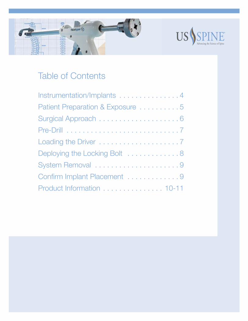
Table of Contents Instrumentation/Implants . . . . . . . . . . . . . . . 4
Patient Preparation & Exposure . . . . . . . . . . 5
Surgical Approach . . . . . . . . . . . . . . . . . . . . 6
Pre-Drill . . . . . . . . . . . . . . . . . . . . . . . . . . . . 7
Loading the Driver . . . . . . . . . . . . . . . . . . . . 7
Deploying the Locking Bolt . . . . . . . . . . . . . 8
System Removal . . . . . . . . . . . . . . . . . . . . . 9
Confirm Implant Placement . . . . . . . . . . . . . 9
Product Information . . . . . . . . . . . . . . . 10-11

Facet Fixation System Technique Guide
Bronze Driver
(60-5000-933)
Magenta Driver
(60-5000-939)
K-Drill
(60-5000-931)
Handpiece
(60-5000-900)
Green Driver
(60-5000-936)
Gold Driver
(60-5000-941)
Bolt Tightener
(60-3035-015)
Ratchet
(60-6000-053)
Instrumentation/Implants
4

Patient Preparation & Exposure1
n Position the patient in prone position on a C-Arm compatible table.
nThe C-Arm position suggested is a very oblique angle; a “modified Ferguson approach” is best utilized.
nMark the midline over the L4 spinous process using fluoroscopy.
nMake a 1 to 11/2 incision over the L4 spinous process. Enlarge the incision on either side if necessary for multiple levels.
nRetract the soft tissue laterally using a tissue retractor to expose the facet. Assess the spinous process of the operative level. Determine if preserving the spinous process will allow for proper trajectory of the handpiece at the angle required to seat the bridge across the thickest portion of the facet joint.
nIf you are able to preserve the spinous process, you can move on to step 2, if not, there are three options for access to the joint.
1) Remove as much spinous process as is required to allow for the Handpiece angle trajectory. In this instance the interspinous ligament may remain intact. (approximately 3-4 mm)
2) Using a Leksell Rongeur, break the base of the spinous process while keeping the supraspinous ligament intact. Bend the spinous process (while attached to the ligament), laterally to allow for insertion of the bridge.
3) Remove the spinous process while preserving the lamina structures.
nMinimize the amount of soft tissue on the superior facet to increase the amount of direct bone contact.
STEP 1A
STEP 1B
5

Facet Fixation System Technique Guide
6
Surgical Approach2
n Determine proper trajectory to position the Handpiece over the desired facet joint.
nBe sure that you are able to visualize the lateral portion of the facet (inferior facet).
nAim the bridge towards the transverse process and position the bridge over the facet joint.
nOnce the bridge is placed over the joint, pull back slightly in order to secure the distal locking washer onto the surface of the superior facet. This will provide proper grip of the implant into the bone,
nThe surgeon may confirm the position of the implant with fluoroscopy.
nThe roll controller at the top of the handpiece can be used to manipulate the washer-angle for proper seating on the facet surface.
nBe sure to position the washers deep into the joint, across the thickest portion of bone.
nWhile maintaining gun-held hand steady, compress the trigger with one smooth controlled movement. This will compress the joint and lock both of the washers onto the facet joint surfaces.
nWith the handpiece clamped into position on the facet joint, determine the proper facet bolt length on the color- coded bolt measurement grid located at the top of the handpiece. The laser etched marking will align with the color and/or number that correspond to the appropriate facet bolt.
STEP 2A
STEP 2B
STEP 2C

Pre-Drill3n To initiate the angle of approach and implant, insert the k-drill into
the back of the handpiece
nTurn the direction selector of the ratchet handle to the left in order to prep the handle for forward drilling.
nAttach the ratchet handle to the proximal end of the k-drill.
nBegin drilling by turning the ratchet handle clockwise.
nTwo full turns of the ratchet handle equates to approximately 1 mm of forward translation of the k-drill.
nContinue to drill until you reach the hard stop. The k-drill will not extend past the distal locking washer
nOnce trajectory and position are appropriate, the direction selector on the ratchet handle should be rotated to the right.
nTurn the ratchet handle counterclockwise to remove the k-drill from the handpiece.
Loading the Driver
nSelect the appropriate facet bolt length previously determined by the bolt measurement grid.
nLoad the corresponding facet bolt onto the driver. With the hex assembly pointing down, grasp the bolt; seat the hex end of the bolt flush into the distal drive assembly shaft. While bolt is seated flush with the shaft, turn the retainer clockwise to engage the threaded portion of the retainer rod into the hex end of the bolt (provisionally tighten to avoid stripping the bolt).
nTo connect the driver to the handpiece, simply slide the screw-end of the assembly into the open proximal end.
nThe direction selector on the ratchet handle should be rotated to the left.
nConnect ratchet handle to the back of the driver
7
4
STEP 3A
STEP 3B
STEP 3C
STEP 4A
STEP 4B

Facet Fixation System Technique Guide
8
Deploying the Locking Bolt5
n Turn the ratchet handle (or driver) to advance the bolt into the joint.
n Confirm bolt placement and trajectory with fluoroscopy.
n Continue to advance the Bolt through the joint into the distal locking washer.
n Confirm with fluoroscopy a second time.
n Two things confirm that the bolt is fully advanced:
1) A visual cue – The square window/opening (posterior to the roll controller) will be filled with yellow color
2) A physical “hard stop” will also indicate that the bolt has fully advanced through the distal locking washer and you are unable to drive any further.
n Two millimeters of the bolt will be protruding through the distal locking washer to ensure proper engagement of the locking mechanism.
n Confirm with fluoroscopy. (key step prior to removal)
STEP 5A
STEP 5B

System is Removed6
n To remove the handpiece assembly, first disengage the bolt from the driver by rotating the retainer counterclockwise until it spins freely.
n Release the clamp by depressing the clamp release.If for some reason the proximal washer does not disengage from the washer tube, pull backward on the roll controller at the top of the handpiece
n Place two fingers on the barrel where the barrel meets the housing while simultaneously axially rotating the handpiece & lifting the gun.
n Pull upward to release the distal locking washer and remove the facet gun from the wound site.
n If necessary, the bolt remover can be used to provide additional tightening.
Confirm Implant Placement
n Confirm the implant position and fixation via fluoroscopy.
n Repeat these steps for the contralateral facet joint or other levels.
7
STEP 6A
STEP 6B
STEP 6C
STEP 7A
9

Facet Fixation System Technique Guide
deScRiPTiOn: The Facet Fixation System consists of titanium alloy bone screws designed to transfix the facet articular process in the spine to enhance spinal fusion and stability. The screws are 4.0mm in diameter and are available partially threaded with a lag region. The screws are available in Ti-6Al-4V ELI alloy. These partially threaded screws are supplied in lengths of 30, 33, 36, 39, and 42 mm. Washers: Washers are available to increase the load bearing area of the screw in contact with the bone. These washers are designed to angulate about the head of the bone screws to provide optimal bony contact over a range of screw trajectories.
uSAGe: The surgeon must be thoroughly knowledgeable not only in the medical and surgical aspects of the implant, but must also be aware of the mechanical and metallurgical limitations of metallic surgical implants. See the technique manual for additional important information.
indicATiOnS: CAUTION: USA Law restricts this device to sale by or on the order of physician. The Facet Fixation System is indicated for the posterior surgical treatment of any or all of the following at the C2 to S1 (inclusive) spinal levels: 1) Trauma, including spinal fractures and/or dislocations; 2) Spondylolisthesis; 3) Spondylolysis; 4) Pseudoarthrosis or failed previous fusions which are symptomatic or which may cause secondary instability or deformity; 5) Degenerative diseases which include: (a) degenerative disc disease (ddd) as defined by neck and/or back pain of discogenic origin as confirmed by patient history with degeneration of the disc as confirmed by radiographic studies and/or (b) degenerative disease of the facets with instability. When properly used, facet screws will provide temporary stabilization as an adjunct to spinal bone grafting processes.
After solid fusion occurs, these devices serve no functional purpose and may be removed. In some cases, removal is indicated because the implants are not intended to transfer or to support forces developed during normal activities. Any decision to remove the device must be made by the physician and the patient. This decision should consider the patient’s general medical condition and the potential risk to the patient of a second surgical procedure.
PRecAuTiOn: The implantation of facet screws should be performed only by experienced spinal surgeons with specific training in the use of this screw spinal system because this is a technically demanding procedure presenting a risk of serious injury to the patient.
POSTOPeRATiVe MOBiLiZATiOn: Until x-rays confirm the maturation of the fusion mass, external immobilization (such as bracing or casting) is recommended. Instructions to the patient to reduce stress on the implants are an equally important part of the attempt to avoid the occurrence of clinical problems that may accompany fixation failure.
cOnTRAindicATiOnS: Disease conditions which have been shown to be safely and predictably managed without the use of internal fixation devices are relative contraindications to the use of these devices. Active systemic infection or infection localized to the site of the proposed implantation are contraindications to implantation. Severe osteoporosis is a relative contraindication because it may prevent adequate fixation of spinal anchors and thus preclude the use of this or any other spinal instrumentation system.
Absence of posterior spinal elements including the pedicle, pars interarticularis, facet joints and the majority of the lamina are contraindications to implantation. Any entity or condition that totally precludes the possibility of fusion, i.e. cancer, kidney dialysis, or osteopenia are relative contraindications. Other relative contraindications include obesity, certain degenerative diseases, or foreign body sensitivity. In addition, the patient’s occupation or activity level or mental capacity may be relative contraindications to this surgery. Specifically, patients who because of their occupation or lifestyle, or because of conditions such as mental illness, alcoholism, or drug abuse, may place undue stresses on the implant during bony healing and may be at higher risk for implant failure.
See also the WARNINGS, PRECAUTIONS AND ADVERSE EFFECTS sections of this insert.
Warnings: The following are specific warnings, precautions, and adverse effects which should be understood by the surgeon and explained to the patient. These warnings do not include all adverse effects which can occur with surgery in general, but are important considerations particular to metallic internal fixation devices. General surgical risks should be explained to the patient prior to surgery. WARNINGS, PRECAUTIONS, AND ADVERSE EFFECTS CONCERNING TEMPORARY METALLIC INTERNAL FIXATION DEVICES
WARninGS 1. CORRECT SELECTION OF THE IMPLANT IS EXTREMELY IMPORTANT. The potential
for satisfactory fixation is increased by the selection of the proper size, shape, and design of the implant. While proper selection can help minimize risks, the size and shape of human bones present limitations on the size, shape, and strength of implants. Metallic internal fixation devices cannot withstand activity levels equal to those placed on normal healthy bone. No implant can be expected to withstand indefinitely the unsupported stress of full weight bearing.
2. IMPLANTS CAN BREAK WHEN SUBJECTED TO THE INCREASED LOAD ASSOCIATED WITH DELAYED UNION OR NONUNION. Internal fixation appliances are load sharing devices which are used to obtain alignment until normal healing occurs. If healing is delayed, or does not occur, the implant may eventually break due to metal fatigue. The degree or success of union, loads produced by weight bearing, and activity levels will, among other conditions, dictate the longevity of the implant. Notches, scratches or bending of the implant during the course of surgery may also contribute to early failure. Patients should be fully informed of the risks of implant failure.
3. MIXING METALS CAN CAUSE CORROSION. There are many forms of corrosion damage and several of these occur on metals surgically implanted in humans. General or uniform corrosion is present on all implanted metals and alloys. The rate of corrosive attack on metal implant devices is usually very low due to the presence of passive surface films. Dissimilar metals in contact, such as titanium and stainless steel, accelerates the corrosion process of stainless steel and more rapid attack occurs. The presence of corrosion often accelerates fatigue fracture of implants. The amount of metal compounds released into the body system will also increase. Internal fixation devices, such as rods, hooks, wires, etc., which come into contact with other metal objects, must be made of like or compatible metals.
4. PATIENT SELECTION. In selecting patients for internal fixation devices, the following factors can be of extreme importance to the eventual success of the procedure.
A. The patient’s weight. An overweight or obese patient can produce loads on the device which can lead to failure of the appliance and the operation.
B. The patient’s occupation or activity. If the patient is involved in an occupation or activity that includes heavy lifting, muscle strain, twisting, or repetitive bending, stooping, running, substantial walking, or manual labor, he/she should not return to these activities until the bone is fully healed. Even with full healing, the patient may not be able to return to these activities successfully.
C. A condition of senility, mental illness, alcoholism, or drug abuse. These conditions, among others, may cause the patient to ignore certain necessary limitations and precautions in the use of the appliance, leading to implant failure or other complications.
D. Certain degenerative diseases. In some cases, the progression of degenerative disease may substantially decrease the expected useful life of the appliance. For such cases, orthopaedic devices can only be considered a delaying technique or temporary remedy.
E. Foreign body sensitivity. The surgeon is advised that no preoperative test can completely exclude the possibility of sensitivity or allergic reaction. Patients can develop sensitivity or allergy after implants have been in the body for a period of time.
F. Smoking. Patients who smoke have been observed to experience higher rates of pseudoarthrosis following surgical procedures where bone graft is used. Additionally, smoking has been shown to cause diffuse degeneration of intervertebral discs. Progressive degeneration of adjacent segments caused by smoking can lead to late clinical failure (recurring pain) even after successful fusion and initial clinical improvement.
10

PRECAUTIONS 1. SURGICAL IMPLANTS MUST NEVER BE REUSED. An explanted metal implant should
never be reimplanted. Even though the device appears undamaged, it may have small defects and internal stress patterns which may lead to early breakage.
2. CORRECT HANDLING OF THE IMPLANT IS EXTREMELY IMPORTANT. The operating surgeon should avoid any notching, scratching, or bending of the devices when contouring. Alterations will produce defects in surface finish and internal stresses which may become the focal point for eventual breakage of the implant. Bending of screws will significantly decrease the fatigue life and may cause failure.
3. CONSIDERATIONS FOR REMOVAL OF THE IMPLANT AFTER HEALING. If the device is not removed after the completion of its intended use, any of the following complications may occur: (1) Corrosion, with localized tissue reaction or pain; (2) Migration of implant position resulting in injury; (3) Risk of additional injury from post-operative trauma; (4) Bending, loosening, and/or breakage, which could make removal impractical or difficult; (5) Pain, discomfort, or abnormal sensations due to the presence of the device; (6) Possible increased risk of infection; and (7) Bone loss due to stress shielding. The surgeon should carefully weigh the risks versus benefits when deciding whether to remove the implant. Implant removal should be followed by adequate postoperative management to avoid refracture. If the patient is older and has a low activity level, the surgeon may choose not to remove the implant thus eliminating the risks involved with a second surgery.
4. ADEQUATELY INSTRUCT THE PATIENT. Postoperative care and the patient’s ability and willingness to follow instructions are among the most important aspects of successful bone healing. The patient must be made aware of the limitations of the implant, and instructed to limit and restrict physical activities, especially lifting and twisting motions and any type of sport participation. The patient should understand that a metallic implant is not as strong as normal healthy bone and could loosen, bend and/or break if excessive demands are placed on it, especially in the absence of complete bone healing. Implants displaced or damaged by improper activities may migrate and damage the nerves or blood vessels. An active, debilitated, or demented patient who cannot properly use weight supporting devices may be particularly at risk during postoperative rehabilitation.
POSSIBLE ADVERSE EFFECTS 1. Nonunion, delayed union. 2. Bending or fracture of implant. 3. Loosening of the implant. 4. Metal sensitivity, or allergic reaction to a foreign body. 5. Infection, early or late. 6. Decrease in bone density due to stress shielding. 7. Pain, discomfort, or abnormal sensations due to the presence of the device. 8. Vascular and/or nerve damage due to surgical trauma or presence of the device.
Neurological difficulties include bowel and/or bladder dysfunction, impotence, retrograde ejaculation, and paraesthesia.
9. Bursitis. 10. Paralysis. 11. Dural tears experienced during surgery could result in the need for further surgery
for dural repair, a chronic CSF leak or fistula, and possible meningitis. 12. Death. 13. Screw back out, possibly leading to implant loosening, and/or reoperation for
device removal. 14. Damage to lymphatic vessels and/or lymphatic fluid exudation. 15. Spinal cord impingement or damage. 16. Fracture of bony structures. 17. Degenerative changes or instability in segments adjacent to fused vertebral levels.
PACKAGING:Packages for each of the components should be intact upon receipt. If a loaner or consignment system is used, all sets should be carefully checked for completeness and all components including instruments should be care¬fully checked to ensure that there is no damage prior to use. Damaged packages or products should not be used, and should be returned to US Spine.
11
CLEANING AND DECONTAMINATION: All instruments must first be cleaned using neutral cleaners before sterilization and introduction into a surgical field or prior to (if applicable) return of the product to US Spine. Cleaning and disinfecting of instruments can be performed with aldehyde-free solvents at higher temperatures. Cleaning and decontamination must include the use of neutral cleaners followed by a deionized water rinse. After cleaning, sterilize as directed prior to use.
Note: The Facet Fixation System Trial Gun should be additionally cleaned with a syringe for solution injection into hard-to-reach parts. Using a syringe, aspirate and squirt the solution several times into the inner barrel. Utilize the flush portals (slots on the top of the trial gun and also the holes at the rear of the proximal washer) to facilitate easier cleaning. Certain cleaning solutions such as those containing formalin, glutaraldehyde, bleach and/or other alkaline cleaners may damage some devices, particularly instruments; these solutions should not be used. All products should be treated with care. Improper use or handling may lead to damage and/or possible improper functioning of the device.
STERILIZATION: Unless marked sterile and clearly labeled as such, the Facet Fixation System components described in this insert are provided non-sterile and must be sterilized prior to use. If the product described in this document is sterilized by the hospital in a tray or case, it must be sterilized in a tray or case provided by US Spine. These products are recommended to be steam sterilized by the hospital using the process parameter below:
METHOD CYCLE TEMPERATURE EXPOSURE TIME DRY TIME
Steam Pre-Vacuum 270° F (132° C) 8 Minutes 40 minutes
(wrapped tray/case)
Remove all packaging materials prior to sterilization. Use only sterile products in the operative field.
IF MORE THAN TWO YEARS HAVE ELAPSED BETWEEN THE DATE OF ISSUE/REVISION AND THE DATE OF CONSULTATION, CONTACT US SPINE FOR CURRENT INFORMATION AT 1-561-367-7463. LIMITED WARRANTY AND DISCLAIMER: US SPINE PRODUCTS ARE SOLD WITH A LIMITED WARRANTY TO THE ORIGINAL PURCHASER AGAINST DEFECTS IN WORKMANSHIP AND MATERIALS. ANY OTHER EXPRESS OR IMPLIED WARRANTIES, INCLUDING WARRANTIES OF MERCHANTABILITY OR FITNESS, ARE HEREBY DISCLAIMED.
In case of complaint, or for supplementary information, or further directions for use of this system, please see the addresses below:
IN THE USA IN THE EUROPEAN UNIONCustomer Service Division RMS UK, LTDUS Spine Inc. 28 Trinity Road3600 FAU Blvd. Suite 100 Nailsea, Somerset BS484NUBoca Raton, Florida 33431 USA United KingdomTelephone: 561-367-7463 Telephone: 044 0125-858891
0459
90-4000-101A© 2009 US Spine, Inc. All rights reserved.

For more information contact a US Spine sales representative at 561.36.SPINE or visit www.us-spine.com
3600 FAU Boulevard, Suite 101 Boca Raton, FL 33431
© 2009 US Spine, Inc. All rights reserved. All trademarks or registered trademarks are the exclusive property of US Spine, Inc. Patents Pending.
90-4000-130A


![Subcondylar/Ramus Fixation Set Technique Guidesynthes.vo.llnwd.net/o16/LLNWMB8/US Mobile/Synthes North Americ… · The Subcondylar/Ramus Fixation Set [115.680] includes specialized](https://img.dokumen.tips/doc/110x75/5ed55669cfcb033b55255b36/subcondylarramus-fixation-set-technique-mobilesynthes-north-americ-the-subcondylarramus.jpg)