Embed Size (px)
Citation preview
Fabrication of custom made ocular prosthesis with three different impression techniques: a report of four cases
Abstract
Loss of eye has a bad effect on the psychology of the patient. Eye prosthesis is fabricated to regain the patient’s confidence by meticulous replacement of the missing eye. Immediate fitting of an anophthalmic socket with an artificial eye may not always be possible, and a delayed prosthesis delivery may result in settling and sinking of the prosthesis into the socket, therefore requiring orbital cavity conformation. It becomes a challenging task for a maxillofacial prosthodontist to fabricate a prosthesis that replicates the healthy side of the face. This article presents a short review of three functional ocular impression techniques to achieve a better fit of the prosthesis to the defect area. These techniques aim to combine cosmetic excellence with comfort for greater benefit to the patients.
Key words: Impression techniques, custom ocular prosthesis
D N EAI NRE TAG LIN
AS NOS IO IAC T
D N EAI NRE TAG LIN
AS NOS IO IAC T
Introduction
Loss of the eye either due to trauma, tumor or any other condition not only causes loss of sensory function but also there is unaesthetic look and has a
(1)psychological effect on the patient . An ocular prosthesis is an artificial replacement for the bulb of the eye (bulbus oculi, eyeball). When the entire content of the orbit (including muscles, fascia, eyelids, conjunctiva and the lacrimal apparatus) is removed, the artificial replacement is referred to as an orbital
(2)prosthesis . Maxillofacial prostheses, which restore and replace stomatognathic and associated facial structures with artificial substitutes, aim to improve the patient’s esthetics, restore and maintain health of the remaining structures and consequently provide
(3)physical and mental well-being . Fabrication of an esthetically pleasing orbital prosthesis is a challenging process. The correct location and the orientation of the ocular component is vital for an esthetically pleasing orbital prosthesis because slight discrepancies in eye position are
(4)immediately noticed . Ocular prosthesis can be either readymade or custom made. Stock eye have some advantages like even distribution of pressure due to equal movement thereby reducing incidence of ulceration, mobility, improved fit, improved facial
(5)contours, esthetics, comfort and adaptation . The custom built, hand painted and individually constructed acrylic resin artificial eye has proved to be
(6)the most satisfactory ocular replacement . One of the most important step in making accurate impression is the close adaptation of the mucosal surface of the ocular prosthesis to the posterior wall of
( 5 )the socket . The combined efforts of the ophthalmologist and the maxillofacial prosthetist are needed to provide a satisfactory ocular prosthesis when trauma or disease causes the loss of an eye. Since every socket differs in size and shape, it is obvious that an individually designed prosthesis,
made from an impression of the socket, is needed to utilize the full movement potential of any implant and also to provide maximum comfort and restore full physiologic function to the accessory organs of the
(7)eye . The aim of the article is to revive three different impression techniques for fabrication of customized ocular prosthesis to achieve a better fit of the prosthesis to the defect area and to improve esthetics.
Case1 A 45 year old patient reported to Department of Prosthodontics, with complaint of unaesthetic appearance due to missing right eye. On examination enucleated right eye socket was found with healthy conjuctival lining and no infection. History revealed traumatic injury (road accident) at the age of 14 during which eye ball was damaged and he was wearing a large size hollow conformer that was esthetically unacceptable since then. The location and movement of the tissue bed, size, shape and dimension of the
81
Nig Dent J Vol. 21 No. 2 July - Dec. 2013
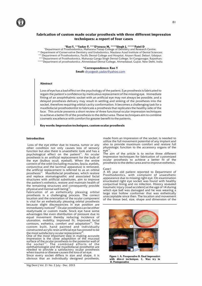
Figure 1. A. Preoperative B. Final impression with direct technique. C. Wax try in D. Postoperative
*Correspondence: Rao YEmail: [email protected]
*Department of Prosthodontics, Maharana Pratap College of Dentistry and Research Centre; ** Department of Conservative Dentistry and Endodontics, Maulana Azad Institute of Dental Sciences; ***.Department of Prosthodontics, Pacific Dental College and Hospital, Airport Road, Debari, Udaipur; ****Department of Prosthodontics, Maharaja Ganga Singh Dental College, Sri Ganganagar, Rajasthan; *****Department of prosthodontics ,Ahmedabad Dental College, Ahmedabad, Gujrat, New Delhi, India
*Rao Y, **Yadav P, ***D'souza M, ****Singh J, *****Patel D
D N EAI NRE TAG LIN
AS NOS IO IAC T
D N EAI NRE TAG LIN
AS NOS IO IAC T
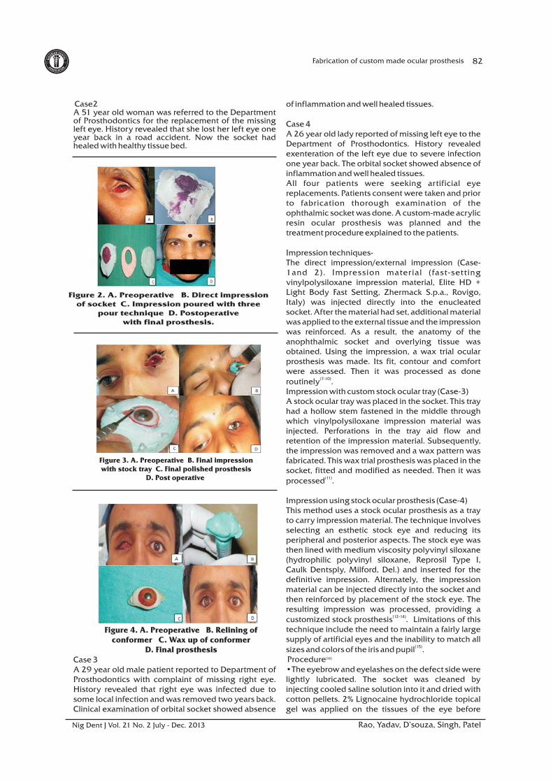
Case2A 51 year old woman was referred to the Department of Prosthodontics for the replacement of the missing left eye. History revealed that she lost her left eye one year back in a road accident. Now the socket had healed with healthy tissue bed.
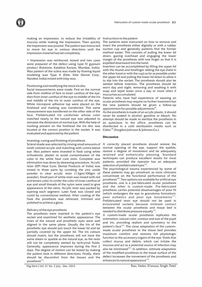
Case 3A 29 year old male patient reported to Department of Prosthodontics with complaint of missing right eye. History revealed that right eye was infected due to some local infection and was removed two years back. Clinical examination of orbital socket showed absence
of inflammation and well healed tissues.
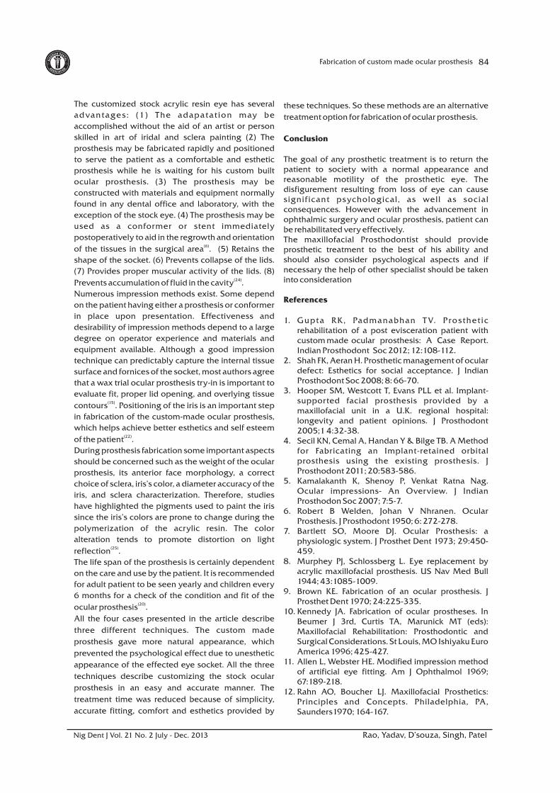
Case 4A 26 year old lady reported of missing left eye to the Department of Prosthodontics. History revealed exenteration of the left eye due to severe infection one year back. The orbital socket showed absence of inflammation and well healed tissues.All four patients were seeking artificial eye replacements. Patients consent were taken and prior to fabrication thorough examination of the ophthalmic socket was done. A custom-made acrylic resin ocular prosthesis was planned and the treatment procedure explained to the patients.
Impression techniques-The direct impression/external impression (Case-1and 2). Impression material (fast-setting vinylpolysiloxane impression material, Elite HD + Light Body Fast Setting, Zhermack S.p.a., Rovigo, Italy) was injected directly into the enucleated socket. After the material had set, additional material was applied to the external tissue and the impression was reinforced. As a result, the anatomy of the anophthalmic socket and overlying tissue was obtained. Using the impression, a wax trial ocular prosthesis was made. Its fit, contour and comfort were assessed. Then it was processed as done
(7-10)routinely . Impression with custom stock ocular tray (Case-3) A stock ocular tray was placed in the socket. This tray had a hollow stem fastened in the middle through which vinylpolysiloxane impression material was injected. Perforations in the tray aid flow and retention of the impression material. Subsequently, the impression was removed and a wax pattern was fabricated. This wax trial prosthesis was placed in the socket, fitted and modified as needed. Then it was
(11)processed .
Impression using stock ocular prosthesis (Case-4) This method uses a stock ocular prosthesis as a tray to carry impression material. The technique involves selecting an esthetic stock eye and reducing its peripheral and posterior aspects. The stock eye was then lined with medium viscosity polyvinyl siloxane (hydrophilic polyvinyl siloxane, Reprosil Type I, Caulk Dentsply, Milford, Del.) and inserted for the definitive impression. Alternately, the impression material can be injected directly into the socket and then reinforced by placement of the stock eye. The resulting impression was processed, providing a
(12-14)customized stock prosthesis . Limitations of this technique include the need to maintain a fairly large supply of artificial eyes and the inability to match all
(15)sizes and colors of the iris and pupil .(16) Procedure
•The eyebrow and eyelashes on the defect side were lightly lubricated. The socket was cleaned by injecting cooled saline solution into it and dried with cotton pellets. 2% Lignocaine hydrochloride topical gel was applied on the tissues of the eye before
82Fabrication of custom made ocular prosthesis
Rao, Yadav, D’souza, Singh, PatelNig Dent J Vol. 21 No. 2 July - Dec. 2013
Figure 2. A. Preoperative B. Direct impression of socket C. Impression poured with three
pour technique D. Postoperative with final prosthesis.
Figure 3. A. Preoperative B. Final impression with stock tray C. Final polished prosthesis
D. Post operative
Figure 4. A. Preoperative B. Relining of conformer C. Wax up of conformer
D. Final prosthesis
D N EAI NRE TAG LIN
AS NOS IO IAC T
D N EAI NRE TAG LIN
AS NOS IO IAC T
making an impression, to reduce the irritability of mucosa while making the impression. Then quickly the impression was poured. The patient was instructed to move his eye in various directions until the impression material had set completely.
• Impression was reinforced, boxed and two casts were prepared of the defect using type IV gypsum product (Kalstone; Kalabhai Karson, Mumbai, India). Wax pattern of the sclera was made by flowing liquid modeling wax Type II (Elite, Elite Dental Corp., Nanded, India) mixed with inlay wax.
Positioning and modifying the stock iris discFacial measurements were made. First on the normal side from midline of face to inner canthus of the eye, then from inner canthus of the eye to middle of the iris and middle of the iris to outer canthus of the eye. White micropore adhesive tap were placed on the forehead and marking was transferred. This same measurement was transferred on the other side of the face. Prefabricated iris conformer whose color matched nearly to the natural eye was adjusted to simulate the dimension of normal eye. With the help of marking present on the forehead the iris disc was located at the correct position in the socket. It was evaluated and approved by the patient.
Investing, curing and finishing of prosthesisScleral shade was selected by mixing small amounts of tooth colored acrylic and matching with contra lateral eye .Wax pattern were invested in flask using white orthodontic plaster to avoid contamination of the color in the white heat cure resin. Complete wax elimination was done by dewaxing procedure. Acrylic resin (DPI Heat Cure; Dental Products of India) was mixed in three parts by combing the zinc oxide powder in clear acrylic resin (1.5gm/100gm of powder). Small part of white resin was mixed with red and brown color to confer the color of inner canthus of eye and small threads of the nylon were used to give appearance of the veins. Acrylic resin was packed by layering each segment. Later flask was closed and cured by conventional method. After cooling of the flask the prosthesis was retrieved, trimmed and polished to achieve a gloss.
Delivery of the eye prosthesis The prosthesis were inserted in the patient’s eye socket and examined for aesthetic appearance. The irises of the natural and prosthetic eyes were well aligned in the same plane. Usually the iris of the prosthetic eye should just touch the lower lid and be partially covered by the upper lid. The iris colours should match, but the prosthesis will not have the same sheen or sparkle as the natural eye, as the resin will not be completely wetted by lachrymal fluids. Generally, appearance improves during the first 2 days. The degree of motion can be tested by making the patient look in different directions. Finally, there should be discomfort from the tissues and the
(17)prosthesis .
Instructions to the patientThe patients were instructed on how to remove and insert the prosthesis either digitally or with a rubber suction cup and generally patients find the former method easier. This consists of pulling the lower lid down, gazing overhead and engaging the lower margin of the prosthesis with one finger so that it is expelled downward into the hand.Insertion can be accomplished by lifting the upper lid with the thumb and forefinger sliding the eye (held in the other hand or with the cup) as far as possible under the upper lid and pulling the lower lid down to allow it to slip into the socket. The prosthesis should also be wetted before insertion. The prosthesis should be worn day and night, removing and washing it with soap and tepid water once a day or more often if mucus has accumulated.Patients who have had experience of wearing an ocular prosthesis may require no further treatment but the new patients should be given a follow-up
(8)appointment for possible adjustments .As the prosthesis is made of an acrylic plastic, it should never be soaked in alcohol, gasoline or bleach. No attempt should be made to sterilize the prosthesis in an autoclave. In the office, prosthesis can be disinfected in a cold sterilization media such as
(18)Cidex (Surgikos Johnson & Johnson Co.).
Discussion
A correctly placed prosthesis should restore the normal opening of the eye, support the eyelids, restore a degree of movement and be adequately retained and esthetically pleasing. Standard techniques can produce excellent results for most patients, provided the operator has an adequate
(19)selection of prefabricated eyes . The psychological trauma experienced by some of these patients may go unnoticed, as most clinicians concentrate on the functional performance of the
(20)prosthesis . Two options are available for artificial eye prosthesis, one is a pre-fabricated ocular prosthesis and the other is custom-made. Pre-fabricated prosthesis carries potential disadvantages of poor fit (which endangers the eye to granuloma formation),
(21 )poor esthetics and poor eye movements . Prefabricated resin eye should not be used in eviscerated sockets because intimate contact between the ocular prosthesis and tissue bed is
(17)needed to distribute pressure equally . A custom-made ocular prosthesis replicates the orientation, natural color, contour and size of the pupil and iris, providing realism and symmetry to the
(22)patient’s face . The close adaptation of the custom-made ocular prosthesis to the tissue bed provides maximum comfort and restores full physiologic function to the accessory organs of the eye. Voids that collect mucus and debris, which can irritate the mucosa and act as a potential source of infection may
(23)also be minimized . In addition, intimate adaptation of the modified prosthesis to the tissue surface of the defect increases the movement of the prosthesis and
(17)enhances its natural appearance .
83Fabrication of custom made ocular prosthesis
Rao, Yadav, D’souza, Singh, PatelNig Dent J Vol. 21 No. 2 July - Dec. 2013
D N EAI NRE TAG LIN
AS NOS IO IAC T
D N EAI NRE TAG LIN
AS NOS IO IAC T
The customized stock acrylic resin eye has several
advantages: (1) The adapatation may be
accomplished without the aid of an artist or person
skilled in art of iridal and sclera painting (2) The
prosthesis may be fabricated rapidly and positioned
to serve the patient as a comfortable and esthetic
prosthesis while he is waiting for his custom built
ocular prosthesis. (3) The prosthesis may be
constructed with materials and equipment normally
found in any dental office and laboratory, with the
exception of the stock eye. (4) The prosthesis may be
used as a conformer or stent immediately
postoperatively to aid in the regrowth and orientation (6)of the tissues in the surgical area . (5) Retains the
shape of the socket. (6) Prevents collapse of the lids.
(7) Provides proper muscular activity of the lids. (8) (24)Prevents accumulation of fluid in the cavity .
Numerous impression methods exist. Some depend
on the patient having either a prosthesis or conformer
in place upon presentation. Effectiveness and
desirability of impression methods depend to a large
degree on operator experience and materials and
equipment available. Although a good impression
technique can predictably capture the internal tissue
surface and fornices of the socket, most authors agree
that a wax trial ocular prosthesis try-in is important to
evaluate fit, proper lid opening, and overlying tissue (15)contours . Positioning of the iris is an important step
in fabrication of the custom-made ocular prosthesis,
which helps achieve better esthetics and self esteem (22)of the patient .
During prosthesis fabrication some important aspects
should be concerned such as the weight of the ocular
prosthesis, its anterior face morphology, a correct
choice of sclera, iris’s color, a diameter accuracy of the
iris, and sclera characterization. Therefore, studies
have highlighted the pigments used to paint the iris
since the iris’s colors are prone to change during the
polymerization of the acrylic resin. The color
alteration tends to promote distortion on light (25)reflection .
The life span of the prosthesis is certainly dependent
on the care and use by the patient. It is recommended
for adult patient to be seen yearly and children every
6 months for a check of the condition and fit of the (20)ocular prosthesis .
All the four cases presented in the article describe
three different techniques. The custom made
prosthesis gave more natural appearance, which
prevented the psychological effect due to unesthetic
appearance of the effected eye socket. All the three
techniques describe customizing the stock ocular
prosthesis in an easy and accurate manner. The
treatment time was reduced because of simplicity,
accurate fitting, comfort and esthetics provided by
these techniques. So these methods are an alternative
treatment option for fabrication of ocular prosthesis.
Conclusion
The goal of any prosthetic treatment is to return the patient to society with a normal appearance and reasonable motility of the prosthetic eye. The disfigurement resulting from loss of eye can cause significant psychological, as well as social consequences. However with the advancement in ophthalmic surgery and ocular prosthesis, patient can be rehabilitated very effectively.The maxillofacial Prosthodontist should provide prosthetic treatment to the best of his ability and should also consider psychological aspects and if necessary the help of other specialist should be taken into consideration
References
1. Gupta RK, Padmanabhan T V. Prosthetic rehabilitation of a post evisceration patient with custom made ocular prosthesis: A Case Report. Indian Prosthodont Soc 2012; 12:108-112.
2. Shah FK, Aeran H. Prosthetic management of ocular defect: Esthetics for social acceptance. J Indian Prosthodont Soc 2008; 8: 66-70.
3. Hooper SM, Westcott T, Evans PLL et al. Implant- supported facial prosthesis provided by a maxillofacial unit in a U.K. regional hospital: longevity and patient opinions. J Prosthodont 2005;1 4:32-38.
4. Secil KN, Cemal A, Handan Y & Bilge TB. A Method for Fabricating an Implant-retained orbital prosthesis using the existing prosthesis. J Prosthodont 2011; 20:583-586.
5. Kamalakanth K, Shenoy P, Venkat Ratna Nag. Ocular impressions- An Overview. J Indian Prosthodon Soc 2007; 7:5-7.
6. Robert B Welden, Johan V Nhranen. Ocular Prosthesis. J Prosthodont 1950; 6: 272-278.
7. Bartlett SO, Moore DJ. Ocular Prosthesis: a physiologic system. J Prosthet Dent 1973; 29:450-459.
8. Murphey PJ, Schlossberg L. Eye replacement by acrylic maxillofacial prosthesis. US Nav Med Bull 1944; 43:1085-1009.
9. Brown KE. Fabrication of an ocular prosthesis. J Prosthet Dent 1970; 24:225-335.
10. Kennedy JA. Fabrication of ocular prostheses. In Beumer J 3rd, Curtis TA, Marunick MT (eds): Maxillofacial Rehabilitation: Prosthodontic and Surgical Considerations. St Louis, MO Ishiyaku Euro America 1996; 425-427.
11. Allen L, Webster HE. Modified impression method of artificial eye fitting. Am J Ophthalmol 1969; 67:189-218.
12. Rahn AO, Boucher LJ. Maxillofacial Prosthetics: Principles and Concepts. Philadelphia, PA, Saunders1970; 164-167.
84Fabrication of custom made ocular prosthesis
Rao, Yadav, D’souza, Singh, PatelNig Dent J Vol. 21 No. 2 July - Dec. 2013
D N EAI NRE TAG LIN
AS NOS IO IAC T
D N EAI NRE TAG LIN
AS NOS IO IAC T
85Fabrication of custom made ocular prosthesis
13. Chalian VA. Treating the patient with facial defects. In Laney WR (ed): Maxillofacial Prosthetics. Littleton, MA, PSG Publishing 1979; 286-290.
14. Welden RB, Niiranen JV. Ocular prosthesis. J Prosthet Dent 1956; 6:272-278.
15. Mathews MF, Smith RM, Sutton AJ, Hudson R. The Ocular Impression: A Review of the Literature and Presentation of an Alternate Technique. J Prosthodon 2000; 9:210-216.
16. Jurel SK, Talwar N, Chand P, Singh RD, Gupta DS. Customization of Stock Eye Prosthesis for a Pediatric Patient by a Simplified Technique. International J Clin Pediac Dent 2012; 5:155-158.
17. Taicher S, Steinberg HM, Tubiana I et al. Modified stock eye ocular prosthesis. J Prosthet Dent 1985; 54:95-98.
18. Raizada K, Rani D. Ocular prosthesis; a review: Contact Lens & Anterior Eye. 2007; 30:152-162.
19. Sykes L.M. Custom made ocular prosthesis. J Prosthet Dent 1996; 75:1-3.
20. Pereira BP, Kour AK, Leow EL, Pho R.WH. Benefits and use of Digital Prostheses. J Hand Surg 1996; 21:222-228.
21. Cain JR. Custom ocular prosthesis. J Prosthet Dent 1982; 48:690-694.
22. Guttal SS, Patil NP, Vernekar N & Porwal A. A simple method of positioning the iris disk on a custom-made ocular prosthesis. A Clinical Report. J Prosthodon 2008; 17: 223-227.
23. Kaur A, Pavaiya A, Singh SV, Raghuvar DS, Chand P A. Simplified approach to fabrication of an ocular prosthesis: A case series. Indian J Dent Res 2010; 21:615-617.
24. Taylor TD. Clinical maxillofacial prosthetics, 3rd edn. Quintessence Publishing Co. Inc, Chicago, 2000; pp 265-276.
25. Marcelo CG, Amalia Moreno, Daniela M dos S, Stefan F de CD, Eduardo PP, Aldieris AP. Contact Lens & Anterior Eye 2010; 33:215-218.
Rao, Yadav, D’souza, Singh, PatelNig Dent J Vol. 21 No. 2 July - Dec. 2013