Embed Size (px)
Citation preview

Prosthetic Management of Patient with Ocular Defect
International Journal of Preventive and Clinical Dental Research, July-September 2017;4(3):1-5 1
IJPCDR
Prosthetic Management of Patient with Ocular Defect1Binoy N Mathews, 2S Sajina, 3Abhinav K Mohan, 4Anil K Subhash
IJPCDR
XXXXXXXXXXX10.5005/jp-journals-00000-0000
1Professor and Head, 2-4Senior Lecturer1,3,4Department of Prosthodontics, Crown, Bridge and Implantology, Mahe Institute of Dental Sciences & Hospital Mahe, Puducherry, India2Department of Pedodontics and Preventive Dentistry, Mahe Institute of Dental Sciences & Hospital, Mahe, Puducherry, India
Corresponding Author:
ABSTRACT
The agony over the loss of an eye and the resulting facial defect has a crippling effect on the psychology of the patient. An artificial prosthesis is probably the only alternative in such cases to help rehabilitate the patients. An ocular prosthesis is undoubtedly a challenge to any maxillofacial prosthodontist because you are attempting to replace a moving organ with a static prosthesis.
Presented here is the case report of a stock acrylic ocular prosthesis which had acceptable fit, retention, and esthetics.
Keywords:
How to cite this article: Mathews BN, Sajina S, Mohan AK, Subhash AK. Prosthetic Management of Patient with Ocular Defect. Int J Prev Clin Dent Res 2017;4(3):1-5.
Source of support: Nil
Conflict of interest: None
INTRODUCTION
Eyes are generally the first feature of the face to be noticed. Removal of this organ due to tumors, trauma, or any other condition not only causes unesthetic look, but also there is loss of function and has a psychologic effect on the patient.1,2 Also, in some cases, age and the medical condition of the patient may contraindicate any major constructive surgery, leaving a huge void in the complete rehabilitation of the patient. An artificial pros-thesis is probably the only alternative in such cases for the psychological well-being of the patient.3
This clinical case report is about the management of a patient with an ocular defect with a stock acrylic ocular prosthesis.
CASE REPORT
A 65-year-old male reported to the prosthodontics depart-ment with the left eye lost due to surgery. On history, it
was found that the patient was suffering from malignant melanoma of the left eye and the eye had to be enucle-ated. It was decided that a stock acrylic ocular prosthesis would be best to meet the needs of the patient (Fig. 1).
After careful examination of the area of the defect, an acrylic ocular prosthesis was planned. Patient was explained about the procedure and its limitations.
First petroleum jelly was applied to the eyebrows for easy removal of the impression material when it sets. A wax frame was fabricated defining the area of the face to be included in the impression. Alginate was mixed in a fluid consistency in a large quantity and poured into
Figs 1A and B: Pretreatment view of the patient with ocular defect
A
B
Binoy N Mathews et al
2
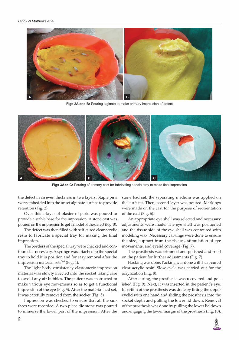
the defect in an even thickness in two layers. Staple pins were embedded into the unset alginate surface to provide retention (Fig. 2).
Over this a layer of plaster of paris was poured to provide a stable base for the impression. A stone cast was poured on the impression to get a model of the defect (Fig. 3).
The defect was then filled with self-cured clear acrylic resin to fabricate a special tray for making the final impression.
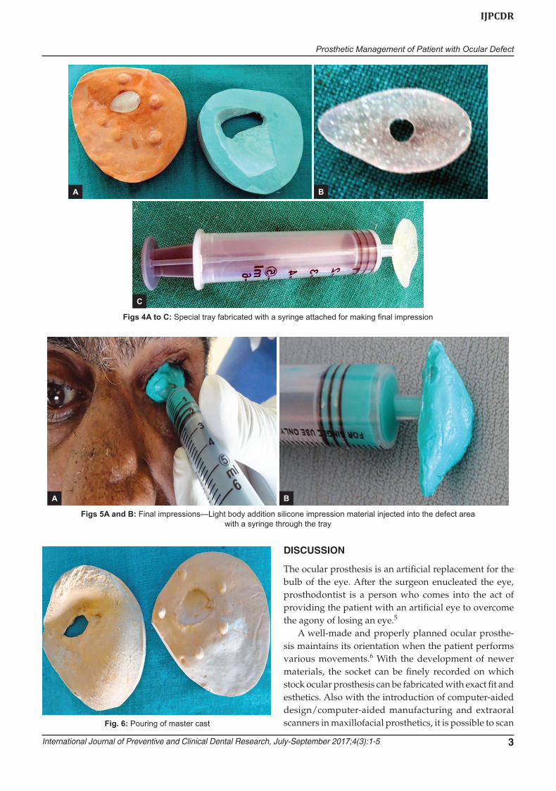
The borders of the special tray were checked and con-toured as necessary. A syringe was attached to the special tray to hold it in position and for easy removal after the impression material sets3,4 (Fig. 4).
The light body consistency elastomeric impression material was slowly injected into the socket taking care to avoid any air bubbles. The patient was instructed to make various eye movements so as to get a functional impression of the eye (Fig. 5). After the material had set, it was carefully removed from the socket (Fig. 5).
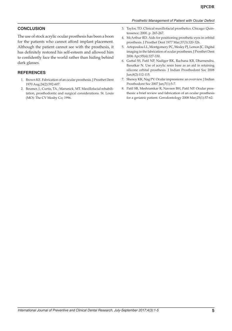
Impression was checked to ensure that all the sur-faces were recorded. A two-piece die stone was poured to immerse the lower part of the impression. After the
stone had set, the separating medium was applied on the surfaces. Then, second layer was poured. Markings were made on the cast for the purpose of reorientation of the cast (Fig. 6).
An appropriate eye shell was selected and necessary adjustments were made. The eye shell was positioned and the tissue side of the eye shell was contoured with modeling wax. Necessary carvings were done to ensure the size, support from the tissues, stimulation of eye movements, and eyelid coverage (Fig. 7).
The prosthesis was trimmed and polished and tried on the patient for further adjustments (Fig. 7).
Flasking was done. Packing was done with heat-cured clear acrylic resin. Slow cycle was carried out for the acrylization (Fig. 8).
After curing, the prosthesis was recovered and pol-ished (Fig. 9). Next, it was inserted in the patient’s eye. Insertion of the prosthesis was done by lifting the upper eyelid with one hand and sliding the prosthesis into the socket depth and pulling the lower lid down. Removal of the prosthesis was done by pulling the lower lid down and engaging the lower margin of the prosthesis (Fig. 10).
Figs 2A and B: Pouring alginate to make primary impression of defect
Figs 3A to C: Pouring of primary cast for fabricating special tray to make final impression
A B
A B C
Prosthetic Management of Patient with Ocular Defect
International Journal of Preventive and Clinical Dental Research, July-September 2017;4(3):1-5 3
IJPCDR
Figs 4A to C: Special tray fabricated with a syringe attached for making final impression
Figs 5A and B: Final impressions—Light body addition silicone impression material injected into the defect area with a syringe through the tray
Fig. 6: Pouring of master cast
DISCUSSION
The ocular prosthesis is an artificial replacement for the bulb of the eye. After the surgeon enucleated the eye, prosthodontist is a person who comes into the act of providing the patient with an artificial eye to overcome the agony of losing an eye.5
A well-made and properly planned ocular prosthe-sis maintains its orientation when the patient performs various movements.6 With the development of newer materials, the socket can be finely recorded on which stock ocular prosthesis can be fabricated with exact fit and esthetics. Also with the introduction of computer-aided design/computer-aided manufacturing and extraoral scanners in maxillofacial prosthetics, it is possible to scan
A B
C
A B

Binoy N Mathews et al
4
the entire defect easily and thus achieve a more precise fit and lifelike appearance for the prosthesis.7
A stock acrylic prosthesis is one of the easiest ways of rehabilitating an ocular defect.6 Acrylic resin has its significance in this procedure since the early 20th century.8
Fig. 9: Final prosthesis Fig. 10: Final prosthesis inserted into the defect
The prosthetic rehabilitation of an ocular defect may be enhanced with the use of implants which can coordi-nate the movements with the natural eye. They are not always possible or feasible because of the requirements of the advanced facilities and expenses encountered.
Figs 7A and B: Wax frame of the prosthesis and trial in the patient
Figs 8A and B: Acrylization of the prosthesis
A B
A B
Prosthetic Management of Patient with Ocular Defect
International Journal of Preventive and Clinical Dental Research, July-September 2017;4(3):1-5 5
IJPCDR
CONCLUSION
The use of stock acrylic ocular prosthesis has been a boon for the patients who cannot afford implant placement. Although the patient cannot see with the prosthesis, it has definitely restored his self-esteem and allowed him to confidently face the world rather than hiding behind dark glasses.
REFERENCES
1. Brown KE. Fabrication of an ocular prosthesis. J Prosthet Dent 1970 Aug;24(2):592-607.
2. Beumer, J.; Curtis, TA.; Marunick, MT. Maxillofacial rehabili-tation, prosthodontic and surgical considerations. St. Louis (MO): The CV Mosby Co; 1996.
3. Taylor, TD. Clinical maxillofacial prosthetics. Chicago: Quin-tessence; 2000. p. 265-267.
4. McArthur RD. Aids for positioning prosthetic eyes in orbital prosthesis. J Prosthet Dent 1977 Mar;37(3):320-326.
5. Artopoulou LL, Montgomery PC, Wesley PJ, Lemon JC. Digital imaging in the fabrication of ocular prostheses. J Prosthet Dent 2006 Apr;95(4):327-330.
6. Guttal SS, Patil NP, Nadiger RK, Rachana KB, Dharnendra, Basutkar N. Use of acrylic resin base as an aid in retaining silicone orbital prosthesis. J Indian Prosthodont Soc 2008 Jun;8(2):112-115.
7. Shenoy KK, Nag PV. Ocular impressions: an overview. J Indian Prosthodont Soc 2007 Jan;7(1):5-7.
8. Patil SB, Meshramkar R, Naveen BH, Patil NP. Ocular pros-thesis: a brief review and fabrication of an ocular prosthesis for a geriatric patient. Gerodontology 2008 Mar;25(1):57-62.