Embed Size (px)
Citation preview

UvA-DARE is a service provided by the library of the University of Amsterdam (http://dare.uva.nl)
UvA-DARE (Digital Academic Repository)
F-actin-rich contractile endothelial pores prevent vascular leakage during leukocytediapedesis through local RhoA signallingHeemskerk, N.; Schimmel, L.; Oort, C.; van Rijssel, J.; Yin, T.; Ma, B.; van Unen, J.; Pitter, B.;Huveneers, S.; Goedhart, J.; Wu, Y.; Montanez, E.; Woodfin, A.; van Buul, J.D.Published in:Nature Communications
DOI:10.1038/ncomms10493
Link to publication
Citation for published version (APA):Heemskerk, N., Schimmel, L., Oort, C., van Rijssel, J., Yin, T., Ma, B., ... van Buul, J. D. (2016). F-actin-richcontractile endothelial pores prevent vascular leakage during leukocyte diapedesis through local RhoAsignalling. Nature Communications, 7, [10493]. DOI: 10.1038/ncomms10493
General rightsIt is not permitted to download or to forward/distribute the text or part of it without the consent of the author(s) and/or copyright holder(s),other than for strictly personal, individual use, unless the work is under an open content license (like Creative Commons).
Disclaimer/Complaints regulationsIf you believe that digital publication of certain material infringes any of your rights or (privacy) interests, please let the Library know, statingyour reasons. In case of a legitimate complaint, the Library will make the material inaccessible and/or remove it from the website. Please Askthe Library: http://uba.uva.nl/en/contact, or a letter to: Library of the University of Amsterdam, Secretariat, Singel 425, 1012 WP Amsterdam,The Netherlands. You will be contacted as soon as possible.
Download date: 13 Oct 2018

ARTICLE
Received 28 Apr 2015 | Accepted 14 Dec 2015 | Published 27 Jan 2016
F-actin-rich contractile endothelial pores preventvascular leakage during leukocyte diapedesisthrough local RhoA signallingNiels Heemskerk1, Lilian Schimmel1, Chantal Oort1, Jos van Rijssel1, Taofei Yin2, Bin Ma3, Jakobus van Unen4,
Bettina Pitter5, Stephan Huveneers1, Joachim Goedhart4, Yi Wu2, Eloi Montanez5, Abigail Woodfin3
& Jaap D. van Buul1
During immune surveillance and inflammation, leukocytes exit the vasculature through
transient openings in the endothelium without causing plasma leakage. However, the exact
mechanisms behind this intriguing phenomenon are still unknown. Here we report that
maintenance of endothelial barrier integrity during leukocyte diapedesis requires local
endothelial RhoA cycling. Endothelial RhoA depletion in vitro or Rho inhibition in vivo provokes
neutrophil-induced vascular leakage that manifests during the physical movement of
neutrophils through the endothelial layer. Local RhoA activation initiates the formation
of contractile F-actin structures that surround emigrating neutrophils. These structures
that surround neutrophil-induced endothelial pores prevent plasma leakage through
actomyosin-based pore confinement. Mechanistically, we found that the initiation of RhoA
activity involves ICAM-1 and the Rho GEFs Ect2 and LARG. In addition, regulation of
actomyosin-based endothelial pore confinement involves ROCK2b, but not ROCK1. Thus,
endothelial cells assemble RhoA-controlled contractile F-actin structures around endothelial
pores that prevent vascular leakage during leukocyte extravasation.
DOI: 10.1038/ncomms10493 OPEN
1 Department of Molecular Cell Biology, Sanquin Research and Landsteiner Laboratory, Academic Medical Centre, University of Amsterdam,1066CX Amsterdam, The Netherlands. 2 Genetics and Developmental Biology, Center for Cell Analyses and Modelling, University of Connecticut HealthCentre, Farmington, Connecticut 06032, USA. 3 Experimental Medicine and Pharmacology, Centre for Microvascular Research, William Harvey ResearchInstitute, Barts and The London School of Medicine and Dentistry, Queen Mary, University of London, Charterhouse Square, London, EC1M 6BQ, UK.4 Molecular Cytology, Swammerdam Institute for Life Sciences, University of Amsterdam, Amsterdam 1098XH, The Netherlands. 5 Department ofAngiogenesis, Walter-Brendel-Center of Experimental Medicine Ludwig-Maximilians University Marchioninistr. 27 81377 Munich, Germany. Correspondenceand requests for materials should be addressed to J.D.v.B. (email: [email protected]).
NATURE COMMUNICATIONS | 7:10493 | DOI: 10.1038/ncomms10493 | www.nature.com/naturecommunications 1

The clinical signs of inflammation, redness, heat, swellingand pain are caused by the acute inflammatoryresponse including increased vasodilatation, enhanced
microvascular permeability and leukocyte recruitment. Duringinflammation the endothelial barrier becomes more permissivefor large molecules, leading to local plasma proteins leakage andoedema formation. Whether leukocyte transendothelial migration(TEM) directly causes increased microvascular permeabilityhas been controversial for decades. Certain studies suggestedleukocyte adhesion and transmigration to be the critical eventsleading to tissue damage and organ failure during inflammationand ischemia reperfusion1,2, since neutrophil depletion orCD11-/CD18-blocking antibodies have been shown to attenuatevascular injury under these circumstances2–5. However, whenmicrovascular permeability was measured simultaneously withleukocyte–endothelial interactions, local plasma leakage siteswere often different from those of leukocyte adhesion ortransmigration6–11. Recently, it has been shown thatintravenous injection of tumour necrosis factor (TNF)-a causedsignificant leukocyte adhesion and transmigration but did notaffect basal microvessel permeability12. Moreover, several studieshave shown that the timing of leukocyte adhesion andtransmigration are not well linked with the evoked permeabilitychange during acute inflammation13–16. Most of theabovementioned studies are descriptive, molecular evidence forthe uncoupling between leukocyte TEM and vascularpermeability has been recently shown by Wessel and colleagues.They mechanistically uncoupled leukocyte extravasation andvascular permeability by showing that opening of endothelialjunctions in those distinct processes are controlled by differenttyrosine residues of VE-cadherin in vivo17. However, how theendothelium maintains a tight barrier during leukocytetransendothelial migration is still unknown18.
Here we investigate the mechanism by which endothelial cells(ECs) prevent vascular leakage during leukocyte TEM. Weexamine the correlation between neutrophil extravasation andthe evoked permeability changes during acute inflammationin vitro and in vivo. Spatiotemporal RhoA activation duringleukocyte crossing is measured using a recently developed RhoAbiosensor19. In addition, we use fluorescently-tagged Lifeact andLifeact-EGFP transgenic knock-in mice to investigate endothelialfilamentous (F)-actin dynamics in remodelling junctions duringneutrophil diapedesis in vitro and in vivo, respectively. We showthat endothelial pore restriction limits vascular leakage duringleukocyte extravasation, which is driven by a basolateralactomyosin-based structure that requires local endothelial RhoAactivation.
ResultsRhoA controls vascular leakage during leukocyte diapedesis. Toinvestigate the molecular mechanism that controls endothelialbarrier function during neutrophil TEM, we simultaneouslymeasured neutrophil transmigration kinetics and fluorescein iso-thiocyanate (FITC)–dextran leakage across TNFa-stimulatedhuman umbilical vein endothelial cells (ECs) towards a C5a gra-dient, for 60 min. Neutrophil transmigration across control ECswas associated with minimal FITC–dextran leakage (Fig. 1a).Increasing neutrophil numbers in the upper compartment up to10-fold did not induce FITC–dextran leakage, indicating that ECsmaintained their barrier function, despite increased numbers oftransmigrating neutrophils (Supplementary Fig. 1a). Toinvestigate the functional role of RhoA in EC barrier maintenanceduring neutrophil TEM, we depleted RhoA using siRNA(Supplementary Fig. 1b). We found that endothelial RhoAdepletion increased FITC–dextran leakage during neutrophil
extravasation, whereas minimal FITC–dextran leakage wasmeasured during neutrophil crossing through control ECs(Fig. 1a,b). Correlation analysis showed that the increase in FITC–dextran leakage was highly correlated to neutrophil transmigration(Fig. 1c). Note that endothelial RhoA depletion did not alterFITC–dextran leakage under basal conditions, which was com-parable to control EC (Fig. 1a,b). Moreover, endothelial resistancemeasured under physiological flow conditions was significantlyreduced during transmigration of neutrophils across Rho-inhib-ited endothelium (Supplementary Fig. 1c). We next investigatedthe role of RhoA in EC barrier maintenance during neutrophilTEM in vivo. Vessel permeability was monitored by Tetra-methylrhodamine (TRITC)–dextran leakage into the cremaster ofC57BL/6 wild type (WT) or LysM–GFP mice during interleukin(IL)-1b and TNF-a-stimulated neutrophil recruitment. Intras-crotal administration of anti-PECAM-1 labelling antibody resultedin a strong labelling of EC junctions in cremasteric venules(Fig. 1d). Administration of IL-1b and TNF-a enhanced leakage ofintravenous TRITC–dextran into the interstitium and neutrophilrecruitment into the cremaster (Fig. 1d; Supplementary Fig. 1d).Rho inhibitor I (C3)-treated animals showed similar extravasatedneutrophil levels, however, TRITC–dextran leakage in those ani-mals was highly increased compared with IL-1b and TNF-aadministration alone (Fig. 1d,e). Although no change in neu-trophil extravasation was measured in the presence or absence ofC3, we cannot exclude that the inhibitor affects other cells. Neu-trophil extravasation and TRITC–dextran leakage in WT micewere not correlated in individual mice, although there was anoverall association between extravasation and permeability,whereas the two processes in Rho-inhibited animals showed ahighly significant correlation (Fig. 1f). Animals treated with C3alone showed unaltered basal vascular permeability(Supplementary Fig. 1e). Thus, neutrophil extravasation andevoked changes in vascular permeability during inflammation arenot correlated. However, when endothelial RhoA is inhibited,neutrophil diapedesis provokes vascular leakage, suggesting thatendothelial RhoA is required to maintain a tight EC barrier duringleukocyte diapedesis in vivo.
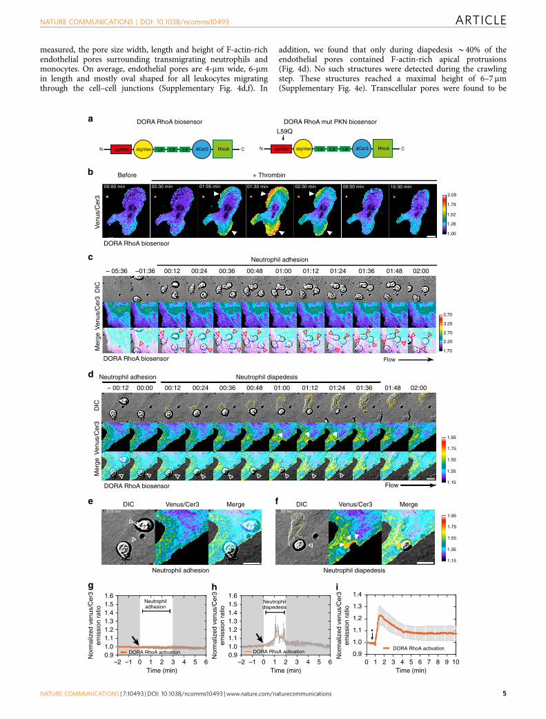
Spatiotemporal RhoA activation during leukocyte diapedesis.To examine spatiotemporal RhoA activation in ECs during ECbarrier maintenance associated with neutrophil TEM, we used arecently developed fluorescence resonance energy transfer(FRET)-based RhoA biosensors called the DimerizationOptimized Reporter for Activation (DORA) RhoA sensors(Fig. 2a)19. DORA RhoA biosensors design were based on thepublished RhoA biosensor20. The ON-state FRET efficiency of theGTPase was improved through modelling of the fluorescentprotein dimers and the GTPase-effector domain complexes.Stable a-helical repeats from ribosomal protein L9, rather than anunstructured linker, were inserted between the fluorescentproteins to disrupt dimerization and diminish FRET efficiencyin the inactive state (Fig. 2a). As a control, DORA RhoA mutantProtein kinase N (PKN) was developed to report misalignment ofCerulean3 (Cer3) and Venus image before and after imageregistration, motion artefacts or pH changes affecting the sensorsfluorescent proteins. Glutamine substitution for a leucine atposition 59 in the PKN domain prevents PKN binding toactivated RhoA21. The characterizations of both DORA RhoAbiosensors are described in online methods (SupplementaryFigs 2 and 3a). From these validation experiments, we concludethat the DORA RhoA biosensor accurately reports RhoAdynamics in ECs downstream from endogenous stimuli such asthrombin (Fig. 2b; Supplementary Fig. 1f).
To study spatiotemporal RhoA activation in ECs duringneutrophil TEM, we expressed the DORA RhoA biosensor in ECs
ARTICLE NATURE COMMUNICATIONS | DOI: 10.1038/ncomms10493
2 NATURE COMMUNICATIONS | 7:10493 | DOI: 10.1038/ncomms10493 | www.nature.com/naturecommunications

and investigated RhoA activation following neutrophil extravasa-tion under physiological flow conditions. Important to note,Venus and Cer3 emission were simultaneously recorded utilizinga double-camera system, since sequential image acquisitionresulted in motion artefacts induced by migrating leukocytesdisplacing fluorescent signals in ECs. We found unaltered RhoAbiosensor activation during neutrophil rolling and crawling overthe endothelium (Fig. 2c,g). Also RhoA activation during the
initial opening of EC junctions was found to be unaltered(Fig. 2d). However, RhoA biosensor activity in the endotheliumwas locally increased at sites were neutrophils breeched theendothelium, between the first and second minute of neutrophildiapedesis (Fig. 2d–f, Supplementary Fig. 2d; SupplementaryMovie 1). On the basis of the normalized ratiometric imaging andthe relative displacement of the sensor, the data showed a 1.2-foldincrease in FRET ratio on diapedesis, comparable to what
a
b c
d e
0
100
200
300
400
500
P= 0.16 NS
IL-1β/TNFα
0
0 20 40
200
400
600
P= 0.006**
IL-1β/TNFα + C3
Neutrophils per 5×104 μm20 20 40
Neutrophils per 5×104 μm2
IL-1
β/T
NF
αIL
-1β/
TN
Fα
+C
3
PECAM-1 TRITC-dextranPMN Merge
0 2 4 6 8 10 1214 1618 200
2
4
6
8
10
12
Time (min)0 2 4 6 8 10 1214 16 18 20
0.80.91.01.11.21.31.41.51.6
Time (min)
Tran
smig
rate
dne
utro
phils
(a.
u)
Dex
tran
leak
age
(a.u
)
siCTRL
siCTRL + PMN
siRhoA + PMN
siRhoA
siCTRL
siRho
A123456789
10111213
T = 20 min
Tra
nsm
igra
ted
neut
roph
ilsfo
ld in
crea
se
NS
siCTRL
siRho
A
siCTRL+
PMN
siRho
A+PM
N1.0
1.1
1.2
1.3
1.4
1.5
1.6
T = 20 min
FIT
C-d
extr
an 7
0 kD
ale
akag
e fo
ld in
crea
se
***
NSNS
0 5 10 15 200.8
1.0
1.2
1.4
1.6
1.8
2.0
T = 5–20min
Neutrophil diapedesisfold increase
FIT
C-d
extr
an 7
0 kD
ale
akag
e fo
ld in
crea
se
0 5 10 15 200.8
1.0
1.2
1.4
1.6
1.8
2.0
T = 5–20min
Neutrophil diapedesisfold increase
FIT
C-d
extr
an 7
0 kD
ale
akag
e fo
ld in
crea
se
f
siCTRL siRhoA
P= 0.004**
P= 0.35 NS
Ext
rava
scul
arT
RIT
C-d
extr
anflu
ores
cenc
e (a
.u.)
Ext
rava
scul
arT
RIT
C-d
extr
anflu
ores
cenc
e (a
.u.)
0
10
20
30
40
50
Neutrophilextravasation
C3
––
+–
++
–+
IL-1β/TNFα
Ext
rava
sate
d ne
utro
phils
(5×
104 μ
m2 )
NATURE COMMUNICATIONS | DOI: 10.1038/ncomms10493 ARTICLE
NATURE COMMUNICATIONS | 7:10493 | DOI: 10.1038/ncomms10493 | www.nature.com/naturecommunications 3

has been observed during RhoA activation after thrombinstimulation (Fig. 2h,i). The negative control DORA RhoAbiosensor (mutant PKN) showed no change in FRET duringleukocyte diapedesis (Supplementary Fig. 3b,c; SupplementaryMovie 2). Importantly, expressing the DORA RhoA biosensors inECs did not interfere with neutrophil TEM. Thus, endothelialRhoA is transiently and locally activated during the final stage ofneutrophil diapedesis, but not during crawling or opening ofendothelial junctions, indicating a role for local RhoA activity inEC barrier maintenance during the final stage of neutrophilextravasation.
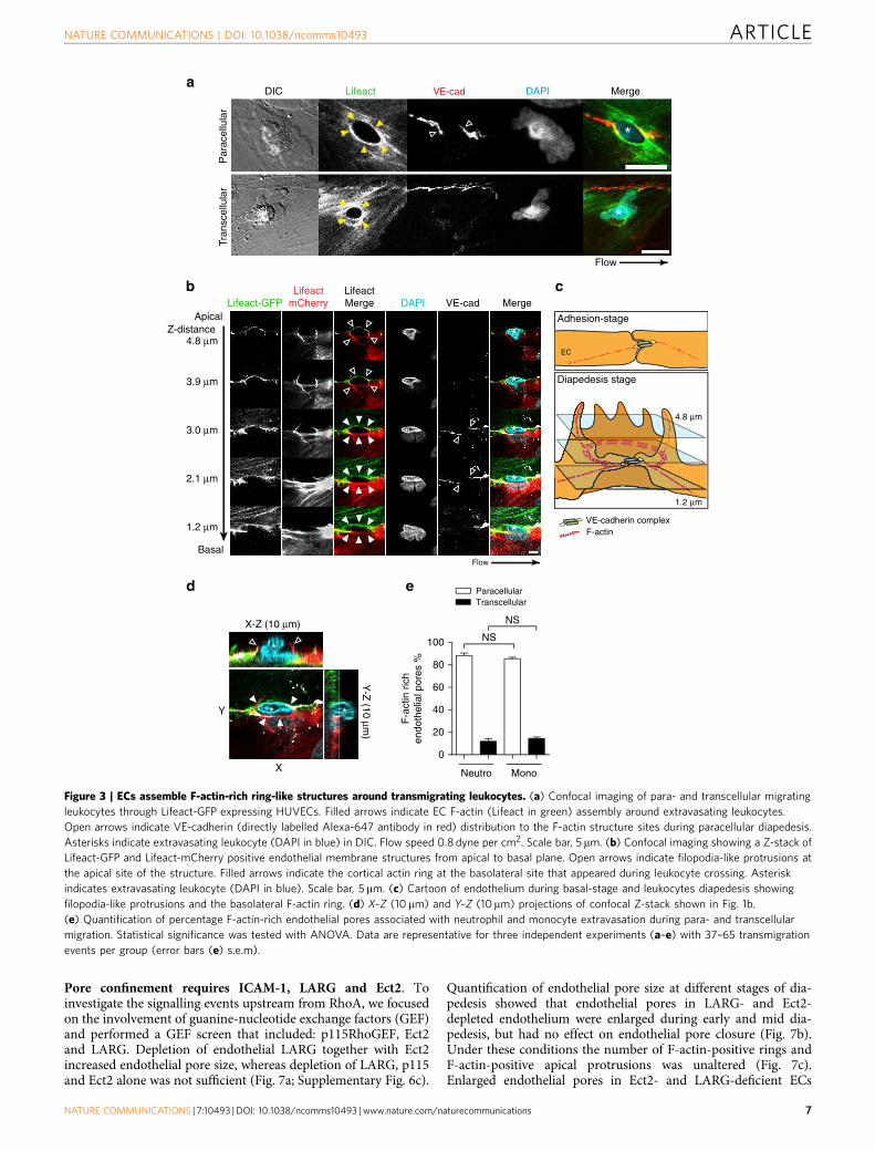
F-actin-rich endothelial pores during diapedesis. To investigateendothelial F-actin dynamics during neutrophil diapedesis, wetransfected ECs with GFP- and/or mCherry-tagged Lifeact22. It isimportant to note that phalloidin staining to visualize F-actincannot be used to investigate endothelial actin structures that are inclose proximity of transmigrating leukocytes, since F-actin in bothleukocytes and ECs are visualized by phalloidin staining, making itimpossible to discriminate between the two (SupplementaryFig. 3d). Transmigrating neutrophils initiated small endothelialpores in the endothelial lining. To study those endothelial pores athigh resolution, transmigrating neutrophils were fixed withformaldehyde when partly breeched the endothelium. Confocalmicroscopy imaging and three-dimensional (3D) reconstructionshowed that ECs assembled F-actin-rich structures aroundendothelial pores through, which neutrophils transmigrated, bothduring transcellular and paracellular migration (Fig. 3a;Supplementary Movies 3–5). During paracellular migration, thejunctional protein VE-cadherin was distributed to the endothelialpore margins (Fig. 3a). Interestingly, using ECs expressing eitherLifeact-GFP or Lifeact-mCherry, we found that paracellular poreswere formed by at least two ECs. At the structures apical site,filopodia-like protrusions were found, whereas at the basolateralsite, a cortical actin ring appeared during leukocyte crossing(Fig. 3b–d). In contrast to VE-cadherin distributed to the poresmargins, the junctional protein PECAM-1 was localized aroundthe basolateral F-actin ring and distributed to apical protrusionssurrounding migrating leukocytes during trans-and paracellularmigration. (Supplementary Fig. 3e,f). Moreover, we found that theadhesion molecule ICAM-1 was localized in the apical protrusionsat endothelial pores (Supplementary Fig. 4a). We found thatB90% of all neutrophils and monocytes used the paracellularroute, whereas B10% migrated transcellular, in line with themigratory preference for neutrophils and monocytes foundin vivo23 (Fig. 3e). Note that all these diapedesis events by eitherneutrophils or monocytes were associated with basolateral F-actin
ring formation around endothelial pores (Fig. 3e). Within theparacellular route of migration, leukocyte transmigration through abi-cellular or a multicellular junction was B50% (SupplementaryFig. 4b). In conclusion, ECs assemble F-actin-rich ring-likestructures around endothelial pores through which neutrophilsand monocytes transmigrate. This data indicate that maintenanceof EC barrier function during leukocyte diapedesis involves actincytoskeleton strengthening around endothelial pores. BasolateralF-actin ring formation may tighten the endothelial barrier duringneutrophil crossing, making the leukocyte-induced endothelialpore impermeable for macromolecules.
F-actin-rich endothelial pores are confined in size. Electronmicroscopy studies showed that ECs maintain intimatecontact with transmigrating neutrophils during the entiretransmigration process15,24. To examine the dynamiccontact between ECs and extravasating neutrophils, weexamined F-actin-enriched endothelial pore shape and size inrelation to neutrophil size. Real-time recordings of transmigratingneutrophils through ECs expressing GFP-tagged Lifeactshowed increased F-actin assembly around endothelial pores(Supplementary Movie 6).
The kinetics of neutrophil diapedesis is on an average 2 minand can be distinguished into early, mid and late diapedesis basedon endothelial pore size and neutrophil morphology (Fig. 4a–c).Endothelial pore formation started when neutrophils partlybreeched the endothelium, defined as early diapedesis. Followingneutrophil diapedesis, most endothelial pores are maximalenlarged 1 min after transmigration was initiated, defined asmid diapedesis (Fig. 4a–c). Subsequently, the endothelial pore isclosed in conjunction with transmigrating neutrophils untilcompletely under the endothelium, a stage defined as latediapedesis (Fig. 4a–c). Real-time imaging of neutrophil diapedesisunder physiological flow conditions showed that neutrophil totalsurface area before TEM was roughly 100 mm2, which wasreduced to o20 mm2 to fit the confined gap in the endotheliumthat had a maximal inner-surface area of 19 mm2 (Fig. 4b,c). Toinvestigate the morphology of de novo formed F-actin-positiverings and F-actin-positive apical protrusions that surroundendothelial pores during neutrophil TEM, we trapped neutrophilsat different stages of diapedesis. Interestingly, de novo formedF-actin-positive rings surrounding endothelial pores were foundthroughout all diapedesis steps, but not during neutrophiladhesion or crawling steps (Fig. 4d; Supplementary Fig. 4c).Quantification of endothelial pore size showed significant largerpores during mid diapedesis than during early and late diapedesiswhen pores open and close, respectively (Fig. 4d). We next
Figure 1 | Impaired endothelial RhoA function results in increased vascular leakage during leukocyte diapedesis in vivo. (a) Extravasation kinetics of
calcein-red-labelled neutrophils and FITC–dextran through TNF-a treated ECs cultured on 3-mm pore permeable filtres. Neutrophils transmigrated towards
a C5a chemotactic gradient in the lower compartment. Four conditions were tested; RhoA depletion (EC)þ neutrophils (Orange line), controlþ neutrophils
(purple line), RhoA depletion (EC) only (red line) and control only (green line). (b) Quantification of FITC–dextran and neutrophil extravasation after
20 min of neutrophil transmigration. Immunoblot of RhoA silencing can be found in supplementary fig. 8. (c) Correlation analysis of dextran and neutrophil
extravasation kinetics through control and RhoA-depleted ECs. (d) Confocal intravital microscopy of 20–80mm diameter cremasteric
venulesin LysM–GFP mice (green neutrophils) immunostained in vivo for EC junctions by intrascrotal injections of fluorescent-labelled PECAM-1 (blue) and
stimulated for four hours with IL-1b and TNF-a only, or with Rho-inhibitor (C3). A second dose of Rho inhibitor was given intrascrotally and TRITC–dextran
(40 kDa) was injected intravenously at T¼ 2 h and allowed to circulate until T¼4 h. Scale bar, 100 mm. (e) Neutrophil extravasation in animals
left unstimulated (control), stimulated with C3 alone, IL-1b/TNFa treated, IL-1b/TNFa treatedþC3 or IL-1b/TNFa treatedþ neutrophil depletion.
(f) Correlation analysis of dextran and neutrophil extravasation kinetics in animals stimulated with IL-1b/TNFa alone or with IL-1b/TNFa treatedþC3.
***Po0.001 control versus RhoA-depleted HUVEC (ANOVA) or P¼0.3504 control versus RhoA-depleted HUVEC (Student’s t-test) (b). r¼0.2547
P¼0.359 (Pearson’s correlation) transmigrated neutrophils versus FITC–dextran leakage in control HUVECs or r¼0.6345 **Po0.01 (Pearson correlation)
transmigrated neutrophils versus FITC–dextran leakage in RhoA-depleted HUVECs (c). P¼0.4230 IL-1b/TNFa versus IL-1b/TNFaþC3 (Student’s t-test)
(e). r¼0.8258 **Po0.01 (Pearson’s correlation) transmigrated neutrophils versus FITC–dextran leakage in IL-1b/TNFaþRho inhibitor treated mice (f).
Data are from three experiments (a–c) or are representative of 5 to 13 (d–e) or 9 (f) independent experiments ((d–f) one mouse per experiment; error bars
(a–c,e,f), s.e.m).
ARTICLE NATURE COMMUNICATIONS | DOI: 10.1038/ncomms10493
4 NATURE COMMUNICATIONS | 7:10493 | DOI: 10.1038/ncomms10493 | www.nature.com/naturecommunications
measured, the pore size width, length and height of F-actin-richendothelial pores surrounding transmigrating neutrophils andmonocytes. On average, endothelial pores are 4-mm wide, 6-mmin length and mostly oval shaped for all leukocytes migratingthrough the cell–cell junctions (Supplementary Fig. 4d,f). In
addition, we found that only during diapedesis B40% of theendothelial pores contained F-actin-rich apical protrusions(Fig. 4d). No such structures were detected during the crawlingstep. These structures reached a maximal height of 6–7 mm(Supplementary Fig. 4e). Transcellular pores were found to be
00:1200:00 00:24 00:36 00:48 01:00 01:12 01:24 01:36 01:48 02:00
DIC
Ven
us/C
er3
Mer
ge
– 00:12
d
DIC Venus/Cer3 Merge
Neutrophil diapedesis
00:12–01:36 00:24 00:36 00:48 01:00 01:12 01:24 01:36 01:48 02:00– 05:36
f
g
–2 –1 0 1 2 3 4 5 60.91.01.11.21.31.41.51.6
Time (min)
Nor
mal
ized
ven
us/C
er3
emis
sion
rat
io
Nor
mal
ized
ven
us/C
er3
emis
sion
rat
io
Nor
mal
ized
ven
us/C
er3
emis
sion
rat
io
DORA RhoA activation
Neutrophiladhesion
–2 –1 0 1 2 3 4 5 60.91.01.11.21.31.41.51.6
Time (min)
Neutrophildiapedesis
DIC
Ven
us/C
er3
Mer
ge
c
e DIC Venus/Cer3 Merge
Neutrophil adhesion
Neutrophil adhesion
Neutrophil diapedesisNeutrophil adhesion
DORA RhoA biosensor
DORA RhoA biosensor
a
cpPKN RhoAN dcpVen dCer3 C cpPKN RhoAN dcpVen dCer3 C
L59Q
DORA RhoA biosensor DORA RhoA mut PKN biosensor
L9 L9 L9L9 L9 L9
b
Flow
Flow
Ven
us/C
er3
Before
00:00 min
–00:12 min 01:12 min
00:30 min 01:05 min 01:30 min 02:30 min 06:00 min 10:30 min
+ Thrombin
DORA RhoA biosensor
0 1 2 3 4 5 6 7 8 9 100.9
1.0
1.1
1.2
1.3
1.4
DORA RhoA activation
Time (min)
*
ih
2.05
1.79
1.52
1.26
1.00
3.70
3.20
2.70
2.20
1.70
1.95
1.75
1.55
1.35
1.15
1.95
1.75
1.55
1.35
1.15
DORA RhoA activation
NATURE COMMUNICATIONS | DOI: 10.1038/ncomms10493 ARTICLE
NATURE COMMUNICATIONS | 7:10493 | DOI: 10.1038/ncomms10493 | www.nature.com/naturecommunications 5

more round or circular shaped and had an average circularity ofabout 1.3 according to the circularity index (SupplementaryFig. 4f). Endothelial pore sizes showed remarkably little variation,despite leukocyte size, type or transmigration route (Fig. 4e).Thus, endothelial pores induced by extravasating neutrophils andmonocytes are confined in size and close directly behindtransmigrated cells. Active endothelial pore confinement andpore closure corroborated earlier findings that showed intimatecontact between neutrophils and ECs during the entire TEMprocess and provides an explanation for limited transendothelialescape of macromolecules during neutrophil crossing.
Pore confinement and pore closure requires endothelial RhoA.Our data showed that increased endothelial RhoA activity duringneutrophil TEM corresponded to endothelial pore restriction andclosure during mid and late diapedesis. To investigate whetherRhoA regulates endothelial pore confinement, we silencedendothelial RhoA using siRNA. RhoA was successfully depletedas shown by western blot analysis (Supplementary Fig. 5a).Confocal microscopy showed that RhoA depletion in ECsreduced Lifeact-GFP accumulation around endothelial pores,whereas Lifeact-GFP in the apical protrusions was still present(Fig. 5a). Basal F-actin rings in RhoA-depleted ECs were sig-nificantly reduced compared to control conditions (Fig. 5b).Endothelial RhoA depletion had no effect on the formation ofF-actin-rich apical protrusions (Fig. 5b). Quantification ofendothelial pore size showed that in the absence of RhoA,endothelial pores were not only larger than endothelial poresformed in control ECs but also did not close properly (Fig. 5c).Note that neutrophil adhesion and transmigration under phy-siological flow conditions were unaltered in RhoA-depleted ECs(Fig. 5d). To study if VE-cadherin signalling regulates endothelialpore size, we depleted VE-cadherin and analysed endothelial poresize. However, VE-cadherin depletion had no effect onendothelial pore size (Fig. 5e; Supplementary Fig. 5b–d). Inconclusion, RhoA facilitates endothelial pore confinement andpore closure during leukocyte diapedesis.
Pore confinement is driven by actomyosin contractility. Toinvestigate how RhoA regulates endothelial pore confinementduring leukocyte diapedesis, we examined RhoA effector myosinII activation. To study myosin II activation we locally measuredmyosin light-chain (MLC) phosphorylation on position Thr18and Ser19. Immunofluorescent staining of pMLC showed anasymmetric phosphorylation pattern in F-actin-rich endothelialpores surrounding transmigrating neutrophils (Fig. 6a). MLC was
particularly phosphorylated at cortical actin bundles as part of theF-actin ring (Fig. 6a; Supplementary Fig. 5e). Note that theuropod of the neutrophil is positive for MLC phosphorylation,most likely to retract its tail during transmigration25. In contrastto local MLC phosphorylation in control ECs, endothelial poresin RhoA-deficient ECs were enlarged and negative for localphospho-MLC (Supplementary Fig. 5e). In addition, wequantified Lifeact-GFP distribution around endothelial poresand found asymmetric F-actin distribution around endothelialpores, indicative of increased tension (Fig. 6b). To corroborateour findings in vivo, we studied F-actin localization duringleukocyte diapedesis in retinal vasculature of Lifeact-EGFP-transgenic knock-in mice26. Lifeact-EGFP expression in theretinas of these mice is largely restricted to the endothelium andthis allowed us to properly visualize F-actin in ECs in situ26,27.We found that endothelial pores induced by transmigratingneutrophils (isolectin B4-positive28) were surrounded by Lifeact-EGFP-positive rings in retinal ECs (Fig. 6c). Quantification ofthese rings showed that endothelial pore size in vivo wascomparable to endothelial pores measured in the in vitro set-up(compare Figs 6d and 4e). Lifeact was present in the basolateralring and in apical protrusions that surrounded transmigratingneutrophils (Supplementary Fig. 5f). These data showed thatapical membrane protrusions in vivo are rich for F-actin andsurround adherent leukocytes. Next, we examined local MLCphosphorylation in WT mice during IL-1b and TNF-a-inducedneutrophil recruitment in cremasteric venules. PECAM-1 wasused as a marker to visualize endothelial pores in vivo23 (Fig. 6e).In line with our in vitro findings, endothelial pores in mousecremaster venules showed asymmetric MLC phosphorylation(Fig. 6f,g). To address the role of ROCK in endothelial poreconfinement we depleted the ROCK isoforms ROCK1 andROCK2b in endothelial cells and examined vascularpermeability during neutrophil diapedesis. In line with RhoAinhibition, silencing ROCK 1 and ROCK2b did not prevent theadhesion or transmigration of neutrophils through theendothelial monolayer (Supplementary Fig. 6a,b). Basalendothelial barrier function in ROCK1 or ROCK2b deficientECs was not affected. However, neutrophil diapedesis throughROCK2b, but not ROCK1 deficient ECs elicited increasedendothelial permeability up to a twofold (SupplementaryFig. 6a,b). These findings may indicate that endothelial poreconfinement is mediated through ROCK2b but not ROCK1.Altogether, local accumulation of F-actin and MLCphosphorylation is associated with neutrophil diapedesisin vitro and in vivo, suggesting that endothelial poreconfinement is driven by local actomyosin contractility.
Figure 2 | Spatiotemporal RhoA activation during neutrophil TEM. Endothelial RhoA is locally and transiently activated during neutrophil extravasation
(a) Schematic illustration of the DORA RhoA sensor design containing Rho effector PKN (red), circular permutated Venus (yellow), structured linker
protein L9 (green), circular permutated Cer3 (blue) and RhoA GTPase (green),left panel. Right panel shows the DORA RhoA mutant PKN biosensor that
was developed as a negative control biosensor, the glutamine was substituted for the leucine at position 59 in the PKN domain. This mutation prevents
binding of PKN to activated RhoA. (b) Time-lapse Venus/Cer3 ratio images of DORA RhoA biosensor simultaneously recorded with an epi-fluorescent
microscope showing spatiotemporal RhoA activation upon thrombin treatment (1 U ml� 1) in HUVECs. Filled arrows indicate RhoA activation. Scale bar,
10mm. Calibration bar shows RhoA activation (Red) relative to basal RhoA activity (Blue). (c) Epi-fluorescent live-cell imaging of HUVEC expressing the
DORA RhoA biosensor during neutrophil adhesion under physiological flow conditions (0.8 dyne per cm2). Red open arrows indicate adherent neutrophils.
Scale bar, 10mm. Calibration bar shows RhoA activation (red) relative to basal RhoA activity (blue). (d) Epi-fluorescent live-cell imaging of HUVECs
expressing the DORA RhoA biosensor during neutrophil TEM. Time-lapse images of DIC (upper) Venus/Cer3 ratio images of DORA RhoA biosensor
(middle) and Merge (bottom) during leukocyte diapedesis. Open arrows indicates adherent neutrophil at the apical side of the endothelium. Filled arrows
indicate local RhoA activation during neutrophil diapedesis. Scale bar, 10mm. (e) Detailed zoom of RhoA activation during neutrophil adhesion (open
arrows) prior diapedesis at time point t¼ �00:12 min. (f) Detailed zoom of local RhoA activation during neutrophil transmigration at time point
t¼01:12 min. Filled arrows indicate local RhoA activation during neutrophil diapedesis. Scale bar, 10 mm. (g) Quantification of temporal RhoA activation
during multiple neutrophil transmigration events starting at time zero (arrow). (h) Quantification of temporal RhoA activation during multiple neutrophil
adhesion events starting at time zero (arrow). (i) Quantification of DORA RhoA biosensor activation after thrombin treatment (1 U ml� 1) in HUVEC.
Asterisk indicates thrombin addition. Data represent mean and s.e.m of 7 experiments (g) 5 experiments and (h) 10 experiments (i).
ARTICLE NATURE COMMUNICATIONS | DOI: 10.1038/ncomms10493
6 NATURE COMMUNICATIONS | 7:10493 | DOI: 10.1038/ncomms10493 | www.nature.com/naturecommunications
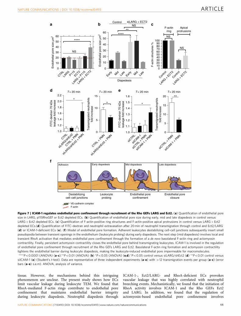
Pore confinement requires ICAM-1, LARG and Ect2. Toinvestigate the signalling events upstream from RhoA, we focusedon the involvement of guanine-nucleotide exchange factors (GEF)and performed a GEF screen that included: p115RhoGEF, Ect2and LARG. Depletion of endothelial LARG together with Ect2increased endothelial pore size, whereas depletion of LARG, p115and Ect2 alone was not sufficient (Fig. 7a; Supplementary Fig. 6c).
Quantification of endothelial pore size at different stages of dia-pedesis showed that endothelial pores in LARG- and Ect2-depleted endothelium were enlarged during early and mid dia-pedesis, but had no effect on endothelial pore closure (Fig. 7b).Under these conditions the number of F-actin-positive rings andF-actin-positive apical protrusions was unaltered (Fig. 7c).Enlarged endothelial pores in Ect2- and LARG-deficient ECs
LifeactDIC VE-cad MergeDAPI
Par
acel
lula
rTr
ansc
ellu
lar
a
bMergeVE-cadDAPI
LifeactMergeLifeact-GFP
LifeactmCherry
Z-distance4.8 μm
3.9 μm
3.0 μm
2.1 μm
1.2 μm
4.8 μm
1.2 μm
Adhesion-stage
Diapedesis stage
EC
VE-cadherin complexF-actin
X-Z (10 μm)
Y-Z
(10 μm)
Y
X
e
c
d
Basal
Apical
*
*
*
*
*
*
*
* *
*
**
Flow
Flow
ParacellularTranscellular
0
20
40
60
80
100
F-a
ctin
ric
hen
doth
elia
l por
es %
Neutro Mono
NS
NS
Figure 3 | ECs assemble F-actin-rich ring-like structures around transmigrating leukocytes. (a) Confocal imaging of para- and transcellular migrating
leukocytes through Lifeact-GFP expressing HUVECs. Filled arrows indicate EC F-actin (Lifeact in green) assembly around extravasating leukocytes.
Open arrows indicate VE-cadherin (directly labelled Alexa-647 antibody in red) distribution to the F-actin structure sites during paracellular diapedesis.
Asterisks indicate extravasating leukocyte (DAPI in blue) in DIC. Flow speed 0.8 dyne per cm2. Scale bar, 5 mm. (b) Confocal imaging showing a Z-stack of
Lifeact-GFP and Lifeact-mCherry positive endothelial membrane structures from apical to basal plane. Open arrows indicate filopodia-like protrusions at
the apical site of the structure. Filled arrows indicate the cortical actin ring at the basolateral site that appeared during leukocyte crossing. Asterisk
indicates extravasating leukocyte (DAPI in blue). Scale bar, 5 mm. (c) Cartoon of endothelium during basal-stage and leukocytes diapedesis showing
filopodia-like protrusions and the basolateral F-actin ring. (d) X–Z (10 mm) and Y–Z (10mm) projections of confocal Z-stack shown in Fig. 1b.
(e) Quantification of percentage F-actin-rich endothelial pores associated with neutrophil and monocyte extravasation during para- and transcellular
migration. Statistical significance was tested with ANOVA. Data are representative for three independent experiments (a–e) with 37–65 transmigration
events per group (error bars (e) s.e.m).
NATURE COMMUNICATIONS | DOI: 10.1038/ncomms10493 ARTICLE
NATURE COMMUNICATIONS | 7:10493 | DOI: 10.1038/ncomms10493 | www.nature.com/naturecommunications 7

c
DIC
Life
act-
GF
P
00:2000:00 00:40 01:00 01:20 01:40 02:00 02:20– 00:20 02:40
b
a
–1 0 1 2 3–5
0
5
10
15
20
Endothelial poreNeutrophil cell body (apical)
Time (min)–1 0 1 2 3
–100
–50
0
50
100
150
200
250
Endothelial pore
Neutrophil cell body (apical)
Neutrophil cell body (under EC)
Time (min)
Are
a μm
2A
rea
μm2
Are
a μm
2
Are
a μm
2
Are
a μm
2
Neutrophil diapedesisNeutrophil diapedesisFlow
e
Endothelialpore size
F-actin rich pore
Cell-cell junction
d
Endothelial pore size
0
10
20
30
40
50
****
**** ****
NS
Adhesion Early diapedesis Mid diapedesis Late diapedesis
0102030405060708090
100
F-a
ctin
str
uctu
res
%
F-actinring
ApicalF-actin
protrusions
****
****
NS NS
NS NS
Adher
ing
Early
diape
desis
Early
diape
desis
Mid
diape
desis
Mid
diape
desis
Late
diap
edes
is
Late
diap
edes
is
Adher
ing
Adher
ing
Early
diape
desis
Early
diape
desis
Mid
diape
desis
Mid
diape
desis
Late
diap
edes
is
Late
diap
edes
is
Adher
ing
Early
diape
desis
Mid
diape
desis
Late
diap
edes
is
Adher
ing0
25
50
75
100
125
Neutrophilcell body apical
Neutrophilcell bodyunder EC
NS****
********
********
NS
NeutroPara
MonoPara
0
10
20
30
40
50
60Endothelial pore size
Figure 4 | Endothelial pores formed during para- and transcellular leukocyte transmigration are confined in size. (a) Epi-fluorescent live-cell imaging of
ECs expressing Lifeact-GFP. Red open arrows indicate F-actin-rich endothelial pore formation during leukocyte diapedesis under physiological flow
conditions (0.8 dyne per cm2). Filled arrows indicate extravasating leukocyte in DIC. Dashed lines indicates neutrophil localization under the endothelium.
Scale bar, 10mm. (b,c) Quantification of size changes occurring in the neutrophil cell body and endothelial pore during neutrophil diapedesis. Endothelial
pore size (red), neutrophil cell body apical (blue) and neutrophil cell body under EC (yellow), diapedesis starts at time zero. (d) Quantification of Neutrophil
size, F-actin-positive ring structures, F-actin positive apical protrusions and endothelial pore size. (e) Quantification of endothelial pore size for neutrophils
and monocytes during paracellular migration. ****Po0.0001 (analysis of variance). Data are representative of four independent experiments (d,e) with 40
transmigration events per group. Data in b and c are representative of 10 transmigration events (error bars (b–e) s.e.m).
ARTICLE NATURE COMMUNICATIONS | DOI: 10.1038/ncomms10493
8 NATURE COMMUNICATIONS | 7:10493 | DOI: 10.1038/ncomms10493 | www.nature.com/naturecommunications

showed increased endothelial permeability during neutrophildiapedesis, whereas basal EC barrier function was not affected(Fig. 7d; Supplementary Fig. 6d–f). Neutrophil diapedesis throughEct2- and LARG-deficient ECs was slightly reduced (Fig. 7d;Supplementary Fig. 6d). To study LARG and Ect2 recruitment tothe intracellular tail of PECAM-1 or ICAM-1 we performedclustering experiments induced by anti-ICAM-1- or anti-PECAM-1-coated beads. We found that LARG and Ect2 arerecruited to the intracellular tail of ICAM-1 (SupplementaryFig. 6g).Whereas PECAM-1 recruited only LARG, but not Ect2 toits intracellular tail on clustering (Supplementary Fig. 6h). Toinvestigate whether ICAM-1 or PECAM-1 initiate and coordinatelocal RhoA activation and endothelial pore confinement duringneutrophil diapedesis, we depleted ICAM-1 and PECAM-1 inECs and examined the extravasation of calcein-red-labelledneutrophils and FITC–dextran across EC and measured endo-thelial pore size. We found that neutrophil transmigration
through ICAM-1-deficient ECs compromised the endothelialbarrier (Fig. 7e; Supplementary Fig. 7a–c), whereas PECAM-1depletion did not alter endothelial pore size or vascular leakage(Supplementary Fig. 7d–e). ICAM-1 and PECAM-1 depletionalone had no effect on endothelial permeability (Fig. 7e;Supplementary Fig. 7d). In agreement with the existing literature,endothelial ICAM-1 depletion significantly reduced the numberof transmigrated neutrophils (Fig. 7e). Neutrophil diapedesisthrough PECAM-1-deficient ECs showed no reduction in trans-migration numbers (Supplementary Fig. 7d). These data point outan important role for ICAM-1, Ect2 and LARG signalling incontrolling RhoA-mediated endothelial pore confinement and ECbarrier protection during neutrophil diapedesis.
DiscussionLeukocytes that cross the endothelium induce large endothelialgaps without provoking leakage of plasma into the underlying
a b
d
c
DIC Lifeact VEcadherin DAPI Merge
DIC Lifeact VEcadherin DAPI Merge
Bas
alA
pica
lB
asal
Api
cal
HUVEC treated with control siRNA
Endogenous RhoA depletion in HUVEC
**
*
*
**
*
Flow
Flow
e
siCTRL
siRho
A
siCTRL
siRho
A0
102030405060708090
100
F-a
ctin
str
uctu
res
%
F-actinring
Apicalprotrusions
NS
****
Early
diape
desis
Mid
diape
desis
Late
diap
edes
is
Early
diape
desis
Mid
diape
desis
Late
diap
edes
is
010203040506070
Are
a μm
2
****
siRhoAsiCTRL
siCTRL
siRho
A0
20
40
60
80
100
Tra
nsm
igra
ted
cells
% NS
siCTRL
siRho
A
0
20
40
60
80
100
Adh
eren
t cel
ls #
NS
shCTRL
shVE-c
adhe
rin0
10
20
30
40
50
60
Endothelialpore size
Are
a μm
2
NS
Figure 5 | RhoA signalling is required for endothelial pore confinement. (a) Confocal imaging of paracellular migrating neutrophils through Lifeact-GFP
expressing HUVECs after 72-h transfection with control siRNA (upper panel) or RhoA siRNA (lower panel) under physiological flow conditions (0.8 dyne
per cm2). Open arrows and filled arrows indicate filopodia-like protrusions at the apical site and the cortical F-actin ring at the basolateral site of the
endothelial pore, respectively. Asterisk indicates extravasating neutrophil (DAPI in blue). VE-cadherin (red). Scale bar, 5 mm. (b) Quantification of F-actin-
positive ring structures and F-actin-positive apical protrusions in control versus RhoA-depleted ECs. (c) Quantification of endothelial pore size during early,
mid and late diapedesis. (d) Quantification of neutrophil adhesion and diapedesis through TNF-a treated ECs under physiological flow conditions after 72 h
transfection with control siRNA (open bar) or RhoA siRNA (filled bar). (e) Quantification of endothelial pore size in control versus VE-cadherin depleted
HUVECs. ****Po0.0001 (analysis of variance). Data are representative of four independent experiments (a–e) with 412 transmigration events per group
(error bars (c,e) s.e.m).
NATURE COMMUNICATIONS | DOI: 10.1038/ncomms10493 ARTICLE
NATURE COMMUNICATIONS | 7:10493 | DOI: 10.1038/ncomms10493 | www.nature.com/naturecommunications 9

LifeactDIC pMLC Merge
Distributionasymmetry
=ROI Intensity – BG
ROI Intensity – BG
F-actin in pore
Sum intensity Z-slices
Cell–cell junction
* *
Flow
Neutro Mono1.0
1.5
2.0
2.5
Dis
trib
utio
n as
ymm
etry
Dis
trib
utio
n as
ymm
etry
NS
PECAM-1pMLC Merge
Cremaster venules
PE
CA
M-1
Neu
trop
hils
Mer
ge
Ret
inal
vas
cula
ture
of li
feac
t-E
GF
P m
icet
Endothelialpore size
pMLC PECAM-10
2
4
6
8Cremasteric venules
Area0
10
20
30
40
50
Are
a μm
2
Isolectin B4Lifeact Merge
Bas
alA
pica
lROI
Isolectin B4Lifeact Merge
ROI
Bas
al
a
b
c
f g
d e
Figure 6 | Endothelial pore confinement is driven by actomyosin contractility. (a) Immunofluorescence analyses of MLC phosphorylation during
neutrophil transmigration. Open and filled arrows indicate Lifeact-mCherry (red) and MLC phosphorylation (green) localization, respectively, during
neutrophil transmigration under physiological flow conditions (0.8 dyne per cm2). Asterisk indicates extravasating leukocyte in DIC. Scale bar, 10mm.
(b) Quantification of F-actin distribution in endothelial pores surrounding transmigrating neutrophils and monocytes. Maximum intensity projection of
F-actin in the endothelial pore was used to quantify F-actin distribution surrounding transmigrating leukocytes. Distribution asymmetry is defined by the
ratio of region of interest ROI-1 and ROI-2 corrected for background. Scale bar, 5 mm. (c) Confocal imaging of F-actin dynamics during leukocyte diapedesis
in retina vasculature of Lifeact-EGFP C57BL6 mice. Filled arrows indicate the vasculature of mice retina, highly expressing Lifeact-GFP. Zoom of ROI, open
arrows indicate the Lifeact-EGFP (green)-positive endothelial pore, filled arrows indicate transmigrating neutrophil. Scale bar, 5 mm. (d) Quantification of
endothelial pore size in retina vasculature. (e) Confocal imaging of PECAM-1 in cremasteric venules during TNF-a and IL-1b induced neutrophil recruitment.
Open arrows indicate PECAM-1 positive endothelial pores that surround extravasating neutrophils (filled arrows). Scale bar, 20 mm. (f) IF analyses of MLC
phosphorylation during neutrophil transmigration into the cremaster of C57BL6 mice. Filled and open arrows indicate phospo-MLC and PECAM-1
localization to endothelial pores, respectively. Scale bar, 5 mm. (g) Quantification of pMLC and PECAM-1 localization in endothelial pores. We quantified
MLC phosphorylation defined as distribution asymmetry. The distribution asymmetry uses the intensity of one ROI versus another ROI as indicated in b.
Because MLC may occur at different heights within the pore we used max projection for this analysis. Data are representative of three independent
experiments (a–g) with 412 transmigration events per group (error bars (b,d,g) s.e.m).
ARTICLE NATURE COMMUNICATIONS | DOI: 10.1038/ncomms10493
10 NATURE COMMUNICATIONS | 7:10493 | DOI: 10.1038/ncomms10493 | www.nature.com/naturecommunications
tissue. However, the mechanisms behind this intriguingphenomenon are unclear. The present study shows how ECslimit vascular leakage during leukocyte TEM. We found thatRhoA-mediated F-actin rings contribute to endothelial poreconfinement that maintains endothelial barrier integrityduring leukocyte diapedesis. Neutrophil diapedesis through
ICAM-1-, Ect2/LARG- and RhoA-deficient ECs provokesvascular leakage that was highly correlated with neutrophilbreeching events. Mechanistically, we found that the initiation ofRhoA activity involves ICAM-1 and the Rho GEFs Ect2and LARG. In addition, we found that the regulation ofactomyosin-based endothelial pore confinement involves
CTRL
LARG
P115ECT2
LARG +
P11
5
LARG +
ECT2
LARG +
ECT2
LARG +
ECT2
0
10
20
30
40
50
60
End
othe
lial p
ore
size
μm
2
End
othe
lial p
ore
size
μm
2
****
NS
Contro
l
Contro
l0
102030405060708090
100
F-a
ctin
str
uctu
res
%
F-actinring
Apicalprotrusions
NS
NS
Early
Mid
Late
Early
Mid
Late
0
10
20
30
40
50
60****
**NS
Diapedesis
Control siLARG + ECT2a cb
fAdhesion Early diapedesis
EC
VE-cadherin complexF-actin
Mid diapedesis Late diapedesis
Destabilizingcell–cell junctions
Endothelial poreconfinement
Endothelial poreclosure
Leukocyteprobing
0
5
10
15
Tra
nsm
igra
ted
neut
roph
ilsfo
ld in
crea
se
Tra
nsm
igra
ted
neut
roph
ilsfo
ld in
crea
se
T= 20 min
*
CTRL
CTRL +
PMN
siLARG/sh
Ect2 +
PM
N
CTRL +
PMN
siLARG/sh
Ect2 +
PM
N
siLARG/sh
Ect21.0
1.2
1.4
1.6
1.8
2.0
2.2
FIT
C-d
extr
an 7
0 kD
ale
akag
e fo
ld in
crea
se
FIT
C-d
extr
an 7
0 kD
ale
akag
e fo
ld in
crea
se
T= 20 min
*
d
siCTRL
siICAM
-1
siCTRL
+ PM
N
siCTRL
+ PM
N
siICAM
-1 +
PM
N
siICAM
-1 +
PM
N
1.0
1.1
1.2
1.3
1.4
1.5
1.6
T= 20 min
*
0
5
10
15
20
T= 20 min
**
e
Figure 7 | ICAM-1 regulates endothelial pore confinement through recruitment of the Rho GEFs LARG and Ect2. (a) Quantification of endothelial pore
size in LARG, p115RhoGEF or Ect2 depleted ECs. (b) Quantification of endothelial pore size during early, mid and late diapedesis in control versus
LARGþ Ect2 depleted ECs. (c) Quantification of F-actin-positive ring structures and F-actin-positive apical protrusions in control versus LARGþ Ect2
depleted ECs.(d) Quantification of FITC–dextran and neutrophil extravasation after 20 min of neutrophil transmigration through control and Ect2/LARG
(d) or ICAM-1-deficient ECs (e). (f) Model of endothelial pore formation. Adherent leukocytes destabilizing cell–cell junctions subsequently insert small
pseudopodia between transient openings in the endothelium (leukocyte probing) during early diapedesis. The next step (mid diapedesis) involves local and
transient RhoA activation that mediates endothelial pore confinement through the formation of a de novo basolateral F-actin ring and actomyosin
contractility. Finally, persistent actomyosin contractility closes the endothelial pore behind transmigrating leukocytes. ICAM-1 is involved in the regulation
of endothelial pore confinement through recruitment of the Rho GEFs LARG and Ect2. Basolateral F-actin ring formation and actomyosin contractility
tightens the endothelial barrier during leukocyte diapedesis, making the leukocyte-induced endothelial pore impermeable for macromolecules.
****Po0.0001 (ANOVA) (a–c).**Po0.01 (ANOVA) (b) *Po0.05 (ANOVA) (e,d).*Po0.05 control versus siLARG/shEct2 (d) **Po0.01 control versus
siICAM-1 (e) (Student’s t-test). Data are representative of three independent experiments (a–e) with 412 transmigration events per group (a–c) (error
bars (a–e) s.e.m). ANOVA, analysis of variance.
NATURE COMMUNICATIONS | DOI: 10.1038/ncomms10493 ARTICLE
NATURE COMMUNICATIONS | 7:10493 | DOI: 10.1038/ncomms10493 | www.nature.com/naturecommunications 11
ROCK2b, but not ROCK1. Our work identifies a novelmechanism that maintains endothelial barrier integrity duringleukocyte extravasation, which is driven by a basolateralactomyosin-based structure that requires spatiotemporal RhoAcycling (Fig. 7f).
Inflammation-driven leukocyte recruitment and vascularpermeability are separable events7,8,17. In line with theseobservations, we discovered that during the TEM processendothelial RhoA plays a central role in EC barriermaintenance, but is redundant for leukocyte transmigration. Inagreement with previous reports, blocking RhoA activity ordepleting RhoA in ECs did not perturb adhesion29 ortransmigration30. In contrast to the general concept that RhoAactivation is required for leukocyte adhesion and opening ofendothelial junctions31–35, we found that endothelial RhoAwas locally and transiently activated during the diapedesisstep and not during neutrophil crawling, firm adhesion oropening of endothelial junctions prior to leukocyte extravasation.These processes require a separate, RhoA-independentmechanism that allows leukocyte–EC adhesion or opening ofendothelial junctions. In agreement with our findings, for bothtransmigration routes, endothelial pore opening is in partmediated by mechanical forces that are generated by migratingleukocytes. Polarized actin polymerization in the leukocyte elicitspulling and pushing forces that support the movement ofimmune cells through the confined endothelial pore36,37.ICAM-1 is known to mediate leukocyte–EC interactions, andcrosslinking ICAM-1 using ICAM-1-coated beads or ICAM-1antibodies results in increased RhoA activation suggesting a rolefor ICAM-1-mediated RhoA activation in leukocyteadhesion32,38–41. However, based on the spatiotemporalactivation of RhoA, we suggest that ICAM-1-mediated RhoAsignalling specifically occurs during the diapedesis step, inagreement with our data that shows ICAM-1 enrichment onlyat diapedesis sites. PECAM-1 was also detected at sites ofdiapedesis, for either paracellular or transcellular migration.Recently, mechanical tension exerted on ICAM-1 and alsoPECAM-1 enhanced RhoA activation and MLCphosphorylation in ECs that was dependent on the recruitmentof the RhoGEF LARG and ICAM-1 clustering41,42. Our workshows that ICAM-1 clustering indeed promotes the recruitmentof LARG and we additionally found Ect2 to be recruited onICAM-1 clustering. Depletion of ICAM-1- and Ect2/LARG inECs compromised the endothelial barrier during neutrophildiapedesis. Indicating that the ICAM-1-LARG/Ect2 signallingaxis is likely to be activated upstream from RhoA activation, toregulate de novo F-actin rearrangements, endothelial confinementand barrier protection during neutrophil crossing. Altogetherthese data suggest that leukocytes exert mechanical forces onendothelial adhesion molecules that modulate endothelial F-actincytoskeleton through mechanotransduction that may causeendothelial confinement.
The relationship between Ect2 and actomyosin contractility hasbeen clearly established by several studies. For instance, Ect2 hasbeen described to be involved in RhoA activation and contractilering formation during cytokinesis43. In addition, it has beenshown that the molecular pathways that regulates local RhoAactivation during cytokinesis are also used to control RhoAdynamics at the zonula adherens in interphase cells44. Althoughno proof for a role of Ect2 in endothelial junction regulation hasbeen described we can speculate that Ect2 mediates similarfunctions in ECs, regulating actomyosin contractility around thepore. The latter is probable, since depletion of LARG and Ect2simultaneously results in larger pores without affecting thenumber of F-actin rings. In agreement with this hypothesisoverall endothelial pore size in RhoA-deficient endothelial cells
was increased due to the lack of basal F-actin ring formation. Inaddition, we observed that RhoA-deficient endothelial cells wereunable to phosphorylate MLC near endothelial pores.Surprisingly, the length-to-width aspect ratio betweenparacellular and transcellular pores was found to be different;however, this was not due a difference in nuclear size, shape orcomposition between neutrophils and monocytes. We speculatethat in case of paracellular migration the amount of VE-cadherindisassembly at the pores margins may regulate pore size, whichmay affect the length/width ratio or circularity. In case oftranscellular migration mechanical forces from the endotheliummight counteract leukocyte-induced forces from all directionsforcing a circular passageway. A physical explanation for circulartranscellular passages may also explained by cellular dewetting45.Despite different length-to-width ratio of the pores, the overallpore size is constant independent of leukocyte type, ortransmigration route. This may indicate that the contractileforces generated during endothelial pore formation are highenough to counteract the mechanical forces generated bytransmigrating leukocytes. Alternatively, the RhoA-inducedbasolateral F-actin ring itself may also add as a limiting factorfor pore confinement on top of the actomyosin-basedcontractility. Endothelial pore confinement is probably notrestricted to neutrophil and monocyte diapedesis but may alsooccur during the diapedesis of other immune cells such asT-lymphocytes. Additional research is required to investigate thishypothesis. Surprisingly, we found that VE-cadherin depletiondid not affect endothelial pore integrity, despite the prominentVE-cadherin localization at the pores margins. It is known thatother junctional molecules such as N-cadherin may take over thefunction of VE-cadherin when VE-cadherin is depleted46. Thefact that we see unaltered pore morphology in VE-cadherindeficient cells makes it conceivable that other molecules likeN-cadherin take over the function of VE-cadherin controlling theintegrity of the endothelial pore at its margins. It is evident thatVE-cadherin plays a dominant role in endothelial barrierformation and regulation of leukocyte traffic through theendothelial barrier. For instance, locking VE-cadherin junctionsreduces the number emigrating leukocytes47 and thephosphorylation of VE-cadherin at Y731 induced by adherentleukocytes prior diapedesis is a necessity for junctionaldestabilization and paracellular diapedesis17. We cannot excludea supportive role for VE-cadherin in endothelial pore integrity,but we can exclude a direct role for VE-cadherin as a signallingmolecule being involved in controlling and coordinating ofendothelial pore confinement. Whether other junctional proteinssuch as JAM-A or CD99, that act distally from ICAM-1, andsignal to RhoA to prevent leakage is currently unknown23,48. Wefound that many F-actin rings comprise apical membraneprotrusions. These projections, also known as ‘dockingstructures’ or ‘transmigratory cups’, have been suggested toanchor endothelial adhesion receptors and therefore controlleukocyte adhesion30,39,49–52. However, the biological function ofthese structures is still under debate. Interestingly, we foundF-actin rings associated with leukocyte diapedesis that containedno apical protrusions suggesting that directional neutrophildiapedesis can occur through other mechanisms than ‘apicalprojection’-guidance for instance transendothelial migration-promoting endothelial chemokines that are locally releasedwithin the endothelial pore53. In agreement with studiesshowing that apical projection assembly requires RhoG andRac1 but not RhoA activity39,54, we still observed apicalmembrane protrusions around migrating leukocytes uponRhoA depletion, whereas the F-actin rings were significantlydecreased. Suggesting that the basolateral F-actin ring and not theapical protrusions in the endothelial pore contribute to vascular
ARTICLE NATURE COMMUNICATIONS | DOI: 10.1038/ncomms10493
12 NATURE COMMUNICATIONS | 7:10493 | DOI: 10.1038/ncomms10493 | www.nature.com/naturecommunications
leakage prevention during TEM. Interestingly, in drosophila,similar actomyosin networks have been found to rapidly closemulticellular wounds by actomyosin contraction55. Studies thatinvestigated the mechanisms by which ECs repair gaps in theendothelial monolayer, show that mechanical inducedmicrowounds in the endothelium are healed by ventrallamellipodia, a mechanism that may also be involved in theclosure of leukocyte-induced endothelial pores56. Our data showthat RhoA-mediated contractile force generation responsible forendothelial pore restriction precedes ventral lamellipodiaformation. Moreover, RhoA-mediated pore constriction in ECsseems to be specific for the closure of leukocyte-inducedendothelial pores, whereas ventral lamellipodia are alsoobserved in maintenance of basal junctional integrity57. On thebasis of electron microscopy studies, it has been suggested thatthe intimate contact between neutrophils and ECs during theentire transendothelial migration process limits leakage of plasmaproteins. Moreover, several studies showed that ECs reseal theendothelial barrier before or in conjunction with neutrophilspenetrating the basal lamina15,58. In agreement with theseultrastructural studies we found that endothelial pores closedbefore or in conjunction with neutrophils that fully breeched theendothelial lining. In the context of EC barrier maintenance it iswell conceivable that endothelial pore confinement and closuredirectly prevents vascular leakage during leukocyte diapedesiswhereas ventral lamellipodia restore junctional homeostasis afterleukocyte crossing. Endothelial LSP1 has been implicated in a rolefor ‘dome’ formation and controlling permeability during TEM58
and has been found to be activated downstream from ICAM-1clustering59. Altogether, this may open up the possibility thatICAM-1 clustering activates RhoA through LSP1. However,future experiments should show if this signalling axis indeed isoperational during TEM.
In conclusion, we have discovered that local RhoA-mediatedF-actin rings in the endothelial lining prevent vascular leakageduring leukocyte diapedesis. Elucidating the molecular andcellular mechanisms of barrier maintenance during leukocytediapedesis may have implications for the development of newtherapies to restore normal homeostatic junctional remodelling tocounteract vascular dysfunction during chronic inflammation.
MethodsDNA and RNA constructs. The DORA RhoA and DORA RhoA mutant PKNbiosensors were a very kind gift of Yi Wu (University of Connecticut Health centre,Farmington, USA). Briefly, circular permutated PKN effector of RhoA coupled todimeric circular permutated Venus is linked via a ribosomal protein-based linker(L9H) with dimeric Cerulean3 (Cer3) coupled to RhoA. The DORA RhoAsequence within a pTriEx-HisMyc backbone is cpPKN(S69-H97-GSG-S14-R68)-KpnI-GS-dcpVen-L9Hx3-BamHI-dCer3(G229)-NheI-RhoA-WT-HindIII. TheDORA RhoA mutant PKN sequence within a pTriEx-HisMyc backbone is cpPKN(S69-H97-GSG-S14-R68, L59Q)-KpnI-GS-dcpVen-L9Hx3-BamHI-dCer3(G229)-NheI-RhoA-WT-HindIII. The Leucine (L) on position 59 in the PKN domain ofthe RhoA control biosensor is substituted for a glutamine (Q). The H1R, p63-RFPand mRFP-RhoGDI-a (pcDNA 3.1) were a kind gift of Joachim Goedhart(Swammerdam Institute for Life Sciences, University of Amsterdam, Amsterdam,the Netherlands). pLenti-Lifeact-mCherry, pLenti-Lifeact-GFP, were a kind gift ofStephan Huveneers (Sanquin, Amsterdam, the Netherlands). shRNA in pLKO.1targeting VE-cadherin (12) B6 (TRCN 54090), GEF-H1 (TRCN 3174), GEF-H1(TRCN 3175), p115RhoGEF (TRCN 33567), and Ect2 (TRCN 47686) werepurchased from Sigma Aldrich mission library. siRNA targeting RhoA (sc-29471)(working concentration 50 nM), ICAM-1 sc-29354 (50 nM), PECAM-1 sc-29445(50 nM), LARG sc-41800 (50 nM), Rock-1 (sc-29473) (50 nM), Rock-2b (sc-29474)(50 nM),and scrambled non-silencing siRNA were purchased from (Santa CruzBiotechnology, Santa Cruz, CA).
Antibodies. Rabbit antibody against GEF-H1 (55B6) (Cat #4076) (1:1000 for WB),phosphor-Myosin light-chain Thr18/Ser19 (Cat #3674) (1:100 for IF),p115RhoGEF (D25D2) (Cat #3669) (1:1,000 for WB), RhoA (67B9) (Cat #2117X)(1:1,000 for WB) and CD31 (PECAM-1) (89C2) (Cat #3528) (1:1000 for WB) werepurchased from Cell Signaling (BIOKE). Polyclonal rabbit antibody against Ect2
(Cat# 07-1364) (1:1,000 for WB) was purchased from Millipore. Polyclonal goatantibody against LARG (Cat#AF4737) (1:1,000 for WB) was purchased from R&Dsystems. Alexa Fluor 405 Phalloidin (1:100 for IF) was purchased from Promokine(Cat# PK-PF405-7-01). Polyclonal goat antibody against VE-cadherin (C-19)(Cat# SC-6458) (1:1,000 for WB), Rock-2 (C-20) sc-1851 (1:1,000 for WB), Rock-1(H-85) sc-5560 (1:1,000 for WB) were purchased from Santa Cruz (Bio-Connect).Polyclonal rabbit antibody against ICAM-1 (Cat #SC-7891) (1:1,000 for WB) waspurchased from Santa Cruz Biotechnology. Monoclonal mouse antibody againstFilamin A (Cat #MCA464S) (1:1,000 for WB) was purchased from Serotec.Monoclonal mouse Alexa Fluor 647 VE-cadherin (55-7H1) ( Cat# 560411)(1:100 for IF) and Alexa Fluor 488 PECAM-1 (Cat# 555445) (1:100 for IF) werepurchased from Becton Dickinson. Monoclonal mouse antibody against Actin(AC-40) (Cat# A3853) (1:1,000 for WB) was purchased from Sigma. The AlexaFluor 405 goat anti-rabbit IgG (Cat# A31556) (1:100 for IF), Alexa Fluor 647chicken anti-goat IgG (Cat# A21469) (1:100 for IF), Alexa Fluor 488 chickenanti-rabbit IgG (Cat# A21441) (1:100 for IF) and Texas red 568 Phalloidin(Cat #T7471) (1:100 for IF) were purchased from Invitrogen. SecondaryHRP-conjugated goat anti-mouse, swine anti-rabbit antibodies (1:3,000 for WB)were purchased from Dako (Heverlee, Belgium). Hoechst 33342 (H-1399) (1:50 forIF) was purchased from Molecular probes (Life Technologies). All antibodies wereused according to manufacturer’s protocol.
Cell cultures and treatments. Pooled human umbilical vein ECs (HUVECs)purchased from Lonza (P938, Cat # C2519A), were cultured on fibronectin(FN)-coated dishes in EGM-2 medium, supplemented with singlequots (Lonza,Verviers, Belgium) HUVECs were cultured until passage 9. HEK-293T weremaintained in DMEM (Invitrogen, Breda, The Netherlands) containing 10% (v/v)heat-inactivated fetal calf serum (Invitrogen, Breda, The Netherlands),300 mg ml� 1 L-glutamine, 100 U ml� 1 penicillin and streptomycin and 1�sodium pyruvate (Invitrogen, Breda, The Netherlands). HeLa cells (AmericanTissue Culture Collection: Manassas, VA, USA) were cultured using DMEMsupplied with Glutamax, 10% fetal bovine serum, Penicillin (100 U ml� 1) andStreptomycin (100 mg ml� 1). Cells were cultured at 37 �C and 5% CO2. HUVECswere treated with 1 U ml� 1 thrombin (Sigma-Aldrich, St Louis, USA) for periodsas indicated, pretreated with 10 ng/ml recombinant TNF-a (PeproTech, Rocky Hill,NJ) 24 h before each leukocyte TEM experiment, For Rho inhibition cells werepreincubated with cell-permeable Rho inhibitor I (C3) (Cytoskeleton, Cat# CT04)for 3 h. Cells were transfected with the expression vectors according to themanufacturer’s protocol with Trans IT-LT1 (Myrus, Madison, WI, USA). Lentiviralconstructs were packaged into lentivirus in Human embryonic kidney (HEK)-293Tcells by means of third generation lentiviral packaging plasmids (Dull et al., 1998;Hope et al. 1990). Lentivirus containing supernatant was collected on day 2 and 3after transfection. Lentivirus was concentrated by Lenti-X concentrator (Clontech,Cat# 631232). Transduced target cells were used for assays after 72 h. Cells weretransfected with siRNA according to manufacturer’s protocol using INTERFERin(Polyplus). HeLa cell were transfected with Lipofectamine and imaged the next day.HeLa cells were treated with 100mM histamine (Sigma-Aldrich, St Louis, USA) and10 mM Pyrilamine (mepyramine; Sigma-Aldrich, St Louis, USA) for periods asindicated.
Neutrophil and monocyte isolation. Polymorphonuclear neutrophils andmonocytes were isolated from whole-blood derived from healthy donors whosigned an informed consent under the rules and legislation in place within theNetherlands and maintained by the Sanquin Medical Ethical Committee. The rulesand legislations are based on the Declaration of Helsinki (informed consent forparticipation of human subjects in medical and scientific research) and guidelinesfor Good Clinical Practice. Whole blood was diluted (1:1) with 5% (v/v) TNC inPBS. Diluted whole blood was pipetted carefully on 12.5 ml Percoll (roomtemperature) 1.076 g ml� 1. Tubes were centrifuged (Rotanta 96R) at 2,000 r.p.m.,slow start, low brake for 20 min. Ring fraction containing lymphocytes andmonocytes was collected and further processed as indicated below*. Aftererythrocyte lysis in an ice-cold isotonic lysis buffer (155 mM NH4CL, 10 mMKHCO3, 0.1 mM EDTA, pH7.4 in Milli-Q(Millipore), neutrophils were centrifugedat 1,500 r.p.m. for five minutes at 4 �C, incubated once with lysis buffer for 5 minon ice, centrifuged again at 1,500 r.p.m. for 5 min at 4 �C, washed once with PBS,centrifuged again at 1,500 r.p.m. for 5 min at 4 �C and resuspended in HEPESmedium (20 mM HEPES, 132 mM NaCl, 6 mM KCL, 1 mM CaCL2, 1 mM MgSO4,1.2 mM K2HPO4, 5 mM glucose (all from Sigma-Aldrich), and 0.4 % (w/v) humanserum albumin (Sanquin Reagents), pH7.4) and kept at room temperature for notlonger than 4 h until use. *Ring fraction was washed three times with MACS buffer(0.5% (v/v) bovine serum albumin (BSA) in PBS, 2 mM EDTA in PBS). Ringfraction was centrifuged at 1,700 r.p.m. for 7 min at 4 �C and low break, andresuspended in 100ml MACS buffer and incubated with 5 ml CD14 microbeads(Miltenyi biotec, # 130-050-201) for 30 min at 4 �C and subsequently washed with5 ml MACS buffer, centrifuged and resuspended in 1 ml MACS buffer. LS columns(Miltenyi biotec, # 130-042-401) were placed in QuadroMACS separator (Miltenyibiotec, # 130-090-976) and cells were subsequently washed three times with 1 mlMACS buffer. Column was removed from the QuadroMACS separator andmonocytes were collected in a collection tube. Neutrophil and monocyte countswere determined by cell counter (Casey).
NATURE COMMUNICATIONS | DOI: 10.1038/ncomms10493 ARTICLE
NATURE COMMUNICATIONS | 7:10493 | DOI: 10.1038/ncomms10493 | www.nature.com/naturecommunications 13

FITC–dextran permeability assay. ECs (n¼ 200,000) were cultured in FN-treated24-well cell culture inserts (Corning FluoroBlok, Falcon, 3.0-mm pore size #351151) and treated with TNF-a overnight. 30 mg FITC–dextran (70 kDa; Sigma) inHEPES medium (20 mM HEPES, 132 mM NaCl, 6 mM KCL, 1 mM CaCL2, 1 mMMgSO4, 1.2 mM K2HPO4, 5 mM glucose (all from Sigma-Aldrich), and 0.4 % (w/v)human serum albumin (Sanquin Reagents), pH7.4) was added to the upper and0.1 nM C5a (Sigma C-5788) in HEPES medium was added to the lowercompartment. FITC–dextran and calcein red–orange (Molecular probes C34851)labelled neutrophil (200,000 cells) extravasation was monitored simultaneously fora period of 60 min with an interval of 1 min by an Infinite F200 pro plate reader(TECAN) at 37 �C. EX BP 485/9 and EM BP 535/20 was used to measureFITC–dextran kinetics. EX BP 535/9 and EM BP 595/20 was used to measureneutrophil (calcein red–orange) transmigration kinetics.
Neutrophil and monocyte TEM under physiological flow. HUVECs werecultured to 70% confluence in FN-coated 6-well plate, and transfected withdifferent expression vectors (for example, DORA RhoA biosensor) according to themanufacturer’s protocol with Trans IT-LT1 (Myrus, Madison, WI, USA) for 24 hor transduced with expression vectors in lentivirus (for example, pLenti-Lifeact-mCherry) for 72 h. HUVECs were cultured in a FN-coated ibidi m-slide VI0.4 (ibidi,Munich, Germany) the day before the experiment was executed and stimulatedovernight with TNFa (10 ng ml� 1). Freshly isolated neutrophils and monocyteswere resuspended at 1� 106 cells per ml in HEPES medium and were incubated for30 min at 37 �C. Cultured HUVECs in ibidi flow chambers were connected to aperfusion system and exposed to 0.5 ml min� 1 HEPES shear flow for 10 min(0.8 dyne per cm2). Actual levels of injected neutrophils ranged between 0.5 and2� 106 dependent on the donor and activity of the neutrophils. Actual levels ofmonocytes ranged between 1.5 and 2.2� 106. Neutrophils or monocyteswere subsequently injected into the perfusion system and real-time leukocyte–endothelial interactions were recorded for 20 min by a Zeiss Observer Z1microscope using a 40� numerical aperture (NA) 1.3 oil immersion objective orsamples were fixed for immunofluorescent staining and subsequent analysis. Alllive imaging was performed at 37 �C in the presence of 5% CO2. Transmigratedneutrophils were distinguished from those adhering to the apical surface of theendothelium by their transition from bright to phase-dark morphology. Percentageadherent or transmigrated neutrophils were manually quantified using the ImageJplug-in Cell Counter (type 1, adherent cells, type 2, transmigrated cells).
Confocal laser scanning microscopy. Cells were cultured in FN-coated ibidim-slide VI0.4 (ibidi, Germany) and transfected or stimulated as indicated. Aftertreatment, cells were washed with cold PBS, containing 1 mM CaCl2 and 0.5 mMMgCl2, and fixed in 4% (v/v) formaldehyde for 10 min. After fixation, cells werepermeabilized in PBS supplemented with 0.5% (v/v) Triton X-100 for 10 minfollowed by a blocking step in PBS supplemented with 2.5% (w/v) BSA. Cells wereincubated with primary and secondary antibodies and after each step washed withPBS. Z-stack image acquisition was performed on a confocal laser scanningmicroscope (LSM510/Meta; Carl Zeiss Micro-Imaging) using a voxel size of0.06� 0.06� 0.48 mm and a 63� NA 1.4 oil immersion objective. Followingacquisition, the sequences of Z-stack images were analysed off-line usingImaris, which renders the optical sections into 3D models enabling analysis ofleukocyte–endothelial interaction dynamics.
Quantification endothelial pore structures. Real-time endothelial pore dynamicsin HUVECs expressing Lifeact-GFP was analysed by ImageJ. Three parameterswere scored; the area of the neutrophil cell body (luminal site), the area of the pore,and the area of the neutrophil cell body under the endothelium (abluminal site).The area (mm2) of each parameter was measured manually by drawing a region ofinterest (ROI) resembling the parameter (for example, pore size) and measured inImageJ using analyse measure. Endothelial pore sizes of fixed HUVECs weremeasured by drawing a ROI resembling various parameters (width, length, height,circularity and pore size). Length of the endothelial pore was defined as the totaldistance of the pore parallel to the junction and width was defined as the totaldistance perpendicular to the junction. Circularity of the pore is defined by thelength over width ratio. Pore size was defined as the total surface area of endothelialpore. Total F-actin in the pore (sum intensity Z-slices) was divided in two equalROI. Distribution asymmetry was defined by ROI (1)-BG over ROI (2)-BG ratio.Leukocytes migrating through the EC body not interrupting junctionalVE-cadherin were scored as transcellular migration, whereas leukocyte migratingbetween ECs interrupting junctional VE-cadherin were scored as paracellularmigration.
Characterization of DORA RhoA biosensor. The design of the DORA RhoAsensor is based on the published RhoA biosensor20. Importantly, the GTPase isplaced at the C terminus of the construct enabling the DORA RhoA biosensor tolocalize at the plasma membrane similar to endogenous RhoA (Fig. 2a). FRETefficiency of the ON-state of the GTPase is improved through modelling of thefluorescent protein dimers and the GTPase-effector domain complexes. Repeats ofstable a-helix from ribosomal protein L9, rather than an unstructured linker, isinserted between the fluorescent proteins to disrupt dimerization and diminish
FRET efficiency in the inactive state (Fig. 2a). In ECs, the DORA RhoA sensoraccurately reported thrombin induced RhoA activation, showing a fast andtransient RhoA activation (Fig. 2b, Supplementary Fig. 1f, SupplementaryMovie 7). Quantification of 10 independent experiments showed reproducibleRhoA activation patterns on thrombin treatment (Supplementary Fig. 1f). As acontrol, DORA RhoA mutant PKN was generated to report misalignment of Cer3and Venus image before and after image registration, motion artefacts or pHchanges affecting the sensors fluorescent proteins. The substitution of glutaminefor the leucine at position 59 in PKN prevents the binding of PKN to activatedRhoA, in the DORA RhoA mutant PKN21. The DORA RhoA mutant PKN showedno change in Venus/Cer3 ratio after thrombin stimulation (Supplementary Fig. 2a;Supplementary Movie 8). Quantification of 10 independent experiments showedno change in Venus/Cer3 ratio after thrombin addition in ECs that expressed theDORA RhoA mutant PKN biosensor (Supplementary Fig. 2a). In addition, Helacells that overexpressed the histamine receptor, histamine stimulation induced afast and sustained RhoA activation (Supplementary Fig. 2b; Supplementary Movie9). Subsequent addition of Pyrilamine reduced RhoA activity to baseline levels,showing that the sensor dynamically reports RhoA activation and inactivation60.The DORA RhoA mutant PKN showed no change in Venus/Cer3 ratio afterhistamine stimulation (Supplementary Fig. 2c; Supplementary Movie 10). To showthat the sensor is cycling between an active and inactive, Rho-GDI-bound form, weco-expressed Rho-GDI and found a reduced FRET ratio, illustrative for binding toRho-GDI (Supplementary Fig. 3a). In conclusion, the DORA RhoA biosensoraccurately reports RhoA dynamics in ECs. Important to note, Venus and Cer3emission were simultaneously recorded utilizing a double-camera system, sincesequential image acquisition results in motion artefacts induced by migratingleukocytes displacing fluorescent signals in the ECs.
Characterization of DORA FRET RhoA-sensor. The intrinsic single-chain natureof the DORA RhoA biosensor rules out the possibility of unequal Cer3 and Venuslocalization within the EC. Therefore, DORA RhoA sensor translocation alwaysresults in a paired translocation of Cer3 and Venus emission. In case the biosensordoes translocate (for example, in response to EC shape alterations induced bymigrating neutrophils lowering the local Venus and Cer3 emission dramatically)sequential imaging may result in erroneous measurements. The delay in theacquired Cer3 and Venus emission image of around 400–800 ms may thereforecontain an intrinsic local CFP-YFP pixel shift that cannot be solved by registrationsoftware. Therefore, quality of the obtained Venus/Cer3 Ratio acquired bysequential image acquisition is dependent on the transmigration speed of theneutrophil. To control for erroneous measurements during sequential imageacquisition one could change the acquisition order of sequential image acquisitionfrom Cer3-Venus to Venus-Cer3, the DORA RhoA activity status will change fromhigh- to a low-activity pattern. If the activity pattern alters, then it indicates that anintrinsic local CFP-YFP pixel shift is present in the sequentially acquired Cer3 andVenus dataset. To circumvent motion artefacts a dual-camera set-up was used toacquire Cer3 and Venus emission images simultaneously. In our set-up, photo-bleaching-rates of Cer3 and Venus are more or less equivalent. Photobleaching(average±s.d.) was determined by the reduction in fluorescence (1-F/F0) in livingcells after 30 min illumination with HXP V at 20%, 800 ms illumination with aninterval of 5 s. dCer3 photo bleaching was 16.3%±7.6%) and dcpVenus bleachingwas 13.3%±7.9%). All ratiometric images and normalized intensity graphs werenot corrected for photo bleaching, since photobleaching kinetics may change whenFRET changes. Cer3 bleed through into the Venus channel is 57%. All normalizedintensity graphs were corrected for bleed through using the equation (YFP-BG)-(0.57*(CFP-BG).
DORA FRET RhoA-sensor analysis. We use a Zeiss Observer Z1 microscope with40� NA 1.3 oil immersion objective, a HXP 120-V excitation light source, aChroma 510 DCSP dichroic splitter, and two Hamamatsu ORCA-R2 digital CCDcameras (2� 2 binning) for simultaneous monitoring of Cer3 and Venusemissions. Image acquisition was performed using Zeiss Zen 2011 microscopesoftware. The lowest achievable HXP excitation power, through a FRET filter cube(Exciter ET 436/20� , and 455 DCLP dichroic mirror (Chroma), the emission filtreis removed) was used to excite the Cer3 donor. The emission is directed to the leftside port by a 100% mirror, to an attached dual-camera adaptor (Zeiss) controllinga 510 DCSP dichroic mirror. Emission wavelengths between 455 and 510 nm aredirected to an emission filter (ET 480/40, Chroma) and then captured by the‘straight’ Hamamatsu ORCA-R2 camera resulting in Cer3 image acquisition. TheEmission wavelength 510 nm and higher are directed to a six positions LEP filterwheel (Ludl Electronic Products) placed in front of the second ‘rear’ HamamatsuORCA-R2 camera. Position 1 in the emission filter wheel is equipped with an ET540/40 m used for venus image acquisition. Position 2 is left empty to allowmCherry image acquisition (EX BP 572/25, BS FT 590, EM BP 629/62, Zeiss).The LEP filter wheel is controlled by the MAC 6000 controller system (LudlElectronic Products). Exposure time of DIC image was set to 76 ms and exposuretime of simultaneous CFP and YFP acquisition was set to ±800 ms, images weresubsequently recorded every 5 s for periods as indicated. Offline ratio analysisbetween Cer3 and Venus images was done utilizing the MBF ImageJ collection(Tony Collins). Individual images in the raw Cer3 and Venus image stacks werebackground (BG)-corrected using the plug-in ‘ROI, BG subtraction from ROI’. An
ARTICLE NATURE COMMUNICATIONS | DOI: 10.1038/ncomms10493
14 NATURE COMMUNICATIONS | 7:10493 | DOI: 10.1038/ncomms10493 | www.nature.com/naturecommunications

ROI in the image were no cells were present was selected as the background. Next,the Cer3 and Venus stacks were aligned using the registration plug-in ‘Registration,MultiStackReg’. A smooth filtre was applied to both image stacks to improve imagequality by reducing the noise. The smooth filtre (mean filtre) was applied over theentire image and replaces each pixel with the average of its 3� 3 neighbourhood.Next, both image stacks were converted to a 32-bit image format, required forsubsequent masking. A user defined threshold was applied exclusively to the Venusimage stack, converting the background pixels to ‘not a number’ (NaN). It allowselimination of artifacts in ratio image stemming from the background noise. Finallythe Venus/Cer3 ratio was calculated using the plug-in ‘Ratio Plus’, and a customlookup table was applied to generate a colour-coded image illustrating the high‘red’ and low ‘blue’ activities. Note that some of the plug-ins, namely Multi-StackReg, and Ratio Plus are not included in the basic MBF ImageJ collection andshould be downloaded from the plug-in page in the ImageJ website(http://rsb.info.nih.gov/ij/plugins/index.html). Normalized intensity graphs; theintensity of an manually selected ROI of interest and the BG of the raw Cer3 andVenus image stacks were measured using the plug-in ROI, Multi Measure inImageJ. The raw Cer3 and Venus intensities were BG subtracted using equationCer3¼ (Cer3 raw�BG), subsequently Venus only was corrected for bleed throughusing the equation VenusC¼ (Venus)� (0.5748*(Cer3). Normalization of theindividual intensity traces was done by dividing the data by the average overacquisition 3 to 13. The Venus/Cer3 ratio was calculated from the normalized datausing Excel.
Confocal intravital microscopy of mouse cremaster muscles. To investigatelocal myosin light-chain phosphorylation during leukocyte diapedesis, whole-mounted cremaster muscles of mice expressing endogenously labelled GFP-leu-kocytes (Lys-EGFP-ki, CX3CR1-EGFP-ki, Tie-2Cre) or C57BL6 mice were fixed,immunostained and analysed by confocal microscopy. Inflammation in mousecremaster muscles was induced by intrascrotal injection (i.s.) of IL-1b/TNF-a(50 ng IL-1b, 300 ng TNF-a in 400ml saline) or saline (400 ml) for 2 h. EC junctionswere labelled through co-administration of Alexa Fluor-555 or 647-labelled anti-PECAM-1 mAb (clone 390; 3 mg i.s.). Anti-PECAM mAb clone 390 was conjugatedto Alexa Fluor-555 using a commercially available kit (Invitrogen, Paisley, UK).The cremasters were surgically exteriorized and TEM was analysed by intravitalmicroscopy. Straight post-capillary venules of 20–40 mm in diameter were selectedfor analysis of leukocyte-vessel wall interactions. Z-stacks of images were capturedby confocal microscopy as described23. The tissues were fixed with 4%paraformaldehyde for 30 min at a time point with several paracellular pores visiblein the PECAM-1 labelling. Subsequently, tissues were permeabilized with 0.5%Triton and blocked with 10% BSA and 10 mg ml� 1 FC block for 2 h. Tissues wereincubated with anti-phospho Myosin light-chain Thr18/Ser19 (Cat #3674)purchased from Cell signaling (BIOKE) antibody for 2 days and a further 2 dayswith the secondary antibody or IgG isotype control (1:100). Transmigrationinduced permeability was examined by looking at fluorescent TRITC–dextranleakage into the cremaster of C57BL/6 WT or LysM–GFP mice during IL-1b andTNFa stimulated neutrophil recruitment. Four different groups were defined;unstimulated, Rho inhibitor I (C3) alone, IL-1b/TNF-a treated, IL-1b/TNF-atreatedþC3. IL-1b (50 ng), TNF-a (300 ng), Rho-inhibitor (1 mg) and anti-PECAM-1 labelling antibody (4mg) were given intrascrotally at T¼ 0 h. A seconddose of C3 (1mg) was given intrascrotally and TRITC–dextran (40 kDa, 200ml/40mM) was injected intravenously at T¼ 2 h and allowed to circulate until T¼ 4 h.At T¼ 4 h animals were sacrificed and tissues were subjected to vascular wash outby perfusion of 10 ml PBS via the left ventricle. Tissues were examined by confocalmicroscopy (Leica SP5, 20� objective) and the level of TRITC–dextranfluorescence by region of interest in the surrounding tissue was quantified using theLAS-AF Lite software. Neutrophil extravasation per 5� 104 mm2 tissue area wasquantified by endogenous GFP fluorescence in the neutrophils of LysM–GFP mice,or by fixation and labelling of neutrophils with anti-MRP14-alexa647 in WT miceafter analysis of TRITC–dextran leakage in unfixed tissues. All in vivo experimentsfollowed UK legislation for the protection of animals and were approved by theEthical Review Process of Queen Mary, University of London.
F-actin visualization in retinal vasculature of Lifeact-GFP mice. Lifeact-EGFPtransgenic mice have been previously described26. All experiments with mice wereperformed in accordance to German guidelines and regulations. Mice wereanalysed at p7 and gender was randomly picked. The protocol was approved by theCommittee on the Ethics of Animal Experiments of the Ludwig-MaximiliansUniversity Munich.
Whole-retina immunohistochemistry. Dissection and labelling of retinas wasperformed as previously described27. Briefly, retinas were fixed for 2 h on ice in 4%paraformaldehyde, incubated in 1% BSA and 0.3% Triton X-100, washed two timesin Pblec (1% Triton X-100, 1 mM CaCl2, 1 mM MgCl2, and 1 mM MnCl2 PBS(pH 6.8)), and incubated overnight with isolectin-B4 (1:50) and antibodies dilutedin Pblec. Images were acquired and processed using a Leica TCS SP5 II microscope,LAS Montage Imaging software (Leica) and the IMARIS Digital Imaging software(Biplane). Biotinylated Griffonia simplicifolia lectin I (IB4), (B-1205) was purchasedfrom Vector Laboratories.
Western blotting. Cells were washed twice with PBS, and lysed with 95 �CSDS-sample buffer containing 4% b-mecapto-ethanol. Samples were boiled at95 �C for 4 min to denature proteins. Proteins were separated on 4–12% NuPAGENovex Bis-Tris gels (Invitrogen), transferred to Immobilon-PVDF transfermembranes (Millipore Corp., Billerica, MA) and subsequently blocked with 2.5%(w/v) BSA in Tris-buffered saline with Tween 20 for 60 min. The immunoblotswere analysed using primary antibodies incubated overnight at 4 �C and secondaryantibodies linked to horseradish peroxidase (HRP) (GE Healthcare, UK), after eachstep immunoblots were washed 6� with Tris-buffered saline with Tween 20.Signals were visualized by enhanced chemiluminescence and light sensitive films(GE Healthcare, UK). The full blots of all immunoblots presented in the study areshown in Supplementary Fig. 8.
Pull-out assay and immunoprecipitation. Volume of 1.2 mg ml� 1 dynabeadsgoat-x-ms IgG (Dynal, Invitrogen) per condition were washed ones with 1 mlbuffer 1 containing PBSþ 2 mM EDTA and 0.1% BSA (Millipore) using a magneticholder. Dynabeads were coated with 1.6 mg a-ICAM-1 CD54 (BBIG-I1/IIC81, R&Dsystems (Cat #BBA9) antibodies per condition and incubated head-over-head at4 �C for 45 min. The beads were washed twice using buffer 1 and resuspended inPBS containing 0.5 MgCl2 and 1 mM CaCl2. Overnight TNF-a treated(10 ng ml� 1) HUVEC (2–5 million cells) were pretreated with 1 mM Ionomycin(Invitrogen, I-24222) for 10 min. 1.2 mg ml� 1 dynabeads per condition were usedto cluster ICAM-1 for 10–30 min. Cells were washed once on ice using PBS con-taining 0.5 MgCl2 and 1 mM CaCl2. Next, cells were lysed in 1 ml cold pH7.4 RIPAbuffer containing 50 mM Tris, 100 mM NaCl, 10 mM MgCl2, 1% NP40, 10% gly-cerol, 0.1% SDS, 1% DOC (Sigma-Aldrich), DNAse inhibitor and protease phos-phatase inhibitor cocktail for 5 min. Cells were scraped together and lysates weretransferred to a new tube. Then, ICAM-1 coated dynabeads were added to non-clustered-control cells. 50 ml whole-cell lysate was taken from all conditions. Beadsand cell-lysates were subsequently incubated head-over-head for 1–2 h at 4 �C.Next cells were washed twice with Ripa buffer and three times with NP-40 lysisbuffer. Beads were resuspended in 30 ml 2� SDS-sample buffer and assessed bywestern blotting.
Electric cell–substrate impedance sensing under flow. Endothelial monolayerintegrity during leukocyte diapedesis was determined by measuring the electricalresistance using Electric Cell–substrate Impedance Sensing (ECIS). Flow chamberelectrode arrays (8F10E; Applied Biophysics, Troy, NY) were pretreated with10 mM L-cysteine (Sigma-Aldrich) for 15 min at 37 �C, subsequently washed twicewith 0.9% NaCl and coated with FN (Sanquin) in 0.9% NaCl for 1 h at 37 �C. Cellswere seeded at 300,000 cells per slide (2.5 cm2) and grown to confluence.Continuous resistance measurements were performed at 37 �C at 5% CO2 with theECIS ZY (Theta) system controller (Applied Biophysics). After forming a stablemonolayer, HUVECs were subjected to flow (0.8 dynes per cm2) for periods asindicated.
Statistics. Endothelial pore analyses between leukocyte subtypes was tested usinga one-way analysis of variance assuming no matching or pairing, comparing themean of each column with the mean of every other column, that were corrected formultiple comparisons by Tukey’s multiple comparisons test, with a single pooledvariance. Statistical comparison between experimental groups was performed bythe Student’s T-test. A two-tailed P value of o0.05 was considered significant.Correlation analysis of dextran and neutrophil extravasation kinetics wasperformed by Pearson’s correlation. Unless otherwise stated, a representativeexperiment out of at least three independent experiments is shown.
References1. Oliver, M. G., Specian, R. D., Perry, M. A. & Granger, D. N. Morphologic
assessment of leukocyte-endothelial cell interactions in mesenteric venulessubjected to ischemia and reperfusion. Inflammation 15, 331–346 (1991).
2. Hernandez, L. a. et al. Role of neutrophils in ischemia-reperfusion-inducedmicrovascular injury. Am. J. Physiol. 253, H699–H703 (1987).
3. Kubes, P., Suzuki, M. & Granger, D. N. Modulation of PAF-induced leukocyteadherence and increased microvascular permeability. Am. J. Physiol. 259,G859–G864 (1990).
4. Sumagin, R., Lomakina, E. & Sarelius, I. H. Leukocyte-endothelial cellinteractions are linked to vascular permeability via ICAM-1-mediatedsignaling. Am. J. Physiol. Heart Circ. Physiol. 295, H969–H977 (2008).
5. Carden, D. L., Smith, J. K. & Korthuis, R. J. Neutrophil-mediated microvasculardysfunction in postischemic canine skeletal muscle. Role of granulocyteadherence. Circ. Res. 66, 1436–1444 (1990).
6. McDonald, D. M., Thurston, G. & Baluk, P. Endothelial gaps as sites for plasmaleakage in inflammation. Microcirculation. 6, 7–22 (1999).
7. Baluk, P., Bolton, P., Hirata, A., Thurston, G. & McDonald, D. M. Endothelialgaps and adherent leukocytes in allergen-induced early- and late-phase plasmaleakage in rat airways. Am. J. Pathol. 152, 1463–1476 (1998).
8. McDonald, D. M. Endothelial gaps and permeability of venules in rat tracheasexposed to inflammatory stimuli. Am. J. Physiol. 266, L61–L83 (1994).
NATURE COMMUNICATIONS | DOI: 10.1038/ncomms10493 ARTICLE
NATURE COMMUNICATIONS | 7:10493 | DOI: 10.1038/ncomms10493 | www.nature.com/naturecommunications 15

9. Baluk, P., Bertrand, C., Geppetti, P., McDonald, D. M. & Nadel, J. A. NK1receptors mediate leukocyte adhesion in neurogenic inflammation in the rattrachea. Am. J. Physiol. 268, L263–L269 (1995).
10. Gawlowski, D. M., Benoit, J. N. & Granger, H. J. Microvascular pressureand albumin extravasation after leukocyte activation in hamster cheek pouch.Am. J. Physiol. 264, H541–H546 (1993).
11. Rosengren, S., Ley, K. & Arfors, K. E. Dextran sulfate prevents LTB4-inducedpermeability increase, but not neutrophil emigration, in the hamster cheekpouch. Microvasc. Res. 38, 243–254 (1989).
12. Zeng, M., Zhang, H., Lowell, C. & He, P. Tumor necrosis factor-alpha-inducedleukocyte adhesion and microvessel permeability. Am. J. Physiol. Heart Circ.Physiol. 283, H2420–H2430 (2002).
13. Valeski, J. E. & Baldwin, a L. Effect of early transient adherent leukocytes onvenular permeability and endothelial actin cytoskeleton. Am. J. Physiol. 277,H569–H575 (1999).
14. Kim, M.-H., Curry, F.-R. E. & Simon, S. I. Dynamics of neutrophilextravasation and vascular permeability are uncoupled during asepticcutaneous wounding. Am. J. Physiol. Cell Physiol. 296, C848–C856 (2009).
15. Lewis, R. E. & Granger, H. J. Diapedesis and the permeability of venousmicrovessels to protein macromolecules: the impact of leukotriene B4 (LTB4).Microvasc. Res. 35, 27–47 (1988).
16. Lewis, R. E., Miller, R. A. & Granger, H. J. Acute microvascular effects of thechemotactic peptide N-formyl-methionyl-leucyl-phenylalanine: Comparisonswith leukotriene B4. Microvasc. Res. 37, 53–69 (1989).
17. Wessel, F. et al. Leukocyte extravasation and vascular permeability are eachcontrolled in vivo by different tyrosine residues of VE-cadherin. Nat. Immunol.15, 223–230 (2014).
18. Vestweber, D., Wessel, F. & Nottebaum, A. F. Similarities and differences in theregulation of leukocyte extravasation and vascular permeability. Semin.Immunopathol. 36, 177–192 (2014).
19. Lin, B., Yin, T., Wu, Y. I., Inoue, T. & Levchenko, A. Interplay betweenchemotaxis and contact inhibition of locomotion determines exploratory cellmigration. Nat. Commun. 6, 6619 (2015).
20. Pertz, O., Hodgson, L., Klemke, R. L. & Hahn, K. M. Spatiotemporal dynamicsof RhoA activity in migrating cells. Nature 440, 1069–1072 (2006).
21. Maesaki, R. et al. The structural basis of Rho effector recognition revealed bythe crystal structure of human RhoA complexed with the effector domain ofPKN/PRK1. Mol. Cell 4, 793–803 (1999).
22. Riedl, J. et al. Lifeact: a versatile marker to visualize F-actin. Nat. Methods 5,605–607 (2008).
23. Woodfin, A. et al. The junctional adhesion molecule JAM-C regulates polarizedtransendothelial migration of neutrophils in vivo. Nat. Immunol. 12, 761–769(2011).
24. Thureson-Klein, A., Hedqvist, P. & Lindbom, L. Leukocyte diapedesis andplasma extravasation after leukotriene B4: lack of structural injury to theendothelium. Tissue Cell 18, 1–12 (1986).
25. Worthylake, R. A., Lemoine, S., Watson, J. M. & Burridge, K. RhoA is requiredfor monocyte tail retraction during transendothelial migration. J. Cell Biol. 154,147–160 (2001).
26. Riedl, J. et al. Lifeact mice for studying F-actin dynamics. Nat. Methods 7,168–169 (2010).
27. Fraccaroli, A. et al. Visualization of endothelial actin cytoskeleton in the mouseretina. PLoS ONE 7, e47488 (2012).
28. Matsumoto, H. et al. Antibodies to CD11b, CD68, and lectin label neutrophilsrather than microglia in traumatic and ischemic brain lesions. J. Neurosci. Res.85, 994–1009 (2007).
29. Strey, A., Janning, A., Barth, H. & Gerke, V. Endothelial Rho signalling is requiredfor monocyte transendothelial migration. FEBS Lett. 517, 261–266 (2002).
30. Carman, C. V. & Springer, T. A. A transmigratory cup in leukocyte diapedesisboth through individual vascular endothelial cells and between them. J. CellBiol. 167, 377–388 (2004).
31. Hixenbaugh, E. A. et al. Stimulated neutrophils induce myosin light chainphosphorylation and isometric tension in endothelial cells. Am. J. Physiol. 273,H981–H988 (1997).
32. Etienne, S. et al. ICAM-1 signalling pathways associated with Rho activation inmicrovascular brain endothelial cells. J. Immunol. 161, 5755–5761 (1998).
33. Wojciak-Stothard, B., Williams, L. & Ridley, A. J. Monocyte adhesion andspreading on human endothelial cells is dependent on Rho-regulated receptorclustering. J. Cell Biol. 145, 1293–1307 (1999).
34. Saito, H., Minamiya, Y., Saito, S. & Ogawa, J. Endothelial Rho and Rho kinaseregulate neutrophil migration via endothelial myosin light chainphosphorylation. J. Leukoc. Biol. 72, 829–836 (2002).
35. Heemskerk, N., Van Rijssel, J. & Van Buul, J. D. Rho-GTPase signalling inleukocyte extravasation: An endothelial point of view. Cell Adh. Migr. 8, 67–75(2014).
36. Rabodzey, A., Alcaide, P., Luscinskas, F. W. & Ladoux, B. Mechanical forcesinduced by the transendothelial migration of human neutrophils. Biophys. J. 95,1428–1438 (2008).
37. Toyjanova, J., Flores-Cortez, E., Reichner, J. S. & Franck, C. Matrix confinementplays a pivotal role in regulating neutrophil-generated tractions, speed andintegrin utilization. J. Biol. Chem. 290, 3752–3763 (2014).
38. Thompson, P. W., Randi, A. M. & Ridley, A. J. Intercellular adhesion molecule(ICAM)-1, but not ICAM-2, activates RhoA and stimulates c-fos and rhoAtranscription in endothelial cells. J. Immunol. 169, 1007–1013 (2002).
39. Van Buul, J. D. et al. RhoG regulates endothelial apical cup assemblydownstream from ICAM1 engagement and is involved in leukocyte trans-endothelial migration. J. Cell Biol. 178, 1279–1293 (2007).
40. Adamson, P., Etienne, S., Couraud, P. O., Calder, V. & Greenwood, J.Lymphocyte migration through brain endothelial cell monolayers involvessignalling through endothelial ICAM-1 via a rho-dependent pathway. J.Immunol. 162, 2964–2973 (1999).
41. Lessey-Morillon, E. C. et al. The RhoA guanine nucleotide exchange factor,LARG, mediates ICAM-1-dependent mechanotransduction in endothelial cellsto stimulate transendothelial migration. J. Immunol. 192, 3390–3398 (2014).
42. Collins, C. et al. Localized tensional forces on PECAM-1 elicit a globalmechanotransduction response via the integrin-RhoA pathway. Curr. Biol. 22,2087–2094 (2012).
43. Su, K.-C., Takaki, T. & Petronczki, M. Targeting of the RhoGEF Ect2 to theequatorial membrane controls cleavage furrow formation during cytokinesis.Dev. Cell 21, 1104–1115 (2011).
44. Ratheesh, A. et al. Centralspindlin and a-catenin regulate Rho signalling at theepithelial zonula adherens. Nat. Cell Biol. 14, 818–828 (2012).
45. Lemichez, E., Gonzalez-Rodriguez, D., Bassereau, P. & Brochard-Wyart, F.Transcellular tunnel dynamics: control of cellular dewetting by actomyosincontractility and I-BAR proteins. Biol. Cell 105, 109–117 (2013).
46. Luo, Y. & Radice, G. L. N-cadherin acts upstream of VE-cadherin in controllingvascular morphogenesis. J. Cell Biol. 169, 29–34 (2005).
47. Schulte, D. et al. Stabilizing the VE-cadherin–catenin complex blocks leukocyteextravasation and vascular permeability. EMBO J. 30, 4157–4170 (2011).
48. Schenkel, A. R., Mamdouh, Z. & Muller, W. A. Locomotion of monocytes onendothelium is a critical step during extravasation. Nat. Immunol. 5, 393–400(2004).
49. Barreiro, O. et al. Dynamic interaction of VCAM-1 and ICAM-1 withmoesin and ezrin in a novel endothelial docking structure for adherentleukocytes. J. Cell Biol. 157, 1233–1245 (2002).
50. Vestweber, D., Zeuschner, D., Rottner, K. & Schnoor, M. Cortactin regulatesthe activity of small GTPases and ICAM-1 clustering in endothelium:Implications for the formation of docking structures. Tissue Barriers 1, e23862(2013).
51. Sandig, M., Negrou, E. & Rogers, K. A. Changes in the distribution of LFA-1,catenins, and F-actin during transendothelial migration of monocytes inculture. J. Cell Sci. 110 Pt 2, 2807–2818 (1997).
52. Millan, J. et al. Lymphocyte transcellular migration occurs through recruitmentof endothelial ICAM-1 to caveola- and F-actin-rich domains. Nat. Cell Biol. 8,113–123 (2006).
53. Shulman, Z. et al. Transendothelial migration of lymphocytes mediated byintraendothelial vesicle stores rather than by extracellular chemokine depots.Nat. Immunol. 13, 67–76 (2011).
54. Van Rijssel, J. et al. The Rho-guanine nucleotide exchange factor Trio controlsleukocyte transendothelial migration by promoting docking structureformation. Mol. Biol. Cell 23, 2831–2844 (2012).
55. Fernandez-Gonzalez, R. & Zallen, J. A. Wounded cells drive rapid epidermalrepair in the early Drosophila embryo. Mol. Biol. Cell 24, 3227–3237 (2013).
56. Martinelli, R. et al. Release of cellular tension signals self-restorative ventrallamellipodia to heal barrier micro-wounds. J. Cell Biol. 201, 449–465 (2013).
57. Abu Taha, A., Taha, M., Seebach, J. & Schnittler, H.-J. ARP2/3-mediatedjunction-associated lamellipodia control VE-cadherin-based cell junctiondynamics and maintain monolayer integrity. Mol. Biol. Cell 25, 245–256 (2014).
58. Kaur, J. et al. Endothelial LSP1 is involved in endothelial dome formation,minimizing vascular permeability changes during neutrophil transmigrationin vivo. Blood 117, 942–953 (2014).
59. Hossain, M., Qadri, S. M., Su, Y. & Liu, L. ICAM-1-mediated leukocyteadhesion is critical for the activation of endothelial LSP1. Am. J. Physiol. CellPhysiol. 304, C895–C904 (2013).
60. Van Unen, J. et al. Plasma membrane restricted RhoGEF activity is sufficientfor RhoA-mediated actin polymerization. Sci. Rep. 5, 14693 (2015).
AcknowledgementsThis work is supported by a LSBR fellowship (grant #1028). J.D.v.B. is a DHF Dekkerfellow (grant #2005T039). J.D.v.B. is supported by a fellowship of the LSBR (project#0903). L.S. and J.v.R. are supported by a grant from the Rembrandt Institute ofCardiovascular Science (RICS). E.M. and B.P. are supported by DFG MO2562/1-1. Wesincerely thank Dr Peter Hordijk and Dr Coert Margadant for critically reading themanuscript.
ARTICLE NATURE COMMUNICATIONS | DOI: 10.1038/ncomms10493
16 NATURE COMMUNICATIONS | 7:10493 | DOI: 10.1038/ncomms10493 | www.nature.com/naturecommunications

Author contributionsN.H., L.S., C.O., J.v.R., T.Y., B.M., J.v.U., B.P., S.H., J.G., Y.W., E.M., A.W., J.D.v.B.performed the experiments; N.H., S.H., J.G., Y.W., E.M., A.W. and J.D.v.B. analysed andinterpreted the data; N.H. and J.D.v.B. did study conception and design; N.H. andJ.D.v.B. wrote the manuscript.
Additional informationSupplementary Information accompanies this paper at http://www.nature.com/naturecommunications
Competing financial interests: The authors declare no competing financial interests.
Reprints and permission information is available online at http://npg.nature.com/reprintsandpermissions/
How to cite this article: Heemskerk, N. et al. F-actin-rich contractile endothelialpores prevent vascular leakage during leukocyte diapedesis through local RhoAsignalling. Nat. Commun. 7:10493 doi: 10.1038/ncomms10493 (2016).
This work is licensed under a Creative Commons Attribution 4.0International License. The images or other third party material in this
article are included in the article’s Creative Commons license, unless indicated otherwisein the credit line; if the material is not included under the Creative Commons license,users will need to obtain permission from the license holder to reproduce the material.To view a copy of this license, visit http://creativecommons.org/licenses/by/4.0/
NATURE COMMUNICATIONS | DOI: 10.1038/ncomms10493 ARTICLE
NATURE COMMUNICATIONS | 7:10493 | DOI: 10.1038/ncomms10493 | www.nature.com/naturecommunications 17