Embed Size (px)
Citation preview
3025Commentary
IntroductionTo fulfill their roles of immune surveillance and pathogenelimination, cells of the immune system (such as blood leukocytes,which include lymphocytes, monocytes, dendritic cells andneutrophils) must continuously traffic throughout the body (vonAndrian and Mackay, 2000). This requires not only locomotion andchemotaxis, but also an explicit propensity to negotiate and crosstissue barriers. Such ‘migratory pathfinding’ represents an importantand rate-limiting aspect of leukocyte trafficking, and is considereda key therapeutic target for inflammatory and immune-mediateddisease (Ley et al., 2007; von Andrian and Mackay, 2000).
Leukocyte trafficking can be broken into two major phases:movement within the vascular and lymphatic circulation, andmigration within tissues. The vascular and lymphatic circulatorysystems are lined by monolayers of endothelial cells that grow onan ablumenal layer of extracellular matrix (the basementmembrane); these cells form organized intercellular junctional zonesthat include adherens junctions, tight junctions and gap junctions(Baluk et al., 2007; Bazzoni and Dejana, 2004; Pepper and Skobe,2003). In this way, the endothelium serves as the principal(selectively permeable) barrier between the circulation and theunderlying tissues. Each phase change during trafficking [i.e.movement into (intravasation) or out of (extravasation) thecirculation] therefore requires that leukocytes cross the endothelium(diapedese) (Fig. 1).
The overall process of leukocyte extravasation has beenintensively studied, whereas that of intravasation remains poorlyunderstood. Extravasation of circulating leukocytes is initiated by
largely selectin-dependent transient rolling-type interactions on thelumenal surface of the endothelium (Luscinskas et al., 1994;Springer, 1994) (Fig. 1B). This facilitates chemokine-dependentactivation of leukocytes, which in turn triggers firm adhesion thatis mediated by the binding of leukocyte integrins (such as LFA1,Mac1 and VLA4) to their endothelial ligands [such as intercellularadhesion molecule (ICAM)1 and 2, and vascular cell adhesionmolecule 1 (VCAM1)] (Fig. 1B) (Carman and Springer, 2003; Luoet al., 2007). Subsequently, lymphocytes undergo actin-dependentpolarization and integrin-dependent lateral migration on the lumenalsurface of the endothelium; this movement seems to allow them toseek out sites that are permissive for diapedesis (Phillipson et al.,2006; Schenkel et al., 2004) (Fig. 1B; Fig. 2). Formally, the basicsteps for intravasation should include interstitial migration towardsthe vessel, migration across the basement membrane and endothelialbarriers (diapedesis), and, ultimately, release of lumenal leukocytesinto the circulation (Fig. 1A). It is now appreciated that diapedesis,whether it occurs during intravasation or extravasation, can occurby two distinct routes or pathways: either via disassembly of theintercellular junction to form a paracellular gap (paracellulardiapedesis) or via the formation of a transcellular pore directlythrough an individual endothelial cell (transcellular diapedesis).
The concept of one cell passing through or, in effect, enteringanother cell (as occurs in transcellular diapedesis) might seem abizarre and unlikely one. In fact, over the past ~100 years, a widerange of such ‘cell-in-cell’ interactions have been documented(Overholtzer and Brugge, 2008). A large number of in vitro andin vivo studies have demonstrated that a variety of viable (i.e. non-
Immune-system functions require that blood leukocytescontinuously traffic throughout the body and repeatedly crossendothelial barriers (i.e. diapedese) as they enter (intravasate)and exit (extravasate) the circulation. The very earliest studiesto characterize diapedesis directly in vivo suggested thecoexistence of two distinct migratory pathways of leukocytes:between (paracellular pathway) and directly through(transcellular pathway) individual endothelial cells. In vivostudies over the past 50 years have demonstrated significantuse of the transcellular diapedesis pathway in bone marrow,thymus, secondary lymphoid organs, various lymphaticstructures and peripheral tissues during inflammation andacross the blood-brain barrier and blood-retinal barrier duringinflammatory pathology. Recently, the first in vitro reports oftranscellular diapedesis have emerged. Together, these in vitroand in vivo observations suggest a model of migratorypathfinding in which dynamic ‘invadosome-like protrusions’
formed by leukocytes have a central role in both identifyingand exploiting endothelial locations that are permissive fortranscellular diapedesis. Such ‘probing’ activity might haveadditional roles in this and other settings.
This article is part of a Minifocus on invadopodia and podosomes.For further reading, please see related articles: ‘Invadosomes at aglance’ by Stefan Linder (J. Cell Sci. 122, 3009-3013), ‘Matrixinvasion by tumour cells: a focus on MT1-MMP trafficking toinvadopodia’ by Renaud Poincloux et al. (J. Cell Sci. 122,3015-3024) and ‘Actin machinery and mechanosensitivity ininvadopodia, podosomes and focal adhesions’ by CorinneAlbiges-Rizo et al. (J. Cell Sci. 122, 3037-3049).
Key words: Leukocyte, Endothelium, Diapedesis, Podosome,Invadopodia, Invadosome, Migration, Transcellular
Summary
Mechanisms for transcellular diapedesis: probing andpathfinding by ‘invadosome-like protrusions’Christopher V. CarmanCenter for Vascular Biology Research, Beth Israel Deaconess Medical Center, Harvard Medical School, 330 Brookline Avenue, Boston, MA 02215,[email protected]
Journal of Cell Science 122, 3025-3035 Published by The Company of Biologists 2009doi:10.1242/jcs.047522
Jour
nal o
f Cel
l Sci
ence
3026
apoptotic) blood leukocytes are internalized into epithelial, liver,Kupffer, follicular-dendritic and thymic nurse cells, as well as intoneurons, astrocytes, megakaryocytes and several kinds of neoplasticcells (Overholtzer and Brugge, 2008). With respect to transcellulardiapedesis specifically, ~45 studies have been publisheddemonstrating significant use of the transcellular route in a widerange of in vivo settings [recently reviewed in detail (Sage andCarman, 2009) and summarized below (Table 1)]. Thus, the premise
that cells move into and through other cells is, indeed, broadlyrelevant. However, the lack of clear evidence for transcellulardiapedesis in initial studies using cultured in vitro endothelial models(Beesley et al., 1979; Burns et al., 2003; Furie et al., 1987;Luscinskas et al., 2002; Muller, 2003; Pawlowski et al., 1988)seemed to largely preclude widespread acceptance, as well asmechanistic investigation, of this pathway.
Recently, the first in vitro observations of transcellular diapedesishave been made. Here, we discuss these alongside previous in vivostudies, and focus on the emerging roles for invadosome-likeprotrusions (ILPs) in transcellular diapedesis. In addition, weconsider distinct settings for transcellular diapedesis and additionalroles for the probing activity of ILPs. Mechanisms for paracellular
Journal of Cell Science 122 (17)
A Intravasation
B Extravasation
D Transcellular diapedesisC Paracellular diapedesis
Vascular lumen
Interstitium
2) Activationand adhesion
3) Lateral migration1) Rolling
EndotheliumLeukocyte Basementmembrane
Vascular lumen Shear
Shear
Interstitium
1) Interstitialmigration 2) Diapedesis
3) Entry to circulation
(migration through a pore in anindividual endothelial cell)
Intact junctionaladhesion complex
Open intercellular gap
4) Diapedesis
Open transcellular pore
(migration between endothelial cells)
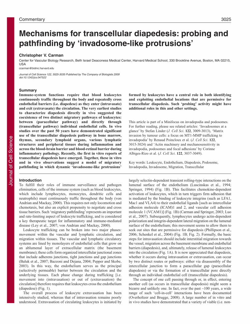
Fig. 1. Paracellular and transcellular routes of leukocyte diapedesis duringintravasation and extravasation. Trafficking of leukocytes throughout the bodyrequires their movement into (intravasation) and out of (extravasation) thevascular and lymphatic circulation. (A) Stages of intravasation. Details of theprocess by which leukocytes (green) enter vascular structures (exemplifiedhere as a post-capillary venule; pink) and lymphatic structures are not wellcharacterized. In general, this is thought to involve steps of interstitialmigration towards the vessel, migration across the basement membrane andendothelial barriers (diapedesis), and, ultimately, release of lumenal leukocytesinto circulation. (B) Stages of extravasation. The leukocyte extravasationprocess has been intensively studied and shown to involve, in the firstinstance, transient rolling-type interactions mediated predominantly byselectins (Springer, 1994). This facilitates chemokine-dependent activationand firm arrest, which is mediated by the binding of leukocyte integrins (e.g.LFA1, Mac1 and VLA4) to endothelial cell-adhesion molecules (e.g. ICAM1,ICAM2 and VCAM1). Subsequently, leukocytes migrate laterally over thesurface of the endothelium, probing for a site to penetrate through it (see alsoFig. 2). Finally leukocytes cross the endothelial barrier (diapedese) and enterthe interstitium. (C,D) The process of diapedesis, whether during intravasationor extravasation, can occur by two distinct pathways: paracellular ortranscellular. (C) Paracellular diapedesis. Leukocytes and endothelial cellscoordinately disassemble endothelial cell-cell junctions and open up a gapbetween two or more endothelial cells (Muller, 2003). (D) Transcellulardiapedesis. Leukocytes migrate directly through individual endothelial cellsvia a transient transcellular pore that leaves endothelial cell-cell junctionsintact. Note that the two individual endothelial cells in C and D aredistinguished by different shades of pink.
*
*
*
**
*
*
6
3
4
5
1
2
Endothelium
Leukocyte
Basementmembrane
Caveolae/VVOs
Invadosome-likeprotrusions
Junctional adhesioncomplex
Lateral migration
Nucleus
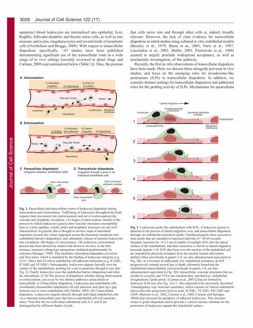
Fig. 2. Leukocytes probe the endothelium with ILPs. A leukocyte (green) isdepicted in the process of lateral migration over, and transcellular diapedesisthrough, an endothelial monolayer (pink). Numbered panels show successivetime points that are intended to represent intervals of ~30-60 seconds.Dynamic insertion (to ~0.2-1 μm in depth) of multiple ILPs into the apicalsurface of the endothelium, and their retraction, is shown as lateral migrationproceeds (panels 1-4). ILPs that form over the nucleus of the endothelial cellare impeded by physical resistance from the nuclear lamina and remainshallow (blue arrowheads in panels 2-4; see also ultrastructural equivalent inFig. 3B). At a location of sufficiently low endothelial resistance, an ILPprogressively extends several μm in depth, ultimately breaching theendothelium transcellularly (red arrowheads in panels 3-6; see alsoultrastructural equivalent in Fig. 3D). Intracellular vesicular structures that aresimilar to caveolae and VVOs are enriched near, and fused to, endothelialinvaginations [‘podo-prints’ (Carman et al., 2007)] that are formed byleukocyte ILPs (see also Fig. 3A,C). Also depicted is the previously described‘transmigratory cup’ structure (asterisks), which consists of vertical endothelialmicrovillus-like projections [rich in actin, ICAM1, VCAM1, PECAM1 andJAM1 (Barreiro et al., 2002; Carman et al., 2003; Carman and Springer,2004)] that surround the periphery of adherent leukocytes. This structureseems to guide diapedesis and to provide a vertical traction substrate for theprotrusion of leukocytes against the endothelial surface.
Jour
nal o
f Cel
l Sci
ence

3027Mechanisms for transcellular diapedesis
diapedesis have been reviewed extensively elsewhere (Burns et al.,2003; Ley et al., 2007; Luscinskas et al., 2002; Muller, 2001;Muller, 2003) and will not be covered here in detail.
In vivo settings for transcellular diapedesisAn overview of leukocyte traffickingAs noted above, immune cells exhibit the distinct ability andrequirement to traffic throughout almost all recesses of the body(von Andrian and Mackay, 2000). Leukocytes originate in the bonemarrow, where they begin their life cycle by migrating into thebloodstream. T and B lymphocytes then enter and exit variouslymphoid organs (including the thymus, lymph nodes, Peyer’spatches, spleen and tonsils) as part of their maturation processesand immune-surveillance functions. Monocytes constitutivelymigrate from the circulation into the peripheral tissues, where theydifferentiate into antigen-presenting cells (APCs), includingmacrophages and dendritic cells. These, in turn, traffic out of thetissue and into the lymphatic system through afferent lymphaticvessels that carry them to secondary lymphoid organs. In cases ofinfection, APCs bearing pathogen-derived antigen interact with andactivate the expansion of antigen-specific lymphocytes. Theselymphocytes then differentiate into effector or memory lymphocytes,
which enter the vascular circulation and join innate immune cells(such as neutrophils) in migrating into the infected or inflamedperipheral tissues. Finally, several recent studies have begun todocument so-called ‘reverse transmigration’, whereby inflammatoryleukocytes are thought to leave the peripheral tissues (during theresolution phase of inflammation) by reversing their migratory pathand undergoing intravasation to re-enter the vascular circulation(Huttenlocher and Poznansky, 2008).
The diapedesis associated with each of the above trafficking stepscan occur in vastly different tissues and involve distinct endothelia(Aird, 2007a; Aird, 2007b), leukocyte subtypes and migrationstimuli. Thus, it should not be surprising that the existing studiessuggest wide variation in the extent to which para- and transcellulardiapedesis routes are used in the various in vivo settings examined,as outlined below. The majority of in vivo reports of transcellulardiapedesis have been conducted using methods that are appropriatefor reasonably conclusive, and often unequivocal, assessment ofthe route of diapedesis [e.g. scanning electron microscopy (SEM),serial-section transmission electron microscopy (TEM), or serial-section confocal fluorescence microscopy in the presence ofjunctional markers; see highlighted references in Table 1]; however,it is important to note when discussing such studies that some have
Table 1. In vivo observations of transcellular diapedesisTissue or structure1 Leukocytes Stimulus2 Species References3
Bone marrow Eosinophils,granulocytes,lymphocytes
Untreated, eotaxin, IL5 Guinea pig, mouse, rat (Becker and De Bruyn, 1976; Campbell, 1972;Chamberlain and Lichtman, 1978; DeBruyn et al., 1971; Muto, 1976; Palframanet al., 1998; Wolosewick, 1984)
Thymus Lymphocytes Untreated Guinea pig, rat (Toro and Olah, 1967; Ushiki, 1986)Lymph-node HEV (a, c,
m, p, pa)Lymphocytes Untreated,
Staphylococcusaureus
Guinea pig, hamster,mouse, rat
(Cho and De Bruyn, 1979; Cho and De Bruyn,1981; Cho and De Bruyn, 1986; Farr and DeBruyn, 1975; Marchesi and Gowans, 1964)
Peyer’s patch HEV Lymphocytes Untreated, intestinalirritant
Guinea pig, mouse, rat (Azzali et al., 2008; Cho and De Bruyn, 1986;Yamaguchi and Schoefl, 1983)
Tonsil HEV Lymphocytes Tonsillitis Human (Indrasingh et al., 2002)Lymphatic structures
(ALPA, ALV,lymphatic sinusoid,SI-lacteals, TAAL)
Lymphocytes,macrophages,neutrophils
Untreated, lymphaticstasis, prolonged fast
Chicken, mouse, rabbit,rat
(Azzali, 1990; Azzali, 1998; Azzali, 2007a;Azzali and Arcari, 2000; Azzali et al.,1990a; Azzali et al., 1990b; Farr et al., 1980;Olah and Glick, 1985)
Mesentery Eosinophils,lymphocytes,monocytes,neutrophils
Mechanical trauma Rat (Marchesi and Florey, 1960)
Pancreas Leukocytes Ischemia Dog (Williamson and Grisham, 1960; Williamson andGrisham, 1961)
Skin Eosinophils, neutrophils C5a, fMLP, NAP, IL1,LTB4
Guinea pig, human,mouse
(Feng et al., 1998; Hoshi and Ushiki, 1999;Schubert et al., 1989)
Liver, lung, spleen,kidney, heart
Lymphocytes IL2
α
Mouse (Fujita et al., 1991)
Cremaster Neutrophils MIP2 Mouse (Phillipson et al., 2006; Phillipson et al., 2008)Blood-brain barrier Lymphocytes,
neutrophils-bungarotoxin, EAE,EAN, post-operative,thalamusdegeneration
Cat, mouse, rabbit, rat (Astrom et al., 1968; Barron et al., 1974;Faustmann and Dermietzel, 1985; Lossinskyet al., 1989; Lossinsky et al., 1991; Matthewsand Kruger, 1973; Raine et al., 1990; Wolburget al., 2005)
Blood-retinal barrier Granulocytes,lymphocytes,macrophages
EAU, IL1 Rat (Bamforth et al., 1997; Greenwood et al., 1994)
1a, axillary; ALPA, absorbing lymphatic peripheral apparatus; ALV, absorbing lymphatic vessel; c, cervical; p, popliteal; pa, para-aortic; m, mesenteric; SI,small intestine; TAAL, tumor-associated absorbing lymphatic. 2C5a, complement component 5a; EAE, experimental autoimmune encephalomyelitis; EAN,experimental autoimmune neuritis; EAU, experimental autoimmune uveoretinitis; fMLP, formyl-Met-Leu-Phe; IL, interleukin; LTB4, leukotriene B4; MIP2,macrophage inflammatory protein 2; NAP, neutrophil-activating peptide. 3Studies providing at least ‘reasonably conclusive’ or unequivocal demonstration oftranscellular diapedesis (via either SEM, serial-section TEM, or serial-section confocal fluorescence microscopy in the presence of junctional markers) arehighlighted in bold.
Jour
nal o
f Cel
l Sci
ence
3028
employed single-section TEM only, which cannot conclusivelydetermine the route of diapedesis.
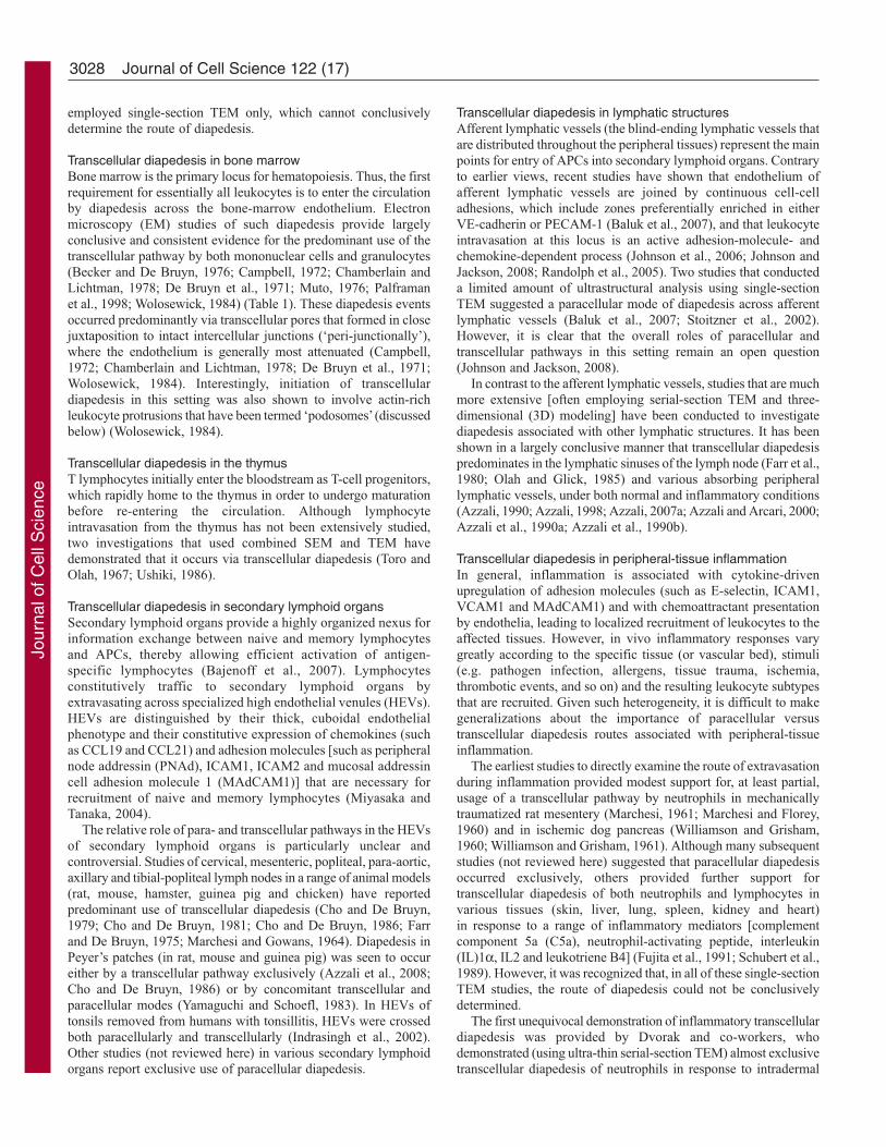
Transcellular diapedesis in bone marrowBone marrow is the primary locus for hematopoiesis. Thus, the firstrequirement for essentially all leukocytes is to enter the circulationby diapedesis across the bone-marrow endothelium. Electronmicroscopy (EM) studies of such diapedesis provide largelyconclusive and consistent evidence for the predominant use of thetranscellular pathway by both mononuclear cells and granulocytes(Becker and De Bruyn, 1976; Campbell, 1972; Chamberlain andLichtman, 1978; De Bruyn et al., 1971; Muto, 1976; Palframanet al., 1998; Wolosewick, 1984) (Table 1). These diapedesis eventsoccurred predominantly via transcellular pores that formed in closejuxtaposition to intact intercellular junctions (‘peri-junctionally’),where the endothelium is generally most attenuated (Campbell,1972; Chamberlain and Lichtman, 1978; De Bruyn et al., 1971;Wolosewick, 1984). Interestingly, initiation of transcellulardiapedesis in this setting was also shown to involve actin-richleukocyte protrusions that have been termed ‘podosomes’ (discussedbelow) (Wolosewick, 1984).
Transcellular diapedesis in the thymusT lymphocytes initially enter the bloodstream as T-cell progenitors,which rapidly home to the thymus in order to undergo maturationbefore re-entering the circulation. Although lymphocyteintravasation from the thymus has not been extensively studied,two investigations that used combined SEM and TEM havedemonstrated that it occurs via transcellular diapedesis (Toro andOlah, 1967; Ushiki, 1986).
Transcellular diapedesis in secondary lymphoid organsSecondary lymphoid organs provide a highly organized nexus forinformation exchange between naive and memory lymphocytesand APCs, thereby allowing efficient activation of antigen-specific lymphocytes (Bajenoff et al., 2007). Lymphocytesconstitutively traffic to secondary lymphoid organs byextravasating across specialized high endothelial venules (HEVs).HEVs are distinguished by their thick, cuboidal endothelialphenotype and their constitutive expression of chemokines (suchas CCL19 and CCL21) and adhesion molecules [such as peripheralnode addressin (PNAd), ICAM1, ICAM2 and mucosal addressincell adhesion molecule 1 (MAdCAM1)] that are necessary forrecruitment of naive and memory lymphocytes (Miyasaka andTanaka, 2004).
The relative role of para- and transcellular pathways in the HEVsof secondary lymphoid organs is particularly unclear andcontroversial. Studies of cervical, mesenteric, popliteal, para-aortic,axillary and tibial-popliteal lymph nodes in a range of animal models(rat, mouse, hamster, guinea pig and chicken) have reportedpredominant use of transcellular diapedesis (Cho and De Bruyn,1979; Cho and De Bruyn, 1981; Cho and De Bruyn, 1986; Farrand De Bruyn, 1975; Marchesi and Gowans, 1964). Diapedesis inPeyer’s patches (in rat, mouse and guinea pig) was seen to occureither by a transcellular pathway exclusively (Azzali et al., 2008;Cho and De Bruyn, 1986) or by concomitant transcellular andparacellular modes (Yamaguchi and Schoefl, 1983). In HEVs oftonsils removed from humans with tonsillitis, HEVs were crossedboth paracellularly and transcellularly (Indrasingh et al., 2002).Other studies (not reviewed here) in various secondary lymphoidorgans report exclusive use of paracellular diapedesis.
Transcellular diapedesis in lymphatic structuresAfferent lymphatic vessels (the blind-ending lymphatic vessels thatare distributed throughout the peripheral tissues) represent the mainpoints for entry of APCs into secondary lymphoid organs. Contraryto earlier views, recent studies have shown that endothelium ofafferent lymphatic vessels are joined by continuous cell-celladhesions, which include zones preferentially enriched in eitherVE-cadherin or PECAM-1 (Baluk et al., 2007), and that leukocyteintravasation at this locus is an active adhesion-molecule- andchemokine-dependent process (Johnson et al., 2006; Johnson andJackson, 2008; Randolph et al., 2005). Two studies that conducteda limited amount of ultrastructural analysis using single-sectionTEM suggested a paracellular mode of diapedesis across afferentlymphatic vessels (Baluk et al., 2007; Stoitzner et al., 2002).However, it is clear that the overall roles of paracellular andtranscellular pathways in this setting remain an open question(Johnson and Jackson, 2008).
In contrast to the afferent lymphatic vessels, studies that are muchmore extensive [often employing serial-section TEM and three-dimensional (3D) modeling] have been conducted to investigatediapedesis associated with other lymphatic structures. It has beenshown in a largely conclusive manner that transcellular diapedesispredominates in the lymphatic sinuses of the lymph node (Farr et al.,1980; Olah and Glick, 1985) and various absorbing peripherallymphatic vessels, under both normal and inflammatory conditions(Azzali, 1990; Azzali, 1998; Azzali, 2007a; Azzali and Arcari, 2000;Azzali et al., 1990a; Azzali et al., 1990b).
Transcellular diapedesis in peripheral-tissue inflammationIn general, inflammation is associated with cytokine-drivenupregulation of adhesion molecules (such as E-selectin, ICAM1,VCAM1 and MAdCAM1) and with chemoattractant presentationby endothelia, leading to localized recruitment of leukocytes to theaffected tissues. However, in vivo inflammatory responses varygreatly according to the specific tissue (or vascular bed), stimuli(e.g. pathogen infection, allergens, tissue trauma, ischemia,thrombotic events, and so on) and the resulting leukocyte subtypesthat are recruited. Given such heterogeneity, it is difficult to makegeneralizations about the importance of paracellular versustranscellular diapedesis routes associated with peripheral-tissueinflammation.
The earliest studies to directly examine the route of extravasationduring inflammation provided modest support for, at least partial,usage of a transcellular pathway by neutrophils in mechanicallytraumatized rat mesentery (Marchesi, 1961; Marchesi and Florey,1960) and in ischemic dog pancreas (Williamson and Grisham,1960; Williamson and Grisham, 1961). Although many subsequentstudies (not reviewed here) suggested that paracellular diapedesisoccurred exclusively, others provided further support fortranscellular diapedesis of both neutrophils and lymphocytes invarious tissues (skin, liver, lung, spleen, kidney and heart)in response to a range of inflammatory mediators [complementcomponent 5a (C5a), neutrophil-activating peptide, interleukin(IL)1α, IL2 and leukotriene B4] (Fujita et al., 1991; Schubert et al.,1989). However, it was recognized that, in all of these single-sectionTEM studies, the route of diapedesis could not be conclusivelydetermined.
The first unequivocal demonstration of inflammatory transcellulardiapedesis was provided by Dvorak and co-workers, whodemonstrated (using ultra-thin serial-section TEM) almost exclusivetranscellular diapedesis of neutrophils in response to intradermal
Journal of Cell Science 122 (17)
Jour
nal o
f Cel
l Sci
ence
3029Mechanisms for transcellular diapedesis
injection of the bacterial chemoattractant formyl-Met-Leu-Phe(fMLP) (Feng et al., 1998). Quantitatively similar, and largelyconclusive, results were obtained in a subsequent study that usedfMLP injected into skin (Hoshi and Ushiki, 1999). Recently, Kubesand co-workers were the first to use serial-section confocalfluorescence microscopy to demonstrate unambiguous transcellulardiapedesis of ~15% of neutrophils in post-capillary venules ofmacrophage inflammatory protein 2 (MIP2)-stimulated mousecremaster muscle (Phillipson et al., 2006).
Transcellular diapedesis across the BBB and BRB duringinflammatory pathologyThe central nervous system (CNS) and retinal parenchyma areimmune-privileged compartments in which leukocyte trafficking istightly limited by the specialized endothelial blood-brain barrier(BBB) and blood-retinal barrier (BRB), respectively. A hallmarkof the BBB and BRB is the exceptionally well-organizedinterendothelial tight junctions, which greatly limit paracellularpermeability (Lightman and Greenwood, 1992; Rubin and Staddon,1999). During inflammatory pathology of the CNS and retina,trafficking of leukocytes across the BBB and BRB is significantlyupregulated. In such settings, many studies have documented apredominant role for transcellular diapedesis pathways (Table 1).
Although diapedesis across the BBB was initially thought to beparacellular in rat models of multiple sclerosis [in whichexperimental autoimmune encephalomyelitis (EAE) was induced](Lampert, 1967), many subsequent studies have strongly supportedtranscellular diapedesis of mononuclear inflammatory cells acrossthe BBB (Lossinsky et al., 1989; Lossinsky et al., 1991; Raine et al.,1990; Wolburg et al., 2005). In a variety of other inflammatorysettings, including acute inflammatory autoimmune neuritis(experimental allergenic neuritis) (Astrom et al., 1968), post-operative neural degeneration (Matthews and Kruger, 1973),corticectomy-induced thalamic nerve degeneration (Barron et al.,1974) and acute inflammatory meningitis, lymphocytes andneutrophils both crossed the BBB largely via transcellular routes.Similarly, in an experimental model of posterior uveitis[experimental autoimmune uveoretinitis (EAU)] in rat and in IL1β-mediated acute inflammation in rat, lymphocytes, granulocytes andmonocytes were shown to cross the BRB exclusively by atranscellular pathway (Bamforth et al., 1997; Greenwood et al.,1994). Interestingly, several authors have noted that, in the BBBor BRB, leukocytes seem to initiate diapedesis by ‘probing’ intoendothelial cells with micron-scale, actin-rich ‘pseudopodia’ or‘processes’ (Bamforth et al., 1997; Greenwood et al., 1994;Lossinsky et al., 1991; Wolburg et al., 2005) that, morphologically,are not unlike invadosomes (discussed below).
The studies summarized above provide extensive demonstrationsof transcellular diapedesis in vivo in essentially all tissues in whichdiapedesis has been examined, although the extent to whichparacellular and transcellular routes dominate varies greatly. It isdifficult to infer mechanisms for transcellular diapedesis from these(largely static and descriptive) studies alone. Nonetheless, whencombined with recent in vitro studies, which are more dynamic andexperimentally tractable, these in vivo observations providesignificant support for roles of specific leukocyte protrusivestructures in transcellular diapedesis, as discussed below.
In vitro studies of transcellular diapedesisThe development of techniques for the isolation and cultureof primary endothelial-cell monolayers in the early 1970s
(Gimbrone et al., 1974; Jaffe et al., 1973) facilitated in vitroinvestigations of diapedesis, which (through the use of single-sectionTEM and light microscopy) initially demonstrated diapedesisthrough paracellular pathways (Beesley et al., 1979; Burns et al.,2003; Furie et al., 1987; Luscinskas et al., 2002; Muller, 2001;Muller, 2003; Pawlowski et al., 1988). However, beginning in2004, a growing collection of investigators started to makeunambiguous observations of transcellular diapedesis in vitro,largely via the use of advanced fluorescence imagingapproaches (Carman et al., 2007; Carman and Springer, 2004;Cinamon et al., 2004; Ferreira et al., 2005; Gerard et al.,2009; Keuschnigg et al., 2009; Marmon et al., 2008; Millanet al., 2006; Nieminen et al., 2006; Riethmuller et al.,2008; Yang et al., 2005). These observations have been made witha range of primary and transformed endothelial cells [humanumbilical-vein endothelial cells (HUVECs), human coronary-artery endothelial cells (HCAECs), human dermal (HDMVECs)and lung (HLMVECs) microvascular endothelial cells, humanlymphatic endothelial cells (HLyECs), and murine brainmicrovascular endothelial cells (bend.3)] and leukocytes(neutrophils, monocytes, and memory and effector lymphocytes).Moreover, these studies have included diverse migratory andinflammatory stimuli [namely, fMLP, IL1β, IL8, platelet-activatingfactor (PAF), stromal cell-derived factor-1 (SDF-1) and TNFα] andboth static and physiologic shear-flow conditions. The quantitativecontribution of the transcellular route in these studies ranged from~5 to ~60% of the total diapedesis.
Mechanisms for transcellular diapedesisAdhesion moleculesThe cell-cell adhesion molecules involved in transcellular diapedesishave begun to be characterized. Several investigations showenrichment of endothelial ICAM1 on transcellular pores ofendothelial cells and enrichment of the integrin LFA1 (the ICAM1receptor) on the closely apposed pore-spanning segments oftransmigrating leukocytes (Carman et al., 2007; Carman andSpringer, 2004; Gerard et al., 2009; Millan et al., 2006; Yang et al.,2005). Numerous studies have also demonstrated so-calledendothelial ‘docking structures’ or ‘transmigratory cups’ enrichedin ICAM1 and VCAM1 that partially ‘embrace’ migratingleukocytes (Barreiro et al., 2002; Carman et al., 2003; Carman andSpringer, 2004; Faustmann and Dermietzel, 1985; Fujita et al., 1991;Millan et al., 2006; Nieminen et al., 2006; Phillipson et al., 2008;Raine et al., 1990; Riethmuller et al., 2008; van Buul et al., 2007;Williamson and Grisham, 1961; Wolburg et al., 2005). These actin-and vimentin-dependent structures seem to have guidance ortraction roles, and are important for both paracellular andtranscellular diapedesis. Finally, a few studies have suggested thatthe ‘junctional’ adhesion molecules that are involved in paracellulardiapedesis [PECAM-1 and JAM-1 (Luscinskas et al., 2002; Muller,2003)] might also be involved in transcellular migration (Carmanet al., 2007; Gerard et al., 2009; Mamdouh et al., 2003).
Integrin-ligand interactions have also been implicated indetermining the preference of leukocytes for paracellular versustranscellular routes. However, consistent roles for integrins are notyet evident. In one in vivo model, knockout of the integrin Mac1led to a large increase (~65% of all extravasations compared with~15% for wild type) in transcellular migration (Phillipson et al.,2006). In addition, an in vitro study demonstrated that neutrophiltranscellular migration was strongly favored by high endothelialICAM1 expression in a manner largely dependent on LFA-1
Jour
nal o
f Cel
l Sci
ence
3030
expressed on neutrophils (Yang et al., 2005). In both cases, alteredadhesion, leading to altered lateral migration (discussed below), wassuggested to be the basis for the change in route, but throughopposite mechanisms [that is, Yang et al. proposed that leukocyte-endothelium adhesion was increased, whereas Phillipson et al.proposed that adhesion was reduced (Yang et al., 2005; Phillipsonet al., 2006)]. Interestingly, a reduction of lymphocyte lateralmigration in the absence of measurable changes in strength ofadhesion (which was achieved by deletion of the Rac1 activatorTiam1) was also found to dramatically upregulate transcellulardiapedesis (Gerard et al., 2009). Importantly, transcellular migrationevents tend to occur near junctions (discussed below), such thatthey often might not be adequately distinguished from paracellularmigration events using light microscopy (Carman and Springer,2008). Thus, the above studies might also reflect changes in locationof transcellular diapedesis events (that is, relatively central versusperi-junctional transcellular diapedesis) (Carman and Springer,2008).
Lateral migrationDuring extravasation, initial adhesion of leukocytes to theendothelium is followed by polarization and integrin-dependentlateral migration (Phillipson et al., 2006; Schenkel et al., 2004).This has been generally interpreted as a mechanism to allowleukocytes to move towards endothelial-cell junctions to enableparacellular diapedesis (Phillipson et al., 2006; Schenkel et al.,2004). However, lateral migration also precedes transcellulardiapedesis (Carman et al., 2007; Cinamon et al., 2004; Millan et al.,2006; Yang et al., 2005) and probably has an analogous role inpositioning leukocytes optimally (that is, at sites where transcellularpore formation can occur most efficiently). A wide range of in vivoEM studies suggest that transcellular diapedesis occurs preferentiallyin close juxtaposition to intact intercellular junctions (peri-junctionally) (Azzali, 1990; Azzali, 1998; Azzali, 2007a; Azzaliand Arcari, 2000; Azzali et al., 2008; Azzali et al., 1990a;Azzali et al., 1990b; Bamforth et al., 1997; Campbell, 1972;Chamberlain and Lichtman, 1978; Cho and De Bruyn, 1981; Choand De Bruyn, 1986; De Bruyn et al., 1971; Farr et al., 1980;Faustmann and Dermietzel, 1985; Feng et al., 1998; Greenwoodet al., 1994; Lossinsky et al., 1989; Lossinsky et al., 1991; Marchesiand Florey, 1960; Marchesi and Gowans, 1964; Wolburg et al., 2005;Wolosewick, 1984) and it has been suggested that leukocytestherefore must somehow ‘seek out these regions’ (Campbell, 1972).This begs the question of how, in the absence of a discrete pre-existing locus (such as endothelial-cell junctions for paracellulardiapedesis), such sites for transcellular pore formation (whether peri-junctional or otherwise) can be identified by the cell.
Probing by ILPsAs discussed below, in vitro studies suggest that ILPs – which areleukocyte structures that are similar to both podosomes andinvadopodia (collectively subtended under the term ‘invadosomes’)– are crucial for transcellular diapedesis (Carman et al., 2007).Invadosomes are classically defined as actin-dependent adhesiveand/or protrusive structures (~500 nm in both diameter and depth)that form specifically on the ventral surface of highly migratoryand invasive cells, such as leukocytes, endothelial cells andtransformed tumor cells (Gimona et al., 2008; Linder, 2009).Although the protrusive structures that are observed in the settingof leukocyte diapedesis (see below) share many features of classicalinvadosomes, other properties are unique or remain undefined
(Box 1). Thus, we refer to these as ‘invadosome-like protrusions’(ILPs) to explicitly denote the current uncertainty of their preciserelationship to the invadosomes that have been defined in othersettings.
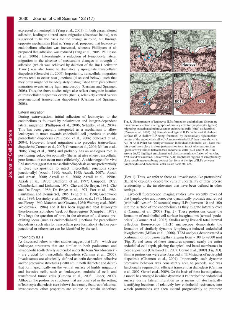
Live-cell fluorescence imaging studies have recently revealedthat lymphocytes and monocytes dynamically protrude and retract(with half-lives of ~20 seconds) many ILPs (between 10 and 100)into the surface of the endothelium as they migrate laterally overit (Carman et al., 2007) (Fig. 2). These protrusions cause theformation of endothelial cell-surface invaginations (termed ‘podo-prints’) (Carman et al., 2007). Studies using live-cell total internalreflection fluorescence (TIRF) microscopy demonstrate theformation of similarly dynamic lymphocyte-induced endothelialinvaginations (Millan et al., 2006). TEM analysis demonstrated acontinuum of protrusion depths (ranging from ~100 to ~2000 nm)(Fig. 3), and some of these structures spanned nearly the entireendothelial-cell depth, placing the apical and basal membranes inclose apposition (Carman et al., 2007; Gerard et al., 2009) (Fig. 3D).Similar protrusions were also observed in TEM studies of neutrophildiapedesis (Cinamon et al., 2004). Importantly, such dynamicprotrusive behavior was consistently seen to precede, and wasfunctionally required for, efficient transcellular diapedesis (Carmanet al., 2007; Gerard et al., 2009). On the basis of these investigations,a model has emerged in which dynamic ILPs ‘probe’ the endothelialsurface during lateral migration as a means of stochasticallyidentifying locations of relatively low endothelial resistance, intowhich protrusions can then extend progressively to promote
Journal of Cell Science 122 (17)
F
E
A
C
B
D
Nucleus
EC2EC1
Fig. 3. Ultrastructure of leukocyte ILPs formed on endothelium. Shown aretransmission electron micrographs of primary effector lymphocytes (green)migrating on activated microvascular endothelial cells (pink) as described(Carman et al., 2007). (A) Formation of typical ILPs on the endothelial-cellsurface. (B) A shallow ILP being ‘frustrated’ by the relatively rigid nuclearlamina of the endothelial cell. (C) A more extended ILP than those shown inA. (D) An ILP that has nearly crossed an individual endothelial cell. Note thatthis event takes place in close juxtaposition to an intact adherens junction(green arrow) formed between two endothelial cells (EC1 and EC2). Bluearrows (A,C) highlight enrichment and plasma-membrane fusion of vesicles,VVOs and/or caveolae. Red arrows (A-D) emphasize regions of exceptionallyclose membrane-membrane contact that form at the tips of ILPs betweenlymphocytes and endothelial cells. Scale bars: 300 nm.
Jour
nal o
f Cel
l Sci
ence
3031Mechanisms for transcellular diapedesis
transcellular pore formation. Interestingly, very recent studies havecharacterized lymphocyte structures termed ‘invasive filopodia’ thatare highly analogous to ILPs (in terms of dynamics, molecularcomposition and morphology), and that were similarly ascribed arole in probing for sites for diapedesis, although not explicitly bythe transcellular route (Shulman et al., 2009).
Importantly, this ILP probing model in its basic form has beenpreviously suggested and supported by diverse observations.Indeed, the first description of a podosome (and use of this term)was made in the context of transcellular migration of lymphocytesand eosinophils across bone-marrow endothelium in vivo(Wolosewick, 1984). The authors predicted that the protrusiveforce supplied by such actin-enriched structures might drivetranscellular pore formation. Shortly thereafter, Marchisio and co-workers conducted the first detailed molecular characterization ofpodosomes in transformed fibroblasts (Tarone et al., 1985).Subsequently, the same group showed that leukocytes (natural
killer cells) that adhered to endothelium in vitro formedpodosomes, and suggested that these functioned in endothelialpenetration during diapedesis (Allavena et al., 1991). Finally,although the structures that were visualized have been referred towith various terms, including ‘microvillus-, filopodium- andfinger-like protrusions’, ‘processes’, ‘pseudopodia’ and ‘probingpseudopods’, many in vivo studies of transcellular diapedesis havealso demonstrated the endothelium-directed protrusion ofstructures with clearly invadosome-like morphology fromlymphocytes, monocytes, neutrophils, eosinophils and acutemyeloid leukemia tumor cells (Astrom et al., 1968; Azzali et al.,2008; Bamforth et al., 1997; Barron et al., 1974; Becker and DeBruyn, 1976; De Bruyn et al., 1989; De Bruyn et al., 1971; Farrand De Bruyn, 1975; Faustmann and Dermietzel, 1985; Feng etal., 1998; Fujita et al., 1991; Greenwood et al., 1994; Lossinskyet al., 1989; Lossinsky et al., 1991; Lossinsky and Shivers, 2004;Marchesi and Florey, 1960; Matthews and Kruger, 1973; Olah
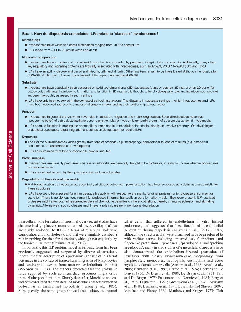
Box 1. How do diapedesis-associated ILPs relate to ‘classical’ invadosomes?
Morphology
• Invadosomes have width and depth dimensions ranging from ~0.5 to several μm
• ILPs range from ~0.1 to ~2 μm in width and depth
Molecular composition
• Invadosomes have an actin- and cortactin-rich core that is surrounded by peripheral integrin, talin and vinculin. Additionally, many otherkey regulatory and signaling proteins are typically associated with invadosomes, such as Arp2/3, WASP, N-WASP, Src and RhoA
• ILPs have an actin-rich core and peripheral integrin, talin and vinculin. Other markers remain to be investigated. Although the localizationof WASP at ILPs has not been characterized, ILPs depend on functional WASP
Substrate
• Invadosomes have classically been assessed on solid two-dimensional (2D) substrates (glass or plastic), 2D matrix or on 2D bone (forosteoclasts). Although invadosome formation and function in 3D matrices is thought to be physiologically relevant, invadosomes have notyet been thoroughly assessed in such settings
• ILPs have only been observed in the context of cell-cell interactions. The disparity in substrate settings in which invadosomes and ILPshave been observed represents a major challenge to understanding their relationship to each other
Function
• Invadosomes in general are known to have roles in adhesion, migration and matrix degradation. Specialized podosome arrays(‘podosome belts’) of osteoclasts facilitate bone resorption. Matrix invasion is generally thought of as a specialization of invadopodia
• ILPs seem to function in probing the endothelial surface and in transcellular diapedesis (clearly an invasive property). On physiologicalendothelial substrates, lateral migration and adhesion do not seem to require ILPs
Dynamics
• The lifetime of invadosomes varies greatly from tens of seconds (e.g. macrophage podosomes) to tens of minutes (e.g. osteoclastpodosomes or transformed-cell invadopodia)
• ILPs have lifetimes from tens of seconds to several minutes
Protrusiveness
• Invadosomes are variably protrusive: whereas invadopodia are generally thought to be protrusive, it remains unclear whether podosomesare necessarily so
• ILPs are defined, in part, by their protrusion into cellular substrates
Degradation of the extracellular matrix
• Matrix degradation by invadosomes, specifically at sites of active actin polymerization, has been proposed as a defining characteristic forthese structures
• ILPs have yet to be assessed for either degradative activity with respect to the matrix (or other proteins) or for protease enrichment orsecretion. There is no obvious requirement for proteases in formal transcellular pore formation – but, if they were present, ILP-localizedproteases might alter local adhesion-molecule and chemokine densities on the endothelium, thereby changing adhesion and signalingdynamics. Alternatively, such proteases might have a role in basement-membrane degradation
Jour
nal o
f Cel
l Sci
ence

3032
and Glick, 1985; Raine et al., 1990; Williamson and Grisham,1960; Wolburg et al., 2005; Wolosewick, 1984) (Table 1). Theactions of these protrusions have often been interpreted as‘probing’ the endothelium and potentially driving transcellularpore formation (Astrom et al., 1968; Bamforth et al., 1997; Farrand De Bruyn, 1975; Greenwood et al., 1994; Lossinsky et al.,1989; Lossinsky et al., 1991; Lossinsky and Shivers, 2004;Marchesi and Florey, 1960; Olah and Glick, 1985; Raine et al.,1990; Wolburg et al., 2005; Wolosewick, 1984). Thus, the abilityand tendency of leukocytes to extend ILPs into the endothelialsurface during diapedesis seems to be broadly relevant.
Mechanotransduction by ILPs?An implicit component of the model presented above is that ILPscan somehow sense mechanical resistance and, in response, altertheir signaling and dynamics. Although this has not been widelyinvestigated, at least two recent studies demonstrate that podosomescan serve as ‘dynamic mechanosensors’ (Collin et al., 2008; Collinet al., 2006). In these two studies, using polyacrylamide collagen-coated substrates of defined rigidity, Collin et al. demonstrated thatthe lifespan and density of fibroblast podosomes depended onsubstrate flexibility and that such mechanosensory properties weremediated in part through myosin-II motor proteins. Theseobservations support the plausibility of a model in which mechanicalprobing of the endothelial surface by ILPs transduces inside-outand outside-in signals that regulate ILP activity and ultimatelyinfluence the efficiency of transcellular diapedesis.
Endothelial membrane fusion and pore formation inresponse to ILPsTo accomplish pore formation, transcellular diapedesis requires thatenergy-dependent membrane-fusion events occur. The protrusiveforces that are supplied by ILPs clearly promote close appositionof apical and basal endothelial membranes (Fig. 3D), and mightalso supply energy that is directly involved in the formal membrane-fusion process. In addition, studies suggest that ILPs somehowtrigger specific proactive responses in the endothelium that facilitatetranscellular pore formation. In vitro studies show enrichment ofthe caveolar marker caveolin-1, plasmalemma vesicle associatedprotein-1 (PV-1), various vesicles and vesiculo-vacuolar organelles(VVOs) and fusogenic proteins (namely, the SNAREs VAMP2 andVAMP3) in the endothelium at sites of ILP protrusionand transcellular pore formation (Carman et al., 2007; Keuschnigget al., 2009; Migliorisi et al., 1987; Millan et al., 2006) (Fig. 2;Fig. 3A,C). Similar observations have been made in vivo (Carmanet al., 2007; De Bruyn et al., 1989; Greenwood et al., 1994; Olahand Glick, 1985), leading one of the research groups to speculatethat “the fusing of the vesicles may form a gradual trans-endothelialchannel in which the pseudopod of the lymphocyte penetrates andby this mechanism the lymphocyte may cross the endothelium”(Olah and Glick, 1985) (Fig. 2). Vesicle fusion might also facilitatelocal delivery of adhesion receptors (Mamdouh et al., 2003; Millanet al., 2006) and chemokines (Middleton et al., 2002) that mightmodify ILP activity and the efficiency of transcellular migration.The functional significance of vesicle-fusion activity was suggestedby studies in which short interfering RNA (siRNA)-mediatedknockdown of caveolin-1 and pharmacologic perturbation of thesoluble N-ethylmaleimide-sensitive factor (NSF)-attachment protein(SNAP)-SNARE membrane-fusogenic machinery in endotheliumsignificantly reduced the efficiency of transcellular diapedesis(Carman et al., 2007; Millan et al., 2006). However, much more
detailed characterization of the molecular mechanisms by whichILPs ‘trigger’ such responses, and of the consequences of thistriggering activity, is still required.
Other settings for transcellular diapedesisCrossing epithelial and other cell typesThe transcellular mode of cell migration might also be used to crossnon-endothelial barriers. For example, it has been demonstratedin vivo that neutrophils can migrate transcellularly through thepericytes that underlie the vascular endothelium (Feng et al., 1998).In addition, extensive migration of leukocytes across epithelial celllayers (e.g. the mucosal epithelia of the intestine, airway and urinarytract) occurs in vivo (Zen and Parkos, 2003). Routes andmechanisms for this migration remain either only partiallyunderstood or uninvestigated (Zen and Parkos, 2003). One recentstudy demonstrated that paracellular diapedesis of neutrophilsoccurs in an in vitro model system (Porter et al., 2008). In analternative (albeit less physiologic) system, we recently found thatlymphocytes could readily form transcellular pores across epithelialcells (in particular, CHO-K1 cells expressing GFP-tagged ICAM1)by means of probing by ILPs (Carman et al., 2007).
Diapedesis of non-leukocyte cell typesTranscellular diapedesis might also be used by non-leukocyte celltypes. For example, a variety of studies have observedreticulocytes performing transcellular intravasation and haveobserved megakaryocytes forming long transcellular processes(which might be important for the release of platelets into thebloodstream), both across bone-marrow endothelium (Becker andDe Bruyn, 1976; Campbell, 1972; Chamberlain and Lichtman,1978; De Bruyn et al., 1971; Muto, 1976). In addition, severalin vivo studies provide strong support for the use of a transcellularmechanism during diapedesis of certain metastatic tumor cell types(Azzali, 2006; Azzali, 2007b; De Bruyn et al., 1989). Finally, stemcells, which are important emerging cellular therapeutics, clearlyundergo extensive trafficking in vivo (Chamberlain et al., 2007;Laird et al., 2008). The routes and mechanisms used by these cellsfor diapedesis await characterization. The degree to whichmechanisms, including ILP probing, that are used by leukocyteswill be relevant to other cell types during transcellular diapedesisremains an open question.
Other possible roles for ILP probingCrossing the basement membrane?In both intravasation and extravasation, the endothelial barrier isonly one of the key obstacles to be overcome. The basementmembrane also provides a formidable and often rate-limitingbarrier. Precisely how the basement membrane is crossed byleukocytes remains poorly defined. Some studies have suggestedthat proteolytic degradation of the basement membrane enables thepassage of leukocytes. This idea is well supported, at least formetastatic tumor cells during their intravasation across lymphaticendothelial layers (Gimona et al., 2008). It seems conceivable that,if proteases are enriched in leukocyte ILPs as they are in classicalinvadosomes (Chavrier, 2009), this could facilitate basement-membrane degradation and crossing. However, the requirement forbasement-membrane degradation during diapedesis remainssomewhat controversial, and several non-proteolytic modes ofbasement-membrane transmigration have been hypothesized (Roweand Weiss, 2008). In this vein, it is interesting to note thatneutrophils extravasating in vivo have been observed to transmigrate
Journal of Cell Science 122 (17)
Jour
nal o
f Cel
l Sci
ence
3033Mechanisms for transcellular diapedesis
preferentially at pre-existing sites of relatively attenuated basementmembrane (Wang et al., 2006). By analogy with their roles inprobing for attenuated regions of endothelium, ILPs might actsimilarly in locating regions of basement membrane that arepermissive for migration.
Interrogating the endothelial surface biochemically?In essence, the pathfinding model discussed above posits that ILPsare sensory probes and organelles, as well as invasive structures.The observations made thus far focus on a putative role for ILPsin sensing the biomechanical properties of cellular, and possiblymatrix, substrates (i.e. a mechanotransduction role). It is interesting,however, to consider whether such a ‘probing’ function might alsoinclude biochemical sensing of the local environment.
The surface of all cells is modified by a gel-like polysaccharidecoating that is termed the glycocalyx, which is made up of diverseproteoglycans and glycoproteins (Reitsma et al., 2007; Weinbaumet al., 2007). On a typical cell, the glycocalyx is at least 45-nmthick, but it can be substantially thicker (up to ~500 nm) in somecases, such as on the lumen-facing surface of endothelial cells(Reitsma et al., 2007; Weinbaum et al., 2007). The glycocalyx canserve as a reservoir of non-covalently immobilized chemokines andother chemoattractants (Middleton et al., 2002; Proudfoot et al.,2003). In addition, the glycocalyx provides a formidable energybarrier (by means of both steric and electrostatic repulsion) to closemembrane-membrane encounter between cells, such that relativelysmall cell-surface adhesion and signaling molecules are effectivelyshielded (Bell, 1978; Bell et al., 1984; Reitsma et al., 2007; Springer,1990; Weinbaum et al., 2007). Forces provided by ILPs seem toprovide sufficient energy to overcome this barrier, driving closemembrane-membrane apposition (see Fig. 3, red arrows) andthereby promoting molecular interactions that might otherwise beinefficient or impossible. Indeed, our recent studies suggest thatrecognition of endothelial major histocompatibility complex(MHC)-antigen complexes by T-cell receptors on lymphocytes isdependent on ILPs (Peter T. Sage and C.V.C., unpublishedobservations). Thus, ILPs might facilitate a kind of informationalscanning of local membrane surfaces, literally allowing cells to geta ‘deeper’ understanding of their local environment.
Conclusions and perspectivesThe significant number of studies in vivo, along with emergingin vitro studies, demonstrates that two physiologically relevantpathways (paracellular and transcellular) for leukocytediapedesis coexist, with greatly varied utilization of each indistinct settings. The cellular and molecular determinants of theobserved route preferences represent a crucial unresolved issue.In contrast to the extensively studied paracellular pathway,mechanisms for transcellular diapedesis are just beginning to beelucidated, and point towards significant roles for leukocyteILPs. These seem to allow for a kind of ‘migratory pathfinding’in which stochastic ‘probing’ of the endothelial cell surface iscoupled to progressive ILP invasion at sites that are permissivefor transcellular pore formation. Permissive sites might includenot only endothelial regions in which the cell is sufficientlyattenuated (that is, sufficiently thin and/or devoid of resistance-producing intracellular components, such as nuclear lamina orother regions dense in cytoskeletal fibers), but also regions thatare responsive to ILP-mediated ‘triggering’ of fusogenic activityin the endothelium. Many important questions regarding themolecular regulation of ILPs and the precise relationship of
these structures to ‘classical’ invadosomes remain to beaddressed. It is particularly important to resolve whether ILPsexhibit localized proteolytic activity as invadosomes do and, ifso, what roles this has in transcellular diapedesis. Finally, thedegree to which the ‘probing activity’ of ILPs has a role inpathfinding in other settings, and the issue of whether ILPs haveadditional ‘probing’ functions (as suggested above), will beimportant additional future areas of inquiry.
Electron micrographs (Fig. 3) were provided through collaborationwith Tracey Sciuto and Ann Dvorak. C.V.C. was supported by a grantfrom the Arthritis Foundation.
ReferencesAird, W. C. (2007a). Phenotypic heterogeneity of the endothelium: I. Structure, function,
and mechanisms. Circ. Res. 100, 158-173.Aird, W. C. (2007b). Phenotypic heterogeneity of the endothelium: II. Representative
vascular beds. Circ. Res. 100, 174-190.Allavena, P., Paganin, C., Martin-Padura, I., Peri, G., Gaboli, M., Dejana, E.,
Marchisio, P. C. and Mantovani, A. (1991). Molecules and structures involved in theadhesion of natural killer cells to vascular endothelium. J. Exp. Med. 173, 439-448.
Astrom, K. E., Webster, H. D. and Arnason, B. G. (1968). The initial lesion inexperimental allergic neuritis: a phase and electron microscopic study. J. Exp. Med. 128,469-495.
Azzali, G. (1990). The passage of macrophage and lymphocytes from the interstitium acrossthe lymphatic endothelium of rat lacteals. Cell Tissue Res. 262, 191-193.
Azzali, G. (1998). Three-dimensional and ultrastructural aspects of the lymphaticvascularization of the vermiform appendix. J. Submicrosc. Cytol. Pathol. 30, 545-553.
Azzali, G. (2006). On the transendothelial passage of tumor cell from extravasal matrixinto the lumen of absorbing lymphatic vessel. Microvasc. Res. 72, 74-85.
Azzali, G. (2007a). The modality of transendothelial passage of lymphocytes and tumorcells in the absorbing lymphatic vessel. Eur. J. Histochem. 51 Suppl. 1, 73-77.
Azzali, G. (2007b). Tumor cell transendothelial passage in the absorbing lymphatic vesselof transgenic adenocarcinoma mouse prostate. Am. J. Pathol. 170, 334-346.
Azzali, G. and Arcari, M. L. (2000). Ultrastructural and three dimensional aspects of thelymphatic vessels of the absorbing peripheral lymphatic apparatus in Peyer’s patches ofthe rabbit. Anat. Rec. 258, 71-79.
Azzali, G., Gatti, R. and Orlandini, G. (1990a). Macrophage migration through theendothelium in the absorbing peripheral lymphatic vessel of the small intestine. J.Submicrosc. Cytol. Pathol. 22, 273-280.
Azzali, G., Orlandini, G. and Gatti, R. (1990b). The migration of lymphocytes andpolymorphonuclear leukocytes across the endothelial wall of the absorbing peripherallymphatic vessel. J. Submicrosc. Cytol. Pathol. 22, 543-549.
Azzali, G., Arcari, M. L. and Caldara, G. F. (2008). The “mode” of lymphocyteextravasation through HEV of Peyer’s patches and its role in normal homing andinflammation. Microvasc. Res. 75, 227-237.
Bajenoff, M., Egen, J. G., Qi, H., Huang, A. Y., Castellino, F. and Germain, R. N.(2007). Highways, byways and breadcrumbs: directing lymphocyte traffic in the lymphnode. Trends Immunol. 28, 346-352.
Baluk, P., Fuxe, J., Hashizume, H., Romano, T., Lashnits, E., Butz, S., Vestweber, D.,Corada, M., Molendini, C., Dejana, E. et al. (2007). Functionally specialized junctionsbetween endothelial cells of lymphatic vessels. J. Exp. Med. 204, 2349-2362.
Bamforth, S. D., Lightman, S. L. and Greenwood, J. (1997). Ultrastructural analysis ofinterleukin-1 beta-induced leukocyte recruitment to the rat retina. Invest. Ophthalmol.Vis. Sci. 38, 25-35.
Barreiro, O., Yanez-Mo, M., Serrador, J. M., Montoya, M. C., Vicente-Manzanares,M., Tejedor, R., Furthmayr, H. and Sanchez-Madrid, F. (2002). Dynamic interactionof VCAM-1 and ICAM-1 with moesin and ezrin in a novel endothelial docking structurefor adherent leukocytes. J. Cell Biol. 157, 1233-1245.
Barron, K. D., Means, E. D., Feng, T. and Harris, H. (1974). Ultrastructure of retrogradedegeneration in thalamus of rat. 2. Changes in vascular elements and transvascularmigration of leukocytes. Exp. Mol. Pathol. 20, 344-362.
Bazzoni, G. and Dejana, E. (2004). Endothelial cell-to-cell junctions: molecularorganization and role in vascular homeostasis. Physiol. Rev. 84, 869-901.
Becker, R. P. and De Bruyn, P. P. (1976). The transmural passage of blood cells intomyeloid sinusoids and the entry of platelets into the sinusoidal circulation; a scanningelectron microscopic investigation. Am. J. Anat. 145, 183-205.
Beesley, J. E., Pearson, J. D., Hutchings, A., Carleton, J. S. and Gordon, J. L. (1979).Granulocyte migration through endothelium in culture. J. Cell Sci. 38, 237-248.
Bell, G. I. (1978). Models for the specific adhesion of cells to cells. Science 200, 618-627.Bell, G. I., Dembo, M. and Bongrand, P. (1984). Cell adhesion. Competition between
nonspecific repulsion and specific bonding. Biophys. J. 45, 1051-1064.Burns, A. R., Smith, C. W. and Walker, D. C. (2003). Unique structural features that
influence neutrophil emigration into the lung. Physiol. Rev. 83, 309-336.Campbell, F. R. (1972). Ultrastructural studies of transmural migration of blood cells in
the bone marrow of rats, mice and guinea pigs. Am. J. Anat. 135, 521-535.Carman, C. V. and Springer, T. A. (2003). Integrin avidity regulation: are changes in
affinity and conformation underemphasized? Curr. Opin. Cell Biol. 15, 547-556.
Jour
nal o
f Cel
l Sci
ence
3034
Carman, C. V. and Springer, T. A. (2004). A transmigratory cup in leukocyte diapedesisboth through individual vascular endothelial cells and between them. J. Cell Biol. 167,377-388.
Carman, C. V. and Springer, T. A. (2008). Trans-cellular migration: cell-cell contacts getintimate. Curr. Opin. Cell Biol. 20, 533-540.
Carman, C. V., Jun, C. D., Salas, A. and Springer, T. A. (2003). Endothelial cellsproactively form microvilli-like membrane projections upon intercellular adhesionmolecule 1 engagement of leukocyte LFA-1. J. Immunol. 171, 6135-6144.
Carman, C. V., Sage, P. T., Sciuto, T. E., de la Fuente, M. A., Geha, R. S., Ochs, H.D., Dvorak, H. F., Dvorak, A. M. and Springer, T. A. (2007). Transcellular diapedesisis initiated by invasive podosomes. Immunity 26, 784-797.
Chamberlain, G., Fox, J., Ashton, B. and Middleton, J. (2007). Concise review:mesenchymal stem cells: their phenotype, differentiation capacity, immunologicalfeatures, and potential for homing. Stem Cells 25, 2739-2749.
Chamberlain, J. K. and Lichtman, M. A. (1978). Marrow cell egress: specificity of thesite of penetration into the sinus. Blood 52, 959-968.
Cho, Y. and De Bruyn, P. P. (1979). The endothelial structure of the postcapillary venulesof the lymph node and the passage of lymphocytes across the venule wall. J. Ultrastruct.Res. 69, 13-21.
Cho, Y. and De Bruyn, P. P. (1981). Transcellular migration of lymphocytes through thewalls of the smooth-surfaced squamous endothelial venules in the lymph node: evidencefor the direct entry of lymphocytes into the blood circulation of the lymph node. J.Ultrastruct. Res. 74, 259-266.
Cho, Y. and De Bruyn, P. P. (1986). Internal structure of the postcapillary high-endothelialvenules of rodent lymph nodes and Peyer’s patches and the transendothelial lymphocytepassage. Am. J. Anat. 177, 481-490.
Cinamon, G., Shinder, V., Shamri, R. and Alon, R. (2004). Chemoattractant signals andbeta 2 integrin occupancy at apical endothelial contacts combine with shear stress signalsto promote transendothelial neutrophil migration. J. Immunol. 173, 7282-7291.
Collin, O., Tracqui, P., Stephanou, A., Usson, Y., Clement-Lacroix, J. and Planus, E.(2006). Spatiotemporal dynamics of actin-rich adhesion microdomains: influence ofsubstrate flexibility. J. Cell Sci. 119, 1914-1925.
Collin, O., Na, S., Chowdhury, F., Hong, M., Shin, M. E., Wang, F. and Wang, N.(2008). Self-organized podosomes are dynamic mechanosensors. Curr. Biol. 18, 1288-1294.
De Bruyn, P. P., Michelson, S. and Thomas, T. B. (1971). The migration of blood cellsof the bone marrow through the sinusoidal wall. J. Morphol. 133, 417-437.
De Bruyn, P. P., Cho, Y. and Michelson, S. (1989). Endothelial attachment andplasmalemmal apposition in the transcellular movement of intravascular leukemic cellsentering the myeloid parenchyma. Am. J. Anat. 186, 115-126.
Farr, A. G. and De Bruyn, P. P. (1975). The mode of lymphocyte migration throughpostcapillary venule endothelium in lymph node. Am. J. Anat. 143, 59-92.
Farr, A. G., Cho, Y. and De Bruyn, P. P. (1980). The structure of the sinus wall of thelymph node relative to its endocytic properties and transmural cell passage. Am. J. Anat.157, 265-284.
Faustmann, P. M. and Dermietzel, R. (1985). Extravasation of polymorphonuclearleukocytes from the cerebral microvasculature: inflammatory response induced by alpha-bungarotoxin. Cell Tissue Res. 242, 399-407.
Feng, D., Nagy, J. A., Pyne, K., Dvorak, H. F. and Dvorak, A. M. (1998). Neutrophilsemigrate from venules by a transendothelial cell pathway in response to FMLP. J. Exp.Med. 187, 903-915.
Ferreira, A. M., McNeil, C. J., Stallaert, K. M., Rogers, K. A. and Sandig, M. (2005).Interleukin-1beta reduces transcellular monocyte diapedesis and compromises endothelialadherens junction integrity. Microcirculation 12, 563-579.
Fujita, S., Puri, R. K., Yu, Z. X., Travis, W. D. and Ferrans, V. J. (1991). An ultrastructuralstudy of in vivo interactions between lymphocytes and endothelial cells in thepathogenesis of the vascular leak syndrome induced by interleukin-2. Cancer 68, 2169-2174.
Furie, M. B., Naprstek, B. L. and Silverstein, S. C. (1987). Migration of neutrophilsacross monolayers of cultured microvascular endothelial cells: an in vitro model ofleucocyte extravasation. J. Cell Sci. 88, 161-175.
Gerard, A., van der Kammen, R. A., Janssen, H., Ellenbroak, S. I. and Collard, J. G.(2009). The rac activator tiam1 controls efficient T-cell trafficking and route oftransendothelial migration. Blood 113, 6138-6147.
Gimbrone, M. A., Jr, Cotran, R. S. and Folkman, J. (1974). Human vascular endothelialcells in culture: growth and DNA synthesis. J. Cell Biol. 60, 673-684.
Gimona, M., Buccione, R., Courtneidge, S. A. and Linder, S. (2008). Assembly andbiological role of podosomes and invadopodia. Curr. Opin. Cell Biol. 20, 235-241.
Greenwood, J., Howes, R. and Lightman, S. (1994). The blood-retinal barrier inexperimental autoimmune uveoretinitis: leukocyte interactions and functional damage.Lab. Invest. 70, 39-52.
Hoshi, O. and Ushiki, T. (1999). Scanning electron microscopic studies on the route ofneutrophil extravasation in the mouse after exposure to the chemotactic peptide N-formyl-methionyl-leucyl-phenylalanine (fMLP). Arch. Histol. Cytol. 62, 253-260.
Huttenlocher, A. and Poznansky, M. C. (2008). Reverse leukocyte migration can beattractive or repulsive. Trends Cell Biol. 18, 298-306.
Indrasingh, I., Chandi, G. and Vettivel, S. (2002). Route of lymphocyte migration throughthe high endothelial venule (HEV) in human palatine tonsil. Ann. Anat. 184, 77-84.
Jaffe, E. A., Nachman, R. L., Becker, C. G. and Minick, C. R. (1973). Culture of humanendothelial cells derived from umbilical veins: identification by morphologic andimmunologic criteria. J. Clin. Invest. 52, 2745-2756.
Johnson, L. A. and Jackson, D. G. (2008). Cell traffic and the lymphatic endothelium.Ann. NY Acad. Sci. 1131, 119-133.
Johnson, L. A., Clasper, S., Holt, A. P., Lalor, P. F., Baban, D. and Jackson, D. G.(2006). An inflammation-induced mechanism for leukocyte transmigration acrosslymphatic vessel endothelium. J. Exp. Med. 203, 2763-2777.
Keuschnigg, J., Henttinen, T., Auvinen, K., Karikoski, M., Salmi, M. and Jalkanen,S. (2009). The prototype endothelial marker PAL-E is a leukocyte trafficking molecule.Blood 114, 478-484.
Laird, D. J., von Andrian, U. H. and Wagers, A. J. (2008). Stem cell trafficking in tissuedevelopment, growth, and disease. Cell 132, 612-630.
Lampert, P. (1967). Electron microscopic studies on ordinary and hyperacute experimentalallergic encephalomyelitis. Acta Neuropathol. 9, 99-126.
Ley, K., Laudanna, C., Cybulsky, M. I. and Nourshargh, S. (2007). Getting to the siteof inflammation: the leukocyte adhesion cascade updated. Nat. Rev. Immunol. 7, 678-689.
Lightman, S. and Greenwood, J. (1992). Effect of lymphocytic infiltration on the blood-retinal barrier in experimental autoimmune uveoretinitis. Clin. Exp. Immunol. 88, 473-477.
Linder, S. (2009). Invadosomes at a glance. J. Cell Sci. 122, 3009-3013.Lossinsky, A. S. and Shivers, R. R. (2004). Structural pathways for macromolecular and
cellular transport across the blood-brain barrier during inflammatory conditions: review.Histol. Histopathol. 19, 535-564.
Lossinsky, A. S., Badmajew, V., Robson, J. A., Moretz, R. C. and Wisniewski, H. M.(1989). Sites of egress of inflammatory cells and horseradish peroxidase transport acrossthe blood-brain barrier in a murine model of chronic relapsing experimental allergicencephalomyelitis. Acta Neuropathol. 78, 359-371.
Lossinsky, A. S., Pluta, R., Song, M. J., Badmajew, V., Moretz, R. C. and Wisniewski,H. M. (1991). Mechanisms of inflammatory cell attachment in chronic relapsingexperimental allergic encephalomyelitis: a scanning and high-voltage electronmicroscopic study of the injured mouse blood-brain barrier. Microvasc. Res. 41, 299-310.
Luo, B. H., Carman, C. V. and Springer, T. A. (2007). Structural basis of integrinregulation and signaling. Annu. Rev. Immunol. 25, 619-647.
Luscinskas, F. W., Kansas, G. S., Ding, H., Pizcueta, P., Schleiffenbaum, B. E., Tedder,T. F. and Gimbrone, M. A., Jr (1994). Monocyte rolling, arrest and spreading on IL-4-activated vascular endothelium under flow is mediated via sequential action of L-selectin, beta 1-integrins, and beta 2-integrins. J. Cell Biol. 125, 1417-1427.
Luscinskas, F. W., Ma, S., Nusrat, A., Parkos, C. A. and Shaw, S. K. (2002). Leukocytetransendothelial migration: a junctional affair. Semin. Immunol. 14, 105-113.
Mamdouh, Z., Chen, X., Pierini, L. M., Maxfield, F. R. and Muller, W. A. (2003).Targeted recycling of PECAM from endothelial surface-connected compartments duringdiapedesis. Nature 421, 748-753.
Marchesi, V. T. (1961). The site of leucocyte emigration during inflammation. Q. J. Exp.Physiol. Cogn. Med. Sci. 46, 115-118.
Marchesi, V. T. and Florey, H. W. (1960). Electron micrographic observations on theemigration of leucocytes. Q. J. Exp. Physiol. Cogn. Med. Sci. 45, 343-348.
Marchesi, V. T. and Gowans, J. L. (1964). The migration of lymphocytes through theendothelium of venules in lymph nodes: an electron microscope study. Proc. R. Soc.Lond. B Biol. Sci. 159, 283-290.
Marmon, S., Cammer, M., Raine, C. S. and Lisanti, M. P. (2008). Transcellular migrationof neutrophils is a quantitatively significant pathway across dermal microvascularendothelial cells. Exp. Dermatol. 18, 88-90.
Matthews, M. A. and Kruger, L. (1973). Electron microscopy of non-neuronal cellularchanges accompanying neural degeneration in thalamic nuclei of the rabbit. I. Reactivehematogenous and perivascular elements within the basal lamina. J. Comp. Neurol. 148,285-312.
Middleton, J., Patterson, A. M., Gardner, L., Schmutz, C. and Ashton, B. A. (2002).Leukocyte extravasation: chemokine transport and presentation by the endothelium. Blood100, 3853-3860.
Migliorisi, G., Folkes, E., Pawlowski, N. and Cramer, E. B. (1987). In vitro studies ofhuman monocyte migration across endothelium in response to leukotriene B4 and f-Met-Leu-Phe. Am. J. Pathol. 127, 157-167.
Millan, J., Hewlett, L., Glyn, M., Toomre, D., Clark, P. and Ridley, A. J. (2006).Lymphocyte transcellular migration occurs through recruitment of endothelial ICAM-1to caveola- and F-actin-rich domains. Nat. Cell Biol. 8, 113-123.
Miyasaka, M. and Tanaka, T. (2004). Lymphocyte trafficking across high endothelialvenules: dogmas and enigmas. Nat. Rev. Immunol. 4, 360-370.
Muller, W. A. (2001). Migration of leukocytes across endothelial junctions: some conceptsand controversies. Microcirculation 8, 181-193.
Muller, W. A. (2003). Leukocyte-endothelial-cell interactions in leukocyte transmigrationand the inflammatory response. Trends Immunol. 24, 327-334.
Muto, M. (1976). A scanning and transmission electron microscopic study on rat bonemarrow sinuses and transmural migration of blood cells. Arch. Histol. Jpn. 39, 51-66.
Nieminen, M., Henttinen, T., Merinen, M., Marttila-Ichihara, F., Eriksson, J. E. andJalkanen, S. (2006). Vimentin function in lymphocyte adhesion and transcellularmigration. Nat. Cell Biol. 8, 156-162.
Olah, I. and Glick, B. (1985). Lymphocyte migration through the lymphatic sinuses ofthe chicken’s lymph node. Poult. Sci. 64, 159-168.
Overholtzer, M. and Brugge, J. S. (2008). The cell biology of cell-in-cell structures. Nat.Rev. Mol. Cell Biol. 9, 796-809.
Palframan, R. T., Collins, P. D., Severs, N. J., Rothery, S., Williams, T. J. and Rankin,S. M. (1998). Mechanisms of acute eosinophil mobilization from the bone marrowstimulated by interleukin 5, the role of specific adhesion molecules andphosphatidylinositol 3-kinase. J. Exp. Med. 188, 1621-1632.
Journal of Cell Science 122 (17)
Jour
nal o
f Cel
l Sci
ence

3035Mechanisms for transcellular diapedesis
Pawlowski, N. A., Kaplan, G., Abraham, E. and Cohn, Z. A. (1988). The selectivebinding and transmigration of monocytes through the junctional complexes of humanendothelium. J. Exp. Med. 168, 1865-1882.
Pepper, M. S. and Skobe, M. (2003). Lymphatic endothelium: morphological, molecularand functional properties. J. Cell Biol. 163, 209-213.
Phillipson, M., Heit, B., Colarusso, P., Liu, L., Ballantyne, C. M. and Kubes, P. (2006).Intraluminal crawling of neutrophils to emigration sites: a molecularly distinct processfrom adhesion in the recruitment cascade. J. Exp. Med. 203, 2569-2575.
Phillipson, M., Kaur, J., Colarusso, P., Ballantyne, C. M. and Kubes, P. (2008).Endothelial domes encapsulate adherent neutrophils and minimize increases in vascularpermeability in paracellular and transcellular emigration. PLoS ONE 3, e1649.
Poincloux, R., Lizárraga, F. and Chavrier, P. (2009). Matrix invasion by tumour cells:a focus on MT1-MMP trafficking to invadopodia. J. Cell Sci. 122, 3015-3024.
Porter, J. C., Falzon, M. and Hall, A. (2008). Polarized localization of epithelial CXCL11in chronic obstructive pulmonary disease and mechanisms of T cell egression. J. Immunol.180, 1866-1877.
Proudfoot, A. E., Handel, T. M., Johnson, Z., Lau, E. K., LiWang, P., Clark-Lewis,I., Borlat, F., Wells, T. N. and Kosco-Vilbois, M. H. (2003). Glycosaminoglycan bindingand oligomerization are essential for the in vivo activity of certain chemokines. Proc.Natl. Acad. Sci. USA 100, 1885-1890.
Raine, C. S., Cannella, B., Duijvestijn, A. M. and Cross, A. H. (1990). Homing to centralnervous system vasculature by antigen-specific lymphocytes. II. Lymphocyte/endothelialcell adhesion during the initial stages of autoimmune demyelination. Lab. Invest. 63,476-489.
Randolph, G. J., Angeli, V. and Swartz, M. A. (2005). Dendritic-cell trafficking to lymphnodes through lymphatic vessels. Nat. Rev. Immunol. 5, 617-628.
Reitsma, S., Slaaf, D. W., Vink, H., van Zandvoort, M. A. and oude Egbrink, M. G.(2007). The endothelial glycocalyx: composition, functions, and visualization. PflugersArch. 454, 345-359.
Riethmuller, C., Nasdala, I. and Vestweber, D. (2008). Nano-surgery at the leukocyte-endothelial docking site. Pflugers Arch. 456, 71-81.
Rowe, R. G. and Weiss, S. J. (2008). Breaching the basement membrane: who, when andhow? Trends Cell Biol. 18, 560-574.
Rubin, L. L. and Staddon, J. M. (1999). The cell biology of the blood-brain barrier. Annu.Rev. Neurosci. 22, 11-28.
Sage, P. T. and Carman, C. V. (2009). Settings and mechanisms for trans-cellulardiapedesis. Front. Biosci. 14, 5066-5083.
Schenkel, A. R., Mamdouh, Z. and Muller, W. A. (2004). Locomotion of monocytes onendothelium is a critical step during extravasation. Nat. Immunol. 5, 393-400.
Schubert, C., Christophers, E., Swensson, O. and Isei, T. (1989). Transendothelialcell diapedesis of neutrophils in inflamed human skin. Arch. Dermatol. Res. 281, 475-481.
Shulman, Z., Shinder, V., Klein, E., Grabovsky, V., Yeger, O., Geron, E., Montresor,A., Bolomini-Vittori, M., Feigelson, S. W., Kirchhausen, T. et al. (2009). Lymphocyte
crawling and transendothelial migration require chemokine triggering of high-affinityLFA-1 integrin. Immunity 30, 384-396.
Springer, T. A. (1990). Adhesion receptors of the immune system. Nature 346, 425-434.Springer, T. A. (1994). Traffic signals for lymphocyte recirculation and leukocyte
emigration: the multistep paradigm. Cell 76, 301-314.Stoitzner, P., Pfaller, K., Stossel, H. and Romani, N. (2002). A close-up view of migrating
Langerhans cells in the skin. J. Invest. Dermatol. 118, 117-125.Tarone, G., Cirillo, D., Giancotti, F. G., Comoglio, P. M. and Marchisio, P. C. (1985).
Rous sarcoma virus-transformed fibroblasts adhere primarily at discrete protrusions ofthe ventral membrane called podosomes. Exp. Cell Res. 159, 141-157.
Toro, I. and Olah, I. (1967). Penetration of thymocytes into the blood circulation. J.Ultrastruct. Res. 17, 439-451.
Ushiki, T. (1986). A scanning electron-microscopic study of the rat thymus with specialreference to cell types and migration of lymphocytes into the general circulation. CellTissue Res. 244, 285-298.
van Buul, J. D., Allingham, M. J., Samson, T., Meller, J., Boulter, E., Garcia-Mata,R. and Burridge, K. (2007). RhoG regulates endothelial apical cup assembly downstreamfrom ICAM1 engagement and is involved in leukocyte trans-endothelial migration. J.Cell Biol. 178, 1279-1293.
von Andrian, U. H. and Mackay, C. R. (2000). T-cell function and migration: two sidesof the same coin. N. Engl. J. Med. 343, 1020-1034.
Wang, S., Voisin, M. B., Larbi, K. Y., Dangerfield, J., Scheiermann, C., Tran, M.,Maxwell, P. H., Sorokin, L. and Nourshargh, S. (2006). Venular basement membranescontain specific matrix protein low expression regions that act as exit points for emigratingneutrophils. J. Exp. Med. 203, 1519-1532.
Weinbaum, S., Tarbell, J. M. and Damiano, E. R. (2007). The structure and function ofthe endothelial glycocalyx layer. Annu. Rev. Biomed. Eng. 9, 121-167.
Williamson, J. R. and Grisham, J. W. (1960). Leucocytic emigration from inflamedcapillaries. Nature 188, 1203.
Williamson, J. R. and Grisham, J. W. (1961). Electron microscopy of leukocyticmargination and emigration in acute inflammation in dog pancreas. Am. J. Pathol. 39,239-256.
Wolburg, H., Wolburg-Buchholz, K. and Engelhardt, B. (2005). Diapedesis ofmononuclear cells across cerebral venules during experimental autoimmuneencephalomyelitis leaves tight junctions intact. Acta Neuropathol. 109, 181-190.
Wolosewick, J. J. (1984). Distribution of actin in migrating leukocytes in vivo. Cell TissueRes. 236, 517-525.
Yamaguchi, K. and Schoefl, G. I. (1983). Blood vessels of the Peyer’s patch in the mouse:III. High-endothelium venules. Anat. Rec. 206, 419-438.
Yang, L., Froio, R. M., Sciuto, T. E., Dvorak, A. M., Alon, R. and Luscinskas, F. W.(2005). ICAM-1 regulates neutrophil adhesion and transcellular migration of TNF-alpha-activated vascular endothelium under flow. Blood 106, 584-592.
Zen, K. and Parkos, C. A. (2003). Leukocyte-epithelial interactions. Curr. Opin. CellBiol. 15, 557-564.
Jour
nal o
f Cel
l Sci
ence