Embed Size (px)
Citation preview

American Journal of Biochemistry and Biotechnology 7 (3): 104-123, 2011 ISSN 1553-3468 © 2011 L. Zhou et al., This open access article is distributed under a Creative Commons Attribution (CC-BY) 3.0 license
Corresponding Author: Abdel Ghaly, Department of Process Engineering and Applied Science, Dalhousie University, Halifax, Nova Acotia, Canada Tel: 902-494-6014
104
Extraction, Purification and
Characterization of Fish Chymotrypsin: A Review
Liang Zhou, Suzanne M. Budge, Abdel E. Ghaly, Marianne S. Brooks and Deepika Dave
Department of Process Engineering and Applied Science, Faculty of Engineering, Dalhousie University, Halifax, Nova Scotia, Canada
Abstract: Problem statement: Solid fish waste is generated from the unwanted parts of fish including heads, tails, fins, frames, offal (guts, kidney and liver) and skin. It accounts for up to 80% of material from production of surimi, 66% from production of fillet and 27% from production of headed and gutted fish. Currently, fish wastes are disposed off in land-based waste disposal systems or at sea generating toxic by-products during the decomposition process. However, fish processing waste can be used to produce commercially valuable by-products, such as chymotrypsin. Approach: A comperehensive review of the literature on the extraction, purification and characeterization of fish chymotrypsin was performed. Results: Chymotrypsin is an endopeptidase secreted by the pancreatic tissues of vertebrates and invertebrates. It has 3 different structures (chymotrypsin A, B and C) varying slightly in solubility, electrophoretic mobility, isoelectric point and cleavage specificity. Only chymotrypsin A and B are found in fish. Compared with mammal chymotrypsin, fish chymotrypsins have similar amino acid composition and molecular weights. Fish chymotrypsins have higher specific activity, especially those from cold-water fish, and low pH and temperature tolerance. The factors affecting the concentration and activity of chymotrypsin in fish are water temperature, fish species, fish age, fish weight and starvation. Chymotrypsin has application in various industries including the food industry, leather production industry chemical industry and medical industry. Conclusion: Extraction techniques for chymotrypsin include: ultra-filtration, ammonium sulphate fractionation precipitation or water-in-oil microemulsions. Purification can be carried out using re-crystallization and gel-filtration, ion-exchange and hydrophobic interaction chromatography. Further studies should focus on the optimization of purifiying chymotrypsin from fish processing wastes. Key words: Chymotrypsin, trypsin, fish waste, extraction, purification, enzyme activity,
ultrafiltration, fractionation, water-in-oil microemulsions, Docosahexaenoic Acid (DHA), Bovine Serum Albumin (BSA), Fish chymotrypsins
INTRODUCTION
Canada has 25% (244,000 km) of the world coastline and 16% of the world’s fresh water (755,000 Km2). In Canada, 80% of total fish landings come from the Atlantic fishery (mostly lobster, crab and shrimp and scallops) while the Pacific fishery accounts for up to 16% (mostly salmon, clams, groundfish and herring roe). The total seafood and fish income in Canada in 2007 was more than $5 billion and the exports were around $3.9 billion (AAC, 2010). A large portion of the fish landed in Canada is processed. The most common fish processing operation includes three steps: (a) removing the viscera, (b) removing the head, tail, fins and skin and (c) removing
the frame and producing fillets. Fish waste is generated from the unwanted parts of the fish which can generally be divided into two streams: (a) solid waste including heads, tails, fins, frames, offal (guts, kidney and liver) and skin and (b) wastewater from cleaning the fish and equipment. Fish processing waste can account for up to 80% of material from production of surimi, 66% from production of fillet and 27% from production of headed and gutted fish (IFC, 2011; Bechtel, 2003). In the tuna canning production, 24-30% of solid waste is generated from bones, viscera, head and skin, while another 30-35% of fish waste is generated in a liquid form from condensate and blood. Currently, fish wastes are disposed off at sea or in land-based waste disposal system (PNPPRC, 2010; AMEC, 2011). Dumping large

Am. J. Biochem. & Biotech., 7 (3): 104-123, 2011
105
volumes of wastes in water lower the level of dissolved oxygen and may generate toxic by-products during the waste decomposition process (Bechtel, 2003; Gumisiriza et al., 2009). Since fish processing waste components (the heads, viscera, skin and frames) are removed in separate processes, each waste stream can be collect separately and used for the production of value added products. Potentially, fish processing wastes contain high value protein which can be reused to produce valuable by-products such as fertilizers, fishmeal and silage (IFC, 2011). Fish wastes are also known to contain highly valued fatty acids including omega-3 fatty acids such as Docosahexaenoic Acid (DHA) and Eicosapentaenoic Acid (EPA), enzymes (pepsin, trypsin, chymotrypsin), collagen and oil (Swatschek et al., 2002; Byun et al., 2003; Kim and Mendis, 2006). These valuable compounds can be used in the medical and food industries while the oil can be converted into biosel and used in the transporttion industry. Therefore, proper utilization of fish wastes can result in a tremendous commercial value (Chong et al., 2002). Industrially, chymotrypsin is always produced from fresh cattle or swine pancreas and is commonly made in either tablet forms for oral consumption or as a liquid for injection. The price of chymotrypsin is related to the purity of the products. Using fish waste rather than fresh cattle or swine pancreas can dramatically lower the cost of production. There have been many studies on chymotrypsin from higher vertebrates such as cattle and swine but there is little information available on fish chymotrypsin (Einarsson et al., 1996). This review will focus on the recovery (extraction, purification and assay) of chymotrypsin from fish waste as a bioactive molecule and its industial and clinical uses.
FUNCTION AND STRYCTURE OF CHYMOTRYPSIN
Chymotrypsin, an endopeptidase, is a digestive enzyme existing in pancreatic tissues of vertebrates and inverbrates which is secreted into the duodenum (Geiger, 1985). Chymotrypsin hydrolyzes amides, ester and peptide ponds in protein with carboxyl side as amino acid residue at P1 position (the first amino acid residue in the N-terminal direction from the cleaced bond). Other peptide amide bonds like tryptophanyl-, tyrosyl- and phenylalanyl- bonds are also attacked by chymotrypsin. It is also reported that leucyl and glutamyl bonds could be cleaved by chymotrypsin (Geiger, 1985; Appel, 1986). Appel (1986) indicated
that the hydrolyzing power increases according to the type of substrate in the following sequence: proteins < peptides < amides < esters < N-tyrosine esters. Chymotrypsin has 245 amino acid residues and 5 pairs of disulfide linkages with a molecular mass around 24000. Generally, the single polypeptide molecular weight is 25-28 kDa (Haard et al., 2000). Chymotrypsin exists in three inactive forms (chymotrypsinogens A, B and C) in the zymogen granules (dense, membrane-bound bodies that are derived from the Golgi body of cells) of the pancreas (Smith et al., 1951; Geiger, 1985; Raae, 1995; Leth-Larsen et al., 1996). The three types of chymotrypsin (A, B, C) have been found in mammals but only two types of chymotrypsin (A and B) have been found in fish (Yang et al., 2009). As a one of the main digestion proteases, chymotrypsin has been widely reported in the gut of a variety of fish including: discus (Symphysodon sp.), carp (Cyprinus carpto), rainbow trout (Oncorhyncus mykiss), coho salmon (O. Kistuch), chinook salmon (O. tshawytscha), gilthead seabream (Sparus awrata) and dentex (Dentes dentex) (Dimes et al., 1994; Alarcón et al., 1998; Chong et al., 2002). Chymotrypsin concentration is 105 times higher in the gut than in non-gut tissue but the activity and concentration vary according to fish species and the environment in which the fish live (Elert et al., 2004; Parra et al., 2007). Fish chymotrypsin usually has two different forms: cationic (chymotrypsin B) and anionic (chymotrypsin A) (Yang et al., 2009). The isoelectric points of fish chymotrypsinogens A and B are at the pHs of 9.1 and 5.2, respectively. The amino acid sequences of chymotrypsins A and B are very similar with only minor differences and the activated enzymes contain three polypeptide chains. The two isoforms of chymotrypsin (A and B) have been found in and extracted from rainbow trout (Kristjansson and Nielson, 1992), grass carp (Fong et al., 1998), Altantic cod (Ásgeirsson et al., 1993), anchovy (Heu et al., 1995), Monterey sardine (Castillo-Yañez et al 2006) and crucian carp (Yang et al., 2009). The inactive form of chymotrypsin (chymotrypsinogen) can be activated by trypsin which partially cleavs it into two parts while still maintaning an S-S bond. In the enzymnogen activation process, chymotrypsinogen goes through different forms of intermediate products (α-chymotrypsin, γ-chymotrypsin, δ-chymotrypsin, π-chymotrypsin) before forming chymotrypsin A. However, the type of intermediate products generated depend on trypsin concentration and reaction velocities as shown in Fig. 1 (Geiger et al., 1985).

Am. J. Biochem. & Biotech., 7 (3): 104-123, 2011
106
Fig. 1: Activation of chymotrypsinogen. Different
chymotrypsins result as reaction products, depending on relative concentrations and reaction velocities (Appel, 1986)
Fig. 2: The catalytic mechanism of chymotrypsin
(Nelson et al., 2008)
CHYMOTRYPSIN SPECIFICITY AND MECHANISM OF HYDROLYSIS
Specificity: Chymotrypsin hydrolyzes peptide bonds with various α-amino acid carbonyl groups and attacks larger nonpolar aromatic groups such as tyrosine,phenylalanine and tryotophan (Folk et al., 1970; Appel et al., Galvão et al., 2001). It also attacks nonpolar groups like leucine but the reaction is slower. Hudáky et al. (1999) reported that chymotrypsins A and B have similar specificities in hydrolyzing peptides with Phe, Trp, Leu and Tyr residues. Raae et al. (1995) found specificity differences between two types of chymotrypsins (ChT1 and ChT2) isolated from cod.
They pointed out that ChT1 can hydrolyze 8 peptide bonds [Leu(6)-Cys(7), Leu(15)-Tyr(16), Tyr(16)-Leu(17), Arg(22)-Gly(23), Phe(25)-Tyr(26),Phe(1)-Val(2), Leu(17)-Val(18) and Tyr(26)-Thr(27)] while ChT2 has only 4 major cleavage sites [Tyr(16)-Leu(17), Arg(22)-Gly(23), Phe(25)-Tyr(26) and Tyr(26)-Thr(27)] and 2 minor cleavage sites which are peptide bonds between [(Phe(1)-Val(2) and the Phe(24)-Phe(25)].The difference in the hydrolysis specificity of chymotrypsin can be used to determine the enzyme activity utilizing specific substrates (Geiger, 1985; Srinivas et al., 2009). However, most substrates used as chymotrypsin substrates can also be hydrolyzed by other proteases like trypsin. Because of this, the use of highly sensitive substrates such as Suc-(Ala)2-Pro-Phe-4-NA and Suc-phe-4-NA has been recommended (Geiger, 1985). Mechanism of catalyzed hydrolysis: The process of chymotrypsin-catalyzed hydrolysis (Fig. 2) can be divided into two steps in which acyl-enzyme plays a role as intermediates (Parker and Wang, 1968; Kallies et al., 1996). In the first step, the substrate binds with the pocket structure of chymotrypsin. The side chain Ser-195 and His-57 attacks the substrate (hydrolyzes it) to form an intermediate. In the second step, solvent water cleaves the acyl-enzyme (ester intermediate) to form carboxylic acid and reforms the enzyme (Kallies et al., 1996). The overall reaction is described by the following equation (Bender and Ferbnj, 1964).
m 1 2K K K2
1
E S ES ES' E P
P
+ ←→ → → ++ (1)
Where: E = The enzyme ES = The enzyme-substrate complex ES’ = The intermediate (acyl-enzyme) which is
found in every case K1, K2 = The first order rate constants for the acylation
and deacylation steps Km = The Michaelis constant P1, P2 = Products (proteins) The reaction is a pH dependent and the optimal pH is approximately 7. Parker and Wang (1968) reported that K2 dramatically increased with an increase in pH when the pH was lower than 7 but once the pH is greater than 7 no significant differences were observed. Similar observations were reported by other researchers (Fersht and Requena, 1971; Lucas et al., 1973; Leslie and Wang, 1968).

Am. J. Biochem. & Biotech., 7 (3): 104-123, 2011
107
Table 1: Effect of various proteinase inhibitors and metal ions on chymotrypsins (Castillo-Yañez et al., 2009; Yang et al., 2009) Residual enzyme activity (%) Chemical Concentration ------------------------------------------------------------------------------------------------------------------------------ (mM) Chy A Chy B Chy I Chy II Bovine chy None 100.0 100.0 100 100 100 TPCK 0.05 35.6 17.5 56 60 53 Chymostatin 0.01 0.0 0.0 - - - Benzamidine 5.00 60.3 62.5 103 71 117 Pefabloc SC 1.00 1.2 1.1 - - - PMSF 1.00 0.4 0.3 1 2 4 SBTI 0.50 - - 0.5 0 17 Pepstatin 0.03 95.9 92.3 - - - EDTA 1.00 99.0 97.0 98 65 96 10.00 98.0 100.0 - - - CaCl2 1.00 107.0 101.0 - - - 5.00 115.0 103.0 - - - MgCl2 1.00 102.0 105.0 - - - 5.00 111.0 110.0 - - - MnCl2 1.00 98.5 51.1 - - - 5.00 85.3 48.9 - - - CdCl2 1.00 86.8 59.1 - - - 5.00 76.8 49.7 - - - FeSO4 1.00 26.4 27.8 - - - 5.00 14.6 21.4 - - - CuCl2 1.00 19.8 21.1 - - - 5.00 0.0 2.5 - - - Chy I and II: 2 types of chymotrypsin isolated from Monterey sardine. TPCK: tosyl-phenylalanine chloromethyl-ketone. PMSF: phenyl-methyl-sulphonyl-fluoride. SBTI: soybean trypsiin inhibitor. -: not tested Inhibitors: Synthetic and natural inhibitors can decrease the ability of chymotrypsin to hydrolyze proteins or peptides by forming enzyme-inhibitor structures (Guha and Sinha, 1984; Makkar et al., 2007). Natural chymotrypsin inhibitors are found in plants such as phaseolus lunatus, black gram, macrotyloma axillare, solanum tuberosum, pigeon pea, chick pea, erythrina caffra and patato (Makkar et al., 2007) or animal sources such as egg whites (Guha and Sinha, 1984). Natural inhibitors have a wide tolerance range of pH but are heat sensitive (Guha and Sinha, 1984; Makkar et al., 2007). Synthetic chymotrypsin inhibitors such as N-Toluenesulfonyle-Phenylalanine Chloromethyl Ketone (TPCK), N-carbobenzoxy-(Gly)2-Phenylalanine Chloromethyl Ketone (ZGGPCK), N-carbobenzoxy -L-phenylalanine Chloromethyl Ketone (ZPCK) and chymostatin are chymotrypsin specific and can dramatically inhibit the catalyzing power of chymotrypsin (Heu et al., 1995; Sabapathy and Teo., 1995; Tokumoto et al., 1999; Santigosa et al., 2008).The effects of inhibitors on the chymotrypsin activity are shown in Table 1. Serine proteinase inhibitors such as Phenylmethanesulfonyl Fluoride (PMSF) and 4-(2-Aminoethyl)-benzenesulfonyl fluoreide (Pefabloc SC) effectively inhibit the activity of
chymotrypsin (Elert et al., 2004; Yang et al., 2009) while asparatic proteinase inhibitors such as pepstatin and the metalloproteinase inhibitor EDTA have slight influences on chymotrypsin (Yang et al., 2009; Heu et al., 1995; Castillo-Yañez et al., 2006). Metal ions such as Mn2+,
Cd2+, Cu2+, Fe2+ can inhibit chymotrypsin but their inhibition abilities are different (Castillo-Yañez et al., 2006; Yang et al., 2009). CHYMOTRYPSIN ACTIVITY AND STABILITY
The pH, temperature, source and storage are the most significant factors influencing the activity and stability of chymotrypsin. pH: The pH range of chymotrypsin activity is 7.5-9.0 (Kristjansson et al., 1992). Castillo-Yañez (2009) studied the effect of pH on activity of sardine and bovine chymotrypsins and found that the activity of chymotrypsin was significantly inhibited when the pH was <6 and the enzyme was denatured at a pH of 4 but was stable at a pH of 8 (Fig. 3). Similar results were obtained by Tsai (1986) for chymotrypsin isolated from P. monodon hepatopancreas. Sabapathy and Teo (1999) found the optimal activity of rabbitfish chymotrypsin to be within the pH range of 7.5-8.5. Lam et al. (1999) reported that the optimal activities for 2 types (CTR1 and CTR2) of chymotrypsin extracted from Locusta migratoria were in the pH ranges of 8-10 and 8-11, respectively. Similar results were reported for European sea bass (Alliot et al., 1973), Dover sole (Clark et al., 1985), skipjack (Joakimsson and Nagayama, 1990), Siganus canaliculatus (Sabapathy and Teo, 1995), anchovy (Heu et al., 1995) and Bluefin tuna (Parra et al., 2007).

Am. J. Biochem. & Biotech., 7 (3): 104-123, 2011
108
Fig. 3: Effect of pH on Chymotrypsin activity at 25°C
(Castillo-Yañez et al., 2009)
Fig. 4: Thermostability of chymotrypsin activities
from the hepatopancreases (Tsai et al., 1986) Temperature: Temperature has a significant effect on the stability of chymotrypsin. Tsai et al. (1986) studied the thermal stability of one type of chymotrypsin isolated from P. monodon and E. superb and found denaturation of the enzymes when the temperature was above 56°C and 45°C, respectively (Fig. 4). Similar results were reported by Heu et al. (1995) for a chymotrypsin extracted from anchovy. Sabapathy and Teo (1995) reported an optimal temperature range of 45-55oC for chymotrypsin extracted from rabbitfish and observed denaturation of the enzyme at a temperature of 60°C. Similar changes were detected for chymotrypsins extracted from Siganus canaliculatus (Sabapathy and Teo, 1995), anchovy (Heu et al., 1995), bluefin tuna (Parra et al., 2007) and Monterey sardine (Castillo-Yañez et al., 2006; Castillo-Yañez et al., 2009). Source: The enzyme activities differ depending on the source from which the enzymes were isolated. Chymotrypsins extracted from marine fish such as the Altantic cod (Gadus morhua), herring (Clupeas harengus), capelin (Mallotus villosus), rainbow trout
(Oncorhynchus mykiss), anchovy (Engraulis japonica) and spiny dogfish (Squalus acanthias) contain higher enzyme activity but are less heat stable than mammalian chymotrypsins (Kristjansson and Nielson, 1992). Carp (warmwater fish) chymotrypsin has similar physical and kinetic properties as mammal chymotrypsins (Cohen et al., 1981a; Cohen et al., 1981b). Kcat/Km, a ratio considered as a measure of enzyme efficiency (Kcat: constant for rate-limiting step; Km: The Michaelis constant). The Kcat/Km of carp is 15 while bovine, anchovy, rainbow trout and monterey sardine chymotrypsins I and II are 21, 165, 62.8, 251 and 100 (Heu et al., 1995; Castillo-Yañez et al., 2006; Castillo-Yañez et al., 2009). Storage: Folk (1970) stated that dry, purified chymotrypsin powder can maintain its activity up to 4 years under frozen conditions without significant losses. He also claimed that chymotrypsin in water or Tris buffer (pH = 6-8.5) solution can be stored up to several weeks in a sterile environment. However, under acidic pH conditions, the enzyme activity is rapidly lost. Tsai et al. (1986) and Castillo-Yañez et al. (2009) reported a 12% loss in chymotrypsin activity in 20 min at a pH of 3. Castillo-Yañez et al. (2006) evaluated chymotrypsin storage in different buffers under various pH conditions and found that the activity is most stable under a pH of 8.0 and in the pH range of 5.0-9.5 no significant differences were observed. No information on the effect of light during storage was found in the literature. DIFFERENCES BETWEEN FISH AND ANIMAL
CHYMOTRYPSINS Chymotrypsins isolated from fish and animals such as bovine and swine have four major differences: pH stability, temperature stability, enzyme activity and molecular weight However, their amino acid compositions are still similar (Heu et al., 1995; Castillo-Yañez et al., 2009). pH Stability: The optimum pH for fish chymotrypsin and mammal chymotrypsin are quite similar (approximately 8.0). Castillo-Yañez et al. (2009)claimed that two types of chymotrypsin isolated from Monterey sardines had an optimum pH similar to that of bovine chymotrypsin (8.0). Sabapathy and Teo (1995) found that the optimum pH for rabbitfish chymotrypsin was 8.0. Castillo-Yañez et al. (2009) reported that fish chymotrypsin was stable only under alkaline pH condition but animal chymotrypsin was stable at both alkaline and acidic pH. For instance, bovine chymotrypsin was found to be stable at a pH of 3.0 (Wilcox, 1970; Bender and Killheffer, 1973) and rat
Am. J. Biochem. & Biotech., 7 (3): 104-123, 2011
109
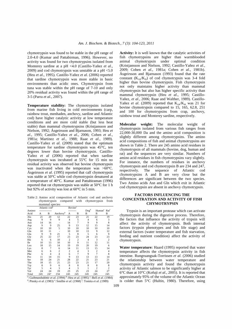
chymotrypsin was found to be stable in the pH range of 2.0-4.0 (Kumar and Pattabirman, 1996). However, no activity was found for two chymotrypsins isolated from Monterey sardine at a pH <4.0 (Castillo-Yañez et al., 2009) and cod chymotrypsin was unstable at a pH <5.0 (Heu et al., 1995). Castillo-Yañez et al. (2006) reported that sardine chymotrypsin was more stable in basic environments than acidic ones. Chymotrypsin from tuna was stable within the pH range of 7-10 and only 20% residual activity was found within the pH range of 3-5 (Parra et al., 2007). Temperature stability: The chymotrypsins isolated from marine fish living in cold environments (carp, rainbow trout, menhaden, anchovy, sardine and Atlantic cod) have higher catalytic activity at low temperature conditions and are more cold stable (but less heat stable) than mammal chymotrypsins (Kristjansson and Nielson, 1992; Ásgeirsson and Bjarnason, 1993; Heu et al., 1995; Castillo-Yañez et al., 2006; Cohen et al., 1981a; Martinez et al., 1988; Raae et al., 1989). Castillo-Yañez et al. (2009) stated that the optimum temperature for sardine chymotrypsin was 45°C, ten degrees lower than bovine chymotrypsin. Castillo-Yañez et al (2006) reported that when sardine chymotrypsin was incubated at 55°C for 15 min no residual activity was observed but bovine chymotrypsin was inactivated when the temperature was >60°C. Ásgeirsson et al. (1995) reported that calf chymotrypsin was stable at 50°C while cod chymotrypsin denatured at a temperature of 40°C. Kumar and Pattabiraman (1996) reported that rat chymotrypsin was stable at 50°C for 1 h but 92% of activity was lost at 60°C in 5 min. Table 2: Amino acid composition of Atlantic cod and anchovy
chymotrypsin compared with chymotrypsin from mammal species
Atlantic coda Bovinec
Amimo -------------- --------- Dogd Humane Ratf
Acid A B Anchovyb A B B B B Ala 21 20 12 22 23 21 22 19 Arg 9 9 9 4 5 4 6 3 Asn 14 16 - 14 7 9 9 8 Asp 9 9 25 9 13 12 15 14 Cys 10 10 5 10 10 10 10 10 Gln 10 11 - 10 10 13 9 12 Glu 9 9 21 5 8 5 6 9 Gly 25 22 17 23 23 24 23 23 His 6 6 10 2 2 4 3 2 Ile 10 13 10 10 9 11 12 11 Leu 18 15 14 19 19 20 19 6 Lys 8 7 6 14 11 13 15 14 Met 6 6 2 2 4 3 2 4 Phe 6 4 4 6 7 9 7 8 Pro 11 14 15 9 13 13 13 14 Ser 18 24 21 28 22 21 23 21 The 18 15 21 23 23 21 17 20 Trp 8 9 6 8 8 8 8 8 Tyr 5 4 3 4 3 1 2 2 Val 24 24 19 23 25 23 24 27 Total 245 247 234 245 245 245 245 245 a: Guðmundsdóttir et al. (1994) b: Heu et al. (1995) c: Bell et al. (1984) d: Pinsky et al. (1983) e: Smillie et al. (1968) f: Tomita et al. (1989)
Activity: It is well known that the catalytic activities of fish chymotrypsins are higher than warmblooded animal chymotrypsin under optimal condition (Kristjansson and Nielson, 1992; Castillo-Yañez et al., 2009; Cohen et al., 1981a; Cohen et al., 1981b). Ásgeirsson and Bjarnason (1993) found that the rate constant (Kcat/Km) of cod chymotrypsin was 3-4 fold higher than bovine chymotrypsin. Fish chymotrypsin not only maintains higher activity than mammal chymotrypsin but also has higher specific activity than mammal chymotrypsin (Heu et al., 1995; Castillo-Yañez, et al., 2006; Raae and Walther, 1989). Castillo-Yañez et al. (2009) reported that Kcat/Km was 21 for bovine chymotrypsin compared to 15, 165, 62.8, 251 and 100 for chymotrypsins from crap, anchovy, rainbow trout and Monterey sardine, respectively. Molecular weight: The molecular weight of chymotrypsin isolated from various fish ranges from 22,000-30,000 Da and the amino acid composition is slightly different among chymotrypsins. The amino acid compositions of fish and animal chymotrypsins are shown in Table 2. There are 245 amino acid residues in chymotrypsin of all mammals (bovine, dog, human and rat) and the sequences are very similar. Numbers of amino acid residues in fish chymotrypsins vary slightly. For instance, the numbers of residues in anchovy chymotrypsin and cod chymotrypsin B are 234 and 247, respectively. The sequence of Atlantic cod chymotrypsins A and B are very close but the differences are significant between the two species. Two Amino acids Asn and Gln which exit in Atlantic cod chymotrypsin are absent in anchovy chymotrypsin.
FACTORS INFLUENCING THE CONCENTRATION AND ACTIVITY OF FISH
CHYMOTRYPSIN Trypsin is an important protease which can activate chymotrypsin during the digestive process. Therefore, the factors that influence the activity of trypsin will affect the activity of chymotrypsin. Both internal factors (trypsin phenotypes and fish life stage) and external factors (water temperature and fish starvation, feeding and nutrient condition) affect the activity of chymotrypsin. Water temperature: Haard (1995) reported that water temperature affects the chymotrypsin activity in fish intestine. Rungruangsak-Torrissen et al. (2006) studied the relationship between water temperature and chymotrypsin activity and found the chymotrypsin activity of Atlantic salmon to be significantly higher at 6°C than at 10°C (Kofuji et al., 2005). It is reported that approximately 95% of the volume of the Atlantic Ocean is colder than 5°C (Hultin, 1980). Therefore, using

Am. J. Biochem. & Biotech., 7 (3): 104-123, 2011
110
Atlantic fish wastes for production of chymotrypsin will offer higher chymotrypsin concentrations because the high latitude and low water temperature. Fish species: Fish species has a significant influence on the concentration and activity of chymotrypsin. Based on their food habit, fish are separated into carnivores, omnivores and herbivores. Fish food habit would affect their gut weight to body weight ratio and the protealytic activity (Chakrabarti et al., 1995; Hidalgo et al., 1999). Hofer and Schiemer (1981) reported that proteolytic activity relates to different feeding habits. Although carnivorous species have smaller guts compared to herbivore species, they have higher activity. Chakrabarti et al. (1995) and Hidalgo et al. (1999) reported similar findings. Fish age and mass: Fish age will influence the chymotrypsin concentration and activity in fish. The chymotrypsin activity is very low (0.085Umg/protein) when the fish hatches, increases very slowly in the first 2-3 weeks, then dramatically increases in the next three days and finally increases at a constant rate (Chakrabarti et al., 1995; Cuvier-Peres and Kestemont, 2001; Chakrabarti et al., 2006). The total chymotrypsin activity is positively related to fish mass which is also correlated to fish age (Dabrowski and Glogowski, 1976; Rungruangsak-Torrissen et al., 2006). Practically, in chymotrypsin manufacturing, fish weight can be a parameter in selecting raw material. Starvation: Chymotrypsin will accumulate in the pancreas tissue when there is no food in the gut. Einarsson et al. (1996) reported that chymotrypsin activity of Atlantic salmon (in pyloriccaeca/pancreas) increased in the first few days but was reduced in the following days. This was explained by the reduced secretion of chymotrypsin from the pancreatic tissue to the intestinal digestion system and the accumulation in the pancreatic tissue. During starvation the level of chymotrypsin in intestinal keeps decreasing. Dabrowski and Glogowski (1976) reported that the starving rainbow trout showed higher chymotrypsin activity in intestine than the fed fish. Therefore, the effect of starvation effect on chymotrypsin activity will depend on the type of fish (Chakrabarti et al., 1995; Dabrowski and Glogowski, 1976).
INDUSTRIAL APPLICATIONS OF CHYMOTRYPSIN
Chymotrypsin has been used extensively in the food processing, leather production, chemical and medical industries.
Food industry: Chymotrypsin can be used to improve the food nutritional value of proteins and to lower the protein denaturation temperature and cleavage specificity (Yamashita et al., 1976; Haard, 1992). It is used in meat tenderization, fermentation, protein hydrolysate production and bone protein removal (Haard, 1998). In the dairy industry, chymotrypsin is used together with trypsin to hydrolyze casein, the main protein content in milk. Hydrolyzed by chymotrypsin, casein functional properties (such as antioxidant activity, angiotensin converting enzyme inhibition and antibacterial ability) are improved (Srinivas and Parksh, 2009). Chymotrypsin is used as an additive with trypsin to control hydrolysis of cheese whey proteins and β-lactoglobulin in the cheese production industry (Galvão et al., 2001). Leather production: In the leather industry, chymotrypsin has been used in production processes including dehairing, bating and soaking. In the bating process, the specific catalystic ability of chymotrypsin has been used in hydrolyzing nonleather-forming proteins such as mucoids, globulin and albumins in raw materials. It can also help tanning materials and other chemicals to penetrate into fibers in order to obtain the desired texture (Kamini et al., 1999; Nathalie et al., 2004; Sandhya et al., 2005; Cera, 2008). Chemical industry: In the detergent industry, chymotrypsin is added into laundry detergent or dish detergents to enhance the decontamination ability of the detergent. The enzyme remains stable in both ionic and non-ionic surfactants and maintains around 80% of its catalyzing ability after 1 h of incubation with chemical detergent (Espósito et al., 2009). Due to the specificity of chymotrypsin on proteins and peptides, it can be used to break down proteinaceous contaminatnts (blood and foods) on cloth efficiently. Another advantage of using chymotrypsin is its ability to work under mild conditions such as low water temperature or natural pH environments which may lower the damage to cloth and body (Kamini et al., 1999). A US patent for detergent (Patent Number: 5, 269, 959) lists chymotrypsin as a Liquid Deep Cleaning Detergent Composition (Schreibman et al., 1993). Another US patent (Patent Number: 20090281010) uses chymotrypsin in an eco-friendly laundry detergent composition comprising natural essence (Carter et al., 2009). Gupta et al. (2002) reported that at least 25% of extracted proteases is used as additives in laundry detergents every year. Espósito (2009) isolated chymotrypsin from fish processing waste and added it to laundry detergent to demonstrate high temperature tolerance and high enzyme stability (only

Am. J. Biochem. & Biotech., 7 (3): 104-123, 2011
111
15% enzyme activity was lost after 30 min incubation less than 60°C). Medical application: Chymotrypsin has been used in the treatment of dyspepsia and anorexia, cataract extraction, infertility and snakebite and as an anti-inflammation drug. It has been applied in several ways including: oral, local injection and atomized inhalation. Dyspepsia and anorexia: Dyspepsia and anorexia are defined as disturbed, difficult or painful digestion. The incomplete protein digestion is dangerous because it may result in allergies or even production of toxic materials by bacterial breakdown of the incompletely digested protein. Chymotrypsin can contribute to proper digestion of proteins (Sims, 2001). Normally, people do not need to supplement with additional proteolytic enzymes because the body can produce them but pancreatic insufficiency does occur because of chemotherapy, physical injuries, chronic stress, cystic fibrosis and acute pancreatitis so extra chymotrypsin supplication may be required (Sims, 2001). Anti-Inflammation and prevention of wound infection: Chymotrypsin has been reported to have significant inhibition against the early stage of inflammation. It is widely used both orally and by injection as a non-steroidal anti-inflammatory drug (NSAID) in the treatment of athletic injuries, wound infections, sciatica and hand trauma (Seppa, 1980). Chymotrypsin dissolves soft fibrin and cleans the proteinecious debris at inflammation sites (Swamy and Patil, 2008). Latha et al. (1997) used a trpsin-chymotrypsin combination in the ratio of 6:1 as an anti-inflammation agent to treat burn patients and found superior results compared to the use of trypsin alone. Cataract extraction: Alpha chymotrypsin is widely used to separate cataractous lens from the zonular attachment sites (Hill, 1960; Rhee et al., 1999; Rich et al., 1974). Chymotrypsin decreases the manipulating force required during the surgery to make it easy to remove the cataract which guarantee a higher success rates in surgery (O’Malley and Straatsma, 1961). It hass been shown that α-chymotrypsin specially attacks certain components of the zonules and does not cause any damage to other tissues in the eye (Hill, 1960; Yorston and Kiku, 2000) claimed that chymotrypsin was the fastest and cheapest way to treat the cataract. Using α-chymotrypsin during the cataract surgery can help avoid significant intraocular pressure (IOP). Rapidly raised IOP may cause pain and comeal oedema to patients (Rich et al., 1974; Passo et al., 1985).
Infertility: α-chymotrypsin can play a significant role in the process of semen liquefaction. It can shorten the semen liquefaction time and make it less viscous without influencing sperm motility. Combined with intrauterine insemination, α-chymotrypsin has been used in infertility treatment experiments on 38 infertile patients and 23% of them became pregnant. Zhang (2005) claimed that twice-weekly injection of 5mg chymotrypsin in 61 cases of infertile patients led to a cure rate of 83.6%. The advantages of using chymotrypsin are shorter and more effective treatment of infertility at a lower cost. Snakebite: Chymotrypsin has been used to treat patients with snakebite with good results (Omogbai et al., 2003). Zhang et al. (2005) studied the effect of local chymotrypsin injection on patients with bites from Chinese cobra and the results showed that it had less primeval effect than local injection of antivenom but better effects than other snakebite drugs. The toxins of snakes usually contain neurotoxin and cytotoxin which can change the permeability of cell membranes and the concentration of ions like Na+, K+ or Ca2+ causing cell death. The low amount of proteases kuronidase in snake toxin can accelerate this process (Zhang et al., 2005). Local chymotrypsin injection can slow down or stop this process because chymotrypsin can cleave the snake toxin protein. The hydrolyzed protein loses its toxic properties and becomes harmless to human (Zhang et al., 2005). Omogbai (2003) reported that the use of chymotrypsin to treat snakebite patients was shown effect especially for those patients with tissue inflammation and edema.
EXTRACTION AND PURIFICATION OF CHYMOTRYPSIN
Generally, the initial recovery steps of chymotrypsinogen (the inactivated zymogen precursor of chymotrypsin) are: (a) extraction which includes preparation of crude material, homogenization using a buffer to extract crude chymotrypsin (chymotrypsingen) from the prepared material and centrifugation to separate the crude chymotrypsin and (b) precipitation or fractionation to collect chymotrypsingen. The processes that can be used extract chymotrypsin from fish waste are shown in Fig. 5-7. Extraction: The fish stomach and intestines are removed from fish and separated as soon as the fish has been killed and washed with cold water or isotonical saline solution to get rid of blood in the tissue. The inhibitors in blood can reduce chymotrypsin activity (Chong et al., 2001; Boeris et al., 2009).

Am. J. Biochem. & Biotech., 7 (3): 104-123, 2011
112
Fig. 5: Extraction of chymotrypsin from fish waste
using ammonium sulphate method
Fig. 6: The extraction of chymotrypsin from fish waste
using reverse micelles method The intestine is then chopped into small pieces and mixed with 50 mM Tris-HCl buffer having a pH of 7.5. The homogenized mixture is centrifuged at 10000 rpm and 4ºC for 15 minutes. Crude chymotrypsin (chymotrypsinogen) is extracted as a supernatant and stored at -80ºC till further purification (Heu et al., 1995; Chong et al., 2001; Li et al., 2005; Castillo-Yañez et al., 2009; Yang et al., 2009).
Fig. 7: The extraction of chymotrypsin from fish waste
using chromatography Purification: The chymotrypsinogen purification processes account for up to 80% of the total cost of chymotrypsin production. Precipitation is the technique most widely used in the chymotrypsin production industry because it can effectively purify and concentrate chymotrypsin at a very low cost (Matsudo et al., 2003; Boeris et al., 2009).Generally, chymotrypsin precipitation is used to obtain crude chymotrypsinogen and can be combined with chymotography techniques for further purification. Purification can be accomplished either by precipitation (using Tris-HCl) and ammonium sulfate fractionation or formation of reverse micelles (Mockel and Barnard, 1969; Simpson, 2000; Wilk, 2001;. Castillo- Yañez et al., 2009; Yang et al., 2009). Ammonium sulfate method: Ammonium sulfate is the most reported polyelectrolyte in chymotrypsinogen production (Chatterjee et al., 2004). Folk (1970) used ammonium sulfate fractionation as second step inchymotrypsin purification process after extracted crude chymotrypsin with acetone powder. Same method had been used by Castillo-Yañez et al. (2006), Lam et al. (1999) and Möckel and Barnard (1969a). In the precipitation process, poly-charged molecules which contain opposite electrical charges to the chymotrypsinogen are added into protein solutions to form a chymotrypsinoge n-polyelectrolyte complex and generate insoluble aggregates. The materials used in the process are salts, non-ionicpolymers, organic solvents and polyelectrolytes which are easy to bioseparate in the downstream process (Boeris et al., 2009; Chatterjeeet al., 2004). Yang (2009) used ammonium

Am. J. Biochem. & Biotech., 7 (3): 104-123, 2011
113
sulfate saturation 30-60% in chymotrypsin extraction; Kristjansson and Nielson (1992), Castillo-Yañez et al. (2006, 2009) and Heu (1995) used ammomium sulfate saturation 30-70% and the recovery yield were in the rang from 55-60%. Boeris (2009) used Polyvinyl Sulfonate (PVS) as polyelectrolytes during the purification process to produce a chymotrypsinogen-PVS precipitate complex in acid pH which decreased the recovery rate of chymotrypsin. Also, increasing the PVS concentration lead to a lower recovery rate but produced a higher purity. Reverse micelles method: Reverse micelles are thermodynamically stable molecules that can extract large biomolecules like proteins through electrostatic interaction that attracts soluble proteins into the inner layer of the reverse micelles (Jolivalt et al., 1990; Hu and Gulari, 1996). Reverse micelles are useful because they can form amphiphilic structures in polar organic media which can be used to extract large amounts of proteins in the aqueous phase without denaturation. In the purification process, selection of surfactants plays a significant role in protein stabilization. Sodium di-2-ethylhexyl sulfosuccinate (AOT) is the most common surfactant used in chymotrypsin purification. It can form reverse micelles without adding co-surfactant (Jolivalt et al., 1990; Hu and Gulari, 1996; Hentsch et al., 1992). pH influences ionic molecular interactions in solution and, therefore, influences the efficiency of extraction by reverse micelles (Jolivalt et al., 1990). The yield of chymotrypsin will increase with an increase in pH and the maximum extraction yield can be reached at an isoelectric point around a pH of 8.5 (Hu and Gulari, 1996). However, the pH of protein aqueous solutions should be held near a pH of 3 because at that pH the autohydrolysis rate is minimized and chymotrypsin is most stable (Hu and Gulari, 1996). Jolivalt et al. (1990) and Hentsch et al. (1992) reported that increasing chloride ion concentration will decrease chymotrypsin yield by competing with chymotrypsin in the extraction process and the effect is particularly significant at low ionic strength. The whole extraction process with reverse micelles can be divided into two steps: forward-extraction and back-extraction. During the forward-extraction step, the aqueous and organic phases are separately prepared and homogenized with an orbital stirrer at 250 rpm for 90 min. After extraction (protein transfer from aqueous to organic phase) the phases are separated by centrifugation at 1500-2000 rpm for 10 min (Jolivalt et al., 1990; Hu and Gulari, 1996). The concentration of protein can be measured with UV spectroscopy at 280 nm. Back-extraction transfers proteins from reverse micelles to aqueous solutions. Back-extraction is
usually very slow and CaCl2 is added into the aqueous phase to assist the process (Hu and Gulari, 1996). The limitations of using reverse micelles in the back-extraction are due to: (a) the difficulty in separating proteins from the AOT reverse micellar phase and (b) the excessive time involved in the process (Hu and Gulari, 1996; Goto et al., 1998). Co-surfactants have been used to improve the extraction properties of AOT and the most common is Dioleyl Phosphoric Acid (DOPLA). This can be added to AOT to form AOT-DOLPA for use in chymotrypsin extract. Goto et al. (1998) reported that the effectiveness of mixed reverse micelles increased with increasing amount of DOLPA added. He also found that 4:1 ratio of AOT: DOLPA was the best for chymotrypsin extraction and that 10 mM of mixed reverse micelles had higher extraction ability than 200 mM of AOT. At low concentrations, the mixed reverse micells are very effective in separating and enriching chymotrypsin. Hu and Gulari (1996) and Goto et al. (1998) reported that mixed reverse micelles not only make chymotrypsin extraction more complete but it, also, shorten the back-extraction time from 24-2 h. Chromatography method: Chromatography is always the last step in the chymotrypsin purification process. Chromatography columns are used to concentrate and ultrafilter the fluid passing though them. The fluids can then be dialyzed against buffer (Folk, 1970). There are four types of chromatography that have been used in the chymotrypsin purification process: (a) ionic chromatography, (b) gel chromatography, (c) affinity chromatography and (d) hydrophobic interaction chromatography. Ionic chromatography is particularly useful because it can be used to both separate cationic and anionic forms of chymotrypsin A and B and to separate chymotrypsin from trypsin. Heu et al. (1995) used gelfiltration chromatography with a Sephadex G-75 column (2.6×75 cm) to first isolate chymotrypsin by molecular size. The crude chymotrypsin solution was further purified using ion chromatograph with a column packed with Diethylaminoethyl (DEAE) cellulose, designed to interact with negatively charged proteins. Ryan (1965) used a similar column to purify chymotrypsin from chicken. Raae and Walther (1989) used both ion chromatograph with a DEAE-Sepharose column and gelfiltration with a Phenyl-Butyl-Amine-Sepharose (PBAS) column to purify chymotrypsin and eluted with 35-40% ethleneglycolin. Ionic chromatography with CM-cellulose was used in the double purification of chymotrypsin and chymotrypsinogen from turtle and fish equilibrated with 0.01M sodium succinate and

Am. J. Biochem. & Biotech., 7 (3): 104-123, 2011
114
0.001M EDTA and eluted by salt gradients (Möckel and Bamard, 1969a; Möckel and Bamard, 1969b). Yang et al. (2009) used ionic chromatography with DEAE-Sephacel to purify chymotrypsin from crucian. The two detected peaks were: the unretained portion was cationic chymotrypsin (B) and the reterned portion was anionic chymotrypsin (A) which was eluted by NaCl at concentration of 0.2M. After gel-filtration on Sephacryl S-200, the two active portions were respectively subjected to hydrophobic interaction chromatography using Phenyl-Sepharose and SP-Sepharose for further purification. Most of the contaminated proteins were removed by washing with ammonium sulfate with concentrations ranging from 0 to 1M. The unretained ionic chymotrypsin B was further purified with a SP-Sepharose cationic exchange column. The results of purification are shown in Fig. 8. The affinity chromatography has been used by Fujiwara et al. (1974), Branchini and Ziolkowski (1979), Nishikata (1983) and Ahn and Chung (1985). In affinity chromatography, a column containing cross-linked insoluble polymer or gel attached with a competitive chymotrypsin inhibitor or ligand is used (Ahn and Chung, 1985; Cuatrecasas, 1970). During the chromatography all other unwanted protein will pass through the column but chymotrypsin will be absorbed and bonded through extended hydrocarbon chains which make the ligand at varying distances from the gel matric backbone. The whole process is pH depended because the affecst the ionization of functional group. Chymotrypsin can be recovered by elution with 0.1 M acetic acid. Fujiwara et al. (1974) used carbobrnzoxyl-L-phenylalanyl-triethylenetetraminyl-Sepharose (Z-L-Phe-T-Sepharose), Cuatrecasas (1970) used Sepharose-bound D-tryptophan methyl ester Nishikata (1983) used sepharose with chymostatin analogue (Gly-Gly-L-Leu-L-phenylalaninal). Ahn and Chung (1985) used 4-phenylbutylamine as a ligand in chymotrypsin purification.
ACTIVATION OF CHYMOTRYPSINOGEN Chymotrypsinogen is an inactive form of chymotrypsin existing in the pancreas and the extraction and purification processes result in pure chymotrypsinogen. Although chymotrypsinogen has enzymatic activity, the low level of activity makes it hard to detect in normal conditions. In order to evaluate the properties of chymotrypsin, chymotrypsinogen activation is a necessary process (Blow, 1976). Trypsin and other bacterial proteases excreted by Bacillus subtilis or Penicillium can be applied as a natural activator of chymotrypsin (Sakota, 1955; Dreyer and Neurrat, 1955; Prokuryakov, 1970). However, the optimal pHs of activators are different.
(a)
(b)
(c)
Fig. 8: Chromatographic purification of crucian carp
chymotrypsins. (a) DEAE-Sepharose chromatography. (b) Phenyl-Sepharose chromatography. (c) SP-Sepharose hromatography. (●): Suc-Leu-Leu-Val-Tyr-AMC hydrolyzing activity. (▲): Boc-Phe-Ser-Arg MAC hydrolyzing activity (Yang et al., 2009)
The optimum pH for trypsin is 7.5, the optimal pH of Aspergillus oryzae is 3.0-5.0 and the optimal pH of Kaufman and Erlanger ranges from 3.2-3.4 (Prokuryakov, 1970). Since chymotrypsinogen is a single polypeptide chain made up of 245 amino acids, different forms of chymotrypsin can be activated by controlling the reaction condition (Boeris et al., 2009). In the whole process, trypsin only hydrolyzes one peptide bond between Arg 15 and Lie Ile 16 and forms π-chymotrypsin (Appel, 1986). The remainder of the N-terminal peptide plays an important role in protecting chymotrypsinogen from other non-specific activators by using disulfide bonds, which are connected to the left molecule (Spilliaert and Gudmundsdottir, 2000). Then, π-chymotrypsin autocatalyzes itself form chymotrypsin and three new disulphide bonds to link the polypeptide chain as shown in Fig. 9. A further

Am. J. Biochem. & Biotech., 7 (3): 104-123, 2011
115
reaction will produce 2 types of chymotrypsin (δ-chymotrypsin and γ- chymotrypsin). Additionally, in the production of δ-chymotrypsin, it will lose the dipeptide Ser14-Arg15 bonds. Several factors contribute to chymotrypsinogen activation rates including concentration of trypsin, incubation time, temperature and pH.
Fig. 9: Activation of chymotrypsinogen. Peptide bonds
split by trypsin and chymotrypsin in an autocatalytic process leading to defined chains and peptides (Berg et al., 2007)
Fig. 10: ChTRP activity from bovine pancreas
homogenate in a medium of sodium citrate-Tris-HCl 50 mM at a pH of 8.2 (Boeris et al., 2009)
Fig. 11: Trypsin Requirement for Chymotrypsinogen
Activation (Glazer and Steer, 1976)
Trypsin concentration: Trypsin is an activator of chymotrypsinogen which can dramatically accelerate the speed of zymogen activation even at a very low concentration (0.01 mg/g of homogenate). The relative activity of chymotrypsin during the zymogen activation process in the presence or absence of trypsin is shown in Fig. 10 (Berg et al., 2007. Glazer and Steer (1976) found that an increase in trypsin concentations at low concentations (0.5-1.0%) did significantly increase the rate of activation but trypsin concentrations above 10% showed little increse in activation (Fig. 11). Thus, properly controlled concentration of trypsin in industry has commercial benefits. Trypsin not only plays a significant role in chymotrypsinogen activation but also contributes to different types of final products. With a large presence of trypsin, π-chymotrypsin is rapidly formed and becomes the predominate product in the process and it further loses its dipeptide Ser 14-Arg 15 bonds. These bonds are autocatalytically produced by δ-chymotrypsin, which is known as rapid activation of chymotrypsinogen (Bettelheim and Neurath, 1954; Appel, 1986). Slow activation occurs in the presence of low concentrations of trypsin. The production of α-chymotrypsin is the major pathway and γ-chymotrypsin will be formed only slowly (Appel, 1986). Incubation time: The incubation time required for chymotrypsinogen activation process depends on the activation method applied. There are two types of activation: classical activation (pH 7.5, 5°C, trypsin free, 48 incubation time) and rapid activation (activated by trypsin with incubation time varying from 1-48 h) (Bettelheim and Neurath, 1954; Miller et al., 1971). With the addition of trypsin, the enzymogen activation significantly increases in 1 h with the decrease of thehomogenate solution viscosity. Sakota (1955) reported that the activity of chymotrypsin increases at a constant rate in the first 10 h followed by an observed decrease in catalytic activity after 24 h. The possible reason for decreased chymotrypsin activity could be that the non-active form of trypsin being self-generating in the solution and destroying the structure of chymotrypsinogen (Sakota, 1955; Boeris et al., 2009). Engel and Alexander (1966) reported that activation of chymotrypsinogen was completed within 30 min. Dreyer and Neurath (1955), Miller et al. (1971), Glazer and Steer (1976) reported that the activity of chymotrypsin was stable at 4ºC and observed decreasing activity under high incubation temperature (Fig. 12).

Am. J. Biochem. & Biotech., 7 (3): 104-123, 2011
116
Fig. 12: The temperature dependence of chymotrypsinogen (Glazer and Steer, 1976)
Fig. 13: pH effect on the activation of
chymotrypsinogen (Guyonnet et al., 1999)
pH: Guyonnet et al. (1999) reported that the optimal pH for chymotrypsinogen activation is 7.5 (Fig. 13) and that the catalyze activity of chymotrypsin decreases with a decrease in pH. Similar results were reported by Engel and Alexander (1966), Bettelheim and Neurath (1954), Glazer and Steer (1976) and Guyonnet et al. (1999).
ASSAYING OF CHYMOTRYPSIN
Enzyme concentration: Three main methods are commonly used to evaluate chymotrypsin concentration: (a) absorbance at 280 nm (b) Bradford method and (c) Lowery method. Protein in solution has a maximum absorbance of ultraviolet light at 280nm. When measuring enzyme concentration, the wavelength must be adjusted to 280 nm and the system calibrated to zero with buffer solution. The absorbance of protein solution is then measured and the concentration (mg/mL) is calculated by the following equations (Layne, 1957; et al.; 2009; Stoscheck, 1990). This method had been used by Yang, Castillo-Yañez et al. (2006, 2009) and Möcke et al. (1969a). For protein mixture:
(cm) length path
∆AUionConcentrat 280= (2)
For protein mixture with possible nucleic acid contamination:
260AUx 0.76 -AU280×1.55ionConcentrat ∆∆=
(3)
In the Bradford method, Coomassie Blue reagent is mixed with the enzyme solution and the absorbance is read at 595 nm after incubation for 15 min. Concentrations are determined relative to standard curve based on Bovine Serum Albumin (BSA). The method had been used by Tsai (1986). The method developed by Lowery et al (1951) is a relatively sensitive method but more complicated and time consuming compared to the Bradford Method. The enzyme solution is treated with Folin-ciocaltea reagents to create a blue compound and allowed to incubate for 10 min. Absorbance is read at 660 nm and standard using BSA is required. There are many substrates that have been reported to affect the results obtained from this method including Tris, EDTA, Sulfhydryl compounds, potassium compounds, disdulfide compounds, carbohydrates, glycerol, Tricine, detergents, most phenols, uric acid, guanine, magnesium and calcium (Olson and Markwell, 2007). This method had been widely used by Yang et al. (2009), Castillo-Yañez et al. (2006, 2009), Lam et al. (1999), Li et al. (2003). Enzyme activity: A number of substrates are used to assay chymotrypsin activity including N-acetyl-L-Tyrosine Ethyl Ester (ATEE), Benzoyl-Tyrosine Ethyl Ester (BTEE) and N-Suc-Ala-Ala-Pro-Phe-p-Nitroanilide (SAAPPNA) (Hummel, 1959; Erlanger et al., 1961; Ramakrishna et al., 1987; Sabapathy and Teo, 1994; Heu et al., 1995; Chong et al., 2002; Chakrabarti et al., 2005; Li et al., 2005; Sveinsdóttir et al., 2006). The activity of chymotrypsin is determined as change in absorbance of chymotrypsin used in the assay per mg protein per min (Chakrabarti et al., 2005). When ATEE is used as substrate, one unit of enzyme activity is defined as the decrease in measured absorbance of 0.0075 min−1 at 237 nm and 25°C. The enzyme is mixed with ATEE solution in potassiium phosphate buffer and the absorbance is measured every half min for 5 min. The activity is calculated using the following Eq. 4.
solutionoriginalenzyme/mlmg0.20.0075
dilution/min∆AUActivity 237
×××
= (4)
When BTEE is used as substrate, one unit of enzyme activity is defined as one unit of enzyme

Am. J. Biochem. & Biotech., 7 (3): 104-123, 2011
117
hydrolyzing one micromole of BTEE per min at a pH of 7.5 and 25°C. The enzyme is added to BTEE solution dissolved in 50mM Tris-HCl buffer (10 mM CaCl2 with a pH of 7.5) at room temperature and the absorbance is measured at 256 nm every half min for 5 min. The activity is calculated using the following Eq. 5 (Hummel, 1959; Sabapathy and Teo, 1994; Li et al., 2005; Parra et al., 2007; Tubio et al., 2009; Boeris et al., 2009).
mixturereaction in theenzyme/mlmg964
1000/min∆AUActivity 256
××
=
(5)
When SAAPNA is used as a substrate, the enzyme activity is defined as one unit of enzyme activity hydrolyzing SAAPNA and releasing one micromole of p-nitroaniline at pH 7.5 and 25°C. The p-nitroaniline molar extinction coefficient is 8800 M−1cm−1. The enzyme is mixed with SAAPNA dissolve in 50 mM Tris-HCl buffer (10 mM CaCl2 with a pH of 7.5) and absorbance is measured at 410 nm every half min for 5 min at room temperature. The activity is calculated using the following Eq. 6 (Hummel, 1959; Chong et al., 2002; Sveinsdóttir et al., 2006; Castillo-Yañez et al., 2006; Castillo-Yañez et al., 2009):
assay in theprotein mg8800
mixturereaction of volume1000/min∆AUActivity 410
×××
= (6)
Molecular weight: Sodium Dodecyl Sulfate Polyacrylamide Gel Electrophoresis (SDS-PAGE) has been widely used to determine molecular weight of chymotrypsin after extraction using zymograms as substrate (Garcia-Carreno et al., 1993; Heu et al., 1995; Chong et al., 2002). The SDS-PAGE consisted of 12% separating gel and 5% stacking gel. The extracted chymotrypsin is mixed with Tris-HCl buffer (pH 6.8) to make a proteinase sample (Chong et al., 2002). A five-microliter mixture of the chymotrypsin sample is loaded into the SDS-PAGE gel and the gel is dipped in a Tris-HCl buffer and 3% casein (pH 7.5) for 30 min at 5°C in order to allow the casein to enter the gel. After incubating the gel at 25°C for 60 min, the gel is washed, stained with Coomassie Brilliant Blue for 2 h and destained. Clear bands on the gel indicating enzyme activity are compared with molecule weight markers to indicate the molecule weight of chymotrypsin (Chong et al., 2002; Chakrabarti et al., 2006). Additionally, Sephacryl S-100 column (0.9×55 cm) gel filtration can also used to determine the molecule weight of chymotrypsin (Heu et al., 1995).
Effect of inhibitors: To study the effect of inhibitors on chymotrypsin activity, purified enzyme is incubated with several specific protease inhibitors such as Ethylenediaminetetraacetic Acid (EDTA), chymotrypsin specific inhibitors such as tosyl-phenylalanine Chloromethyl-Ketone (TPCK), serine protease inhibitors such as Soybean, Trypsin Inhibitor (SBTI) and Phenyl-Methyl-Sulphonyl-Fluoride (PMSF), benzamidine, 4-(2-Aminoethyl)-Benzenesulfonyl Fluoreide (AEBSF), leupeptin, benzamidine, ela-statinal or Tosyl-L-Lysine Chloromethyl Ketone (TLCK) (Lam et al., 1999; Castillo-Yañez et al., 2006; Castillo-Yañez et al., 2009; Yang et al., 2009). After incubation, substrate solutions are added and the residual activity is measured. The percentage activity was calculated using the activity of the blank as 100% (Lam et al., 1999; Castillo-Yañez et al., 2006; Castillo-Yañez et al., 2009; Yang et al., 2009). Isoelectric point: The Isoelectric Point (IP) of chymotrypsin is always measured by analytical electrofocusing in thin-layer polyacrylamide flat get (LKB ampholyne PAG plate) containing ampholyne in the pH range of 3.5-9.5 (Castillo-Yañez et al., 2006; Castillo-Yañez et al., 2009). Purified protein is stained by Coomassie Brilliant Blue. The result is compared with isoelectric focusing calibration kit with 11 IP known proteins (Gildberg et al., 1990; Castillo-Yañez et al., 2006; Castillo-Yañez et al., 2009).
CONCLUSION Chymotrypsin is an important digestive enzyme which widely exists in mammal pancreatic tissues and fish guts (intestine). It is a endopeptide compound with 245 amino acid and molecular weights ranging for 22.000-30,000 Da. Chymotrypsin specifically hydrolyzes peptide bounds with α-amino acid carbonyl groups, nonpolar aromatic group and nonpolar groups, it is inhibited by synthetic chymotrypsin inhibitors such as TPCK, ZGGPCK, ZPCK, chymostatin and PMSF as well as natural inhibitors from egg white, lima beans, potatoes and pigeonpea. Three types of chymotrypsin (A, B and C) have been found in mammal pancreatic tissue but only two chymotrypsins (A and B) are found in fish. The optimal pH range for fish chymotrypsin is between 7.5-11 which is slightly higher than mammalian chymotrypsin. Fish chymotrypsin is more stable in basic environment than the acidic one. The optimal temperature for fish chymotrypsin is lower than mammal chymotrypsin. Most cold-water fish chymotrypsins have been reported to have 3-5 fold higher catalytic activity than mammalian chymotrypsin. The factors affecting

Am. J. Biochem. & Biotech., 7 (3): 104-123, 2011
118
activity and the amount of chymotrypsin in fish are water temperature, fish species, fish age, fish weight, fish weight, starvation and nutrition. Buffer extraction, ammonium sulphate precipitation and chromatography are usually used to produce chymotrypsin from fish. Each method has its own merit and demerit. Buffer extraction and ammonium sulphate precipitation can be used in large scale applications but the enzyme purity and activity is very low which can only be used in the leather or chemical industry. For the food and clinical industry, they require the use of higher purity and activity chymotrypsin, thus chromatography is needed. However, chromatography is very costly and it can only be applied on a small scale. Reverse micelles has been used for commercial chymotrypsin purification which can give a higher purity on a larger scale but no reports about purifying the chymotrypsin from raw materials have been found. Further study should focus on the optimization of purifying chymotrypsin from fish processing waste.
ACKNOWLEDGMENTS This research was supported by the Natural Sciences and Engineering Research Council (NSERC) of Canada.
REFERENCES AAC. Canada’s fish and seafood industry.
http://www.ats.agr.gc.ca/supply/3301_e.htm (September 25, 2010).
Ahn, S.H. and I.J. Chung, 1985. pH effect on the separation of α-chymotrypsin by affinity chromatography. Kor. J. Chem. Eng., 2: 11-15. Doi: 10.1007/BF02697544
Alarcón, F.J., M. Diaz, F.J. Moyano and E. Abellan, 1998. Characterization and functional properties of digestive proteases in two sparids; gilthead seabream Sparus aurata. and common dentex. Fish Physiol. Biochem., 19: 257-267. DOI: 10.1023/A:1007717708491
Alliot, E., R. Febvre, R. Metailler, A. Pastoureaud, 1973. Besoins nutritifs du bar (dicentrarchus labrax l.) Etude du taux de protéine et du taux de lipide dans le régime. http://archimer.ifremer.fr/doc/1973/publication-5996.pdf (March 28, 2011).
AMEC. Management of wastes from atlantic seafood processing operations. 2003; http://aczisc.dal.ca/nparpt.pdf (March 28, 2011).
Appel, W., 1986. Chymotrypsin: molecular and catalytic properties. Clinic. Biochem., 19: 317-322. DOI:10.1016/S0009-9120(86)80002-9
Ásgeirsson, B. J.B. Bjarnason, 1993. Properties of elastase from atlantic cod, a cold-adapted proteinase. Biochim. Biophy. Acta., 1164: 91-100. DOI: 10.1016/0167-4838(93)90116-9
Ásgeirsson, B., R. Gartemink, J.F. Chlebowski, 1995. Alkaline phosphatase from Atlantic cod (Gadusmorhua). Kinetic and structural properties which indicate adaptation to low temperatures. Comp. Biochem. Physiol. B Biochem. Mol. Biol., 110: 315-329. DOI: 10.1016/0305-0491(94)00171-P
Bechtel, P.J., 2003. Properties of different fish processing by-products from pollock, cod and salmon. J. Food Processing Preservation., 27: 101-116. DOI: 10.1111/j.1745-4549.2003.tb00505.x
Bell, G., C. Quinto, M. Quiroga, P. Valenzuela, C. Craik and W. Rutter, 1984. Isolation and sequence of a rat chymotrypsin B gene. J. Biol. Chem., 259: 14265-14270. PMID:6209274
Bender, M. and C.K. Ferbnj, 1964. The current status of the chymotrypsin mechanism. J. Amer. Chem. Soc., 86: 3704-3714. DOI: 10.1021/ja01072a020
Bender, M.L. and J.V. Killheffer, 1973. Chymotrypsins. Crit. Rev. Biochem., 1: 149-199. PMID: 4372018
Berg, J.M., L.T. John and L. Stryer, 2007. Biochemistry 5th Edn., W.H. Freeman: New York, ISBN-13: 978-0716787242
Bettelheim, F.R. and H. Neurath, 1954. The rapid activation of chymotrypsinogen. J. Biol Chem., 212: 241-253. http://hwmaint.jbc.org/cgi/reprint/212/1/241.pdf (Septrmber 16, 2011)
Blow, D.M., 1976. Structure and mechanism of chymotrypsin. Acc. Chem. Res., 9: 145-152. DOI: 10.1021/ar50100a004
Boeris, V., D. Romanini, B. Farruggia and G. Pico, 2009. Purification of chymotrypsin from bovine pancreas using precipitation with a strong anionic polyelectrolyte. Proc. Biochem., 44: 588-592. DOI:10.1016/j.procbio.2009.02.009
Branchini, B. and R. Ziolkowski, 1979. The separation of chymotrypsin and chymotrypsinogen: An affinity chromatography experiment for biological chemistry students. J. Chem. Educ., 56: 281. DOI: 10.1021/ed056p281
Byun, H.G., P.J. Park, N.J. Sung, S.K. Kim, 2003. Purification and characterization of a serine proteinase from the tuna pyloric caeca. J. Food Biochem., 26: 479-494. DOI: 10.1111/j.1745-4514.2002.tb00768.x

Am. J. Biochem. & Biotech., 7 (3): 104-123, 2011
119
Carter, D.L., P. Lam, T. Bastigkeit, T. Mikkelsen and D. Wood, 2009. Eco-friendly laundry detergent compositions comprising natural essence. Patent application number: 20090281010; http://www.faqs.org/patents/app/20090281010 (January 13, 2011).
Castillo-Yañez, F.J., R. Pacheco-Aguilar and F.L. Garcia-Carreno, 2006. Purification and biochemical characterization of chymotrypsin from the viscera of Monterey sardine (Sardinops sagax caeruleus). Food Chem., 99: 252-259.
Castillo-Yañez, F.J., R. Pacheco-Aguilar, M.E. Lugo-Sanchez, G. Garcia-Sanchez and I.E. Quintero-Reyes, 2009. Biochemical characterization of an isoform of chymotrypsin from the viscera of Monterey sardine (Sardinops sagax caerulea) and comparison with bovine chymotrypsin. Food Chem., 112: 634-639. DOI:10.1016/j.foodchem.2005.06.052
Cera, E.D. Serine Proteases. 2008; http://www.scitopics.com/Serine_Proteases.html (November 27, 2010).
Chakrabarti, I., M.A. Gani, K.K. Chaki, R. Sur and K.K. Misra, 1995. Digestive enzyme in 11 freshwater teleost fish species in relation to food habit and niche segregation. Comp. Biochem. Physiol. A Physiol., 112: 167-177. DOI: 10.1016/0300-9629(95)00072-F
Chakrabarti, R., R.M. Rathore, P. Mittal and S. Kumar, 2006. Functional changes in digestive enzymes and characterization of proteases of silver carp (♂) and bighead carp (♀) hybrid, during early ontogeny. Aquac., 253 : 694-702. DOI: 10.1016/j.aquaculture.2005.08.018
Chatterjee, S., S. Chatterjee, B.P. Chatterjee and A.K. Guha, 2004. Clarification of fruit juice with chitosan. Proc. Biochem., 39: 2229-2232. DOI: 10.1016/S0044-8486(01)00630-5.
Chong, A.S.C., R. Hashim, L. Chow-Yang and A.B. Ali, 2002. Partial characterization and activities of proteases from the digestive tract of discus fish Symphysodon aequifasciata. Aquaculture, 203: 321-333. DOI: 10.1016/S0044-8486(01)00630-5
Clark, J., N.L MacDonald and J.R. Stark, 1985. Metabolish in marine flatfish- II. Protein digestion in dover sole (Solea Solea L.). Comp. Biochem. Physiol. B Comp. Biochem., 81: 217-222. DOI: 10.1016/0305-0491(85)90186-5
Cohen, T., A. Gertler and Y. Birk, 1981a. Pancreatic proteolytic enzymes from carp (Cyprinus carpio)-I. Purification and physical properties of trypsin, chymotrypsin, elastase and carboxypeptidase. Comparative Biochemistry and Physiology Part B: Comp. Biochem., 69: 639-646. DOI: 10.1016/0305-0491(81)90364-3
Cohen, T., A. Gertler and Y. Birk, 1981b. Pancreatic proteolytic enzymes from carp (Cyprinus carpio)-II. Kinetic properties and inhibition studies of trypsin, chymotrypsin and elastase. Comp. Biochem. Physiol. B Comp. Biochem., 69: 647-653. DOI: 10.1016/0305-0491(81)90365-5
Cuatrecasas, P., 1970. Protein purification by affinity chromatography derivatizations of afarose and polyacrylamide beads. J. Biol. Chem., 245: 3059-3065.
Cuvier-Peres, A. and P. Kestemont, 2001. Development of some digestive enzymes in Eurasian perch larvae Perca fluviatilis. Fish Physiol. Biochem., 24: 279-285. DOI: 10.1023/A:1015033300526
Dabrowski, K., J. Glogowski, 1976. Study on the role of exogenous proteolytic enzymes in digestion process in fish. Hydrobiologia., 54: 129-134. DOI: 10.1007/BF00034986
Dimes, L.E., F.L. Garcia-Carreno and N.F. Haard, 1994. Estimation of protein digestibility: III. Studies on the digestive enzymes from the pyloric ceca of rainbow trout and salmon. Comp. Biochem. Physiol. A Physiol., 109: 349-360. DOI: 10.1016/0300-9629(94)90138-4
Dreyer, W.J. and H. Neurrat, 1955. The activation of chymotrypsinogen isolation and identification of a peptide librated during activation. J. Biol. Chem., 217: 527-540. PMID: 13271414
Einarsson, S., P.S. Davies and C. Talbot, 1996. The effect of feeding on the secretion of pepsin, trypsin and chymotrypsin in the Atlantic salmon, Salmo salar L. Fish Physiol. Biochem.. 15: 439-446. DOI: 10.1007/BF01875587
Elert, E.V., M.K. Agrawal, C. Gebauer, H. Jaensch and B. Ulrike 2004. Protease activity in gut of daphnia magna: evidence for trypsin and chymotrypsin enzymes. Comp. Biochem. Physiol. B Biochem. Mol. Biol., 137: 287-296. DOI:10.1016/j.cbpc.2003.11.008
Engel, A. and B. Alexander, 1966. Activation of ahymotrypsinogen-A by thrombin preparations. Biochem., 5: 3590-3598. DOI: 10.1021/bi00875a030
Erlanger, B.F., N. Kokowsky and W. Cohen, 1961. The preparation and properties of two new chromogenic substrates of trypsin. Arch. Biochem. Biophys., 95: 271-278. PMID:13890599
Espósito, T.S., I.P.G. Amaral, G.B. Oliveira, J.L.B. Carvalho and R.S. Bezerra, 2009. Fish processing waste as a source of alkaline proteases for laundry detergent. Food Chem., 112: 125-130. DOI:10.1016/j.foodchem.2008.05.049

Am. J. Biochem. & Biotech., 7 (3): 104-123, 2011
120
Fersht, A.R. and Y. Requena, 1971. Mechanism of the .alpha.-chymotrypsin-catalyzed hydrolysis of amides. pH dependence of kc and km. kinetic detection of an intermediate. J. Amer. Chem. Soc., 93: 7079-7087. DOI: 10.1021/ja00754a066
Folk, J.E., 1970. Chymotrypsin C (porcine pancreas). Meth. Enzymol., 19: 109-112. DOI:10.1016/0076-6879(70)19008-2
Fong, W.P., E.Y Chan and K.K. Lau, 1998. Isolation of two chymotrypsinsfrom grass carp. Biochem. Mol. Bio. Int., 45: 409-418. PMID: 9678263
Fujiwara, A., K. Osue and D. Tsuru, 1974. Affinity chromatography of α-chymotrypsin, subtilisin and metalloendopeptidases on carbobenzoxy-L-phenylalanyl-triethylenetetraminyl-sepharose. J. Biochem., 77: 739-743. PMID:238966
Galvão, C.M., A.F. Silva, M.F. Custodio, R. Monti and R.L. Giordano, 2001. Controlled hydrolysis of cheese whey proteins using trypsin and α-chymotrypsin. Appl. Biochem. Biotech., 91-93:761-776. PMID: 11963904
Garcia-Carreno, F.L., L.E. Dimes and N.F. Haard, 1993. Substrategel electrophoresis for composition and molecular weight of protienases or proteinaceous proteinase inhibitors. Analyt. Biochem., 214: 65-69. PMID:8250256
Geiger, R., 1985. Chymotrypsin. In Methods of Enzymatic Analysis, 3rd, Bergmeyer, H.U. Ed.; VCH Pub: Deerfield, 5: 99-118.
Gildberg, A., R.L. Olsen and J.B. Bjarnason, 1990. Catalytic properties and chemical composition of pepsins from Atlantic cod (gadus morhua). Comp. Biochem. Physiol. B., 96: 323-330. doi:10.1016/0305-0491(90)90382-4
Glazer, G. and M.L. Steer, 1977. Requirements for activation of trypsinogen and chymotrypsinogen in rabbit pancreatic juice. Anal. Biochem., 77: 130-140. DOI:10.1016/0003-2697(77)90297-4
Goto, M., Y. Ishikawa, T. Ono, F. Nakashio and T.A. Hatton, 1998. Extraction and activity of chymotrypsin using AOT-DOLPA mixed reversed micellar systems. Biotechnol Prog., 14: 729-734. DOI: 10.1021/bp9800790
Guðmundsdóttir, Á., S. Óskarsson, A.E. Eakin, C.S. Craik and J.B. Bjarnason, 1994. Atlantic cod cDNA encoding a psychrophilic chymotrypsinogen. Biochem. Biophys. Acta (BBA)-Gene. Struct. Express., 1219: 211-214. doi:10.1016/0167-4781(94)90274-7
Guha, M.K. and N.K. Sinha, 1984. Purification and characterization of chymotrypsin inhibitors from marine turtle egg white. J. Biosci., 6 :155-163. DOI: 10.1007/BF02702636
Gumisiriza, R., A.M. Mashandete, M.S.T. Rubindamayugi, F. Kansiime and A.K. Kivaisi, 2009. Enhancement of anaerobic digestion of Nile perch fish processing wastewater. Afr. J. Biotechnol., 8: 328-333. ISSN 1684-5315
Gupta, R., Q.K. Beg and P. Larenz, 2002. Bacterial alkaline proteases: molecular approaches and industrial applications. Appl. Microbiol. Biotechnol., 59: 15-32. PMID: 12073127
Guyonnet, V., F. TlÇuscik, P.L. Long, A. Polanowski and J. Travis, 1999. Purification and partial characterization of the pancreatic proteolytic enzymes trypsin, chymotrypsin and elastase from the chicken. J. Chrom. A., 852: 217-225. DOI:10.1016/S0021-9673(99)00355-6
Haard, N.F. and B.K. Simpson, 2000. Eds. Seafood Enzymes: Utilization and Influence on Postharvest Seafood Quality, Marcel Dekker, Inc, New York. ISBN-13: 978-0824703264
Haard, N.F., 1992. A review of proteolytic enzymes from marine organisms and their application in the food industry. J. Aqua. Food Prod. Technol., 1: 17-36. DOI:10.1300/J030v01n01_05
Haard, N.F., 1995. Digestibility and in Citro Evaluation of Plant for Salmonis Feed.Lim. In Nutrition and utilization technology in aquaculture; Lim, C. and Sessa, D.J., Eds. AOCS Press: Champaign, Ill, 199-218.
Haard, N.F., 1998. Specialty enzymes from marine organisms. Food Technol., 52: 64-67. ISSN: 0015-6639
Hentsch, M., P. Menoud, L. Steiner, E. Flaschel and A. Renken, 1992. Optimization of the surfactant (AOT) concentration in a reverse micellar extraction process. Biotrchnol. Tech.., 6: 359-364. DOI: 10.1007/BF02439326
Heu, M.S., H.R Kim and J.H. Pyeun, 1995. Comparison of trypsin and chymotrypsin from theviscera of anchovy, Engraulis japonica. Comp. Biochem. Physiol. B Biochem. Mol. Biol., 112: 557-67. PMID: 8529032
Hidalgo, M.C., E. Urea and A. Sanz, 1999. Comparative study of digestive enzymes in fish with different nutritional habits. Proteolytic and amylase activities. Aquac., 170: 267-283. DOI:10.1016/S0044-8486(98)00413-X
Hill, H.F. and W. Me, 1960. Technique of cataract extraction with alpha-chymotrypsin. Arch. Ophthalmol., 64: 601-605. http://archopht.ama-assn.org/cgi/content/summary/64/4/601 (September 17, 2011)
Hofer, R. and F. Schiemer, 1981. Proteolytic activity in the digestive tract of several species of fish with different feeding habits. Oecolog., 48: 342-345. DOI: 10.1007/BF00346492

Am. J. Biochem. & Biotech., 7 (3): 104-123, 2011
121
Hu, Z.Y. and E. Gulari, 1996. Communication to the editor protein extraction using the sodium bis(2-ethyIhexyI) phosphate (NaDEHP) reverse micellar system. Biotechnol. Bioengin., 50: 203-206. http://deepblue.lib.umich.edu/bitstream/2027.42/37936/1/9_ftp.pdf (September 17, 2011)
Hudáky, P., G. Kaslik, I. Venekei and L. Gráf, 1999. The differential specificity of chymotrypsin A and B is determined by amino acid 226. Eur. J. Biochem., 259: 528-533. DOI: 10.1046/j.1432-1327.1999.00075.x
Hultin, H.O., 1980. Enzymes from organisms acclimated to low temperatures. In Enzymes: The Interface between Technology and Economics, Danehy, J.P. and B. Wolnak Eds; Marcel Decker, New York, pp: 161-178. ISBN 13: 9780824769307
Hummel, B.C.W., 1959. A modified spectrophotometric determination of chymotrypsin, trypsin and thrombin. Ca. J. Biochem Physiol., 37: 1393-1399. PMID:14405350
IFC Environment, Health and Safety Guidelines for Fish Processing. 2007; http://www.ifc.org/ifcext/enviro.nsf/AttachmentsByTitle/gui_fishproc/$FILE/fishprocessing.pdf (April 6, 2011).
Joakimsson, K.G. and F. Nagayama, 1990. Partial purificationand characterisation of proteinases from the pyloric caeca of skipjack, Euthynnus pelamis. J. Tokyo Univ. Fish., 77: 95-104.
Jolivalt, C., M. Minier, H. Renon, 1990. Extraction of α-chymotrypsin using reverse micelles. J. Colloid. Interface Sci., 135: 85-96. DOI:10.1016/0021-9797(90)90290-5
Kallies, B. and R. Mitzner, 1996. Substrate specifity of chymotrypsin. Study of induced strain by molecular mechanics. J. Mol. Mod., 2: 149-159. . DOI: 10.1007/s0089460020149
Kamini, N.R., C. Hemachander, J. Mala and S. Geraldine, 1999. Microbial enzyme technology as an alternative to conventional chemicals in leather industry. http://www.ias.ac.in/currsci/jul10/articles16.htm (June, 7, 2010).
Kim, S. and E. Mendis, 2006. Bioactive compounds from marine processing byproducts-a review. Food Res. Int., 39: 383-393. DOI:10.1016/j.foodres.2005.10.010
Kofuji, P.Y.M., A. Akimoto, H. Hosokawa and T. Masumoto, 2005. Seasonal changes in proteolytic enzymes of yellowtail Seriola quinqueradiata (Temminck and Schlegel; Carangidae) fed extruded diets containing different protein and energy levels. Aquac. Res., 36: 696-703. 10.1111/j.1365-2109.2005.01276.x
Kristjansson, M.M. and H.H. Nielson, 1992. Purification and characterization of two chymotrypsin-like protease from the pyloric ceca of rainbow trout (Oncorhychus mykiss). Comp Biochem Physiol., 101: 247-253. PMID:1499272
Kumar, R.S. and T.N. Pattabirman, 1996. Purification and characterization of chymotrypsin-like enzyme from rat plasma. Ind. J. Clinic. Biochem.. 11: 152-157. DOI: 10.1007/BF02896434
Lam, W., G.M. Coast and R.C. Rayne, 1999. Isolation and characterization of two chymotrypsins from the midgut of Locusta migratoria. Ins. Biochem. Mol. Biol., 29: 653-660. DOI:10.1016/S0965-1748(99)00049-1
Latha, B., M. Ramakrishnan, V. Jayaraman and M. Babu, 1997. Serum Enzymatic changes modulated using trypsin: chymotrypsin preparation during burn wounds in humans. Burns., 23: 560-564. DOI:10.1016/S0305-4179(97)00080-6
Layne, E., 1957. Spectrophotometric and turbidimetric methods for measuring proteins. Meth. Enzymol., 10: 447-455. doi:10.1016/S0076-6879(57)03413-8
Leslie, P., J.H. Wang, 1968. On the mechanism of action at the acylation step of the α-chymotrypsin-catalyzed hydrolysis of anilides. J. Bio. Chem., 243: 3729-3734. http://www.jbc.org/content/243/13/3729.full.pdf+html (April 6, 2011)
Leth-Larsen, R., B. Àsgeirsson, N.M. Thorolfsson and P. HØjrup, 1996. Structure of chymotrypsin variant B from Atlantic cod, Gadus morhua. Biochim. Biophys. Acta., 1297: 49-56. DOI:10.1016/0167-4838(96)00088-X
Li, Z.Y., W. Youravong and A.H. Kittikun, 2005. Separation of proteases from yellow tuna spleen by ultra filtration. Biores. Technol., 97: 2364-2370. DOI:10.1016/j.biortech.2005.10.019
Lowery, O.H., N.J. Rosebrough, A.L. Farr and R.J. Randall, 1951. Protein measurement with the folin phenol reagent. J. Biol. Chem., 193: 265-275. http://www.jbc.org/content/193/1/265.full.pdf+html (September 15, 2011)
Lucas, E.C., M. Caplow and K.J. Bush, 1973. Chymotrypsin catalysis. evidence for a new intermediate III. J. Amer. Chem. Soc., 95: 2670-2673. DOI: 10.1021/ja00789a043
Makkar, H.P.S., P. Siddhuraju and K. Becker, 2007. Chymotrypsin inhibitor. Meth. Mol. Biol., 393: 7-9. DOI: 10.1007/978-1-59745-425-4_2
Martinez, A., R.L. Olsen and J.L. Serra, 1988. Purification and characterization of two trypsin-like enzymes from the digestive tract of anchovy Engraulis encrasicholus. Comp. Biochem. Physiol. B Comp. Biochem., 91: 677-684. DOI: 10.1007/978-1-59745-425-4_2

Am. J. Biochem. & Biotech., 7 (3): 104-123, 2011
122
Matsudo, T., K. Ogawaand, E. Kokufuta, 2003. Complex formation of protein with different water-soluble synthetic polymers. Biomacromol., 4: 1794-1799. DOI: 10.1021/bm0341935
Miller, D.D., T.A. Horbett, D.C. Teller, 1971. Reevaluation of the activation of bovine chymotrypsinogen A. Biochem., 10: 4641-4648. PMID: 5168678
Möckel, W. and E.A. Barnard, 1969a. Isolation and properties of some reptilian and fish chymotrypsin. Biochim. Biophy. Acta (BBA)-Enzymol., 178: 354-363. . DOI:10.1016/0005-2744(69)90402-1
Möckel, W., E.A. Barnard, 1969b. Isolation and Properties of Two Chymotrypsins From the Turle Pseudemys Elegans. Biochim Biophy. Acta (BBA)-Enzymol., 191: 370-378. DOI:10.1016/0005-2744(69)90256-3
Nathalie, R.L.F., M. Desjardins and P.U. Blier, 2004. Enhancement of profitability perspectives of wolffish cultivation by the extraction of high value biomolecules. In Sea Food Quality and Safety: Advances in the New Millennium, Shahidi, F.; Simpson, B.K. Eds; DEStech Publications.Inc, St. John’s, NL, Canada, pp: 61-62. ISBN: 0-9683220-2-6
Nelson, L.N., A.L. Lehninger, M.M. Cox and Chymotrypsin mechanism. 2008. http://www.sumanasinc.com/webcontent/animations/content/chymotrypsin.html (March 4, 2010).
Nishikata, M., 1983. Affinity chromatography of chymotrypsin on a sepharose derivative coupled with a chymostatin analogue. J. Biochem., 93: 73-79. PMID: 6841337
O’Malley, M.M. and B.R. Straatsma, 1961. Experimentally induced adverse effect of alpha-chymotrypsin. Arch. ophthalmol., 66: 539-544. PMID:14480893
Olson, B.J.S.C. and J. Markwell, 2007. Assays for determination of protein concentration. In Current Protocols in Protein Science: John Wiley and Sons, Inc., New York, Unit 3.4. Doi: 10.1002/0471140864.ps0304s48
Omogbai, E.K.I., Z.A.M. Nworgu, M.A. Imhafidon, A.A. Ikpeme and D.O. Ojo et al., 2003. Snake Bites in Nigeria: A Study of the Prevalence and Treatment in Benin City. Trop. J. Pharm. Res., 1: 39-44. http://www.bioline.org.br/pdf?pr02006 (September 15, 2011)
Parker, L. and J.H. Wang, 1968. On the mechanism of action at the acylation step of the α-chymotrypsin-catalyzed hydrolysis of anilides. J. Biol. Chem., 243: 3729-3734. http://www.jbc.org/content/243/13/3729.full.pdf+html (September 15, 2011)
Parra, A.M.D., A. Rosas, J.P. Lazo and M.T. Viana, 2007. Partial characterization of the digestive enzymes of Pacific bluefin tuna (Thunnus orientalis) under culture conditions. Fish Physiol. Biochem., 33: 223-231. DOI: 10.1007/s10695-007-9134-9
Passo, M.S., J.T. Ernest and T.K. Goldstick, 1985. Hyaluronate increases intraocular pressure when used in cataract extraction. Br. J. Ophthalmol., 69: 572-575. DOI: 10.1136/bjo.69.8.572
Pinsky, S.D., K.S. Laforge, V. Luc, G. Scheele, 1983. Identification of cDNA clones encoding secretory isoenzyme forms: Sequence determination of canine pancreatic prechymotrypsinogen 2 mRNA. Proc. Natl. Acad. Sci. USA., 80: 7486-7490. PMCID: PMC389976
PNPPRC. Pollution prevention opportunities in the fish processing industry. 1993. http://www.fpeac.org/seafood/P2FishProcessing.pdf (October 10, 2010).
Prokuryakov, M.T., 1970. Interaction between pepsin and chymotrypsinogen. Bull. Exp. Biol. Med.., 69: 239-240. DOI: 10.1007/BF00801502
Raae, A.J. and B.T. Walther, 1989. Purification and characterization of chymotrypsin, trypsin and elastase like proteinases from cod (Gadus morhua L.). Comp. Biochem. Physiol. B Comp. Biochem., 93: 317-324.
Raae, A.J., R. Flengsrudt and K. Sletten, 1995. Chymotrypsin isoenzymes in Atlantic cod; differences in kinetics and substrate specificity. Comp. Biochem. Physiol.. B Biochem. Mol. Biol., 112: 393-398. DOI:10.1016/0305-0491(89)90087-4
Ramakrishna, M., H.O. Hultin and W.F. Racicot, 1987. Some kinetic properties of dogfish chymotrypsin. Comp. Biochem. Physiol. B Biochem. Mol. Biol., 87: 25-30.
Rhee, D.J., V.A., Deramo, B.P. Connolly and M.H. Blecher, 1999. Intraocular pressure trends after supranormal pressurization to aid closure of sutureless cataract wounds. J. Catar. Refr. Surg., 25: 546-549. PMID: 10198861
Rich, W.J., N.D. Radtke and B.E. Cohan, 1974. Early ocular hypertension after cataract Extraction. Br. J. Ophthalmol., 58: 725-731. PMCID: PMC1215009
Rungruangsak-Torrissen, K., R. Mossi, L.H. Andresen, A. Berg and R. Waagb, 2006. Different expressions of trypsin and chymotrypsin in relation to growth in Atlantic salmon (Salmo salar L.). Fish Physiol. Biochem., 32: 7-23. . PMID: 20035474
Ryan, C.A., 1965. Chicken chymotrypsin and turkey trypsin part I: purification. Arch. Biochem. Biophy., 110: 169-174. DOI:10.1016/0003-9861(65)90169-4

Am. J. Biochem. & Biotech., 7 (3): 104-123, 2011
123
Sabapathy, U., L.H. Teo, 1995. Some properties of the intestinal proteases of the rabbitfish, Siganus canaliculatus (Park). Fish Physiol. Biochem., 14: 215-221. DOI: 10.1007/BF00004312
Sakota, N., 1955. On the mechanism of the activation of α-chymotrypsinogen to α-chymotrypsin. II. The changes in amino-terminal amino acids of α-chymotrypsinogen during the activation by trypsin. J. Biochem., 42: 69-75.
Sandhya, S., S. Padmavathy, K. Swaminathan, Y.V. Subrahmanyam and S.N. Kaul, 2005. Microaerophilic-aerobic sequential batch reactor for treatment of azo dyes containing simulated wastewater. Process Biochem., 40: 885-890. doi:10.1016/j.procbio.2004.02.015
Santigosa, E., J. Sànchez, F. Médale, S. Kaushik, J. Pèrez-Sànchez and M.A. Gallardo, 2008. Modifications of digestive enzymes in trout (Oncorhynchus mykiss) and sea bream (Sparus aurata) in response to dietary fish meal replacement by plant protein sources. Aquac., 282: 68-74. ISSN 0044-8486
Schreibman, G. Liquid deep cleaning detergent composition. United States Patent number: 5269959. 1993; http://www.freepatentsonline.com/5269959.html (April 11, 2011).
Seppa, H., 1980. The role of chymotrypsin-like protease of rat mast cells in inflammatory vasopermeability and fibrinolysis. Inflamm., 4: 1-8.
Sims, J. Applied kinesiology. 2001; http://www.encyclopedia.com/doc/1G2-3435100048.html (January 14, 2011).
Smillie, L.B., A. Furka, N. Nagabhushan, K.J. Stevenson and C.O. Parkes, 1968. Structure of chymotrypsinogen B compared with chymotrypsinogen A and trypsinogen. Nature., 218: 343–346. DOI: 10.1038/218343a0
Smith, E.L., D.M. Brown, M. Laskowski, 1951. Molecular weights of chymotrypsinogen and chymotrypsin A and B. J. Biol. Chem., 191: 639-650. PMID: 14861210
Spilliaert, R. and À. Gudmundsdottir, 2000. molecular cloning of the Atlantic cod chymotrypsinogen B. Mich. Comp. Genom., 5: 41-50. DOI:10.1089/10906590050145258
Srinivas, S. and V. Parksh, 2009. Bioactive peptides from bovine milk a-casein: isolation, characterization and multifunctional properties. Int. J. Pept. Res. Therapeut., 16: 7-15. DOI: 10.1007/s10989-009-9196-x
Stoscheck, C., 1990. Quantification of protein. Meth. Enzymol., 182: 50-68. PMID:2314256
Sveinsdóttir, H., H. Thorarensen and Á. Gudmundsdóttir, 2006. Involvement of trypsin and chymotrypsin activities in Atlantic cod (Gadus morhua) embryogenesis. Aquac., 260: 307-314. DOI:10.1016/j.aquaculture.2006.06.009
Swamy, A.H.M.V. and P.A. Patil, 2008. Effect of some clinically used proteolytic enzymes on inflammation in rats. Ind. J. Pharm. Sci., 70: 114-117. DOI: 10.4103/0250-474X.40347
Swatschek, D., W. Schatton, J. Kellerman, W.E.G. Muller and J. Kreuter, 2002. Marine sponge collagen: isolation, characterization and effects on the skin parameters on pH, moisture and sebum. Eur. J. Pharm. Biopharm., 53: 107-113. PMID: 11777758
Tokumoto, T., 1999. Nature and role of proteasomes in maturation of fish oocytes. Int. Rev. Cytol., 186:261-294. PMID: 9770302
Tomita, N., Y. Yizumoto, A. Horii, S. Doi and H. Yokouchi et al., 1989. Molecular cloning and nucleotide sequence of human pancreatic prechymotrypsinogen cDNA. Biochem. Biophy. Res. Comm., 158: 569-575.
Tsai, I.H., K.L. Chuang and J.L. Chuang, 1986. Chymotrypsins in digestive tracts of crustacean decapods (shrimps). Comp. Biochem. Physiol. B Comp. Biochem., 85: 235-239. DOI:10.1016/0305-0491(86)90248-8
Tubio, G., G.A. Picó and B.B. Nerli, 2009. Extraction of trypsin from bovine pancreas by applying polyethyleneglycol/sodium citrate aqueous two-phase systems. J. Chrom. B Analyt. Technol. Biomed. Life Sci., 877: 115-120. PMID: 19109075
Wilcox, P.E., 1970. Chymotrypsinogens-chymotrypsins. Meth. Enzymol., 19: 64-108. . DOI: 10.1016/0076-6879(70)19007-0
Yamashita, M., S. Arai and M. Fujimaki, 1976. Plastein reaction for food protein improvement. J. Agric. Food Chem., 24: 1100-1104. DOI: 10.1021/jf60208a038
Yang, F., W.J. Su, B.J. Lu, T. Wu and L.C. Sun et al., 2009. Purification and characterization of chymotrypsins from the hepatopancreas of crucian carp (Carassius auratus). Food Chem., 116: 860-866. DOI:10.1016/j.foodchem.2009.03.035
Zhang, Y., Y.S. Tan, L.D. Wang, H.H. Chen and J.X. Li et al., 2005. Treatment of local tissue damages caused by snakebite (Chinese cobra). J. Snake., 17: 69-71. www.journal.shouxi.net/upload/pdf/328/5070/232919_5055.pdf