Embed Size (px)
Citation preview
Citation: Abdel-Hamid NM, Abdel-Fattah SM, Nazmy MH and Mahmoud AS. Extracellular Glycosaminoglycans in Management of Primary Hepatocellular Carcinoma Rather than Alpha Fetoprotein. J Dis Markers. 2016; 3(1): 1036.
J Dis Markers - Volume 3 Issue 1 - 2016ISSN : 2380-0682 | www.austinpublishinggroup.com Abdel-Hamid et al. © All rights are reserved
Journal of Disease MarkersOpen Access
Abstract
Objectives: Extracellular matrix (ECM) is an essential player at various stages of carcinogenesis. Current study aims to evaluate diagnostic value of components of ECM, released to the serum, i.e. total glycosaminoglycans (TGAGs), total sialic acid (TSA) and free glucosamine (FGA) in primary HCC patients solely or confounded by other conditions (i.e. diabetes mellitus (DM), hepatitis C virus (HCV) or bilharziasis (B).
Design and Methods: Our study was conducted upon 40 HCC patients: 32 (80%) males, 8 (20%) females, among these samples, patients with ascites, single/or multiple HCC lesions, as shown in demographic Table.
Results: Liver and renal indices were significantly disturbed in HCC patients. Significant elevations of AFP, TGAGS and FGA, non-significant increases in TSA in HCC patients compared to normal control. These parameters except AFP showed significant persistent higher levels during cancer progression. AFP showed irrelevant changes to the stages of HCC lesion. HCC patients with HCV, DM or B showed significantly higher levels of AFP than with HCC solely. Both TGAGs and FGA showed the highest diagnostic accuracy over AFP, but TSA showed the lowest value.
Conclusion: TGAGs and FGA may be regarded as cost-effective and more accurate diagnostic tools during primary HCC progression, whether solely, or commixed by other diseases.
Keywords: Hepatocellular carcinoma (HCC); Alpha-fetoprotein; Glycosaminoglycans (TGAGs); Total sialic acid (TSA); Free glucosamine (FGA)
Proteoglycans possess a protein core to which glycosaminoglycan (GAG) chains are covalently linked [5]. GAGs are heteropolysaccharides present in all mammalian tissue, consisted of repeating disaccharide units with either sulphated or non-sulphated monosaccharides [6]. Recently, GAGs were counted among key macromolecules that affect cell properties and functions, acting directly on cell receptors or via interactions with growth factors. Altered structure of GAGs was observed in several diseases, announcing for their importance for disease diagnosis and progression, as well as pharmacological targets. Structural characteristics of GAGs and enzymes involved in their biosynthesis and degradation, are involved in cell signalling, cell function and cancer progression [7,8].
Sialic acid (SA), an important component of nine carbon atom ketoses, is an acetylated derivative of neuraminic acid (2-keto-5-amino-3, 5-dideoxy-D-nonulosonic acid) [9,10]. In addition, its negative charge due to a carboxyl group, contributes to the biophysical features of several biological roles, such as cell-to-cell recognition and transformation to malignancy [11]. D-Glucosamine (2-amino-2-deoxy-D-glucose) is an amino monosaccharide component of glycoproteins, glycolipids and GAGs [12]. It is synthesized in the phosphorylated form, glucosamine-6-phosphate from fructose-6-phosphate and glutamine by glucosamine fructose-6-phosphate
AbbreviationsAFP: Alpha Fetoprotein; ECM: Extracellular Matrix; FGA: Free
Glucosamine; FSA: Free Sialic Acid; HCC: Hepatocellular Carcinoma; TGAGs: Total Glycosaminoglycans; TSA: Total Sialic Acid
IntroductionHepatocellular carcinoma (HCC) is a multistage disease not only
because different molecular aetiologies underlie the same clinical outcome, but also, variable cell types, in addition to cancer cells, and non-cellular components need to play together to support the survival, growth and invasion of cancer [1]. Extracellular matrix (ECM), a major component of the local microenvironment, was considered as an principal factor at various stages of the carcinogenesis. Its functional diversity and dynamic nature, allows the ECM to participate in cell behaviour and developmental processes, whose disruption may be a critical step in cancer management [2]. The ECM is composed of proteins, glycoproteins, proteoglycans and polysaccharides with different physical and biochemical properties [3]. These components make up basement membrane, which is expressed by epithelial, endothelial and stromal cells to separate epithelium or endothelium from stroma and interstitial matrix, which is primarily made by stromal cells [4].
Research Article
Extracellular Glycosaminoglycans in Management of Primary Hepatocellular Carcinoma Rather than Alpha FetoproteinAbdel-Hamid NM1*, Abdel-Fattah SM2, Nazmy MH3 and Mahmoud AS3
1Biochemistry Department, Faculty of Pharmacy, Kafrelsheikh University, Kafrelsheikh, Egypt 2National Cancer Institute, Cairo University, Cairo, Egypt3Biochemistry Department, Faculty of Pharmacy, Minia University, Minia, Egypt
*Corresponding author: Nabil Mohie Abdel-Hamid, Ex-Dean of College of Pharmacy, Kafrelsheikh University, Egypt
Received: February 16, 2016; Accepted: March 17, 2016; Published: March 21, 2016
J Dis Markers 3(1): id1036 (2016) - Page - 02
Abdel-Hamid NM Austin Publishing Group
Submit your Manuscript | www.austinpublishinggroup.com
amidotransferase, which is the first and rate-limiting step of the hexosamine biosynthetic pathway. In humans, the endogenous production of glucosamine is in ranges from 4 to 20 g/day [13]. Exogenous glucosamine is actively transported into the animal cells by glucose transporters, phosphorylated to glucosamine-6-P by hexokinase. Glucosamine-6-P is converted either back to fructose-6-P by deamination for glycolysis pathway [14], or to UDP-N-acetyl glucosamine which serves as a donor of N-acetyl-glucosamine for O- or N-linked protein glycosylation [15].
The current study aims to re-examine possible disruptions in certain individual components of ECM (i.e. total glycosaminoglycans (TGAGs), total sialic acid (TSA) and free glucosamine (FGA), and to evaluate their possible diagnostic value in primary HCC patients, as , we previously studied these figures at an experimental level.
Patients and MethodsSixty patients were recruited from Minia oncology institute
through September 2013 to April 2014. All patients were fully aware of the purpose of the study and signed a written consent. The study protocol was approved by the Ethics Committee of the Faculty of Pharmacy, Minia University, Egypt, in accordance with the Helsinki Declaration of 1975. Twenty age and sex matched apparently healthy subjects were taken as controls. This study included 40 patients with histologically proven primary HCC aged 28 to 62 years (32 males and 8 females). All studied patients and controls were subjected to the following investigations:
Medical and demographic data and abdominal ultrasonography (US). Other co-morbid diseases such as diabetes mellitus, HCV and bilharziasis. Complete clinical and general examination. Laboratory investigations, serum albumin, AST, and ALT, blood urea, creatinine, hepatitis C virus antibodies (anti-HCV) and proposed markers: AFP, TGAGs, FGA and TSA.
General characteristics of study population The study included 40 HCC patients: 32(80%) males and 8(20%)
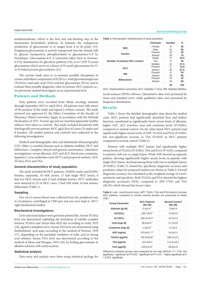
females, separately, 10 with ascites, 17 had single HCC lesion, 8 had two HCC lesions and 15 had multiple lesions. HCV antibodies were detected in 25 of HCC cases, 5 had DM while 14 had urinary bilharziasis (Table 1).
SamplingFive ml of venous blood were collected from the peripheral vein
in vacutainers, centrifuged at 1500 rpm and sera were kept at -80 Co right biochemical studies.
Biochemical investigationsLiver and renal indices were got from patient files. Serum TGAGs
level was determined exploiting the formation of soluble complex between TGAGs and Alcian blue 8GX dye according to Gold, 1979 [16], against a standard curve. Serum TSA level was determined using thiobarbituric acid assay according to the method of Warren, 1959 [17], depending on the periodate oxidation of sialic acid in strong acid solution. Serum FGA level was determined according to the method of Elson and Morgan, 1933 [18], by boiling glucosamine in alkaline solution with acetyl acetone.
Statistical analysisData entry and analysis were done using statistical package for
social sciences (SPSS) software. Quantitative data were presented by mean and standard error, while qualitative data were presented by frequency distribution.
Results Table 1 shows the detailed demographic data about the studied
cases. HCC patients had significantly disturbed liver and kidney function, manifested as significantly lower serum levels of albumin, higher AST, ALT activities, urea and creatinine levels (P<0.001), compared to normal control. On the other hand, HCC patients had significantly higher serum levels of AFP, TGAGS and FGA (P<0.001) but non-significant increase in TSA (P>0.05) in HCC patients compared to normal control (Table 2).
Patients with multiple HCC lesions had significantly higher serum levels of TGAGs (P<0.001), TSA and FGA (P<0.05), compared to patients with two or single lesion. While AFP showed an opposite pattern, showing significantly higher serum levels in patients with single HCC lesion, declined among those with two or multiple lesions (P<0.001), (Table 3). Sensitivity, specificity, diagnostic accuracy and predictive values for proposed markers are shown in Table 4. Overall diagnostic accuracy was calculated as the weighted average of a test’s sensitivity and specificity. Both TGAGs and FGA showed the highest diagnostic accuracies (91%), compared to AFP (75%) and TSA (48.5%) which showed the lowest value.
Character Number %
SEX FemaleMale
832
2080
Ascites PresentAbsent
1030
2575
Number of primary HCC LesionsSingleTwo
Multiple
178
15
42.520
37.5
HCV PresentAbsent
535
12.587.5
DM PresentAbsent
832
2080
Bilharziasis PresentAbsent
1426
3565
40 100
Table 1: Demographic characteristics of study population.
HCC: Hepatocellular Carcinoma; HCV: Hepatitis C Virus; DM: Diabetes Mellitus.
Group Parameter HCC Patients(No:40)
Normal Control(No:20)
Albumin (g/ dl) 2.4±0.4*** 3.8±0.4
AST(U/L) 138.7±8.9*** 12.9±2.5
ALT(U/L) 104.5±13.4*** 16.2±2.4
Urea (mg/ dl) 56.6±8.6*** 27.2±3.8
Creatinine (mg/ dl) 1.5±0.3*** 0.7±0.2
AFP (ng/ml) 675.9±17.7** 10.9±1.3
TGAGs (µg/ml) 730.9±18.8*** 335.2±13.1
TSA (µg/ml) 121.4±8.1 111.3±10.2
FGA (µg/ml) 118.5±5.6*** 68.8±2.8
Table 2: Liver, renal function tests, AFP, TGAG, TSA and FGA levels in primary HCC patients, compared to normal controls (results are expressed as Mean ±SE).
Differences between groups were analysed by one way ANOVA: P > 0.05: Not significant, *significant at P<0.05, **significant at P< 0.01, ***highly significant at P < 0.001: significant.

J Dis Markers 3(1): id1036 (2016) - Page - 03
Abdel-Hamid NM Austin Publishing Group
Submit your Manuscript | www.austinpublishinggroup.com
Discussion The current study was executed for the resumption of our earlier
work on experimental animals, which demonstrated significant changes in serum and tissue levels of certain individual components of ECM in chemically induced –HCC in rats against AFP, and it was assumed that, these parameters might provide useful diagnostic markers for early prediction of HCC [19,20].
Routine laboratory indices in the present work introduce a predictive image regarding hepatocellular e status during different stages of the disease. Disrupted composition of ECM directly affect cancer metastasis. Thus, variations in the assembly of ECM can greatly alter its biochemical contributions, catalyse the oncogenic transformation of growth factor impacts and perturb cellular malignant transformation [21,22].
Hereby, the current study showed promising diagnostic potential of TGAGS and FGA but not TSA over AFP in HCC patients. Additionally, AFP showed a highest level in patients with single, rather than two or multiple HCC lesions.
Current concerns about AFP accuracy have been raised. Up to 20% of HCC do not produce AFP, even among large mass holders [23,24]. On the other hand, serum AFP levels were elevated in non-cancerous liver diseases such as cirrhotic HCV patients without evidence of HCC [25].
During the last decade many studies have elucidated the various valuable biological actions of ECM molecules, specifically GAGs, in tissue development and homeostasis as well as pathological processes, showing useful diagnostic and prognostic values in various cancers as well [5,26,27].
In liver diseases, the changes in the concentration of sialylated glycoproteins in the blood affect the concentration of TSA [28]. Elevated levels of TSA have been reported in hepatitis and various
cancers such as lymphoma, malignant melanoma, lung cancer and gastrointestinal cancers [29].
This study is one of few studies which evaluated serum FGA in HCC. All previous studies discussed the possible anti-cancer potential of FGA rather that its diagnostic value. Although anticancer activity of GA was first demonstrated more than 50 years ago [30], the possible molecular mechanisms which mediate this effect still poorly understood [31]. GA is toxic to several malignant cell lines in vitro, and an effective lytic agent for several types of transplanted tumours in vivo, with little toxicity to normal host tissues [32]. This was confirmed in SMMC-7721 hepatoma [33] and K562 leukemia cells [34].
In the present work, both TGAG and TSA were much better than AFP in their diagnostic utility and predictive efficacy in assuring cancer diagnosis. TGAG, TSA and FGA continued to change parallel to malignant lesion progress, while AFP showed confusing correlation to this variable. This action is in accordance with a similar presumptions elsewhere [35].
In conclusion, ECM abnormalities including TGAGS and FGA but not TSA in primary HCC patients may have higher priority over AFP as diagnostic tools, being more accurate, feasible and cost-effective. Substitution of TSA with FSA may be a suitable candidate for larger prospective studies for both diagnosis and follow up of HCC.AFP perturbations during hepatocarinogenesis always mislead diagnosis, this why we stress on selecting both TGAGs and FGA in all expected steps for diagnosis and follow up of HCC management. Routine biochemical investigations only share as a guide for the deteriorative and/ or response to therapy, though our suggested parameters keep more accurate correlation to disease stages.
Ethical ApprovalThe whole work was early approved by the College Committee of
Research and Ethics.
Authors’ ContributionsAll authors equally contributed to the article, however, the
corresponding author is the team leader and suggested the research goal.
References1. Lu P, Weaver VM, Werb Z. The extracellular matrix: a dynamic niche in
cancer progression. J Cell Biol. 2012; 196: 395-406.
2. Whittaker CA, Bergeron KF, Whittle J, Brandhorst BP, Burke RD, Hynes RO. The echinoderm adhesome. Dev Biol. 2006; 300: 252-266.
3. Ozbek S, Balasubramanian PG, Chiquet-Ehrismann R, Tucker RP, Adams JC. The evolution of extracellular matrix. Mol Biol Cell. 2010; 21: 4300-4305.
4. Egeblad M, Rasch MG, Weaver VM. Dynamic interplay between the collagen scaffold and tumor evolution. Curr Opin Cell Biol. 2010; 22: 697-706.
5. Theocharis AD, Skandalis SS, Tzanakakis GN, Karamanos NK. Proteoglycans in health and disease: novel roles for proteoglycans in malignancy and their pharmacological targeting. FEBS J. 2010; 277: 3904-3923.
6. Afratis N, Gialeli C, Nikitovic D, Tsegenidis T, Karousou E, Theocharis AD, et al. Glycosaminoglycans: key players in cancer cell biology and treatment. FEBS J. 2012; 279: 1177-1197.
7. Iozzo RV, Schaefer L. Proteoglycans in health and disease: novel regulatory signaling mechanisms evoked by the small leucine-rich proteoglycans. FEBS J. 2010; 277: 3864-3875.
Group Parameter Multiple lesions(No:15)
Two lesions(No:8)
Single lesion(No:17)
AFP (ng/ml) 140.8±18.9 45.2±4.6 *** 1402.5±24.5
TGAGs (µg/ml) 860.3±15.1 *** 728.5±27.9 *** 617.9±27.4
TSA (µg/ml) 138.2±15.6 * 118.3±12.2 108.1±8.7
FGA (µg/ml) 135.7±12.4 * 108.2±13.1 108.1±14.5
Table 3: Internal variations of serum AFP, TGAGs , TSA and FGA levels in relation to number of primary HCC lesions (results are expressed as Mean ±SE).
Differences between groups were analysed by one way ANOVA: P > 0.05: Not significant, *significant at P<0.05, **significant at P< 0.01, ***highly significant at P < 0.001: significant.
Group Parameter NPV PPV Diagnostic
Accuracy Specificity Sensitivity
AFP (ng/ml) 52.9% 92.3% 75% 90% 60%
TGAGs (µg/ml) 74.1% 100% 91% 100% 82%
TSA (µg/ml) 30.7% 65.9% 48.5% 20% 77%
FGA (µg/ml) 74.1% 100% 91% 100% 82%
Table 4: Sensitivity, specificity, diagnostic accuracy and predictive values for serum AFP, TGAGs, TSA and FGA levels.
N.B: Overall diagnostic accuracy was calculated as the weighted average of a test’s sensitivity and specificity. PPV: Positive predictive value. NPV: Negative predictive value.

J Dis Markers 3(1): id1036 (2016) - Page - 04
Abdel-Hamid NM Austin Publishing Group
Submit your Manuscript | www.austinpublishinggroup.com
8. N Abdel-Hamid, El-Moselhy, MA and Fawzy MA. Novel Panel of Early Diagnostic Markers for Experimental Hepatocellular Carcinoma. Journal of Health Science. 2012; 2: 14-18.
9. VP Bhavanandan and M Sheykhnazari. Adaptation of the periodate-resorcinol method for determination of sialic acids to a microassay using microtiter plate reader. Anal Biochem. 1993; 213: 438-440.
10. Varki NM, Varki A. Diversity in cell surface sialic acid presentations: implications for biology and disease. Lab Invest. 2007; 87: 851-857.
11. Narayanan S. Sialic acid as a tumor marker. Ann Clin Lab Sci. 1994; 24: 376-384.
12. Anderson JW, Nicolosi RJ, Borzelleca JF. Glucosamine effects in humans: a review of effects on glucose metabolism, side effects, safety considerations and efficacy. Food Chem Toxicol. 2005; 43: 187-201.
13. Uldry M, Ibberson M, Hosokawa M, Thorens B. GLUT2 is a high affinity glucosamine transporter. FEBS Lett. 2002; 524: 199-203.
14. H Wolosker, D Kline, Y Bian, S Blackshaw, AM Cameron, TJ Fralich, et al. Molecularly cloned mammalian glucosamine-6-phosphate deaminase localizes to transporting epithelium and lacks oscillin activity. FASEB J. 1998; 12: 91-99.
15. L Wells, and GW Hart. O-GlcNAc turns twenty: functional implications for post-translational modification of nuclear and cytosolic proteins with a sugar. FEBS Lett. 2003; 546: 154-158.
16. E Gold. A simple spectrophotometric method for estimating glycosaminoglycan concentrations. Analytical Biochemistry. 1979; 99: 183-188.
17. WARREN L. Sialic acid in human semen and in the male genital tract. J Clin Invest. 1959; 38: 755-761.
18. Elson LA, Morgan WT. A colorimetric method for the determination of glucosamine and chondrosamine. Biochem J. 1933; 27: 1824-1828.
19. N Abdel-Hamid. Premalignant variations in extracellular matrix composition in chemically induced hepatocellular carcinomain rats. J Memb Biol. 2009; 230: 155-162.
20. NMM Abdel-Hamid. Novel biochemical pathways for 5-Fluorouracil in managing experimental hepatocellular carcinoma in rats. J Membr Biol. 2010; 234: 29-34.
21. Provenzano PP, Eliceiri KW, Campbell JM, Inman DR, White JG, Keely PJ. Collagen reorganization at the tumor-stromal interface facilitates local invasion. BMC Med. 2006; 4: 38.
22. Levental KR, Yu H, Kass L, Lakins JN, Egeblad M, Erler JT, Fong SF. Matrix
crosslinking forces tumor progression by enhancing integrin signaling. Cell. 2009; 139: 891-906.
23. J Lopez, V Thambyrajah, M Balasegaram and J Timor. Appropriate cut-off levels for serum alpha-fetoprotein in hepatocellular carcinoma. Diag Oncol J. 1994; 4: 9l-287.
24. N Abdel-Hamid. Priority considerations in early laboratory diagnosis of hepatocellular carcinoma. International Journal of Integrative Biology. 2008; 3: 196-201.
25. Colombo M. Screening for cancer in viral hepatitis. Clin Liver Dis. 2001; 5: 109-122.
26. Karamanos NK, Tzanakakis GN. Glycosaminoglycans: from “cellular glue” to novel therapeutical agents. Curr Opin Pharmacol. 2012; 12: 220-222.
27. A Ibrahim, H Attia, A Rabea, and A El-Gayar. Serum levels of glycosaminoglycans (GAGs) and insulin like growth factor-1 (IGF-1) as diagnostic markers for early hepatocellular carcinoma in cirrhoticpatients with or without diabetes. J Med Lab Diag. 2013; 4: 8-20.
28. Arif S, Najeeb-ul-Haq, Hanif R, Khan AS, Jamil-ur-Rehman, Mufti TA. Variations of serum sialic acid level in liver cirrhosis. J Ayub Med Coll Abbottabad. 2005; 17: 54-57.
29. Kökoğlu E, Sönmez H, Uslu E, Uslu I. Sialic acid levels in various types of cancer. Cancer Biochem Biophys. 1992; 13: 57-64.
30. Quastel JH, Cantero A. Inhibition of tumour growth by D-glucosamine. Nature. 1953; 171: 252-254.
31. Dahmer S, Schiller RM. Glucosamine. Am Fam Physician. 2008; 78: 471-476.
32. Molnar Z, Bekesi JG . Cytotoxic effects of D-glucosamine on the ultrastructures of normal and neoplastic tissues in vivo. Cancer Res. 1972; 32: 756-765.
33. Zhang L, Liu WS, Han BQ, Peng YF, Wang DF. Antitumor activities of D-glucosamine and its derivatives. J Zhejiang Univ Sci B. 2006; 7: 608-614.
34. Z Wang, R Liang, GS Huang, Y Piao, YQ Zhang, AQ Wang, et al. Glucosamine sulfate-induced apoptosis in chronic myelogenous leukemia K562 cells is associated with translocation of cathepsin D and downregulation of Bcl-xL. Apoptosis. 2006; 11: 1851-1860.
35. Friedman SJ, Skehan P. Membrane-active drugs potentiate the killing of tumor cells by D-glucosamine. Proc Natl Acad Sci U S A. 1980; 77: 1172-1176.
Citation: Abdel-Hamid NM, Abdel-Fattah SM, Nazmy MH and Mahmoud AS. Extracellular Glycosaminoglycans in Management of Primary Hepatocellular Carcinoma Rather than Alpha Fetoprotein. J Dis Markers. 2016; 3(1): 1036.
J Dis Markers - Volume 3 Issue 1 - 2016ISSN : 2380-0682 | www.austinpublishinggroup.com Abdel-Hamid et al. © All rights are reserved