Embed Size (px)
Citation preview
189© The Author(s), under exclusive license to Springer Nature Switzerland AG 2021 G. Lauc, I. Trbojević-Akmačić (eds.), The Role of Glycosylation in Health and Disease, Advances in Experimental Medicine and Biology 1325, https://doi.org/10.1007/978-3-030-70115-4_9
Glycosaminoglycans in Neurodegenerative Diseases
Weihua Jin, Fuming Zhang, and Robert J. Linhardt
Abstract
Glycosaminoglycans (GAGs) are linear poly-saccharides that consist of alternating disac-charides sequences of uronic acids and/or galactose hexamino sugars most of which are sulfated. GAGs are ubiquitously expressed on the cell surface, in the intracellular milieu and in the extracellular matrix of all animal cells.
Thus, GAGs exhibit many essential roles in a variety of physiological and pathological pro-cesses. The targets of GAGs are GAG-binding proteins and related proteins that are of sig-nificant interest to both the academic commu-nity and in the pharmaceutical industry. In this review, the structures of GAGs, their binding proteins, and analogs are presented that fur-ther the development of GAGs and their ana-logs for the treatment of neurodegenerative diseases agents.
Keywords
Glycosaminoglycans · Neurodegenerative diseases · Heparan sulfate · Alzheimer’s disease · Parkinson’s disease
9.1 Introduction: Neurodegenerative Diseases and Glycosaminoglycans
Neurodegenerative diseases (NDDs) are a hetero-geneous group of disorders that are characterized by the progressive degeneration of the structure and function of the central nervous system or the peripheral nervous system (Nature Springer 2020a). More people are living longer so that more people are of higher risk of being affected by NDDs. Research shows that genes and envi-
W. Jin College of Biotechnology and Bioengineering, Zhejiang University of Technology, Hangzhou, China
Department of Chemical and Biological Engineering, Center for Biotechnology and Interdisciplinary Studies, Rensselaer Polytechnic Institute, Troy, NY, USA e-mail: [email protected]
F. Zhang (*) Department of Chemical and Biological Engineering, Center for Biotechnology and Interdisciplinary Studies, Rensselaer Polytechnic Institute, Troy, NY, USAe-mail: [email protected]
R. J. Linhardt (*) Department of Chemical and Biological Engineering, Center for Biotechnology and Interdisciplinary Studies, Rensselaer Polytechnic Institute, Troy, NY, USA
Department of Biological Science, Departments of Chemistry and Chemical Biology and Biomedical Engineering, Center for Biotechnology and Interdisciplinary Studies, Rensselaer Polytechnic Institute, Troy, NY, USAe-mail: [email protected]
9
190
ronment contribute to NDDs (Nature Springer 2020b), however, it is still generally unknown which gene or which compounds impact NDDs. There are limited medicines for the treatment of the physical or mental symptoms related with NDDs and there is an urgent need to uncover and improve our understanding of the causes and cures of NDDs.
NDDs include Alzheimer’s disease (AD), Parkinson’s disease (PD), dementia with Lewy bodies (DLB), multiple system atrophy (MSA), prion diseases, Huntington’s disease (HD), amy-otrophic lateral sclerosis (ALS), ataxia telangi-ectasia, multiple sclerosis, spinocerebellar ataxia (SCA), HIV-associated neurocognitive disorders (HAND), Pick’s disease, Krabbe’s disease, Kennedy’s disease, primary lateral sclerosis (PLS), Cockayne syndrome, spinal muscular atrophy (SMA), tabes dorsalis, progressive supranuclear palsy (PSP), and Pelizaeus-Merzbacher disease (Appel et al. 1996; Hardy 2000).
There are millions of people suffering from NDDs throughout the world. Alzheimer’s disease and Parkinson’s disease are the most common NDDs. The number of Americans age 65 and older with Alzheimer’s dementia is expected to grow from 5.8 million to 13.8 million by mid- century (Alzheimer’s Association 2020). The number of deaths associated with AD was 0.12 million in 2018 making AD as the sixth leading cause of death in the United States and the fifth leading cause of death among 65 and older Americans (Alzheimer’s Association 2020). This situation has been getting worse as the num-ber of deaths resulting from AD has increased by 146.2% between 2000 and 2018 (Alzheimer’s Association 2020). The total payments in 2020 for health care, long-term care, and hospice ser-vices for people aged 65 and older with dementia in the United State is estimated as $305 billion (Alzheimer’s Association 2020). The number of people living with PD in the United States is pre-dicted to rise from 0.93 million in 2020 to 1.2 million by 2030 (Marras et al., 2018). There are more than 10 million people, who are living with PD around the world. Like AD, the inci-dence of PD increases with age. An estimated 4% PD people are diagnosed before 50, which is
15 years younger than AD patients are identified (Marras et al. 2018). The combined direct and indirect cost of PD is approximately $52 billion per year in the United States (Marras et al. 2018).
The extracellular matrix (ECM) plays key roles in regulating the development, function, and homeostasis of all eukaryotic cells (Mouw et al. 2014; Nita et al. 2014; Barros et al. 2011; Vieira et al. 2018; Song and Dityatev 2018). The diverse functions of tissues are reflected and facilitated through the complex chemical compo-sition and organization of ECM, which result from a biochemical and biophysical interplay between the various cells in each tissue and the evolving microenvironment (Mouw et al. 2014; Naba et al. 2012). In the central nervous system, ECM contains basal lamina, proteoglycans (PGs), collagens, fibronectin, and elastin (Mouw et al. 2014; Cui et al. 2013; Lau et al. 2013; Hubert et al. 2009; Barros et al. 2011; Ma et al. 2020). These components are also regulated by biochemical mediators, such as interleukins, ara-chidonic acid and derivatives, interferons, platelet- derived growth factor (PDGF), fibroblast growth factor (FGF), transforming growth factor (TGF), and epidermal growth factor (Carmen et al. 2019). Therefore, the dysfunction of ECM in the brain leads to neurodevelopment disorders, psychiatric dysregulation, and neurodegenerative diseases (Wang and Ding 2014; Ariga et al. 2010; Hillen et al. 2018; Fawcett et al. 2019).
The most important components of the ECM are proteoglycans (PGs). PGs are a subset of heavily glycosylated glycoproteins. The “core proteins” include decorin, versican, testican, per-lecan, bikunin, neurocan, aggrecan, brevican, fri-bromodulin, and lumican with their covalently attached carbohydrate portion being glycosami-noglycans (GAGs) (Timpl 1989; Carmen et al. 2019; Ariga et al. 2010; Wang and Ding 2014; Castillo et al. 1997; Snow et al. 1995; Smith et al. 2015; Hayes and Melrose 2018; Volpi 2006).
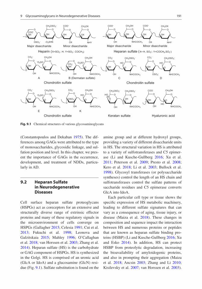
GAGs are a family of unbranched polysaccha-rides, including a repeating disaccharide unit (Fig. 9.1). The disaccharide unit usually contains an amino sugar (N-acetylglucosamine [GlcNAc] or N-acetylgalactosamine) (GalNAc) along with an uronic sugar (glucuronic acid [GlcA] or idu-ronic acid [IdoA]) or galactose (Gal)
W. Jin et al.
191
(Constantopoulos and Dekaban 1975). The dif-ferences among GAGs were attributed to the type of monosaccharides, glycosidic linkage, and sul-fation position and level. In this chapter, we pres-ent the importance of GAGs in the occurrence, development, and treatment of NDDs, particu-larly in AD.
9.2 Heparan Sulfate in Neurodegenerative Diseases
Cell surface heparan sulfate proteoglycans (HSPGs) act as coreceptors for an extensive and structurally diverse range of extrinsic effector proteins and many of these regulatory signals in the microenvironment of cells converge on HSPGs (Gallagher 2015; Celesia 1991; Cui et al. 2013; Fukuchi et al. 1998; Leonova and Galzitskaia 2015; Mahley 1996; O’Callaghan et al. 2018; van Horssen et al. 2003; Zhang et al. 2014). Heparan sulfate (HS) is the carbohydrate or GAG component of HSPGs. HS is synthesized in the Golgi. HS is comprised of an uronic acid (GlcA or IdoA) and a glucosamine (GlcN) resi-due (Fig. 9.1). Sulfate substitution is found on the
amine group and at different hydroxyl groups, providing a variety of different disaccharide units in HS. The structural variation in HS is attributed to a variety of sulfotransferases and C5 epimer-ase (Li and Kusche-Gullberg 2016; Xu et al. 2011; Peterson et al. 2009; Presto et al. 2008; Kero et al. 2018; Li et al. 2003; Bullock et al. 1998). Glycosyl transferases (or polysaccharide syntheses) control the length of an HS chain and sulfotransferases control the sulfate patterns of saccharide residues and C5 epimerase converts GlcA into IdoA.
Each particular cell type or tissue shows the specific expression of HS metabolic machinery, leading to different sulfate signatures that can vary as a consequence of aging, tissue injury, or disease (Maiza et al. 2018). These changes in composition and sequence impact the interaction between HS and numerous proteins or peptides that are known as heparan sulfate binding pro-teins (HSBP) (Li and Kusche-Gullberg 2016; Xu and Esko 2014). In addition, HS can protect HSBP from proteolytic degradation, increasing the bioavailability of amyloidogenic proteins, and also in prompting their aggregation (Maiza et al. 2018; Ancsin 2003; Zhang and Li 2010; Kisilevsky et al. 2007; van Horssen et al. 2003).
OOCO2-OH
OOH
CH2OH
NHCOCH3
O
-O3SOOO
CO2-
OH
OOH
CH2OH
NHCOCH3
O
-O3SO
OCOO-
OSO3-
OHO
OCH2OSO3
-
-O3SHN
OHO
O
COO-
OX
OHO
OCH2OX
NHY
OXO
Major disaccharide Minor disaccharide
OCOO-
OH
OH
O
O
CH2OH
O
NHCOCH3
HO
Heparin (X=SO3-, H; Y=SO3
-, COCH3)
OO
O
HO
OH
CH2OSO3-
NHCOCH3
OH
O
CH2OSO3-
OCOO-
OH
OHO
OCH2OH
NHCOCH3
OHO
O
COO-
OX
OHO
OCH2OX
NHY
OHO
Major disaccharide Minor disaccharide
Heparan sulfate (X= H, SO3-; Y=COCH3,SO3
-)
OOCO2
-
OH
OOH
CH2OSO3-
NHCOCH3
O
HO OO
CO2-
OH
OOSO3
-
CH2OSO3-
NHCOCH3
O
HO
OOCO2
-
OH
OOH
CH2OSO3-
NHCOCH3
O
-O3SOOO
CO2-
OSO3-
OOH
CH2OH
NHCOCH3
O
HO
A B (Dermatan sulfate)Chondroitin sulfate Chondroitin sulfate
Chondroitin sulfateE K
Keratan sulfate Hyaluronic acid
C D
Fig. 9.1 Chemical structures of various glycosaminoglycans
9 Glycosaminoglycans in Neurodegenerative Diseases

192
Protein aggregation is a process in which mis-folded proteins interact together to form well- structured fibrils, leading to the formation of filaments, known as amyloids (Maiza et al. 2018). Accumulation of amyloids in the brain tissue is related to many neurodegenerative diseases, including AD, PD, and HD (Som Chaudhury and Das Mukhopadhyay 2018; Ross and Poirier 2004; Collinge 2016; Peng et al. 2020).
AD is characterized by two major types of brain lesions and involves two main proteins, Aβ and Tau. Aβ forms amyloid plaques by extracel-lular accumulation while Tau makes neurofibril-lary tangles through intraneuronal accumulation (Scheltens et al. 2016).
Snow et al. (1990) first described HS implica-tion in amyloid plaque formation in the brains of AD patients. Many studies have subsequently demonstrated that HS plays a vital role in the aggregation of Aβ peptides (Castillo et al. 1997; Snow et al. 1994; Bame et al. 1997; Cotman et al. 2000; van Horssen et al. 2003; Patey 2006; O’Callaghan et al. 2008; Geneste et al. 2014; Fu et al. 2016; Liu et al. 2016; Nguyen and Rabenstein 2016; Vera et al. 2017; Maiza et al. 2018; Wesen et al. 2018). For example, the over-expression of HS degrading heparanase, decrease the number of Aβ amyloid plaques without alter-ing the production and proportion of Aβ40 and Aβ42 peptides, which are derived from the sequential cleavage of amyloid precursor protein by β- and γ-secretases (Jendresen et al. 2015; Nagai et al. 2007; Schworer et al. 2013; Cheng et al. 2014). Additionally, HS interacts with resi-dues 12–18 (VHHQKLV) in Aβ40 and Aβ42 and also breaks the anionic bridge of Aβ42 between lysine 28 and alanine 42, leading to the accelera-tion of the aggregation process (Zhang et al. 2014). Moreover, sulfation content and pattern of HS also influence Aβ peptide aggregation. Highly sulfated HS deposits in both Aβ40 and Aβ42 amyloids while under sulfated HS only deposits in Aβ40 amyloid, suggesting that high sulfation content will prompt the aggregation of the Aβ42 peptide in the AD’s brains (Maiza et al. 2018). On the aspects of sulfation pattern, it was found that different isoforms of amyloids require an HS with different sulfation patterns to promote the
interaction between HS and Aβ. For example, Aβ amyloid fibrils require N- and 2-O-sulfation while Aβ monomer requires 6-O-sulfation (Lindahl et al. 1999). The secondary structure of Aβ peptides is impacted by the degree of polym-erization (DP), disaccharide sequences, sulfate content, and sulfation pattern suggesting that HS is involved in the Aβ aggregation process in the AD brain (Maiza et al. 2018).
Unlike Aβ, Tau is a soluble protein, which does not aggregate in vitro without the incorpora-tion of polyanionic molecules. An abnormally phosphorylated Tau protein (P-Tau) is found in AD brain. P-Tau deposits inside neurons along with HS forming paired helical filaments and grow into neurofibrillary tangles (NFTs). This suggests that HS is part of the Tau aggregation process and also regulates the P-Tau aggregation process (Snow et al. 1990; Konno et al. 2004; Iqbal et al. 2010). In addition, HSPGs also par-ticipate in the spreading of Tau-related proteins (Holmes et al. 2013; Goedert and Spillantini 2017). HS plays a vital role in the process of Tau misfolding, phosphorylation, aggregation, and spreading in the AD brain. This has been a hot research topic in elucidating the role of HS struc-ture on Tau pathology. For example, the 3-O-sulfated HS might act as a molecular chaper-one for abnormal Tau phosphorylation (Sepulveda-Diaz et al. 2015; Alavi Naini and Soussi-Yanicostas 2018; Thacker et al. 2014; Zhao et al. 2019). In addition, 6-O-sulfated hepa-ran sulfate is required for interaction with Tau and also for Tau internalization (Zhao et al. 2017; Rauch et al. 2018). Moreover, it was reported that knockouts of HS extension enzymes (polysac-charide synthases) exostosin 1 (EXT1), exostosin 2 (EXT2), exostosin 3 (EXT3), N-deacetylase, N-sulfotransferase (NDST1), and 6-O-sulfotransferase (HS6ST2) significantly reduce Tau uptake, suggesting that Tau aggre-gates display specific interactions with HSPGs that depend on chain length and the position of sulfate groups (Stopschinski et al. 2018; Garcia et al. 2017). HS accumulates with protein depos-its not only in AD, but also in other neurodegen-erative diseases, which is summarized in Table 9.1.
W. Jin et al.
193
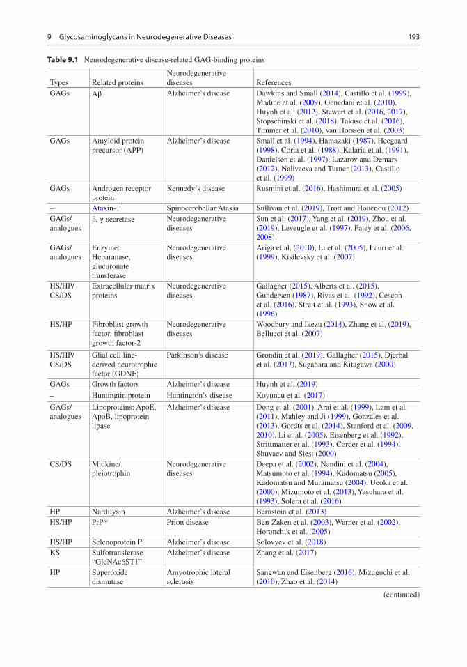
Table 9.1 Neurodegenerative disease-related GAG-binding proteins
Types Related proteinsNeurodegenerative diseases References
GAGs Aβ Alzheimer’s disease Dawkins and Small (2014), Castillo et al. (1999), Madine et al. (2009), Genedani et al. (2010), Huynh et al. (2012), Stewart et al. (2016, 2017), Stopschinski et al. (2018), Takase et al. (2016), Timmer et al. (2010), van Horssen et al. (2003)
GAGs Amyloid protein precursor (APP)
Alzheimer’s disease Small et al. (1994), Hamazaki (1987), Heegaard (1998), Coria et al. (1988), Kalaria et al. (1991), Danielsen et al. (1997), Lazarov and Demars (2012), Nalivaeva and Turner (2013), Castillo et al. (1999)
GAGs Androgen receptor protein
Kennedy’s disease Rusmini et al. (2016), Hashimura et al. (2005)
– Ataxin-1 Spinocerebellar Ataxia Sullivan et al. (2019), Trott and Houenou (2012)GAGs/analogues
β, γ-secretase Neurodegenerative diseases
Sun et al. (2017), Yang et al. (2019), Zhou et al. (2019), Leveugle et al. (1997), Patey et al. (2006, 2008)
GAGs/analogues
Enzyme: Heparanase, glucuronate transferase
Neurodegenerative diseases
Ariga et al. (2010), Li et al. (2005), Lauri et al. (1999), Kisilevsky et al. (2007)
HS/HP/CS/DS
Extracellular matrix proteins
Neurodegenerative diseases
Gallagher (2015), Alberts et al. (2015), Gundersen (1987), Rivas et al. (1992), Cescon et al. (2016), Streit et al. (1993), Snow et al. (1996)
HS/HP Fibroblast growth factor, fibroblast growth factor-2
Neurodegenerative diseases
Woodbury and Ikezu (2014), Zhang et al. (2019), Bellucci et al. (2007)
HS/HP/CS/DS
Glial cell line- derived neurotrophic factor (GDNF)
Parkinson’s disease Grondin et al. (2019), Gallagher (2015), Djerbal et al. (2017), Sugahara and Kitagawa (2000)
GAGs Growth factors Alzheimer’s disease Huynh et al. (2019)
– Huntingtin protein Huntington’s disease Koyuncu et al. (2017)
GAGs/analogues
Lipoproteins: ApoE, ApoB, lipoprotein lipase
Alzheimer’s disease Dong et al. (2001), Arai et al. (1999), Lam et al. (2011), Mahley and Ji (1999), Gonzales et al. (2013), Gordts et al. (2014), Stanford et al. (2009, 2010), Li et al. (2005), Eisenberg et al. (1992), Strittmatter et al. (1993), Corder et al. (1994), Shuvaev and Siest (2000)
CS/DS Midkine/pleiotrophin
Neurodegenerative diseases
Deepa et al. (2002), Nandini et al. (2004), Matsumoto et al. (1994), Kadomatsu (2005), Kadomatsu and Muramatsu (2004), Ueoka et al. (2000), Mizumoto et al. (2013), Yasuhara et al. (1993), Solera et al. (2016)
HP Nardilysin Alzheimer’s disease Bernstein et al. (2013)HS/HP PrPSc Prion disease Ben-Zaken et al. (2003), Warner et al. (2002),
Horonchik et al. (2005)HS/HP Selenoprotein P Alzheimer’s disease Solovyev et al. (2018)KS Sulfotransferase
“GlcNAc6ST1”Alzheimer’s disease Zhang et al. (2017)
HP Superoxide dismutase
Amyotrophic lateral sclerosis
Sangwan and Eisenberg (2016), Mizuguchi et al. (2010), Zhao et al. (2014)
(continued)
9 Glycosaminoglycans in Neurodegenerative Diseases
194
9.3 Heparin in Neurodegenerative Diseases
Heparin (HP) (Fig. 9.1) has a high negative charge density (about 3.3 negative charges per disaccharide), especially compared to the related HS (about 0.8 sulfate groups per disaccharide in typical HS) (Weiss et al., 2017). HP is composed of 90% L-IdoA and 10% D-GlcA while HS con-sists of primarily GlcA (Capila and Linhardt 2002). Moreover, HP has an average molecular weight of about 15 kDa, ranging from 5 to 40 kDa while HS has a 30 kDa average molecular weight, ranging from 5 to 50 kDa. HP has a lower molec-ular weight, a higher sulfate content, and a higher IdoA content than HS. HS is ubiquitously expressed in all animal cells while HP is pro-duced and stored selectively in the secretory granules of connective-tissue mast cells (Weiss et al. 2017; Guyton and Hall 2006).
HP, an anticoagulant drug, is used as a sur-rogate for HS in most research (Xu and Esko 2014) because HP is commercially available in ton quantities while HS is only available in gram quantities. Moreover, the higher sulfation level in HP results in its tight binding to HSBPs. Currently, HP has been reported to bind to more than 300 secreted and membrane associated human proteins and, in most cases, the natural ligand is HS (Xu and Esko 2014; Ori et al. 2011). These HSBPs can be divided into several major categories, chemokines and cytokines (~60), growth factors and morphogens in devel-opment and tissue repair (~50), blood coagula-tion factors (~25), extracellular structural proteins (~25), complement proteins (~20),
single- transmembrane signaling receptor (~15), cell adhesion proteins (~10), and other proteins (5–10), such as proteins in intracellular gran-ules, lipid-binding proteins, and “helper pro-teins” (Xu and Esko 2014). These HSBPs play key roles in NDDs (summarized in Table 9.1). There are many excellent reviews (Alavi Naini and Soussi- Yanicostas 2018; Muramatsu 1993; Stutzmann et al. 2002; Ma et al. 2007; Dudas et al. 2008; Bergamaschini et al. 2009; Ariga et al. 2010; Dudas and Semeniken 2012; Szczubialka et al. 2012; Wang and Ding 2014; Woodbury and Ikezu 2014; Lima et al. 2017) on HP or HS and NDDs.
9.4 Chondroitin Sulfate and Dermatan Sulfate in Neurodegenerative Diseases
Chondroitin sulfate (CS) was first found from cartilage by Fisher and Boedecker in 1861, iso-lated in purer form by Krukenburg in 1884, eluci-dated the full structure of CS by Levene and Forge until 1915 (Djerbal et al. 2017). The differ-ent forms are attributed to sulfation pattern and epimerization pattern, which divided CS into six forms (Fig. 9.1), namely chondroitin-4-sulfate (CS-A), chondroitin-6-sulfate (CS-C), chondroitin- 2,6-sulfate (CS-D), chondroitin- 4,6- sulfate (CS-E), chondroitin-3-sulfate (CS-K), and chondroitin sulfate B (CS-B), which is the product of uronic acid epimerization and also named dermatan sulfate (DS).
CS is found in the ECM, at the cell surface, associated with the plasma membrane in most
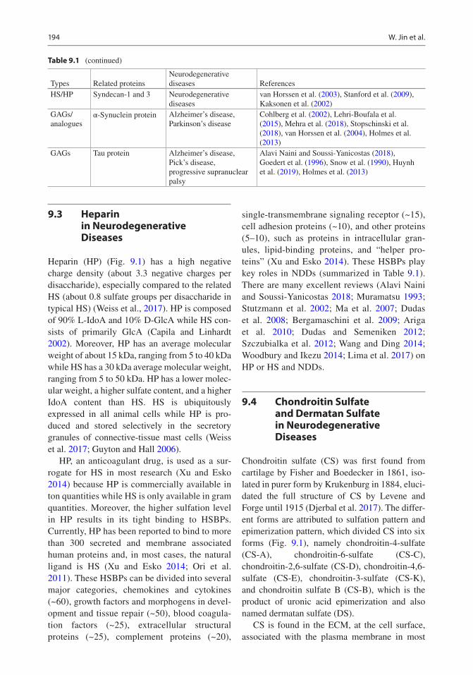
Table 9.1 (continued)
Types Related proteinsNeurodegenerative diseases References
HS/HP Syndecan-1 and 3 Neurodegenerative diseases
van Horssen et al. (2003), Stanford et al. (2009), Kaksonen et al. (2002)
GAGs/analogues
α-Synuclein protein Alzheimer’s disease, Parkinson’s disease
Cohlberg et al. (2002), Lehri-Boufala et al. (2015), Mehra et al. (2018), Stopschinski et al. (2018), van Horssen et al. (2004), Holmes et al. (2013)
GAGs Tau protein Alzheimer’s disease, Pick’s disease, progressive supranuclear palsy
Alavi Naini and Soussi-Yanicostas (2018), Goedert et al. (1996), Snow et al. (1990), Huynh et al. (2019), Holmes et al. (2013)
W. Jin et al.
195
animal tissues, and also, in the case of CS-E, in the intracellular granules of certain cells like mast cells (Yamada et al. 2011; Stevens et al. 1988; Thompson et al. 1988; Farrugia et al. 2016). The expression of CS is different in differ-ent tissues and the highest level is found in the ECM in cartilage and in the central nervous sys-tem (Djerbal et al. 2017). CS is synthesized in the Golgi. Like HS, CS is also linked to the “core proteins” to form chondroitin sulfate proteogly-cans (CSPGs) in the ECM and at the cell surface, and are involved in the formation, development, and maintenance of brain morphology and func-tion (Djerbal et al. 2017; Egea et al. 2010; Kastana et al., 2019; Khan et al. 2020; Kwok et al., 2008; Maeda et al. 2010; Malavaki et al. 2008; Malmstrom et al. 2012; Mikami and Kitagawa 2013, 2015; Purushothaman et al. 2012; Rani et al. 2018; Rauvala et al. 2017; Avram et al. 2014; Carulli et al. 2005; Maeda 2010; Oohira et al. 2004; Sugahara and Mikami 2007). The ratio of CS to HS in the central ner-vous system (CNS) is 9:1 while it is 7:3 in the perineuronal net (PNN) matrix. There are many proteins that interact with various forms of CS (depending on the different sulfate content, pat-tern, and uronic acid epimerization) to accom-plish their functions in promoting growth, differentiation, guidance, and plasticity. These proteins can be divided into several categories (Djerbal et al. 2017), growth factors (neuro-trophic factors, FGF, and Midkine and pleiotro-phin), receptors (receptor protein tyrosine phosphatases and Nogo receptors NgR1 and NgR3), cell adhesion molecules, guidance pro-teins (semaphoring), extracellular matrix pro-teins (collagen VI, laminin and fibronectin), and pathological protein (amyloid precursor protein). These CS binding proteins and their functions in the NDDs are summarized in Table 9.1.
9.5 Hyaluronic Acid in Neurodegenerative Diseases
Hyaluronic acid (HA) was firstly discovered by Meyer and John Palmer in 1934 from vitreous body in cow’s eye. As the simplest structure
among GAGs, HA is composed of GlcA and GlcNAc (Fig. 9.1) with a molecular weight range from 5000 to 20 million Da (Carmen et al. 2019; Toole 2004). HA is synthesized on the cell membrane without any modification by a class of integral membrane proteins, named hyaluronan synthases (HAS1, HAS2, and HAS3) (Toole 2004). More than half of HA is localized in the skin, about 25% is in the skel-eton and supporting structures and less than 10% is in the skeletal muscle (Reed et al. 1988).
The sulfate moiety is necessary for the forma-tion of amyloid fibrils, as confirmed the lack of fibril formation in the presence of HA (Ariga et al. 2010) and it is suggested that HA might inhibit the fibril formation and be useful for treat-ing AD. There is a positive correlation between HA accumulation and AD neuropathology, sug-gesting that HA synthesis and metabolism play a role in AD (Reed et al. 2019). More specifically, HA reduces Aβ42 oligomer uptake and supports neuron cell survival (Bejoy et al. 2018). During the progression of AD, the abolishing of axonal- localization of HAS1 and an increased expres-sion of HAS3 results in the upregulation of short-chain HA production and the reorganiza-tion of the ECM, providing biochemical and physical support to aggrecan-based perineuronal nets and regulation of neuronal plasticity (Li et al. 2017).
9.6 Keratan Sulfate in Neurodegenerative Diseases
Keratan sulfate (KS) was first isolated from the cornea and is found in brain, skeletal, and nervous tissues (Meyer et al. 1953; Funderburgh 2000; Kleene and Schachner 2004; Lindahl et al. 1996). KS consists of disaccharide unit [Gal and GlcNAc], sulfated at C6 of both Gal and GlcNAc residues (Fig. 9.1). KS is covalently attached to a “core protein” through its reducing end in both an O-linkage to N-acetylated galactosamine (GalNAc) or N-linkage Fuc-Man-GlcNAc-linked oligosaccharides and can be terminated with a sialic acid residue or a Gal or GalNAc at the non-
9 Glycosaminoglycans in Neurodegenerative Diseases
196
reducing end of KS (Funderburgh 2000). KS is synthesized by Golgi-resident enzymes, such as galactosyltransferase, N-acetylglucosaminyl transferase, and Gal/GlcNAc/GalNAc sulfotrans-ferases (Uchimura and Rosen 2006; Christner et al. 1979; Funderburgh 2000). KS plays an indispensable, suppressive role in ALS pathology progression by microglial activation and prolifer-ation, suggesting that KS might be a new target for the treatment of ALS (Foyez et al. 2015; Hirano et al. 2013). Deficiency of a sulfotransfer-ase “GlcNAc6ST1” synthesizing sialylated KS can modulate AD pathology, suggesting that GlcNAc6ST1 also might be a good target (Zhang et al. 2017).
9.7 Glycosaminoglycans and Their Analogues in Neurodegenerative Diseases
Aβ is an important target for treating AD. Although Aβ self-aggregates to form amyloid fibrils in vitro, amyloid aggregation and fibril for-mation can be enhanced in the presence of PGs or GAGs (Castillo et al. 1998, 1999). Their binding sites in Aβ at the 13–16 amino acid region (His- His- Gln-Lys), particular of His 13, are important for the interaction between GAGs with Aβ (Ariga et al., 2010). The sequence of the effectiveness of GAGs on prompting fibril formation is HP > HS > CS = DS. In HP the sequence of the effectiveness based on sulfate content and posi-tion is HP > N-desulfated N-acetylated HP > com-pletely desulfated N-desulfated HP > completely desulfated N-acetylated HP (Castillo et al. 1998, 1999). Fibril formation can be inhibited by low molecular weight heparin (LMWH, MW 4000–6000), suggesting that GAG analogs might be used in inhibiting fibril formation. Research on NDDs have examined “neuroparin,” a low molec-ular weight GAG (Ma et al. 2003; Dudas et al. 2008), a specific disaccharide (CSPG-DS) (Rolls et al. 2004), and HP oligosaccharides (Ariga et al. 2010). There are also some Aβ related tar-gets, including ApoE and β-secretase (Shuvaev and Siest 2000; Leveugle et al. 1997; Patey et al. 2006, 2008). There are also some GAG analogs,
such pentosan polysulfate, that have been used to target Aβ (Ariga et al. 2010).
Tau is also an important target for treating AD. Unlike Aβ, Tau needs polyanionic molecules to aggregate. In addition, Tau phosphorylation plays an important role in AD progression. Therefore, Tau is an important target for curing AD. In terms of GAGs and their analogs that influence Tau phosphorylation by the proline-directed protein kinases NCLK, GSK3β and MAP kinase, HP, dextran sulfate, pentosan polysulfate, and HS show efficacy, while KS, HA, and dextran and poly-L-glutamic acid are ineffective. It is inter-esting to note that CS and DS had intermediate effects when Tau was phosphorylated by NCLK and GSK3β, but no effect when Tau was modified by MAP kinase (Hasegawa et al. 1997). There are a number of reviews on the GAGs analogs and their biological activities (Mende et al. 2016; Morla 2019; Zhang et al. 2020; Fraser et al. 2001; Arlov and Skjak-Braek 2017; Coombe and Kett 2012).
9.8 Conclusion
This chapter described the importance of GAGs in NDDs. The data presented probably underrep-resents the significance of GAGs, as GAGs inter-act with many proteins. In addition, there are many GAGs attached to a variety of core pro-teins. These core proteins can also exhibit impor-tant roles in the progression of NDDs. There is still much to uncover about the role of GAGs in NDDs. The development of effective compounds to target the interactions of GAGs for the treat-ment of NDDs still represents a formidable challenge.
Compliance with Ethical Standards
Funding: This research was funded through grants from the NIH (DK111958, CA231074, AG062344 and AG069039 to RL).
Disclosure of interests: All authors declare they have no conflict of interest.
Ethical approval: This article does not contain any stud-ies with animals performed by any of the authors.
W. Jin et al.

197
References
Alavi Naini SM, Soussi-Yanicostas N (2018) Heparan sulfate as a therapeutic target in Tauopathies: insights from Zebrafish. Front Cell Dev Biol 6:163
Alberts B, Johnson A, Lewis J, Morgan D, Raff M, Roberts K et al (2015) Cell junctions and the extracel-lular matrix. In: Molecular biology of the cell. Garland Science, New York
Alzheimer’s Association (2020) Alzheimer’s disease facts and figures (2020). Alzheimers Dement
Ancsin JB (2003) Amyloidogenesis: historical and mod-ern observations point to heparan sulfate proteogly-cans as a major culprit. Amyloid 10(2):67–79
Appel SH, Smith RG, Le WD (1996) Immune-mediated cell death in neurodegenerative disease. Adv Neurol 69:153–159
Arai H, Kashiwagi S, Nagasaka Y, Uchida K, Hoshii Y, Nakamura K (1999) Oxidative modification of apo-lipoprotein E in human very-low-density lipopro-tein and its inhibition by glycosaminoglycans. Arch Biochem Biophys 367(1):1–8
Ariga T, Miyatake T, Yu RK (2010) Role of proteogly-cans and glycosaminoglycans in the pathogenesis of Alzheimer’s disease and related disorders: amy-loidogenesis and therapeutic strategies – a review. J Neurosci Res 88(11):2303–2315
Arlov O, Skjak-Braek G (2017) Sulfated alginates as hep-arin analogues: a review of chemical and functional properties. Molecules 22(5):778
Avram S, Shaposhnikov S, Buiu C, Mernea M (2014) Chondroitin sulfate proteoglycans: structure-function relationship with implication in neural development and brain disorders. Biomed Res Int 2014:642798
Bame KJ, Danda J, Hassall A, Tumova S (1997) Abeta(1- 40) prevents heparanase-catalyzed degra-dation of heparan sulfate glycosaminoglycans and proteoglycans in vitro. A role for heparan sulfate proteoglycan turnover in Alzheimer’s disease. J Biol Chem 272(27):17005–17011
Barros CS, Franco SJ, Muller U (2011) Extracellular matrix: functions in the nervous system. Cold Spring Harb Perspect Biol 3(1):a005108
Bejoy J, Song L, Wang Z, Sang QX, Zhou Y, Li Y (2018) Neuroprotective activities of heparin, Heparinase III, and hyaluronic acid on the Abeta42-treated fore-brain spheroids derived from human stem cells. ACS Biomater Sci Eng 4(8):2922–2933
Bellucci C, Lilli C, Baroni T, Parnetti L, Sorbi S, Emiliani C et al (2007) Differences in extracellular matrix pro-duction and basic fibroblast growth factor response in skin fibroblasts from sporadic and familial Alzheimer’s disease. Mol Med 13(9–10):542–550
Ben-Zaken O, Tzaban S, Tal Y, Horonchik L, Esko JD, Vlodavsky I et al (2003) Cellular heparan sulfate par-ticipates in the metabolism of prions. J Biol Chem 278(41):40041–40049
Bergamaschini L, Rossi E, Vergani C, De Simoni MG (2009) Alzheimer’s disease: another target for heparin therapy. ScientificWorldJournal 9:891–908
Bernstein HG, Stricker R, Dobrowolny H, Steiner J, Bogerts B, Trubner K et al (2013) Nardilysin in human brain diseases: both friend and foe. Amino Acids 45(2):269–278
Bullock SL, Fletcher JM, Beddington RS, Wilson VA (1998) Renal agenesis in mice homozygous for a gene trap mutation in the gene encoding heparan sulfate 2-sulfotransferase. Genes Dev 12(12):1894–1906
Capila I, Linhardt RJ (2002) Heparin-protein interactions. Angew Chem Int Ed Engl 41(3):391–412
Carmen L, Maria V, Morales-Medina JC, Vallelunga A, Palmieri B, Iannitti T (2019) Role of proteoglycans and glycosaminoglycans in Duchenne muscular dys-trophy. Glycobiology 29(2):110–123
Carulli D, Laabs T, Geller HM, Fawcett JW (2005) Chondroitin sulfate proteoglycans in neural devel-opment and regeneration. Curr Opin Neurobiol 15(1):116–120
Castillo GM, Ngo C, Cummings J, Wight TN, Snow AD (1997) Perlecan binds to the beta-amyloid proteins (A beta) of Alzheimer’s disease, accelerates A beta fibril formation, and maintains A beta fibril stability. J Neurochem 69(6):2452–2465
Castillo GM, Cummings JA, Yang W, Judge ME, Sheardown MJ, Rimvall K et al (1998) Sulfate content and specific glycosaminoglycan backbone of perlecan are critical for perlecan’s enhancement of islet amy-loid polypeptide (amylin) fibril formation. Diabetes 47(4):612–620
Castillo GM, Lukito W, Wight TN, Snow AD (1999) The sulfate moieties of glycosaminoglycans are critical for the enhancement of beta-amyloid protein fibril forma-tion. J Neurochem 72(4):1681–1687
Celesia GG (1991) Alzheimer’s disease: the proteogly-cans hypothesis. Semin Thromb Hemost 17(Suppl 2):158–160
Cescon M, Chen P, Castagnaro S, Gregorio I, Bonaldo P (2016) Lack of collagen VI promotes neurodegenera-tion by impairing autophagy and inducing apoptosis during aging. Aging (Albany NY) 8(5):1083–1101
Cheng F, Cappai R, Lidfeldt J, Belting M, Fransson LA, Mani K (2014) Amyloid precursor protein (APP)/APP-like protein 2 (APLP2) expression is required to initiate endosome-nucleus-autophagosome trafficking of glypican-1-derived heparan sulfate. J Biol Chem 289(30):20871–20878
Christner JE, Distler JJ, Jourdian GW (1979) Biosynthesis of keratan sulfate: purification and properties of a galactosyltransferase from bovine cornea. Arch Biochem Biophys 192(2):548–558
Cohlberg JA, Li J, Uversky VN, Fink AL (2002) Heparin and other glycosaminoglycans stimulate the forma-tion of amyloid fibrils from alpha-synuclein in vitro. Biochemistry 41(5):1502–1511
Collinge J (2016) Mammalian prions and their wider relevance in neurodegenerative diseases. Nature 539(7628):217–226
Constantopoulos G, Dekaban AS (1975) Chemical defi-nition of the mucopolysaccharidoses. Clin Chim Acta 59(3):321–336
9 Glycosaminoglycans in Neurodegenerative Diseases
198
Coombe DR, Kett WC (2012) Heparin mimetics. Handb Exp Pharmacol 207:361–383
Corder EH, Saunders AM, Risch NJ, Strittmatter WJ, Schmechel DE, Gaskell PC Jr et al (1994) Protective effect of apolipoprotein E type 2 allele for late onset Alzheimer disease. Nat Genet 7(2):180–184
Coria F, Castano E, Prelli F, Larrondo-Lillo M, van Duinen S, Shelanski ML et al (1988) Isolation and character-ization of amyloid P component from Alzheimer’s disease and other types of cerebral amyloidosis. Lab Investig 58(4):454–458
Cotman SL, Halfter W, Cole GJ (2000) Agrin binds to beta-amyloid (Abeta), accelerates abeta fibril forma-tion, and is localized to Abeta deposits in Alzheimer’s disease brain. Mol Cell Neurosci 15(2):183–198
Cui H, Freeman C, Jacobson GA, Small DH (2013) Proteoglycans in the central nervous system: role in development, neural repair, and Alzheimer’s disease. IUBMB Life 65(2):108–120
Danielsen B, Sorensen IJ, Nybo M, Nielsen EH, Kaplan B, Svehag SE (1997) Calcium-dependent and -inde-pendent binding of the pentraxin serum amyloid P component to glycosaminoglycans and amyloid pro-teins: enhanced binding at slightly acid pH. Biochim Biophys Acta 1339(1):73–78
Dawkins E, Small DH (2014) Insights into the physi-ological function of the beta-amyloid precursor protein: beyond Alzheimer’s disease. J Neurochem 129(5):756–769
Deepa SS, Umehara Y, Higashiyama S, Itoh N, Sugahara K (2002) Specific molecular interactions of oversulfated chondroitin sulfate E with various heparin- binding growth factors. Implications as a physiological bind-ing partner in the brain and other tissues. J Biol Chem 277(46):43707–43716
Djerbal L, Lortat-Jacob H, Kwok J (2017) Chondroitin sulfates and their binding molecules in the central ner-vous system. Glycoconj J 34(3):363–376
Dong J, Peters-Libeu CA, Weisgraber KH, Segelke BW, Rupp B, Capila I et al (2001) Interaction of the N-terminal domain of apolipoprotein E4 with heparin. Biochemistry 40(9):2826–2834
Dudas B, Semeniken K (2012) Glycosaminoglycans and neuroprotection. Handb Exp Pharmacol 207:325–343
Dudas B, Rose M, Cornelli U, Pavlovich A, Hanin I (2008) Neuroprotective properties of glycosaminoglycans: potential treatment for neurodegenerative disorders. Neurodegener Dis 5(3–4):200–205
Egea J, Garcia AG, Verges J, Montell E, Lopez MG (2010) Antioxidant, antiinflammatory and neuropro-tective actions of chondroitin sulfate and proteogly-cans. Osteoarthr Cartil 18(Suppl 1):S24–S27
Eisenberg S, Sehayek E, Olivecrona T, Vlodavsky I (1992) Lipoprotein lipase enhances binding of lipoproteins to heparan sulfate on cell surfaces and extracellular matrix. J Clin Invest 90(5):2013–2021
Farrugia BL, Whitelock JM, O’Grady R, Caterson B, Lord MS (2016) Mast cells produce a unique chondroitin sulfate epitope. J Histochem Cytochem 64(2):85–98
Fawcett JW, Oohashi T, Pizzorusso T (2019) The roles of perineuronal nets and the perinodal extracellu-
lar matrix in neuronal function. Nat Rev Neurosci 20(8):451–465
Foyez T, Takeda-Uchimura Y, Ishigaki S, Narentuya, Zhang Z, Sobue G et al (2015) Microglial keratan sulfate epitope elicits in central nervous tissues of transgenic model mice and patients with amyotrophic lateral sclerosis. Am J Pathol 185(11):3053–3065
Fraser PE, Darabie AA, McLaurin JA (2001) Amyloid- beta interactions with chondroitin sulfate-derived monosaccharides and disaccharides. Implications for drug development. J Biol Chem 276(9):6412–6419
Fu Y, Zhao J, Atagi Y, Nielsen HM, Liu CC, Zheng H et al (2016) Apolipoprotein E lipoprotein particles inhibit amyloid-beta uptake through cell surface heparan sul-phate proteoglycan. Mol Neurodegener 11(1):37
Fukuchi K, Hart M, Li L (1998) Alzheimer’s disease and heparan sulfate proteoglycan. Front Biosci 3:d327–d337
Funderburgh JL (2000) Keratan sulfate: structure, biosyn-thesis, and function. Glycobiology 10(10):951–958
Gallagher J (2015) Fell-Muir lecture: Heparan sulphate and the art of cell regulation: a polymer chain conducts the protein orchestra. Int J Exp Pathol 96(4):203–231
Garcia B, Martin C, Garcia-Suarez O, Muniz-Alonso B, Ordiales H, Fernandez-Menendez S et al (2017) Upregulated expression of Heparanase and Heparanase 2 in the brains of Alzheimer’s disease. J Alzheimers Dis 58(1):185–192
Genedani S, Agnati LF, Leo G, Buzzega D, Maccari F, Carone C et al (2010) Beta-amyloid fibrillation and/or hyperhomocysteinemia modify striatal patterns of hyaluronic acid and dermatan sulfate: possible role in the pathogenesis of Alzheimer’s disease. Curr Alzheimer Res 7(2):150–157
Geneste A, Guillaume YC, Magy-Bertrand N, Lethier L, Gharbi T, Andre C (2014) The protease activity of transthyretin reverses the effect of pH on the amyloid- beta protein/heparan sulfate proteoglycan interaction: a biochromatographic study. J Pharm Biomed Anal 97:88–96
Goedert M, Spillantini MG (2017) Propagation of Tau aggregates. Mol Brain 10(1):18
Goedert M, Jakes R, Spillantini MG, Hasegawa M, Smith MJ, Crowther RA (1996) Assembly of microtubule- associated protein tau into Alzheimer-like filaments induced by sulphated glycosaminoglycans. Nature 383(6600):550–553
Gonzales JC, Gordts PL, Foley EM, Esko JD (2013) Apolipoproteins E and AV mediate lipoprotein clearance by hepatic proteoglycans. J Clin Invest 123(6):2742–2751
Gordts P, Foley EM, Lawrence R, Sinha R, Lameda-Diaz C, Deng L et al (2014) Reducing macrophage proteo-glycan sulfation increases atherosclerosis and obesity through enhanced type I interferon signaling. Cell Metab 20(5):813–826
Grondin R, Littrell OM, Zhang Z, Ai Y, Huettl P, Pomerleau F et al (2019) GDNF revisited: a novel mammalian cell-derived variant form of GDNF increases dopamine turnover and improves brain bio-distribution. Neuropharmacology 147:28–36
W. Jin et al.
199
Gundersen RW (1987) Response of sensory neurites and growth cones to patterned substrata of laminin and fibronectin in vitro. Dev Biol 121(2):423–431
Guyton AC, Hall JE (2006) Textbook of medical physiol-ogy. Elsevier Saunders, Philadelphia
Hamazaki H (1987) Ca2+-mediated association of human serum amyloid P component with hepa-ran sulfate and dermatan sulfate. J Biol Chem 262(4):1456–1460
Hardy J (2000) Pathways to primary neurodegenerative disease. Ann N Y Acad Sci 924:29–34
Hasegawa M, Crowther RA, Jakes R, Goedert M (1997) Alzheimer-like changes in microtubule-associated protein Tau induced by sulfated glycosaminoglycans. Inhibition of microtubule binding, stimulation of phosphorylation, and filament assembly depend on the degree of sulfation. J Biol Chem 272(52):33118–33124
Hashimura K, Sudhir K, Nigro J, Ling S, Williams MR, Komesaroff PA et al (2005) Androgens stimu-late human vascular smooth muscle cell proteogly-can biosynthesis and increase lipoprotein binding. Endocrinology 146(4):2085–2090
Hayes AJ, Melrose J (2018) Glycans and glycosaminogly-cans in neurobiology: key regulators of neuronal cell function and fate. Biochem J 475(15):2511–2545
Heegaard NH (1998) A heparin-binding peptide from human serum amyloid P component characterized by affinity capillary electrophoresis. Electrophoresis 19(3):442–447
Hillen AEJ, Burbach JPH, Hol EM (2018) Cell adhesion and matricellular support by astrocytes of the tripartite synapse. Prog Neurobiol 165–167:66–86
Hirano K, Ohgomori T, Kobayashi K, Tanaka F, Matsumoto T, Natori T et al (2013) Ablation of kera-tan sulfate accelerates early phase pathogenesis of ALS. PLoS One 8(6):e66969
Holmes BB, DeVos SL, Kfoury N, Li M, Jacks R, Yanamandra K et al (2013) Heparan sulfate proteo-glycans mediate internalization and propagation of specific proteopathic seeds. Proc Natl Acad Sci U S A 110(33):E3138–E3147
Horonchik L, Tzaban S, Ben-Zaken O, Yedidia Y, Rouvinski A, Papy-Garcia D et al (2005) Heparan sulfate is a cellular receptor for purified infectious pri-ons. J Biol Chem 280(17):17062–17067
Hubert T, Grimal S, Carroll P, Fichard-Carroll A (2009) Collagens in the developing and diseased nervous sys-tem. Cell Mol Life Sci 66(7):1223–1238
Huynh MB, Villares J, Diaz JE, Christiaans S, Carpentier G, Ouidja MO et al (2012) Glycosaminoglycans from aged human hippocampus have altered capacities to regulate trophic factors activities but not Abeta42 pep-tide toxicity. Neurobiol Aging 33(5):1005.e11–1005.e22
Huynh MB, Ouidja MO, Chantepie S, Carpentier G, Maiza A, Zhang G et al (2019) Glycosaminoglycans from Alzheimer’s disease hippocampus have altered capacities to bind and regulate growth factors activi-ties and to bind tau. PLoS One 14(1):e0209573
Iqbal K, Wang X, Blanchard J, Liu F, Gong CX, Grundke- Iqbal I (2010) Alzheimer’s disease neurofibrillary degeneration: pivotal and multifactorial. Biochem Soc Trans 38(4):962–966
Jendresen CB, Cui H, Zhang X, Vlodavsky I, Nilsson LN, Li JP (2015) Overexpression of heparanase lowers the amyloid burden in amyloid-beta precursor protein transgenic mice. J Biol Chem 290(8):5053–5064
Kadomatsu K (2005) The midkine family in cancer, inflammation and neural development. Nagoya J Med Sci 67(3–4):71–82
Kadomatsu K, Muramatsu T (2004) Midkine and pleiotro-phin in neural development and cancer. Cancer Lett 204(2):127–143
Kaksonen M, Pavlov I, Voikar V, Lauri SE, Hienola A, Riekki R et al (2002) Syndecan-3-deficient mice exhibit enhanced LTP and impaired hippocampus- dependent memory. Mol Cell Neurosci 21(1):158–172
Kalaria RN, Galloway PG, Perry G (1991) Widespread serum amyloid P immunoreactivity in cortical amy-loid deposits and the neurofibrillary pathology of Alzheimer’s disease and other degenerative disorders. Neuropathol Appl Neurobiol 17(3):189–201
Kastana P, Choleva E, Poimenidi E, Karamanos N, Sugahara K, Papadimitriou E (2019) Insight into the role of chondroitin sulfate E in angiogenesis. FEBS J 286(15):2921–2936
Kero D, Bilandzija TS, Arapovic LL, Vukojevic K, Saraga- Babic M (2018) Syndecans and enzymes involved in Heparan sulfate biosynthesis and degradation are dif-ferentially expressed during human Odontogenesis. Front Physiol 9:732
Khan AR, Yang X, Du X, Yang H, Liu Y, Khan AQ et al (2020) Chondroitin sulfate derived theranostic and therapeutic nanocarriers for tumor-targeted drug deliv-ery. Carbohydr Polym 233:115837
Kisilevsky R, Ancsin JB, Szarek WA, Petanceska S (2007) Heparan sulfate as a therapeutic target in amy-loidogenesis: prospects and possible complications. Amyloid 14(1):21–32
Kleene R, Schachner M (2004) Glycans and neural cell interactions. Nat Rev Neurosci 5(3):195–208
Konno T, Oiki S, Hasegawa K, Naiki H (2004) Anionic contribution for fibrous maturation of protofibrillar assemblies of the human tau repeat domain in a fluoroalcohol solution. Biochemistry 43(42):13613–136120
Koyuncu S, Fatima A, Gutierrez-Garcia R, Vilchez D (2017) Proteostasis of huntingtin in health and disease. Int J Mol Sci 18(7):1568
Kwok JC, Afshari F, Garcia-Alias G, Fawcett JW (2008) Proteoglycans in the central nervous system: plastic-ity, regeneration and their stimulation with chondroiti-nase ABC. Restor Neurol Neurosci 26(2–3):131–145
Lam V, Takechi R, Pallebage-Gamarallage MM, Galloway S, Mamo JC (2011) Colocalisation of plasma derived apo B lipoproteins with cerebral proteoglycans in a transgenic-amyloid model of Alzheimer’s disease. Neurosci Lett 492(3):160–164
9 Glycosaminoglycans in Neurodegenerative Diseases
200
Lau LW, Cua R, Keough MB, Haylock-Jacobs S, Yong VW (2013) Pathophysiology of the brain extracellu-lar matrix: a new target for remyelination. Nat Rev Neurosci 14(10):722–729
Lauri SE, Kaukinen S, Kinnunen T, Ylinen A, Imai S, Kaila K et al (1999) Regulatory role and molecular interactions of a cell-surface heparan sulfate proteo-glycan (N-syndecan) in hippocampal long-term poten-tiation. J Neurosci 19(4):1226–1235
Lazarov O, Demars M (2012) All in the family: how the APPs regulate neurogenesis (review). Front Neurosci 6:81
Lehri-Boufala S, Ouidja MO, Barbier-Chassefiere V, Henault E, Raisman-Vozari R, Garrigue-Antar L et al (2015) New roles of glycosaminoglycans in alpha- synuclein aggregation in a cellular model of Parkinson disease. PLoS One 10(1):e0116641
Leonova EI, Galzitskaia OV (2015) Role of syndecan-2 in amyloid plaque formation. Mol Biol (Mosk) 49(1):89–98
Leveugle B, Ding W, Durkin JT, Mistretta S, Eisle J, Matic M et al (1997) Heparin promotes beta-secretase cleavage of the Alzheimer’s amyloid precursor pro-tein. Neurochem Int 30(6):543–548
Li JP, Kusche-Gullberg M (2016) Heparan sulfate: bio-synthesis, structure, and function. Int Rev Cell Mol Biol 325:215–273
Li JP, Gong F, Hagner-McWhirter A, Forsberg E, Abrink M, Kisilevsky R et al (2003) Targeted disruption of a murine glucuronyl C5-epimerase gene results in hep-aran sulfate lacking L-iduronic acid and in neonatal lethality. J Biol Chem 278(31):28363–28366
Li JP, Galvis ML, Gong F, Zhang X, Zcharia E, Metzger S et al (2005) In vivo fragmentation of heparan sulfate by heparanase overexpression renders mice resistant to amyloid protein A amyloidosis. Proc Natl Acad Sci U S A 102(18):6473–6477
Li Y, Li ZX, Jin T, Wang ZY, Zhao P (2017) Tau pathol-ogy promotes the reorganization of the extracellular matrix and inhibits the formation of Perineuronal nets by regulating the expression and the distribu-tion of hyaluronic acid synthases. J Alzheimers Dis 57(2):395–409
Lima M, Rudd T, Yates E (2017) New applications of heparin and other glycosaminoglycans. Molecules 22(5):749
Lindahl B, Eriksson L, Spillmann D, Caterson B, Lindahl U (1996) Selective loss of cerebral kera-tan sulfate in Alzheimer’s disease. J Biol Chem 271(29):16991–16994
Lindahl B, Westling C, Gimenez-Gallego G, Lindahl U, Salmivirta M (1999) Common binding sites for beta- amyloid fibrils and fibroblast growth factor-2 in hepa-ran sulfate from human cerebral cortex. J Biol Chem 274(43):30631–30635
Liu CC, Zhao N, Yamaguchi Y, Cirrito JR, Kanekiyo T, Holtzman DM et al (2016) Neuronal heparan sulfates promote amyloid pathology by modulat-ing brain amyloid- beta clearance and aggregation in Alzheimer’s disease. Sci Transl Med 8(332):332ra44
Ma Q, Schultz C, Neville B, Jeske W, Hoppensteadt D, Cornelli U et al (2003) Pharmacodynamics and phar-macokinetics of C3, a heparin-derived oligosaccha-ride mixture, in non-human primates. Thromb Res 112(4):249–255
Ma Q, Cornelli U, Hanin I, Jeske WP, Linhardt RJ, Walenga JM et al (2007) Heparin oligosaccharides as potential therapeutic agents in senile dementia. Curr Pharm Des 13(15):1607–1616
Ma J, Ma C, Li J, Sun Y, Ye F, Liu K et al (2020) Extracellular matrix proteins involved in Alzheimer’s disease. Chemistry 26(53):12101–12110
Madine J, Clayton JC, Yates EA, Middleton DA (2009) Exploiting a (13)C-labelled heparin analogue for in situ solid-state NMR investigations of peptide-glycan interactions within amyloid fibrils. Org Biomol Chem 7(11):2414–2420
Maeda N (2010) Structural variation of chondroitin sul-fate and its roles in the central nervous system. Cent Nerv Syst Agents Med Chem 10(1):22–31
Maeda N, Fukazawa N, Ishii M (2010) Chondroitin sul-fate proteoglycans in neural development and plastic-ity. Front Biosci (Landmark Ed) 15:626–644
Mahley RW (1996) Heparan sulfate proteoglycan/low density lipoprotein receptor-related protein path-way involved in type III hyperlipoproteinemia and Alzheimer’s disease. Isr J Med Sci 32(6):414–429
Mahley RW, Ji ZS (1999) Remnant lipoprotein metabo-lism: key pathways involving cell-surface heparan sul-fate proteoglycans and apolipoprotein E. J Lipid Res 40(1):1–16
Maiza A, Chantepie S, Vera C, Fifre A, Huynh MB, Stettler O et al (2018) The role of heparan sulfates in protein aggregation and their potential impact on neu-rodegeneration. FEBS Lett 592(23):3806–3818
Malavaki C, Mizumoto S, Karamanos N, Sugahara K (2008) Recent advances in the structural study of functional chondroitin sulfate and dermatan sul-fate in health and disease. Connect Tissue Res 49(3):133–139
Malmstrom A, Bartolini B, Thelin MA, Pacheco B, Maccarana M (2012) Iduronic acid in chondroitin/der-matan sulfate: biosynthesis and biological function. J Histochem Cytochem 60(12):916–925
Marras C, Beck JC, Bower JH, Roberts E, Ritz B, Ross GW et al (2018) Prevalence of Parkinson’s disease across North America. NPJ Parkinsons Dis 4:21
Matsumoto K, Wanaka A, Takatsuji K, Muramatsu H, Muramatsu T, Tohyama M (1994) A novel family of heparin-binding growth factors, pleiotrophin and mid-kine, is expressed in the developing rat cerebral cortex. Brain Res Dev Brain Res 79(2):229–241
Mehra S, Ghosh D, Kumar R, Mondal M, Gadhe LG, Das S et al (2018) Glycosaminoglycans have vari-able effects on alpha-synuclein aggregation and dif-ferentially affect the activities of the resulting amyloid fibrils. J Biol Chem 293(34):12975–12991
Mende M, Bednarek C, Wawryszyn M, Sauter P, Biskup MB, Schepers U et al (2016) Chemical synthesis of glycosaminoglycans. Chem Rev 116(14):8193–8255
W. Jin et al.
201
Meyer K, Linker A, Davidson EA, Weissmann B (1953) The mucopolysaccharides of bovine cornea. J Biol Chem 205(2):611–616
Mikami T, Kitagawa H (2013) Biosynthesis and func-tion of chondroitin sulfate. Biochim Biophys Acta 1830(10):4719–4733
Miyata S, Kitagawa H (2015) Mechanisms for modulation of neural plasticity and axon regeneration by chon-droitin sulphate. J Biochem 157(1):13–22
Mizuguchi S, Capretta A, Suehiro S, Nishiyama N, Luke P, Potter RF et al (2010) Carbon monoxide-releasing molecule CORM-3 suppresses vascular endothelial cell SOD-1/SOD-2 activity while up-regulating the cell surface levels of SOD-3 in a heparin-dependent manner. Free Radic Biol Med 49(10):1534–1541
Mizumoto S, Fongmoon D, Sugahara K (2013) Interaction of chondroitin sulfate and dermatan sulfate from vari-ous biological sources with heparin-binding growth factors and cytokines. Glycoconj J 30(6):619–632
Morla S (2019) Glycosaminoglycans and glycosamino-glycan Mimetics in cancer and inflammation. Int J Mol Sci 20(8):1963
Mouw JK, Ou G, Weaver VM (2014) Extracellular matrix assembly: a multiscale deconstruction. Nat Rev Mol Cell Biol 15(12):771–785
Muramatsu T (1993) Midkine (MK): a retinoic acid- responsive, heparin-binding growth factor in relation-ship with differentiation, development, cancer and neural function. Seikagaku 65(12):1494–1504
Naba A, Clauser KR, Hoersch S, Liu H, Carr SA, Hynes RO (2012) The matrisome: in silico definition and in vivo characterization by proteomics of normal and tumor extracellular matrices. Mol Cell Proteomics 11(4):M111.014647
Nagai N, Habuchi H, Kitazume S, Toyoda H, Hashimoto Y, Kimata K (2007) Regulation of heparan sulfate 6-O-sulfation by beta-secretase activity. J Biol Chem 282(20):14942–149451
Nalivaeva NN, Turner AJ (2013) The amyloid precursor protein: a biochemical enigma in brain development, function and disease. FEBS Lett 587(13):2046–2054
Nandini CD, Mikami T, Ohta M, Itoh N, Akiyama-Nambu F, Sugahara K (2004) Structural and functional char-acterization of oversulfated chondroitin sulfate/dermatan sulfate hybrid chains from the notochord of hagfish. Neuritogenic and binding activities for growth factors and neurotrophic factors. J Biol Chem 279(49):50799–50809
Nature Springer (2020a) https://www.nature.com/subjects/neurodegenerative- diseases. Accessed 8 Apr 2020
Nature Springer (2020b) https://www.niehs.nih.gov/research/supported/health/neurodegenerative/index.cfm. Accessed 8 Apr 2020
Nguyen K, Rabenstein DL (2016) Interaction of the heparin-binding consensus sequence of beta-amyloid peptides with heparin and heparin-derived oligosac-charides. J Phys Chem B 120(9):2187–2197
Nita M, Strzalka-Mrozik B, Grzybowski A, Mazurek U, Romaniuk W (2014) Age-related macular degenera-tion and changes in the extracellular matrix. Med Sci Monit 20:1003–1016
O’Callaghan P, Sandwall E, Li JP, Yu H, Ravid R, Guan ZZ et al (2008) Heparan sulfate accumulation with Abeta deposits in Alzheimer’s disease and Tg2576 mice is contributed by glial cells. Brain Pathol 18(4):548–561
O’Callaghan P, Zhang X, Li JP (2018) Heparan sulfate proteoglycans as relays of Neuroinflammation. J Histochem Cytochem 66(4):305–319
Oohira A, Ida M, Matsui F (2004) Development and regeneration of the central nervous system and neu-ral chondroitin sulfate proteoglycans. Tanpakushitsu Kakusan Koso 49(15 Suppl):2342–2347
Ori A, Wilkinson MC, Fernig DG (2011) A systems biology approach for the investigation of the hepa-rin/heparan sulfate interactome. J Biol Chem 286(22):19892–19904
Patey SJ (2006) The role of heparan sulfate in the genera-tion of Abeta. Drug News Perspect 19(7):411–416
Patey SJ, Edwards EA, Yates EA, Turnbull JE (2006) Heparin derivatives as inhibitors of BACE-1, the Alzheimer’s beta-secretase, with reduced activity against factor Xa and other proteases. J Med Chem 49(20):6129–6132
Patey SJ, Edwards EA, Yates EA, Turnbull JE (2008) Engineered heparins: novel beta-secretase inhibi-tors as potential Alzheimer’s disease therapeutics. Neurodegener Dis 5(3–4):197–199
Peng C, Trojanowski JQ, Lee VM (2020) Protein trans-mission in neurodegenerative disease. Nat Rev Neurol 16(4):199–212
Peterson S, Frick A, Liu J (2009) Design of biologically active heparan sulfate and heparin using an enzyme- based approach. Nat Prod Rep 26(5):610–627
Presto J, Thuveson M, Carlsson P, Busse M, Wilen M, Eriksson I et al (2008) Heparan sulfate biosynthesis enzymes EXT1 and EXT2 affect NDST1 expression and heparan sulfate sulfation. Proc Natl Acad Sci USA 105(12):4751–4756
Purushothaman A, Sugahara K, Faissner A (2012) Chondroitin sulfate “wobble motifs” modulate main-tenance and differentiation of neural stem cells and their progeny. J Biol Chem 287(5):2935–2942
Rani A, Patel S, Goyal A (2018) Chondroitin sulfate (CS) Lyases: structure, function and application in thera-peutics. Curr Protein Pept Sci 19(1):22–33
Rauch JN, Chen JJ, Sorum AW, Miller GM, Sharf T, See SK et al (2018) Tau internalization is regulated by 6-O Sulfation on Heparan Sulfate Proteoglycans (HSPGs). Sci Rep 8(1):6382
Rauvala H, Paveliev M, Kuja-Panula J, Kulesskaya N (2017) Inhibition and enhancement of neural regen-eration by chondroitin sulfate proteoglycans. Neural Regen Res 12(5):687–691
Reed RK, Lilja K, Laurent TC (1988) Hyaluronan in the rat with special reference to the skin. Acta Physiol Scand 134(3):405–411
Reed MJ, Damodarasamy M, Pathan JL, Chan CK, Spiekerman C, Wight TN et al (2019) Increased hyal-uronan and TSG-6 in association with neuropatho-logic changes of Alzheimer’s disease. J Alzheimers Dis 67(1):91–102
9 Glycosaminoglycans in Neurodegenerative Diseases
202
Rivas RJ, Burmeister DW, Goldberg DJ (1992) Rapid effects of laminin on the growth cone. Neuron 8(1):107–115
Rolls A, Avidan H, Cahalon L, Schori H, Bakalash S, Litvak V et al (2004) A disaccharide derived from chondroitin sulphate proteoglycan promotes central nervous system repair in rats and mice. Eur J Neurosci 20(8):1973–1983
Ross CA, Poirier MA (2004) Protein aggrega-tion and neurodegenerative disease. Nat Med 10(Suppl):S10–S17
Rusmini P, Crippa V, Cristofani R, Rinaldi C, Cicardi ME, Galbiati M et al (2016) The role of the protein quality control system in SBMA. J Mol Neurosci 58(3):348–364
Sangwan S, Eisenberg DS (2016) Perspective on SOD1 mediated toxicity in amyotrophic lateral sclerosis. Postepy Biochem 62(3):362–369
Scheltens P, Blennow K, Breteler MM, de Strooper B, Frisoni GB, Salloway S et al (2016) Alzheimer’s dis-ease. Lancet 388(10043):505–517
Schworer R, Zubkova OV, Turnbull JE, Tyler PC (2013) Synthesis of a targeted library of heparan sulfate hexa- to dodecasaccharides as inhibitors of beta- secretase: potential therapeutics for Alzheimer’s disease. Chemistry 19(21):6817–6823
Sepulveda-Diaz JE, Alavi Naini SM, Huynh MB, Ouidja MO, Yanicostas C, Chantepie S et al (2015) HS3ST2 expression is critical for the abnormal phosphorylation of tau in Alzheimer’s disease-related tau pathology. Brain 138(Pt 5):1339–1354
Shuvaev VV, Siest G (2000) Heparin specifically inhibits binding of apolipoprotein E to amyloid beta-peptide. Neurosci Lett 280(2):131–134
Small DH, Nurcombe V, Reed G, Clarris H, Moir R, Beyreuther K et al (1994) A heparin-binding domain in the amyloid protein precursor of Alzheimer’s dis-ease is involved in the regulation of neurite outgrowth. J Neurosci 14(4):2117–2127
Smith PD, Coulson-Thomas VJ, Foscarin S, Kwok JC, Fawcett JW (2015) “GAG-ing with the neuron”: the role of glycosaminoglycan patterning in the central nervous system. Exp Neurol 274(Pt B):100–114
Snow AD, Mar H, Nochlin D, Sekiguchi RT, Kimata K, Koike Y et al (1990) Early accumulation of heparan sulfate in neurons and in the beta-amy-loid protein- containing lesions of Alzheimer’s disease and Down’s syndrome. Am J Pathol 137(5):1253–1270
Snow AD, Sekiguchi R, Nochlin D, Fraser P, Kimata K, Mizutani A et al (1994) An important role of heparan sulfate proteoglycan (Perlecan) in a model system for the deposition and persistence of fibrillar A beta- amyloid in rat brain. Neuron 12(1):219–234
Snow AD, Kinsella MG, Parks E, Sekiguchi RT, Miller JD, Kimata K et al (1995) Differential binding of vascular cell-derived proteoglycans (perlecan, bigly-can, decorin, and versican) to the beta-amyloid pro-tein of Alzheimer’s disease. Arch Biochem Biophys 320(1):84–95
Snow DM, Brown EM, Letourneau PC (1996) Growth cone behavior in the presence of soluble chondroitin sulfate proteoglycan (CSPG), compared to behavior on CSPG bound to laminin or fibronectin. Int J Dev Neurosci 14(3):331–349
Solera C, Macchione G, Maza S, Kayser MM, Corzana F, de Paz JL et al (2016) Chondroitin sulfate Tetrasaccharides: synthesis, three-dimensional structure and interaction with Midkine. Chemistry 22(7):2356–2369
Solovyev N, Drobyshev E, Bjorklund G, Dubrovskii Y, Lysiuk R, Rayman MP (2018) Selenium, selenopro-tein P, and Alzheimer’s disease: is there a link? Free Radic Biol Med 127:124–133
Som Chaudhury S, Das Mukhopadhyay C (2018) Functional amyloids: interrelationship with other amyloids and therapeutic assessment to treat neuro-degenerative diseases. Int J Neurosci 128(5):449–463
Song I, Dityatev A (2018) Crosstalk between glia, extracellular matrix and neurons. Brain Res Bull 136:101–108
Stanford KI, Bishop JR, Foley EM, Gonzales JC, Niesman IR, Witztum JL et al (2009) Syndecan-1 is the primary heparan sulfate proteoglycan mediating hepatic clear-ance of triglyceride-rich lipoproteins in mice. J Clin Invest 119(11):3236–3245
Stanford KI, Wang L, Castagnola J, Song D, Bishop JR, Brown JR et al (2010) Heparan sulfate 2-O-sulfotransferase is required for triglyceride-rich lipoprotein clearance. J Biol Chem 285(1):286–294
Stevens RL, Fox CC, Lichtenstein LM, Austen KF (1988) Identification of chondroitin sulfate E proteoglycans and heparin proteoglycans in the secretory granules of human lung mast cells. Proc Natl Acad Sci U S A 85(7):2284–2287
Stewart KL, Hughes E, Yates EA, Akien GR, Huang TY, Lima MA et al (2016) Atomic details of the interac-tions of Glycosaminoglycans with amyloid-beta fibrils. J Am Chem Soc 138(27):8328–8331
Stewart KL, Hughes E, Yates EA, Middleton DA, Radford SE (2017) Molecular origins of the compatibility between Glycosaminoglycans and Abeta40 amyloid fibrils. J Mol Biol 429(16):2449–2462
Stopschinski BE, Holmes BB, Miller GM, Manon VA, Vaquer-Alicea J, Prueitt WL et al (2018) Specific glycosaminoglycan chain length and sulfation pat-terns are required for cell uptake of tau versus alpha- synuclein and beta-amyloid aggregates. J Biol Chem 293(27):10826–10840
Streit A, Nolte C, Rasony T, Schachner M (1993) Interaction of astrochondrin with extracellular matrix components and its involvement in astrocyte process formation and cerebellar granule cell migration. J Cell Biol 120(3):799–814
Strittmatter WJ, Saunders AM, Schmechel D, Pericak- Vance M, Enghild J, Salvesen GS et al (1993) Apolipoprotein E: high-avidity binding to beta- amyloid and increased frequency of type 4 allele in late-onset familial Alzheimer disease. Proc Natl Acad Sci U S A 90(5):1977–1981
W. Jin et al.
203
Stutzmann JM, Mary V, Wahl F, Grosjean-Piot O, Uzan A, Pratt J (2002) Neuroprotective profile of enoxaparin, a low molecular weight heparin, in in vivo models of cerebral ischemia or traumatic brain injury in rats: a review. CNS Drug Rev 8(1):1–30
Sugahara K, Kitagawa H (2000) Recent advances in the study of the biosynthesis and functions of sul-fated glycosaminoglycans. Curr Opin Struct Biol 10(5):518–527
Sugahara K, Mikami T (2007) Chondroitin/dermatan sul-fate in the central nervous system. Curr Opin Struct Biol 17(5):536–545
Sullivan R, Yau WY, O’Connor E, Houlden H (2019) Spinocerebellar ataxia: an update. J Neurol 266(2):533–544
Sun L, Zhou R, Yang G, Shi Y (2017) Analysis of 138 pathogenic mutations in presenilin-1 on the in vitro production of Abeta42 and Abeta40 pep-tides by gamma-secretase. Proc Natl Acad Sci USA 114(4):E476–E485
Szczubialka K, Kaminski K, Zasada K, Karewicz A, Nowakowska M (2012) Heparin – a key drug in the treatment of the circulatory degenerative diseases: controlling its action with polymers. Curr Pharm Des 18(18):2591–2606
Takase H, Tanaka M, Yamamoto A, Watanabe S, Takahashi S, Nadanaka S et al (2016) Structural requirements of glycosaminoglycans for facilitating amyloid fibril formation of human serum amyloid A. Amyloid 23(2):67–75
Thacker BE, Xu D, Lawrence R, Esko JD (2014) Heparan sulfate 3-O-sulfation: a rare modification in search of a function. Matrix Biol 35:60–72
Thompson HL, Schulman ES, Metcalfe DD (1988) Identification of chondroitin sulfate E in human lung mast cells. J Immunol 140(8):2708–2713
Timmer NM, Schirris TJ, Bruinsma IB, Otte-Holler I, van Kuppevelt TH, de Waal RM et al (2010) Aggregation and cytotoxic properties towards cultured cerebrovas-cular cells of Dutch-mutated Abeta40 (DAbeta(1-40)) are modulated by sulfate moieties of heparin. Neurosci Res 66(4):380–389
Timpl R (1989) Structure and biological activity of basement membrane proteins. Eur J Biochem 180(3):487–502
Toole BP (2004) Hyaluronan: from extracellular glue to pericellular cue. Nat Rev Cancer 4(7):528–539
Trott A, Houenou LJ (2012) Mini-review: spinocerebel-lar ataxias: an update of SCA genes. Recent Pat DNA Gene Seq 6(2):115–121
Uchimura K, Rosen SD (2006) Sulfated L-selectin ligands as a therapeutic target in chronic inflammation. Trends Immunol 27(12):559–565
Ueoka C, Kaneda N, Okazaki I, Nadanaka S, Muramatsu T, Sugahara K (2000) Neuronal cell adhesion, medi-ated by the heparin-binding neuroregulatory fac-tor midkine, is specifically inhibited by chondroitin sulfate E. Structural ans functional implications of the over-sulfated chondroitin sulfate. J Biol Chem 275(48):37407–37413
van Horssen J, Wesseling P, van den Heuvel LP, de Waal RM, Verbeek MM (2003) Heparan sulphate proteogly-cans in Alzheimer’s disease and amyloid-related disor-ders. Lancet Neurol 2(8):482–492
van Horssen J, de Vos RA, Steur EN, David G, Wesseling P, de Waal RM et al (2004) Absence of heparan sul-fate proteoglycans in Lewy bodies and Lewy neu-rites in Parkinson’s disease brains. J Alzheimers Dis 6(5):469–474
Vera C, Alvarez-Orozco JA, Maiza A, Chantepie S, Chehin RN, Ouidja MO et al (2017) Heparan sul-phates, amyloidosis and neurodegeneration. Rev Neurol 65(10):457–468
Vieira MS, Santos AK, Vasconcellos R, Goulart VAM, Parreira RC, Kihara AH et al (2018) Neural stem cell differentiation into mature neurons: mechanisms of regulation and biotechnological applications. Biotechnol Adv 36(7):1946–1970
Volpi N (2006) Therapeutic applications of glycosamino-glycans. Curr Med Chem 13(15):1799–1810
Wang P, Ding K (2014) Proteoglycans and glycosamino-glycans in misfolded proteins formation in Alzheimer’s disease. Protein Pept Lett 21(10):1048–1056
Warner RG, Hundt C, Weiss S, Turnbull JE (2002) Identification of the heparan sulfate binding sites in the cellular prion protein. J Biol Chem 277(21):18421–18430
Weiss RJ, Esko JD, Tor Y (2017) Targeting heparin and heparan sulfate protein interactions. Org Biomol Chem 15(27):5656–5668
Wesen E, Gallud A, Paul A, Lindberg DJ, Malmberg P, Esbjorner EK (2018) Cell surface proteoglycan- mediated uptake and accumulation of the Alzheimer’s disease peptide Abeta(1-42). Biochim Biophys Acta Biomembr 1860(11):2204–2214
Woodbury ME, Ikezu T (2014) Fibroblast growth factor-2 signaling in neurogenesis and neurodegeneration. J Neuroimmune Pharmacol 9(2):92–101
Xu D, Esko JD (2014) Demystifying heparan sulfate- protein interactions. Annu Rev Biochem 83:129–157
Xu Y, Masuko S, Takieddin M, Xu H, Liu R, Jing J et al (2011) Chemoenzymatic synthesis of homoge-neous ultralow molecular weight heparins. Science 334(6055):498–501
Yamada S, Sugahara K, Ozbek S (2011) Evolution of glycosaminoglycans: comparative biochemical study. Commun Integr Biol 4(2):150–158
Yang G, Zhou R, Zhou Q, Guo X, Yan C, Ke M et al (2019) Structural basis of Notch recognition by human gamma-secretase. Nature 565(7738):192–197
Yasuhara O, Muramatsu H, Kim SU, Muramatsu T, Maruta H, McGeer PL (1993) Midkine, a novel neurotrophic factor, is present in senile plaques of Alzheimer dis-ease. Biochem Biophys Res Commun 192(1):246–251
Zhang X, Li JP (2010) Heparan sulfate proteoglycans in amyloidosis. Prog Mol Biol Transl Sci 93:309–334
Zhang GL, Zhang X, Wang XM, Li JP (2014) Towards understanding the roles of heparan sulfate proteo-glycans in Alzheimer’s disease. Biomed Res Int 2014:516028
9 Glycosaminoglycans in Neurodegenerative Diseases
204
Zhang Z, Takeda-Uchimura Y, Foyez T, Ohtake-Niimi S, Narentuya, Akatsu H et al (2017) Deficiency of a sulfotransferase for sialic acid-modified glycans miti-gates Alzheimer’s pathology. Proc Natl Acad Sci U S A 114(14):E2947–E2954
Zhang F, Zheng L, Cheng S, Peng Y, Fu L, Zhang X et al (2019) Comparison of the interactions of different growth factors and Glycosaminoglycans. Molecules 24(18):3360
Zhang X, Lin L, Huang H, Linhardt RJ (2020) Chemoenzymatic synthesis of Glycosaminoglycans. Acc Chem Res 53(2):335–346
Zhao D, Zhang S, Meng Y, Xiongwei D, Zhang D, Liang Y et al (2014) Polyanion binding acceler-
ates the formation of stable and low-toxic aggre-gates of ALS- linked SOD1 mutant A4V. Proteins 82(12):3356–3372
Zhao J, Huvent I, Lippens G, Eliezer D, Zhang A, Li Q et al (2017) Glycan determinants of heparin-tau inter-action. Biophys J 112(5):921–932
Zhao J, Zhu Y, Song X, Xiao Y, Su G, Liu X et al (2019) Rare 3-O-sulfation of Heparan sulfate enhances tau interaction and cellular uptake. Angew Chem Int Ed Engl 58:2–11
Zhou R, Yang G, Guo X, Zhou Q, Lei J, Shi Y (2019) Recognition of the amyloid precur-sor protein by human gamma-secretase. Science 363(6428):eaaw0930
W. Jin et al.