Embed Size (px)
Citation preview

External Skeletal Fixation in Small Animal Fracture
Repair Russell Yeadon
Vets Now Referrals, Swindon

Sponsor this evening

INTRODUCTION Fracture planning
• Relies on understanding of different mechanisms of bone healing
• Prediction of predominant mode of bone healing for any given fracture determines:
– rate of bone healing
– degree of stability required
• Management plan can then be determined

Why use an ESF?

Plan
• Quick review of bone biology – Direct healing
– Indirect healing
• External Skeletal Fixation – Major types, configurations and mechanics
– Application techniques
• Examples of different applications

Bone biology

Stages of fracture healing
Inflammation
Repair
Remodeling

Mechanisms of bone formation
3 main types of bone formation – different involvement in different types of bone healing:
1. Cutting cones
2. Intramembranous bone formation
3. Endochondral bone formation
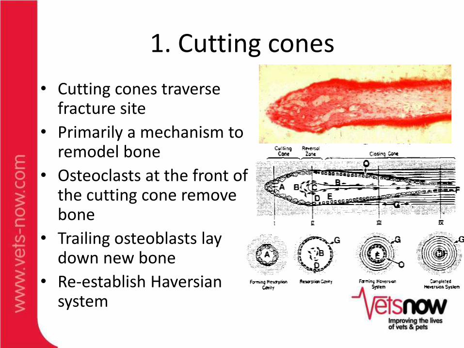
1. Cutting cones
• Cutting cones traverse fracture site
• Primarily a mechanism to remodel bone
• Osteoclasts at the front of the cutting cone remove bone
• Trailing osteoblasts lay down new bone
• Re-establish Haversian system

Haversian System
• Osteon with central Haversian canal containing
– cells
– vessels
– nerves
• Volkmann’s canal
– connects osteons
osteon
Haversian
canal
osteocyte
Volkmann’s
canal

2. Intramembranous (periosteal) bone formation
• Mechanism by which long bone grows in width
• Osteoblasts differentiate directly from pre-osteoblasts (periosteum and endosteum) and lay down seams of osteoid
• Does NOT involve cartilage precursor / matrix

3. Endochondral bone formation • Mechanism by which long bone grows in length
• Osteoblasts line a cartilage precursor
• Chondrocytes hypertrophy, degenerate and calcify (low oxygen tension)
• Vascular invasion of cartilage occurs followed by ossification (increasing oxygen tension)
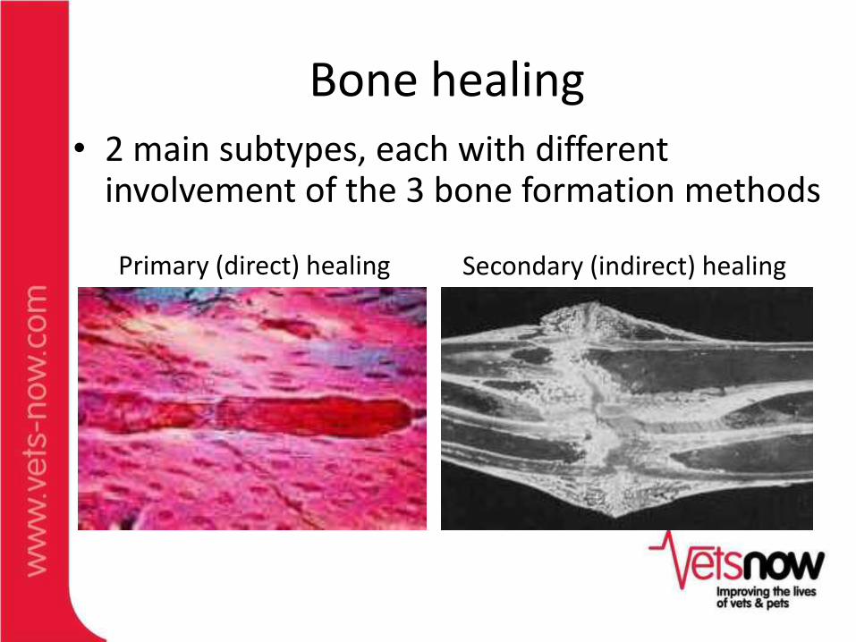
Bone healing
Primary (direct) healing
Secondary (indirect) healing
• 2 main subtypes, each with different involvement of the 3 bone formation methods

Primary (direct) bone healing Orthopaedic carpentry

Primary (direct) bone healing
• Two subtypes
A. Contact healing
(no gap)
B. Gap healing
(<200-500μm gap)
• Both require absolute stability (<2% inter-fragmentary strain)

Definition • Inter-fragmentary strain
– Defined as the % deformation of the fracture gap
= [Distance of deformation ÷ original fracture gap] x100
Strain (ε) Δl / l
l l Δl

A. Contact healing • Bone ends in direct contact
• Re-establishes cortical lamellar bone structure
– Cutting cones cross fracture line
– Leave osteon “pegs” crossing fracture line
– Haversian system re-established
• Resorption and formation occur simultaneously
– rate ~50μm per day
– slow process (months-years)
• Begins immediately when local conditions appropriate (contact and stability)

B. Gap healing • Fracture gap <200-500μm • Gap fills with haematoma / loose connective tissue • <2 weeks after fracture, vascular supply re-
established • Gap fills directly with woven bone
– intramembranous ossification – direct recruitment / differentiation of osteoblasts
– i.e. no cartilaginous anlage • Woven bone gradually lamellar bone by cutting
cones (remodelling)

Traditional AO principles
1. Rigid internal fixation
2. Accurate anatomic reduction
3. Atraumatic operative technique
4. Early mobilization
• Aimed at maximising primary bone healing
• Permits load sharing between implants and reconstructed bone column

AO1. Rigid internal fixation
• Most fractures that have full open reduction should have rigid internal fixation

AO2. Anatomic reduction
• Important in articular fractures
• Avoid callus formation

AO3. Atraumatic operative technique
• Necessary for successful management of all fractures (and surgery in general)
• Halsted’s principles
a) gentle handling of tissue
b) aseptic technique
c) sharp anatomic dissection of tissue
d) careful hemostasis
e) obliteration of dead space
f) avoidance of tension

Limitations to direct bone healing
• Requires minimal / no fracture gap – Accurate reduction ± compression
• Requires absolute stability (<2% strain) – Rigid fixation (internal)
• Slow to achieve maximum strength – Durable fixation – Susceptible to failure for many months – Problematic if implant removal required

Is accurate anatomical reconstruction possible here?

Is bulky internal fixation desirable here?

Anatomic reduction
• Not necessary for all long bone fracture repair
• Cost/benefit ratio of reconstructing all the fragments
• Must preserve the neuro-vascular supply to bone and surrounding soft tissue
Sequestrum
Cloaca
Involucrum

Anatomic reduction
• Load sharing between reconstructed bone column and implants

Indirect bone healing Orthopaedic gardening

Indirect healing
1. Inflammation • Haematoma • Inflammatory cells cleanse fracture
site of necrotic debris, bacteria, etc • Vascular thrombosis bony
necrosis at edges of fracture – Increases fracture gap decreases
relative strain…
• Increased capillary permeability • Release of inflammatory, angiogenic
and osteoinductive factors • Early granulation

Indirect healing
2. Soft callus formation • Endochondral ossification when
inter-fragmentary strain <10% – Chondrocytes type II collagen
matrix (cartilage callus) • Intramembranous ossification
(periosteal callus) • Soft callus starts to stabilise fracture,
– ↑ instability bigger callus

Indirect bone healing
3. Hard callus • Endochondral ossification
progresses as inter-fragmentary strain reduced to ~2% – cartilage callus converts
to woven bone • Intramembranous
ossification progresses throughout periosteal callus

Indirect bone healing
4. Remodelling • Cutting cones cross
fracture site – remodel woven bone
callus – re-establish lamellar
bone cortices • Intramedullary canal re-
established • Responds to Wolff’s law
(remodels in response to mechanical stimuli)

Biologic fracture management
• Atraumatic surgical technique top priority
• Preserve vitality of bones and soft tissue – Neurovascular supply – Periosteum – Endosteum
• Preserve fracture haematoma – chemical mediators – early soft callus

Many inflammatory, angiogenic and osteoinductive factors expressed early in healing process

Biologic fracture management AO principles
• Rigid internal fixation • Accurate anatomic
reduction • Atraumatic operative
technique • Early mobilization
• Not new concept • Reprioritizes AO principles
Biologic principles • Adequate fixation • Accurate bone axial / length orientation • Atraumatic operative
technique • Early mobilization

• Optimise secondary (indirect) bone healing
– Requires less rigid stabilisation
• Callus development additional stabilisation
– No load sharing by bone column initially – need strong stabilisation
– BUT faster than primary (direct) healing so need less durable stabilisation
Biologic fracture management
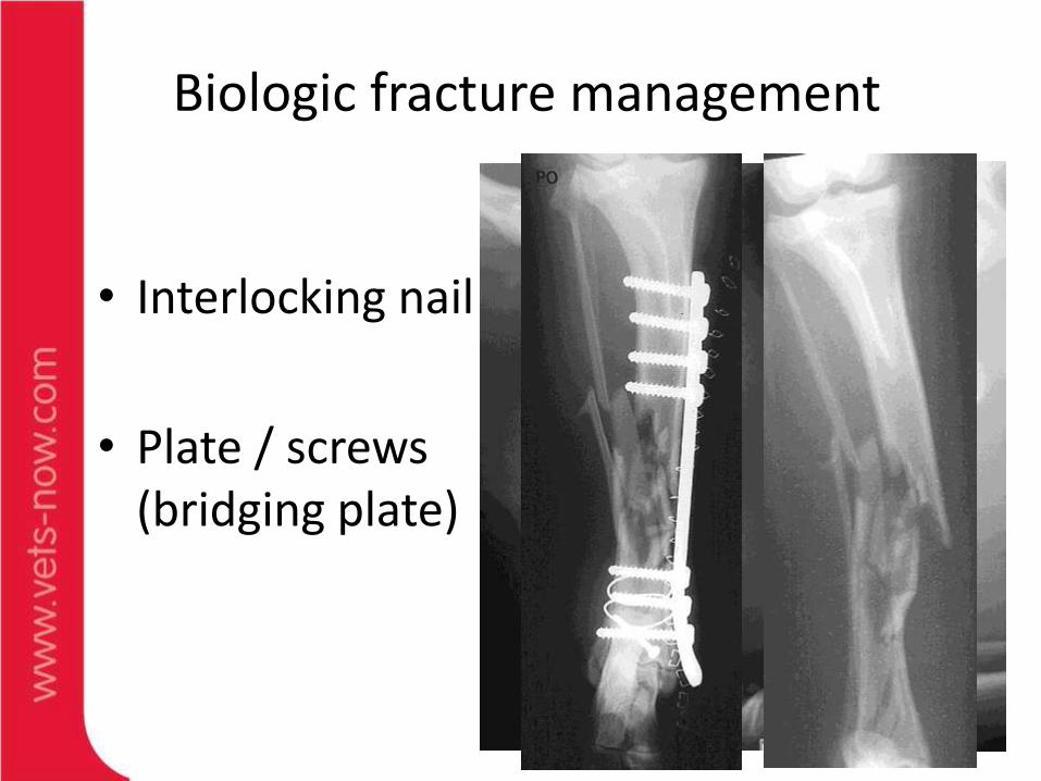
• Interlocking nail
• Plate / screws (bridging plate)
Biologic fracture management

• ESF
• ESF + IM pin
Biologic fracture management

Fracture forces – a reminder
Tension
Rotation
Axial compression
Bending

EXTERNAL SKELETAL FIXATION
An Introduction

Plan
• Quick review of bone biology – Direct healing
– Indirect healing
• External Skeletal Fixation – Major types, configurations and mechanics
– Application techniques
• Examples of different applications

ESF
• Transcutaneous fixation pins inserted into bone – connected by external
rods, bars, or columns

Advantages of ESF
• Rigidity versatile depending on construct
• No implants at fracture site
• Applied closed or through minimal approach – no disturbance of soft tissues
• Versatile construction for varying anatomy / fracture configuration
• Can be combined with other fixation methods
• No residual implants (removed at fracture union)

Types of ESF
• Linear
• Ring
• Acrylic / freeform
• [Pinless]

Linear ESF

ESF Components
• Fixation pins/wires
• Connecting bars/rods
• Connecting clamps
• Special components

Fixation pins / wires
• Smooth
• Threaded
– End
– Central
– Positive profile
– Negative profile
• Half pin
• Full pin
• Wires
• ALL penetrate BOTH cortices of the bone

Connecting bars
• Stainless steel
• Aluminum
• Titanium
• Carbon fiber

Clamps • Single
– Pin to rod
• Double
– Rod to rod
• Acrylic / resin boluses
Kirschner-Ehmer Imex SK
Synthes Meynard Securos

IMEX SK External Fixation System
• Accepts varied fixation pin diameter (including positive profile pins)
• Can be assembled and disassembled independent of frame construct

Type I
Type 1A
• Unilateral uniplanar
Type 1B
• Unilateral biplanar
Least rigid frame types

Type II
• Bilateral uniplanar
• Full pins
• May include additional half pins
• Limited to use below elbow / stifle
• More rigid than type 1

Type III
• Bilateral biplanar
• I + II = III
• Most rigid linear frame

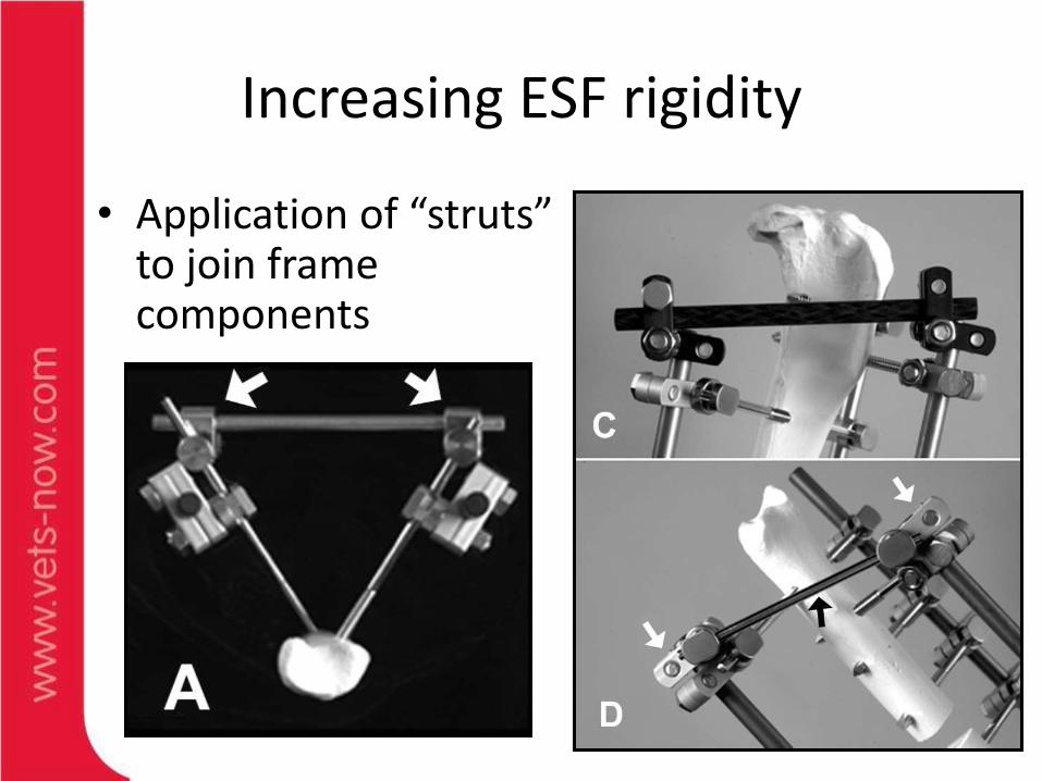
Increasing ESF rigidity

Increasing ESF rigidity
• Double bar / clamp
• Increase pin / bar diameter
– Aim for 20-25% bone diameter for fixation pin
• Reduce bone-bar distance
• Pin distribution
– >1cm from fracture site
– Even spread across proximal and distal fragments
• Number of pins (up to 4 per fragment)

Increasing ESF rigidity
• Tie-in IM Pin
Increasing ESF rigidity
• Application of “struts” to join frame components

Special components

Fixation Pins
• Weakest link is the bone-pin interface

Fixation Pins: Design
Positive Profile Pins
• Superior stiffness and axial extraction characteristics (site dependent)
• Pre-drill (0.1mm <pin core diameter)
• Low insertion speed (<150rpm)

Fixation Pins: Design
Negative Profile Pins:
• Ellis Pins
• Thread-shaft interface is stress riser
• Stress riser effect mitigated if interface is positioned in medullary canal
Partially threaded Steinmann pin
Ellis pin

Fixation Pins: Design
Tapered thread run-out pins (IMEX “Duraface”)
• 55% stiffer than equivalent diameter +ve profile pins

Fixation Pins: Application •Pre-drilling pilot hole
–↓mechanical damage
–↓ heat generation
Ring sequestrum
Bone necrosis occurs @ 56° for 10 secs

No escape channel for debris – low RPM
DRILL BIT
FIXATION PIN
Twist channel in drill bit escape of debris while drilling minimizing frictional heat – high RPM

Fixation Pins: Application
• Fixation pin < 30% bone diameter
• Beveled tip of half pins should completely penetrate far cortex

Fixation Pins: Application
• Small / stab incisions
• Not through traumatic / surgical wounds
• Space pins appropriately
• Distance bars / clamps from skin
• Post-op swelling
• Joint movement (esp elbow and hock)
• “Safe corridors”
• Neurovascular bundles
• Large muscle masses

Circular / ring ESF

cESF
• Graduated from medical school in 1944
• Staff surgeon at Hospital for War Invalids, Western Siberia
• Faced with consequences (nonunion fractures, bone defects and osteomyelitis) of WWII
Gavriil A Ilizarov, MD

cESF
• Lack of facilities, equipment and antibiotics

cESF
• Ilizarov Method revolutionized
–limb lengthening
–deformity correction
–bone transport
–soft tissue expansion
–cranio-facial reconstruction
–fracture management

cESF
• Small diameter fixation wires
• One wire either side of each ring
• Wires tensioned to improve stiffness (depending on ring / patient size)

Fixation Elements: Wires
• 1.6 and 1.0mm diameter wires
• Single lip cutting point
• Stopper or olive wires

Wire placement • Minimal soft tissue penetration
• Perpendicular wire placement
• Wires flat against rings – Above
–Below
–Washer spacers / bolts / posts
•Wire tensioning

Supporting Elements: Rings
• High strength tempered aluminum alloy
• Full, 5/8, 1/3 ring arches
• Assorted other shapes

Ring diameter is single most important variable affecting biomechanics of cESF
• Selection based on patient dimensions
• Minimum ring diameter
–Must leave 1-2cm clearance between soft tissues & rings
Supporting Elements: Rings

Connecting Elements
• Linear motors
• Threaded rods

Assembly Elements
• Fixation bolts
• Washers
• Hinges

cESF advantages
• Small fragment stabilisation – Juxta-articular fractures
• Versatile • Self-dynamising
– Controlled axial micromotion – Excellent resistance to bending and rotation forces
• Minimal soft tissue disruption – Especially if applied closed

13.4 13.4 18.1 18.1
Rigid Micromotion
Fracture Management

cESF application
•“Far-near-near-far” fixation
•Two ring blocks separated by linear motors
Far
Far
Near
Near
Proximal
ring block
Distal
ring
block


Post-op management •Minimize skin-pin movement
•Prevent self-trauma from wire tips / sharp ends
•Radiography <q6-8 weeks
•Periodic checking apparatus tightness

Distraction osteogenesis
• Law of Tension Stress
–slow steady tension stimulates cellular proliferation
–terminates when cells occupy their genetic space constraints
• Abbott 1939
• Ilizarov 1954

Distraction osteogenesis
Intramembranous bone formation
Endochondral bone formation
Similarities with fracture healing & embryonic bone development

Distraction osteogenesis

Bone transport
•Large bone defects
•Segment of bone is transported
–regenerate bone forms in its wake

Hybrid ESF

Hybrid ESF
• Combines circular and linear components
• Uses threaded pins & transfixation wires

Hybrid ESF Advantages: • Quicker / simpler to apply • Increased versatility • Anatomical sites where full rings impractical
– Femur – Humerus – Near joints
Disadvantage: • Lose some benefits of axial micromotion from cESF (depending on construct)

Linear-Circular Hybrid Constructs
1A 1B i/m pin tie-in

hESF application

Acrylic / freeform ESF

Acrylic / epoxy resin ESF
Mix PMMA powder / solvent – fill tubing in semi-liquid phase prior to curing

Acrylic ESF: Advantages
• Pins any diameter
• Pins in almost any orientation
• Largely radiolucent
• Minimal distance between connecting column and bone
• Lightweight
• Limited inventory
• Cheap
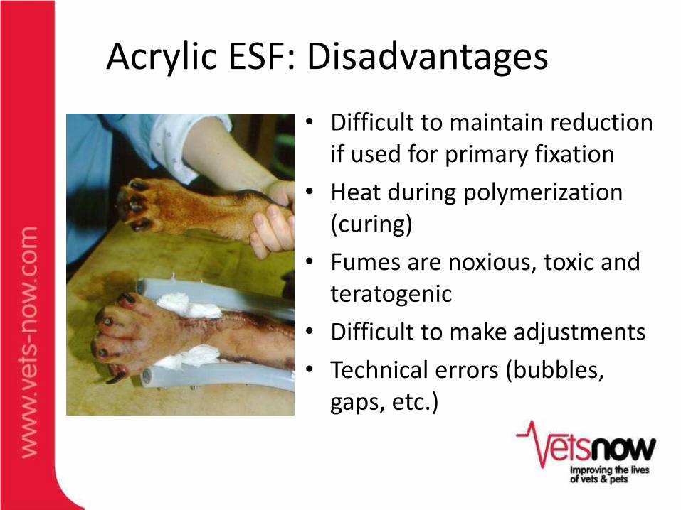
Acrylic ESF: Disadvantages
• Difficult to maintain reduction if used for primary fixation
• Heat during polymerization (curing)
• Fumes are noxious, toxic and teratogenic
• Difficult to make adjustments
• Technical errors (bubbles, gaps, etc.)

“Pinless” ESF

Extreme biologic ESF • Pinless ESF – does not penetrate periosteum – minimal
vascular compromise or potential for tracking infection • Current system (Synthes) too large for small animal
applications
• [Reported for mandibular fractures of cattle]

ESF application and management
A few key points

Carpentry vs gardening


ESF Application •Hanging limb prep?
•Fluoroscopy?
•Proximal and distal pins inserted first to re-establish limb axis and length

Destabilization
• Destabilization <4 weeks: – ↑ callus, ↓mechanical strength
• Destabilization at 6 weeks: – no ↑ callus, but ↑ mechanical
strength
• Destabilization at 12 weeks: – no ↑ callus or mechanical
strength
• 6w typically recommended • Better to remove frame components than
fixation pins

Aftercare
• Bandaging
• Cleaning???
• Antibiotics???

Pin tract discharge
Bone resorption
Pin loosening
Soft tissue liquefaction
Local bacterial
infection
Case selection
• Will fracture healing occur before the ESF fails? – All ESFs will have some complication if left in situ
long enough – Fracture biology – Patient signalment
• Is ESF mechanically appropriate?
• Additional considerations (owner factors,
lifestyle, fixator management, personal experience / preference) Internal fixation often more appropriate

How do we choose ESF configuration?
• BIOMECHANICS and BIOLOGY
• Fracture type, location, configuration
• Patient age, weight, and activity
• Other injuries
• Surgeon skill, experience, and equipment

Plan
• Quick review of bone biology – Direct healing
– Indirect healing
• External Skeletal Fixation – Major types, configurations and mechanics
– Application techniques
• Examples of different applications

Long bone fractures

Comminuted humeral fracture - ESF


Radius / ulna

Radius / ulna

Femur

Femur

Tibia

Tibia

Tibia

Tibia

Other fracture applications

Mandibular fractures

Tarso-metatarsal luxation (Plantar ligament intact)

Tarso-metatarsal luxation

Tarso-metatarsal luxation

Tarso-metatarsal luxation

MT/MC Fractures

MT/MC Fractures

Surgical technique • Dorsal approach • K-wires directed retrograde
through the fracture/luxation – exit MT bones at dorsal aspect of
distal articular surfaces – driven distally until pin tip barely
visible at fracture site
• Fractures/luxations reduced • K-wires driven proximally
into the MT – up to or through the TMT joints

Surgical technique • One or two pins placed
transversely across bases of the MT or distal row of tarsal bones
• Pin ends contoured over dorsal MT
• Epoxy resin compressed over ends of K-wires to anchor frame – wooden tongue depressors used
to space bolus away from skin • allow for post operative swelling • prevent thermal injury during curing

Pelvic ESF

Pelvic ESF
“No other type of fracture lends itself
to iatrogenic trauma with so little to
show for surgical interference as does
the fractured pelvis” (Whittick,1974)
“With a little thought, patience and
practice, the KE splint may successfully
be used for the repair of pelvic fractures”
(Knowles, 1949)

How does ESF management of pelvic fractures compare to conventional plate/screws?
•Mechanical Factors
Application of EF to ilial fracture is superior to plate/screws in terms of yield and failure and is of comparable stiffness

Hip luxation
• Coxo-femoral luxation: customized hinge units mounted from frame pin driven into the proximal femur

Spinal application


Non-fracture applications

Hock instability

Stifle Disruption

Stifle Disruption

Limb deformity management

Can get complicated…

Pedal Arch Wire Scaffold (PAWS)

Neurogenic injury subsequent to sensory neuropathy (trophic ulceration)

PAWS used to protect foot during renervation


Take home messages - ESF
• Choice of fixation technique based on understanding of specific fracture biology and mechanics – Careful case selection
– Preservation of biologic potential essential
• Extremely versatile
• Familiarity with range of implants / ESF systems (and respective properties) important

Questions