Embed Size (px)
Citation preview

Expression and Activation of the Nerve Growth Factor ReceptorTrkA in Serous Ovarian Carcinoma
Ben Davidson,1 Reuven Reich,2 Philip Lazarovici,2
Jahn M. Nesland, Martina Skrede, Bjorn Risberg,Claes G. Trope, and Vivi Ann FlørenesDepartments of Pathology [B. D., J. M. N., M. S., B. R., V. A. F.] andGynecologic Oncology [C. G. T.], The Norwegian Radium Hospital,Montebello N-0310 Oslo, Norway, and Department of Pharmacologyand Experimental Therapeutics, School of Pharmacy, Faculty ofMedicine, The Hebrew University of Jerusalem, Jerusalem 91120,Israel [R. R., P. L.]
ABSTRACTPurpose: The purpose is to analyze the possible corre-
lation between expression and activation of the high-affinitynerve growth factor (NGF) receptor TrkA, cell cycle proteinexpression, and disease outcome in serous ovarian carci-noma. In addition, we wished to study the possible linkbetween expression of NGF, a novel angiogenic factor and itsreceptor TrkA, and the expression of factors involved inangiogenesis in effusions and solid tumors.
Experimental Design: Sections from 80 malignant effu-sions and 65 corresponding solid tumors were evaluated forprotein expression of NGF, TrkA, and phospho-TrkA (p-TrkA). Effusions were additionally studied for expression ofp53, p21WAF1/CIP1, Ki-67, and the Mr 85,000-cleaved frag-ment of poly(ADP-ribose) polymerase (p85-PARP) usingimmunohistochemistry (IHC). Thirty-two effusions werestudied for TrkA, p-TrkA, p53, and p21WAF1/CIP1 expressionusing immunoblotting. mRNA expression of basic fibroblastgrowth factor (bFGF), interleukin 8, and vascular endothe-lial growth factor (VEGF) was studied in 63 effusions and allsolid tumors using in situ hybridization. Protein expressionof bFGF, interleukin 8, and VEGF was additionally studiedin 30 effusions using IHC.
Results: NGF, TrkA, and p-TrkA were expressed incarcinoma cells in effusions in 60 of 80 (75%), 64 of 80(80%), and 15 of 80 (19%) specimens, respectively. In solidtumors, p-TrkA expression was more frequent (52 of 65tumors; 80%) and was accompanied by p-TrkA expressionin endothelial cells. NGF colocalized with bFGF protein(P � 0.016) and mRNA (P � 0.032) in effusions, and with
VEGF (P < 0.001) and bFGF (P � 0.008) in solid tumors. Insurvival analysis, expression of p85-PARP (P � 0.017) andcytoplasmic TrkA (P < 0.001) in effusions predicted betteroutcome, whereas membrane expression of p-TrkA in solidtumors correlated with poor survival (P � 0.004). Diffuseexpression of p53 and Ki-67 was often seen using IHC,whereas p21WAF1/CIP1 and p85-PARP expression was infre-quent and focal. None of these correlated with NGF or TrkAexpression or activity.
Conclusions: Coexpression of NGF with molecules in-volved in angiogenesis and p-TrkA expression in endothelialcells suggest that the proangiogenic role attributed to NGFin vitro and in vivo may be relevant in clinical cancer.Expression of p85-PARP as a marker of apoptosis andcytoplasmic expression of TrkA (probably representing non-glycosylated receptor) predict better outcome, whereas p-TrkA activation correlates with poor outcome in advancedstage serous ovarian carcinoma.
INTRODUCTIONNeurotrophins are a family of growth factors, consisting, at
present, of the prototype compound NGF3 and additional mem-bers such as brain-derived neurotrophic factor, and the neuro-trophins NT-3, NT-4, and NT-6 (1–4). Neurotrophins bind tothe TrkA, TrkB, and TrkC receptors. NGF binds to the specifichigh-affinity tyrosine kinase receptor TrkA. TrkA receptorspossess a tyrosine kinase catalytic domain. The structure of theTrkA-NGF complex has recently been published and containsone conserved motif, common to all neurotrophins, and onespecific motif for TrkA-NGF binding (5). NGF ligand bindingto TrkA receptors activates TrkA autophosphorylation at severalsites such as tyrosine 490, promoting Src homology and colla-gen binding and phosphorylation, which in turn leads to cou-pling of growth factor receptor binding protein to son of sev-enless complexes and activation of Ras (6, 7). The net result inthe central nervous system is differentiation and survival ofneuronal cells, mainly of sympathetic and sensory systems (2,7). Inhibition of Ras activity leads to neuronal death, whereasdeletion in neurofibromatosis-1, a Ras inhibitor, allows forneuronal survival in the absence of neurotrophins in vitro (7).Ras is able to activate two major intracellular signal transduc-tion pathways: the mitogen activated protein kinase pathwayand the phosphoinositol-3-kinase/AKT pathway (3, 7). AKTappears to be the main (although not the only) target for phos-
Received 11/26/02; revised 2/13/03; accepted 2/20/03.The costs of publication of this article were defrayed in part by thepayment of page charges. This article must therefore be hereby markedadvertisement in accordance with 18 U.S.C. Section 1734 solely toindicate this fact.1 R. R. and P. L. are affiliated with the David R. Bloom Center forPharmacy at the Hebrew University.2 To whom requests for reprints should be addressed, at Department ofPathology, The Norwegian Radium Hospital, Montebello N-0310 Oslo,Norway. Phone: 47-22934871; Fax: 47-22508554; E-mail: [email protected].
3 The abbreviations used are: NGF, nerve growth factor; PARP, poly-(ADP-ribose) polymerase; VEGF, vascular endothelial growth factor;IL, interleukin; bFGF, basic fibroblast growth factor; IHC, immunohis-tochemistry; IB, immunoblotting; FIGO, International Federation ofGynecologists and Obstetricians; ISH, in situ hybridization; OS, overallsurvival.
2248 Vol. 9, 2248–2259, June 2003 Clinical Cancer Research
Cancer Research. on November 27, 2018. © 2003 American Association forclincancerres.aacrjournals.org Downloaded from

phoinositol-3-kinase signaling and is responsible for 80% ofNGF-induced survival in sympathetic neurons, mainly throughphosphorylation of Bad and Forkhead 1 (7).
NGF signaling is able to induce survival and proliferationbut can also induce differentiation without proliferation (8). Thisis mediated by TrkA signaling, which has been shown to inhibitcell proliferation through a variety of mechanisms, includingincreased nuclear expression of p53 and direct activation of thecyclin-dependent kinase inhibitor p21WAF1/CIP1 promoter inPC-12 cells (9–11). TrkA itself has been shown to associatewith p53 and was phosphorylated in presence of the latter evenin the absence of NGF (12, 13).
In addition to these well-defined effects of TrkA-mediatedsignaling in neural networks, recent data has suggested thatNGF may have additional biological roles by demonstrating thatNGF is an angiogenic factor in human endothelial umbilicalvein cells (14). This finding supports earlier findings that havedocumented the proangiogenic effects of NGF in the developingcentral nervous system (15), NGF-induced expression of inflam-matory markers in skin vessels (16), and NGF-mediatedincrease in vascularization of brain malignancies such as glio-mas (17).
Although originally isolated from neural tissues, a growingbody of evidence supports a role for Trk receptors in tumori-genesis in nonneural tumors, primarily carcinomas (18–20).Rearrangement or mutation of the TrkA gene, resulting in con-stitutive activation of the receptor, has been observed in colonand thyroid papillary carcinomas, as well as in acute myeloidleukemia (reviewed in Ref. 18). Reduced TrkB and elevatedTrkA and TrkC expression showed an association with tumorprogression in medullary thyroid carcinoma (21). Conversely,the expression of TrkA as a proto-oncogene has been associatedwith better prognosis in neuroblastoma (22, 23). In addition tothe alteration in structural and expression of Trk receptors,intracellular signaling appears to be dysregulated in cancer cells(18). These studies suggest a novel emerging concept in cancerthat NGF and other neurotrophins may be involved in thegrowth of certain nonneural tumors by paracrine and/or auto-crine regulation via humoral-tumoral-stromal Trk interaction.
Ovarian carcinoma is the most lethal gynecologic tumor.Although recent advances in chemotherapy regimens have led tosomewhat prolonged disease-free survival, essentially no effecton long-term survival has been achieved. Although this mainlyreflects the late detection of this tumor, it also owes to our poorunderstanding of changes undergone by ovarian cancer cellsalong tumor progression because most studies focus exclusivelyon primary tumors. As part of our efforts to define eventsinvolved in ovarian cancer evolution and metastasis, we haverecently reported on the expression of the NGF receptors TrkAand the low-affinity neurotrophin receptor p75 in effusions,primary and metastatic tumors of serous ovarian carcinomapatients (19). Expression of TrkA predominated in both solidtumors and effusions, but cancer cells in the latter compartmentshowed reduced expression of TrkA and elevated expression ofp75 compared with solid tumors. Activation (autophosphoryla-tion) of TrkA, using an antibody against the tyrosine 490-phosphorylated form of TrkA, was found in a few effusions.
The objective of this study was to further investigate thebiological role of the NGF-TrkA axis in ovarian carcinoma by
studying NGF and TrkA protein expression in effusions andsolid tumors from a new cohort of 72 ovarian carcinoma pa-tients, with longer follow-up periods compared with the previ-ous study. In addition, p-TrkA expression was analyzed in solidtumors for the first time. Finally, the possible correlation be-tween NGF, TrkA, and p-TrkA expression and the expression ofangiogenic molecules, p53, p21WAF1/CIP1, the Mr 85,000-cleaved product of PARP as an indicator of apoptosis (24, 25)and of the proliferation marker Ki-67 was studied.
MATERIALS AND METHODSEffusion Specimens
The material consisted of 80 fresh nonfixed peritoneal andpleural effusions submitted to the Division of Cytology, Depart-ment of Pathology, Norwegian Radium Hospital, during theperiod of January 1998 to November 2000. Specimens wereobtained preoperatively, intraoperatively, or at disease recur-rence from 69 patients diagnosed with serous ovarian carcinomaand 3 patients diagnosed with primary peritoneal carcinoma.Effusion specimens consisted of 57 peritoneal and 23 pleuraleffusions. All effusion specimens, as well as relevant clinicaldata, were obtained from the Department of Gynecologic On-cology, Norwegian Radium Hospital. Specimens submitted toour laboratory arrived within minutes after tapping and wereprocessed immediately. Cells were suspended and frozen inRPMI � DMSO at �70°C. Smears and cellblock sections fromall specimens underwent morphological evaluation by threeexperienced cytopathologists and were additionally character-ized using immunocytochemistry with broad antibody panelsagainst cancer and mesothelial epitopes, as detailed previously(26–28). Clinicopathological data are presented in Table 1.
Tumor SpecimensSixty-five surgical specimens, consisting of primary tu-
mors (n � 28) and metastatic lesions (n � 37) of the above
Table 1 Clinicopathological data of the study cohort
Parameter No. of specimens Percent
Effusion sitePeritoneal 57 71Pleural 23 29
GradeI 5 6II 33 41III 42 53
Chemotherapya
Yes 37 46No 43 54
FIGO stageb
III 36 50IV 36 50
Ageb
31–40 2 241–50 7 951–60 20 3061–70 25 3571–80 17 2381–90 1 1
a Before sampling.b For a total of 72 patients.
2249Clinical Cancer Research
Cancer Research. on November 27, 2018. © 2003 American Association forclincancerres.aacrjournals.org Downloaded from

patients were additionally studied. Metastases consisted of 19omental, 9 peritoneal, and 6 intestinal biopsies, as well as 3lesions from other sites. Formalin-fixed, paraffin-embedded tis-sue blocks were obtained from archival material at the Depart-ment of Pathology, Norwegian Radium Hospital. All tissuespecimens underwent microscopic confirmation of diagno-sis, tumor type, and histological grade, following establishedcriteria (29).
Immunohistochemical AnalysisTrkA, pTrkA, and NGF. Sections from all 80 malig-
nant effusions and 65 solid tumors were stained for these pro-teins. Staining for TrkA protein was performed using the veryselective 203 anti-TrkA antibody developed at National CancerInstitute, NIH (Frederick, MD) and kindly donated by David R.Kaplan (30), which has been extensively used in many studies.Staining for NGF was performed using an antibody describedpreviously (31). This rabbit polyclonal antibody selectivelycross-reacts with proNGF and mature NGF but lacks any cross-reactivity with other neurotrophins such as brain-derived neu-rotrophic factor, ciliary neuropathic factor, NT-3, or NT-4 asverified by ELISA assay. Pretreatment consisted of microwaveoven antigen retrieval for 4 � 5 min in citrate buffer. Stainingusing both antibodies was done using the Envision peroxidasesystem (Dako, Glostrup, Denmark).
For p-TrkA, a mouse monoclonal IgG1 antibody was raisedagainst a decapeptide corresponding to an amino acid sequencecontaining a phosphorylated tyrosine residue (tyrosine 490) ofhuman TrkA. The antibody was isolated from a serum-freehybridoma culture medium (32) by sequential affinity chroma-tography on unphosphorylated and phosphorylated peptide-Sepharose gels. The antibody reacts specifically with phospho-rylated tyrosine 490 of human TrkA by Western blotting andimmunohistochemistry and is noncross-reactive to phosphoryl-ated TrkB or TrkC. The antibody was concentrated to 150–200�g/ml in PBS containing 0.1% sodium azide and 0.1% gelatinand stored at 4°C. Staining procedure was identical to the onedescribed above for TrkA. Positive immunostaining with thisantibody indicates that the TrkA receptor was autophosphory-lated at tyrosine 490, as shown in our previous report (19).Positive control for these antibodies consisted of a specimenof immature ovarian teratoma containing primitive neuralelements.
Cell Cycle Proteins. All 80 effusions were stained forthe proliferation marker Ki-67 (clone Ki-S5; Dako), the p85fragment of PARP (Promega, Madison, WI), p53 (DO-1 anti-body; Santa Cruz Biotechnology, Santa Cruz, CA) andp21WAF1/CIP1 (Oncogene Research Products, San Diego, CA).Staining for Ki-67 and p21WAF1/CIP1 was performed using thebiotin streptavidin peroxidase method (supersensitive immuno-detection LP000-UL; BioGenex, San Ramon, CA). Staining forp85-PARP and p53 was performed using the Envision method(Dako). Microwave pretreatment in citrate buffer (pH 6) wasused for all antibodies, 4 � 5 min for PARP and Ki-67, 2 � 5min for p53 and p21WAF1/CIP1.
Angiogenic Proteins. Angiogenic protein expressionwas studied in 30 effusions. Polyclonal antibodies againstVEGF (Zymed, San Francisco, CA), IL-8 (Biosource Interna-tional, Camarillo, CA), and bFGF (Sigma, Saint Louis, MO)were used. Using the antibodies directed against VEGF andbFGF, pretreatment consisted of microwave oven antigen re-trieval for 4 � 5 min in citrate buffer. No pretreatment was usedusing the anti-IL-8 antibody (33).
Negative controls in all reactions consisted of sections thatunderwent a similar staining procedure, with the exclusion ofprimary antibody application or that were stained with mousemyeloma protein of the same isotype as the primary antibodyused. Two ovarian carcinoma biopsies in which immunoreac-tivity for the studied antigens was previously demonstrated wereused as positive controls.
Evaluation of IHC Results. Membrane and cytoplasmimmunoreactivity was scored for TrkA, whereas only the latterwas evaluated for NGF staining. p-TrkA expression was scoredat the membrane, cytoplasm, and nucleus. Nuclear immunore-activity was interpreted as positive for cell cycle proteins,whereas cytoplasmic expression of angiogenic molecules wasthe parameter scored. The extent of staining was scored usingthe following scale: 0 � no staining; 1 � staining of 0–5% oftumor cells; 2 � staining of 6–25% of cells; 3 � staining of26–75% of tumor cells; and 4 � staining of 76–100% of tumorcells. A minimum of 500 cells, when present, was evaluated.Evaluation was done without knowledge of patient clinical data.
IBThirty-two malignant effusions (21 peritoneal, 11 pleural)
were studied. Frozen samples were thawed, washed twice in
Table 2 NGF, TrkA, p-TrkA and cell cycle protein expression in 80 effusions
MoleculeCellularlocation
Percentage of stained carcinoma cells
0% 1–5% 6–25% 26–75% 76–100%
TrkA Membrane 47 (59%) 20 (25%) 8 (10%) 5 (6%) 0 (0%)TrkA Cytoplasm 16 (20%) 0 (0%) 6 (7%) 11 (14%) 47 (59%)p-TrkAa Membrane 69 (86%) 7 (9%) 3 (4%) 1 (1%) 0 (0%)p-TrkAa Nucleus 72 (90%) 5 (6%) 1 (1%) 2 (3%) 0 (0%)NGF Cytoplasm 20 (25%) 9 (11%) 14 (18%) 16 (20%) 21 (26%)Ki-67 Nucleus 2 (3%) 18 (22%) 22 (28%) 33 (41%) 5 (6%)p85-PARP Nucleus 38 (48%) 36 (45%) 5 (6%) 1 (1%) 0 (0%)p53 Nucleus 21 (27%) 16 (20%) 10 (12%) 23 (29%) 10 (12%)p21WAF1/CIP1 Nucleus 56 (70%) 23 (29%) 1 (1%) 0 (0%) 0 (0%)
a Total � 15 positive specimens, of which 7 showed membrane expression alone, 4 nuclear expression alone, and 4 immunoreactive at both sites.
2250 TrkA, Angiogenic Genes, and Cell Cycle Proteins
Cancer Research. on November 27, 2018. © 2003 American Association forclincancerres.aacrjournals.org Downloaded from

Fig. 1 IHC results in effusions. A, TrkA staining in a peritoneal effusion. The majority of carcinoma cells show cytoplasmic immunoreactivity,whereas some cells show additional localization to the cell membrane. B, p-TrkA membrane immunoreactivity in a peritoneal effusion. C, TrkAtranslocation to the nucleus in 5–25% of cancer cells in a peritoneal effusion. D, NGF expression in a peritoneal effusion. All tumor cells showcytoplasmic immunoreactivity. E, Ki-67 expression in a peritoneal effusion. Five to 25% of tumor cells show nuclear staining. F, p53 accumulationin the specimen shown in A. Seventy-five to 100% of tumor cells show nuclear staining. G, focal expression of p85-PARP in a peritoneal effusion.Less than 5% of tumor cells show nuclear staining but many reactive cells surrounding tumor cells express p85-PARP. H, focal expression ofp21WAF1/CIP1 in a peritoneal effusion. Less than 5% of tumor cells show nuclear staining. I, another peritoneal effusion, showing no immunoreactivityfor p21WAF1/CIP1 in tumor cells, whereas reactive cells, including both leukocytes and reactive mesothelium, are stained.
2251Clinical Cancer Research
Cancer Research. on November 27, 2018. © 2003 American Association forclincancerres.aacrjournals.org Downloaded from

cold PBS, and lysed in NP40 ice-cold lysis phosphorylationbuffer [1% NP40, 10% glycerol, 20 mM Tris-HCl (pH 7.5), 137mM NaCl, 100 mM NaF, 1 mM sodium vanadate, 1 mM phenyl-methylsulfonyl fluoride, and 10 �g/ml each of leupeptin, pep-statin, and aprotinin] to preserve the integrity and phosphoryl-ation activity of samples proteins. The lysates were thensonicated and clarified by centrifugation, and protein quantifi-cation was done by Bradford analysis. Twenty-five �g of totalcellular protein were loaded into each lane, separated by elec-trophoresis through SDS-12% PAGE, blotted onto immobilon-P-membranes (Millipore Corporation, Bedford, MA) andblocked in 5% dry milk in tris buffered saline-tween. The filterswere subsequently hybridized with the antibodies against TrkA,p-TrkA, p53, and p21WAF1/CIP1 used for IHC. A mouse mono-clonal antibody against �-tubulin (clone 57; Oncogene) wasused as loading control. After washing in TBS-T, bound anti-body was visualized using peroxidase-conjugated antimouseIgG and the enhanced chemiluminescence detection system(Amersham Pharmacia, Buckinghamshire, United Kingdom).Negative controls consisted of antibody in the absence of lysate.Positive controls for the IB procedure consisted of the WM35melanoma cell line (34).
Blocking/Competition Experiment. To verify the spec-ificity of the TrkA reaction, a specific blocking antibody wasused. Five-10-fold excess of blocking peptide in 500 �l of PBSwas used. Incubation was for 2 h in room temperature.
Oligonucleotide ProbesSpecific antisense oligonucleotide DNA probes for mRNA
transcripts of VEGF, bFGF and IL-8 were obtained from Re-search Genetics (Huntsville, AL). The probe sequences (5�-3�)were as follows (35): bFGF, 5�-CGGGAAGGCGCCGCTGC-CGCC3�; IL-8, 5�-CTCCACAACCCTCTGCACCC-3�; andVEGF, 5�-TGGTGATGTTGGACTCCTCAGTGGGCU-3�.
A poly d(T)20 oligonucleotide (Research Genetics) wasused to verify the integrity and lack of degradation of mRNA ineach sample. DNA probes for VEGF, bFGF, and IL-8 werehyperbiotinylated. Stock dilution was prepared with a resultingequal concentration for all probes. The stock dilution was di-luted with probe diluent (Research Genetics) immediately be-fore use. Specific sense oligonucleotides were used for theevaluation of nonspecific activity for each probe.
mRNA ISHmRNA expression of angiogenic molecules was studied in
63 effusions and 65 corresponding solid tumors. Sections (4-�m
thick) of formalin-fixed, paraffin-embedded specimens weremounted on ProbeOn Plus slides (Fisher Scientific, Pittsburgh,PA). Sectioning was performed in RNase-free water. ISH wascarried out using the microprobe manual staining system (FisherScientific; Ref. 36). Hybridization of the probes was carried outas described previously (37). A positive enzymatic reaction inthis assay stained dark blue. Known positive controls were usedin each hybridization reaction. These consisted of two ovariancarcinomas for which positive hybridization was reproducible ina previous study. Controls for endogenous alkaline phosphatasefor all probes included treatment of the sample in the absence ofthe probe and use of chromogen alone.
Evaluation of ISH Results. The presence mRNA ofangiogenic molecules in carcinoma and stromal cells wasscored. Staining extent was scored as detailed for IHC. Inaddition, staining intensity was scored as absent (n � 0), weakto moderate (n � 1), or intense (n � 2). Evaluation was donewithout knowledge of patient clinical data or other expressiondata.
Statistical AnalysisStatistical analysis was performed applying the SPSS-PC
package (version 10.1; SPSS, Chicago, IL). P � 0.05 wasconsidered statistically significant. Full clinical and pathologicaldata were available for all patients. Studies of the associationbetween molecule expression in effusions and clinicopatholog-
Fig. 2 IB results for seven effusions using the antibodies against TrkA,p-TrkA, p21WAF1/CIP1, and p53. The glycosylated form of TrkA andpTrkA (Mr 140,000) is strongly expressed in one effusion (case no. 4)and weaker in two additional ones. With use of blocking peptide forTrkA, elimination of its expression in all positive cases is seen.p21WAF1/CIP1 shows weak expression in all but one specimen, whereasp53 is strongly expressed in the majority of specimens. �-Tubulin wasused as loading control. The seven specimens shown in this figure werechosen with the objective of showing some negative specimens and allthree pTrkA-positive effusions because only 3 of 32 cases were positivefor this receptor.
Table 3 TrkA, pTrkA, p53 and p21WAF1/CIP1 protein expression in32 effusions using immunoblottinga
Molecule
Expression
Absent Weak Moderate Strong Total
TrkA 23 7 2 0 32p-TrkA 29 3 0 0 32p53 1 13 14 4 32p21WAF1/CIP1 3 13 9 6 31b
a Data refer only to expression at predicted molecular sizes (see“Discussion” section).
b One failed reaction.
2252 TrkA, Angiogenic Genes, and Cell Cycle Proteins
Cancer Research. on November 27, 2018. © 2003 American Association forclincancerres.aacrjournals.org Downloaded from

ical parameters were undertaken using the two-sided 2 test.These consisted of analyses of the association between IHCresults and effusion site, FIGO stage, tumor grade, and chemo-therapy status (pre- versus posttreatment specimen). Analysis ofthe association between TrkA, p-TrkA, and NGF expression andthat of cell cycle and angiogenic molecules were similarlyperformed using the two-sided 2 test. Univariate survival anal-yses were executed using the Kaplan-Meier method and log-rank test. Expression categories in these tests were clustered soas to allow for a sufficient number of cases to be included ineach category.
RESULTSTrk-A, p-TrkA, and NGF Expression Is Independent of
Cell Cycle Protein Expression in Effusions. In this analysis,we investigated the possible correlation between TrkA and NGFexpression and cell cycle protein expression to define whetherthe findings in cells of neural origin are of relevance in ovariancarcinoma. Although NGF and TrkA protein expression wasrelatively frequent in carcinoma cells in effusions, p-TrkA wasfound in only a minority of specimens (Table 2; Fig. 1A–D). Aspredicted for metastatic cancer cells, Ki-67 expression and p53accumulation were frequently found (Table 2; Fig. 1, E and F).Conversely, p21WAF1/CIP1 and p85-PARP expression, whenpresent, were limited to few (�5%) tumor cells (Table 2; Fig. 1,G and H), whereas staining of reactive cells, mostly lympho-cytes, was distinctly more common (not scored; Fig. 1I). Whenthe presence of proteins at their predicted molecular weight wasevaluated, IB demonstrated expression of TrkA in 9 of 32,p-TrkA in 3 of 32, p21WAF1/CIP1 in 31 of 32, and p53 in 28 of31 cases (one failed reaction for p53; Table 3; Fig. 2). Addi-tional truncated forms were seen for p53 (data not shown). Useof blocking peptide for TrkA resulted in elimination of itsexpression in all positive cases (Fig. 2). Ovarian carcinoma cellsin pleural (n � 23) and peritoneal (n � 57) effusions showedcomparable expression of all markers. Similarly, no associationwas seen between protein expression of the studied moleculesand patient age, FIGO stage, tumor grade, the extent of residualdisease, or chemotherapy status (data not shown). Trk-A, p-TrkA, and NGF expression showed no association with that ofp53, Ki-67, p21WAF1/CIP1, or p85-PARP.
Trk-A Activation Is a Frequent Event in Solid OvarianCarcinoma. Because activation of TrkA plays a significantrole in the clinical behavior of various solid tumors and becauseNGF is angiogenic, we wished to investigate the expression ofthese molecules in solid primary and metastatic tumors. TrkAprotein membrane and cytoplasmic expression was detected in
carcinoma cells in 57 of 65 (88%) and 63 of 65 (97%) solidtumors, respectively (Table 4; Fig. 3A). Although these figuresexceeded those seen in effusions in both this and our earlierreport, the difference was still more pronounced using the p-TrkA antibody because p-TrkA membrane and cytoplasmicexpression was seen in 50 of 65 (77%) and 52 of 65 (80%)tumors, respectively (Table 4; Fig. 3B). As in effusions, a smallsubset of carcinomas (n � 6) showed unequivocal nuclearimmunoreactivity (Table 4; Fig. 3C). Interestingly, intratumoraland peritumoral vessels showed intense membrane expressionof p-TrkA in 60 specimens, including cases that were pTrkA-negative in tumor cells (Fig. 3D). NGF expression was seen incancer cells in 52 of 65 (80%) tumors (Table 4; Fig. 3E).
NGF and TrkA Are Coexpressed with AngiogenicGenes. Because NGF is an angiogenic molecule, we wished tostudy the possible relationship between NGF and TrkA expres-sion and the synthesis of three angiogenic molecules that havebeen previously studied in this cohort. mRNA expression ofbFGF predominated in both biopsy and effusion specimens,followed by IL-8, whereas VEGF mRNA expression was infre-quent, as previously reported (Refs. 33, 38; Table 5; Fig. 3, F–I).In effusions, NGF expression correlated with that of bFGFprotein (P � 0.016), as well as with intense bFGF mRNAexpression (P � 0.032; Table 6). In solid tumors, the presenceof NGF in tumor cells correlated with high VEGF mRNAexpression in both tumor (P � 0.002 for both intensity andextent) and stromal (P � 0.001 for intensity, P � 0.002 forextent) cells (Table 7). In addition, NGF and bFGF mRNAshowed significant coexpression in tumor cells (P � 0.008; datanot shown). Finally, TrkA membrane expression in tumor cellscorrelated with intense stromal expression of VEGF mRNA(P � 0.017; Table 8).
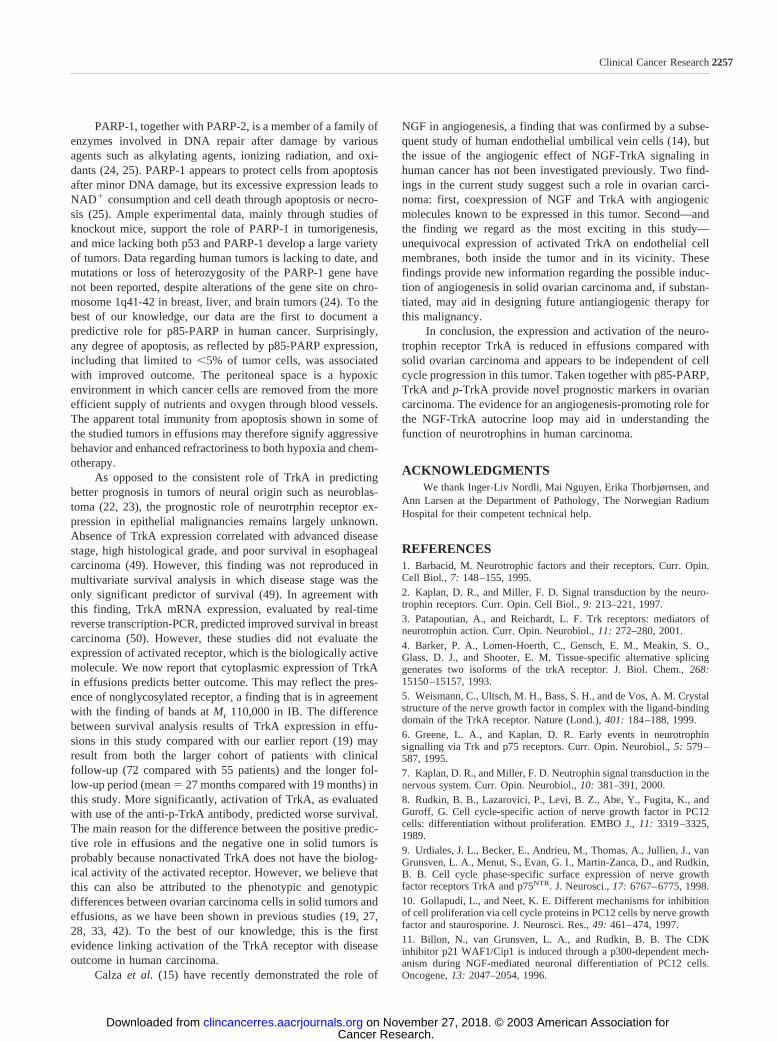
TrkA, p-TrkA, and p85-PARP Are Novel Predictors ofDisease Outcome in Ovarian Cancer. In the final part of theanalysis, we wished to evaluate the potential prognostic role ofthe molecules studied. In this cohort, no patients were lost tofollow-up. Follow-up period ranged from 1 to 83 months(mean � 27 months). Forty-nine patients died of disease, 18patients were alive with disease, and 5 patients were free ofdisease at the time of last follow-up. In survival analysis, cyto-plasmic expression of TrkA (P � 0.001) and nuclear expressionof p85-PARP (P � 0.017) in tumor cells in effusions predictedbetter outcome (Fig. 4, A and B). In solid tumors, expression ofp-TrkA at both the membrane (P � 0.004) and cytoplasm (P �0.042) predicted poor survival (Fig. 4, C and D). As previouslyreported, angiogenic gene expression showed no correlationwith disease outcome (P 0.05, data not shown). Neither did
Table 4 NGF, TrkA and p-TrkA expression in 65 solid tumors
MoleculeCellularlocation
Percentage of stained carcinoma cells
0% 1–5% 6–25% 26–75% 76–100%
TrkA Membrane 8 (12%) 30 (46%) 16 (25%) 9 (14%) 2 (3%)TrkA Cytoplasm 2 (3%) 2 (3%) 6 (9%) 14 (22%) 41 (63%)p-TrkA Membrane 15 (23%) 30 (46%) 16 (25%) 4 (6%) 0 (0%)p-TrkA Cytoplasm 13 (20%) 11 (17%) 21 (32%) 9 (14%) 11 (17%)p-TrkA Nucleus 59 (90%) 4 (6%) 0 (0%) 1 (2%) 1 (2%)NGF Cytoplasm 13 (20%) 7 (11%) 17 (26%) 8 (12%) 20 (31%)
2253Clinical Cancer Research
Cancer Research. on November 27, 2018. © 2003 American Association forclincancerres.aacrjournals.org Downloaded from

Fig. 3 Immunohistochemistry and mRNA ISH in solid tumors. A–F, TrkA, p-TrkA, and NGF. A, TrkA protein membrane expression in a primaryserous ovarian carcinoma. All cells express TrkA. B, p-TrkA protein membrane expression in a primary serous ovarian carcinoma. C, p-TrkA proteinnuclear expression in a cutaneous metastasis. The majority of tumor cells are positive. This pattern was observed in six carcinomas. D, p-TrkA proteinmembrane expression in peritumoral vessels in the vicinity (left) of a primary carcinoma. Unequivocal membrane expression is seen. E, vascularp-TrkA expression at a peritumoral site in the tumor shown in C. F, NGF expression in cancer cells in a uterine metastasis of a serous ovariancarcinoma. All cells show cytoplasmic immunoreactivity. G–I, expression of mRNA of angiogenic molecules. G, d(T) control for mRNA integrityin a primary ovarian serous carcinoma. All cells are labeled (nitroblue tetrazolium-5-bromo-4-chloro-3-indolyl phosphate staining). Inset shows anintestinal metastasis negative for VEGF. Cells are counterstained with nuclear fast red. H, weak diffuse VEGF mRNA expression in both the tumorand the stromal cell compartments. I, intense expression (dark blue) of the bFGF in the two compartments.
2254 TrkA, Angiogenic Genes, and Cell Cycle Proteins
Cancer Research. on November 27, 2018. © 2003 American Association forclincancerres.aacrjournals.org Downloaded from

the expression of p53, Ki-67, or p21WAF1/CIP1 show such cor-relation (P 0.05, data not shown).
DISCUSSIONOverexpression of growth factor receptors such as tyrosine
kinase receptors is one of the phenotypic changes providingcancer cells with the ability to override normal cell growthregulatory mechanisms. Expression of TrkA, the high-affinityreceptor for NGF, has been documented in several epithelialmalignancies, including ovarian carcinoma (18–21) and recent
studies in murine models have demonstrated the role of neuro-trophin signaling in tumor growth and metastasis (39, 40).However, the biological roles of the NGF-TrkA autocrine loopin ovarian carcinoma are poorly defined at present. In this study,we attempted to study the correlation between these moleculesand disease outcome, as well as their association with expres-sion of cell cycle molecules and angiogenic factors.
We have previously reported on the reduced expression ofTrkA in ovarian carcinoma cells in effusions compared withcorresponding solid tumors (19). In this study, a similar andmore pronounced phenomenon was seen for p-TrkA. However,unlike experimental models (39, 40), this down-regulation doesnot appear to be related to reduced cell proliferation, as evi-denced both by the lack of correlation with Ki-67 counts and bythe generally high expression of this marker in effusions. Thepossibility that the reduced expression of TrkA is associatedwith the alternative expression of other tyrosine kinases recep-tors such as epidermal growth factor, c-erbB-2, and fibroblastgrowth factor receptors in effusions deserves additionalresearch.
Interestingly, nuclear carcinoma cell-specific expression ofp-TrkA was detected in a few specimens, both primary andmetastatic. The biological significance of this finding, which iswell documented for other kinases involved in signal transduc-tion, is unknown to us at present and requires additional inves-tigation.
The comparison between pleural and peritoneal carcinomacells did not reveal differences in the expression of TrkA,p-TrkA, NGF, or the cell cycle markers detailed below, inagreement with our previous studies of ovarian carcinoma cellsin effusions (19, 27, 28, 33, 41–47). The present findings for
Table 5 Angiogenic gene expression in 63 effusions and 65 solid tumorsa
Molecule Specimen Cells
Percentage of stained cells Staining intensity
0% 1–25% 26–100% Absent Weak Intense
VEGF Effusions Carcinoma 41 (66%) 11 (17%) 11 (17%) 41 (66%) 18 (28%) 4 (6%)IL-8 Effusions Carcinoma 14 (22%) 22 (35%) 27 (43%) 14 (22%) 25 (40%) 24 (38%)bFGF Effusions Carcinoma 3 (5%) 13 (21%) 47 (74%) 3 (5%) 26 (41%) 34 (54%)VEGF Solid tumors Carcinoma 35 (54%) 15 (23%) 15 (23%) 35 (54%) 28 (43%) 2 (3%)IL-8 Solid tumors Carcinoma 14 (21%) 13 (20%) 38 (59%) 14 (21%) 44 (68%) 7 (11%)bFGF Solid tumors Carcinoma 1 (1%) 18 (28%) 46 (71%) 1 (1%) 29 (45%) 35 (54%)VEGF Solid tumors Stromal 33 (51%) 17 (26%) 15 (23%) 33 (51%) 32 (49%) 0 (0%)IL-8 Solid tumors Stromal 18 (28%) 15 (23%) 32 (49%) 18 (28%) 42 (65%) 5 (7%)bFGF Solid tumors Stromal 2 (3%) 20 (31%) 43 (66%) 2 (3%) 35 (54%) 28 (43%)
a The percentage of stained cells in shown as 0, 1–25%, or 26–100% of cells.
Table 6 The association between protein expression of NGF andmRNA expression of bFGF in carcinoma cells in effusions (n � 63
specimens)
NGF
bFGF
TotalaAbsent Weak Intense
Absent 1 10 2 13Focal (1–25%) 2 6 13 21Diffuse (26–100%) 0 10 19 29Total 3 26 34 63
a P � 0.032.
Table 7 The association between expression of NGF in carcinomacells and mRNA expression of VEGF in solid tumors (n � 65
specimens)
NGF
A. VEGF tumor cells
TotalaAbsentFocal
(1–25%)Diffuse
(26–100%)
Absent 12 0 1 13Focal (1–25%) 6 9 9 24Diffuse (26–100%) 17 6 5 28Total 35 15 15 65
NGF
B. VEGF stromal cells
TotalaAbsentFocal
(1–25%)Diffuse
(26–100%)
Absent 12 0 1 13Focal (1–25%) 5 9 10 24Diffuse (26–100%) 16 8 4 28Total 33 17 15 65
a P � 0.002.
Table 8 The association between membrane expression of TrkA incarcinoma cells and stromal mRNA expression of VEGF in solid
tumors (n � 65 specimens)
TrkA
VEGF stromal cells
TotalaAbsent Weak
Absent 7 1 8Focal (1–25%) 24 22 46Diffuse (26–100%) 2 9 11Total 33 32 65
a P � 0.017.
2255Clinical Cancer Research
Cancer Research. on November 27, 2018. © 2003 American Association forclincancerres.aacrjournals.org Downloaded from

NGF and TrkA provide additional evidence in support of ourhypothesis that ovarian carcinoma cells in pleural effusionsclosely resemble those in peritoneal effusions and possess nophenotypic or genotypic advantages that can support the classi-fication of isolated pleural effusion as FIGO stage IV disease.Furthermore, carcinoma cells at both effusion sites undergosignificant biological alterations compared with cells at theprimary tumor site, suggesting that the cells in ascites are trulymetastatic.
p53 and p21WAF1/CIP1 are two powerful regulators of thecell cycle, the latter through inhibition of the cyclin-dependentkinase 2, involved in the G1-S transition (48). The role of theNGF-TrkA autocrine loop in activation and expression of p53
and p21WAF1/CIP1 has been documented in PC12 cells (9–11).Our findings do not support such role in ovarian carcinoma cellsbecause expression of p53 and p21WAF1/CIP1 did not correlatewith NGF and TrkA expression or with activation of TrkA. Weobserved an almost universally low expression of p21WAF1/CIP1
in cancer cells in our material, with expression in blots reflectinginflammatory and mesothelial cell contribution. This was ac-companied by high expression of cyclin-dependent kinase 2 inall effusions using IB (data not shown). Blots for p53 haveshown additional truncated forms, possibly representing mu-tated protein, in many specimens. It therefore appears that thedysregulation of cell cycle events in ovarian carcinoma in effu-sions also involves the dissociation from NGF-TrkA signaling.
Fig. 4 Kaplan-Meier survival curves showing the correlation between TrkA, p85-PARP, and p-TrkA and disease outcome. A, the correlationbetween cytoplasmic expression of TrkA in carcinoma cells in effusions and OS. Patients with tumors showing expression in �25% of cancer cells(n � 22, discontinuous line) had a median OS of 20 months compared with 31 months for patients with tumors showing 25% expression (n � 58,continuous line; P � 0.001). The death of the last patient among the poor survivors, 65 months after diagnosis, has been censored but is shown inB. B, the correlation between p85-PARP expression in effusions and OS. Patients with specimens showing any number of p85-PARP-positive cells(n � 42, continuous line) had a median OS of 37 months, compared with 23 months for patients with p85-PARP-negative tumors (n � 38,discontinuous line; P � 0.017). C, the correlation between membrane expression of p-TrkA in carcinoma cells in solid tumors and OS. Patients withp-TrkA-negative tumors (n � 15, continuous line) had a median OS of 83 months compared with 24 months for patients with tumors showing stainingof 1–5% of cells (n � 30, discontinuous line) and 25 months for patients with expression exceeding 5% of cells (n � 20, dotted line; P � 0.004).D, the correlation between cytoplasmic expression of p-TrkA in carcinoma cells in solid tumors and OS, showing results that are comparable withthose in C. Patients with p-TrkA-negative tumors (n � 13, continuous line) had a median OS of 46 months compared with 24 months for patientswith tumors showing staining of 1–5% of cells (n � 32, discontinuous line) and 23 months for patients with expression exceeding 5% of cells (n �20, dotted line; P � 0.042).
2256 TrkA, Angiogenic Genes, and Cell Cycle Proteins
Cancer Research. on November 27, 2018. © 2003 American Association forclincancerres.aacrjournals.org Downloaded from
PARP-1, together with PARP-2, is a member of a family ofenzymes involved in DNA repair after damage by variousagents such as alkylating agents, ionizing radiation, and oxi-dants (24, 25). PARP-1 appears to protect cells from apoptosisafter minor DNA damage, but its excessive expression leads toNAD� consumption and cell death through apoptosis or necro-sis (25). Ample experimental data, mainly through studies ofknockout mice, support the role of PARP-1 in tumorigenesis,and mice lacking both p53 and PARP-1 develop a large varietyof tumors. Data regarding human tumors is lacking to date, andmutations or loss of heterozygosity of the PARP-1 gene havenot been reported, despite alterations of the gene site on chro-mosome 1q41-42 in breast, liver, and brain tumors (24). To thebest of our knowledge, our data are the first to document apredictive role for p85-PARP in human cancer. Surprisingly,any degree of apoptosis, as reflected by p85-PARP expression,including that limited to �5% of tumor cells, was associatedwith improved outcome. The peritoneal space is a hypoxicenvironment in which cancer cells are removed from the moreefficient supply of nutrients and oxygen through blood vessels.The apparent total immunity from apoptosis shown in some ofthe studied tumors in effusions may therefore signify aggressivebehavior and enhanced refractoriness to both hypoxia and chem-otherapy.
As opposed to the consistent role of TrkA in predictingbetter prognosis in tumors of neural origin such as neuroblas-toma (22, 23), the prognostic role of neurotrphin receptor ex-pression in epithelial malignancies remains largely unknown.Absence of TrkA expression correlated with advanced diseasestage, high histological grade, and poor survival in esophagealcarcinoma (49). However, this finding was not reproduced inmultivariate survival analysis in which disease stage was theonly significant predictor of survival (49). In agreement withthis finding, TrkA mRNA expression, evaluated by real-timereverse transcription-PCR, predicted improved survival in breastcarcinoma (50). However, these studies did not evaluate theexpression of activated receptor, which is the biologically activemolecule. We now report that cytoplasmic expression of TrkAin effusions predicts better outcome. This may reflect the pres-ence of nonglycosylated receptor, a finding that is in agreementwith the finding of bands at Mr 110,000 in IB. The differencebetween survival analysis results of TrkA expression in effu-sions in this study compared with our earlier report (19) mayresult from both the larger cohort of patients with clinicalfollow-up (72 compared with 55 patients) and the longer fol-low-up period (mean � 27 months compared with 19 months) inthis study. More significantly, activation of TrkA, as evaluatedwith use of the anti-p-TrkA antibody, predicted worse survival.The main reason for the difference between the positive predic-tive role in effusions and the negative one in solid tumors isprobably because nonactivated TrkA does not have the biolog-ical activity of the activated receptor. However, we believe thatthis can also be attributed to the phenotypic and genotypicdifferences between ovarian carcinoma cells in solid tumors andeffusions, as we have been shown in previous studies (19, 27,28, 33, 42). To the best of our knowledge, this is the firstevidence linking activation of the TrkA receptor with diseaseoutcome in human carcinoma.
Calza et al. (15) have recently demonstrated the role of
NGF in angiogenesis, a finding that was confirmed by a subse-quent study of human endothelial umbilical vein cells (14), butthe issue of the angiogenic effect of NGF-TrkA signaling inhuman cancer has not been investigated previously. Two find-ings in the current study suggest such a role in ovarian carci-noma: first, coexpression of NGF and TrkA with angiogenicmolecules known to be expressed in this tumor. Second—andthe finding we regard as the most exciting in this study—unequivocal expression of activated TrkA on endothelial cellmembranes, both inside the tumor and in its vicinity. Thesefindings provide new information regarding the possible induc-tion of angiogenesis in solid ovarian carcinoma and, if substan-tiated, may aid in designing future antiangiogenic therapy forthis malignancy.
In conclusion, the expression and activation of the neuro-trophin receptor TrkA is reduced in effusions compared withsolid ovarian carcinoma and appears to be independent of cellcycle progression in this tumor. Taken together with p85-PARP,TrkA and p-TrkA provide novel prognostic markers in ovariancarcinoma. The evidence for an angiogenesis-promoting role forthe NGF-TrkA autocrine loop may aid in understanding thefunction of neurotrophins in human carcinoma.
ACKNOWLEDGMENTSWe thank Inger-Liv Nordli, Mai Nguyen, Erika Thorbjørnsen, and
Ann Larsen at the Department of Pathology, The Norwegian RadiumHospital for their competent technical help.
REFERENCES1. Barbacid, M. Neurotrophic factors and their receptors. Curr. Opin.Cell Biol., 7: 148–155, 1995.2. Kaplan, D. R., and Miller, F. D. Signal transduction by the neuro-trophin receptors. Curr. Opin. Cell Biol., 9: 213–221, 1997.3. Patapoutian, A., and Reichardt, L. F. Trk receptors: mediators ofneurotrophin action. Curr. Opin. Neurobiol., 11: 272–280, 2001.4. Barker, P. A., Lomen-Hoerth, C., Gensch, E. M., Meakin, S. O.,Glass, D. J., and Shooter, E. M. Tissue-specific alternative splicinggenerates two isoforms of the trkA receptor. J. Biol. Chem., 268:15150–15157, 1993.5. Weismann, C., Ultsch, M. H., Bass, S. H., and de Vos, A. M. Crystalstructure of the nerve growth factor in complex with the ligand-bindingdomain of the TrkA receptor. Nature (Lond.), 401: 184–188, 1999.6. Greene, L. A., and Kaplan, D. R. Early events in neurotrophinsignalling via Trk and p75 receptors. Curr. Opin. Neurobiol., 5: 579–587, 1995.7. Kaplan, D. R., and Miller, F. D. Neutrophin signal transduction in thenervous system. Curr. Opin. Neurobiol., 10: 381–391, 2000.8. Rudkin, B. B., Lazarovici, P., Levi, B. Z., Abe, Y., Fugita, K., andGuroff, G. Cell cycle-specific action of nerve growth factor in PC12cells: differentiation without proliferation. EMBO J., 11: 3319–3325,1989.9. Urdiales, J. L., Becker, E., Andrieu, M., Thomas, A., Jullien, J., vanGrunsven, L. A., Menut, S., Evan, G. I., Martin-Zanca, D., and Rudkin,B. B. Cell cycle phase-specific surface expression of nerve growthfactor receptors TrkA and p75NTR. J. Neurosci., 17: 6767–6775, 1998.10. Gollapudi, L., and Neet, K. E. Different mechanisms for inhibitionof cell proliferation via cell cycle proteins in PC12 cells by nerve growthfactor and staurosporine. J. Neurosci. Res., 49: 461–474, 1997.11. Billon, N., van Grunsven, L. A., and Rudkin, B. B. The CDKinhibitor p21 WAF1/Cip1 is induced through a p300-dependent mech-anism during NGF-mediated neuronal differentiation of PC12 cells.Oncogene, 13: 2047–2054, 1996.
2257Clinical Cancer Research
Cancer Research. on November 27, 2018. © 2003 American Association forclincancerres.aacrjournals.org Downloaded from

12. Montano, X. p53 associates with trk tyrosine kinase. Oncogene, 15:245–246, 1997.
13. Brown, A., Browes, C., Mitchell, M., and Montano, X. c-abl isinvolved in the association of p53 and trk A. Oncogene, 19: 3032–3040,2000.
14. Cantarella, G., Lempereur, L., Presta, M., Ribatti, D., Lombardo,G., Lazarovici, P., Zappala, G., Pafumi, C., and Bernardini, R. Nervegrowth factor-endothelial cell interaction leads to angiogenesis in vitroand in vivo. FASEB J., 16: 1307–1309, 2002.
15. Calza, L., Giardino, L., Guiliani, A., Aloe, L., and Levi-Montalcini,R. Nerve growth factor control of neuronal expression of angiogenic andvasoactive factors. Proc. Natl. Acad. Sci. USA, 98: 4160–4165, 2001.
16. Raychaudhuri, S. K., Raychaudhuri, S. P., Weltman, H., and Farber,E. M. Effects of nerve growth factor on endothelial cell biology:proliferation and adherence molecule expression on human dermalmicrovascular endothelial cells. Arch. Dermatol. Res., 293: 291–295,2002.
17. Goldbrunner, R. H., Wagner, S., Roosen, K., and Tonn, J. C.Models for assessment of angiogenesis in gliomas. J. Neuro-oncol., 50:53–62, 2000.
18. Nakagawara, A. Trk receptor tyrosine kinases: A bridge betweencancer and neural development. Cancer Lett., 169: 107–114, 2001.
19. Davidson, B., Lazarovici, P., Ezersky, A., Nesland, J. M., Berner,A., Risberg, B., Trope, C. G., Kristensen, G. B., Goscinski, M., van dePutte, G., and Reich, R. Expression levels of the NGF receptors TrkAand p75 in effusions and solid tumors of serous ovarian carcinomapatients. Clin. Cancer Res., 7: 3457–3464, 2001.
20. Shibayama, E., and Koizumi, H. Cellular localization of the Trkneurotrophin receptor family in human non-neuronal tissues. Am. J.Pathol., 148: 1807–1818, 1996.
21. McGregor, L. M., McCune, B. K., Graff, J. R., McDowell, P. R.,Romans, K. E., Yancopoulos, G. D., Ball, D. W., Baylin, S. B., andNelkin, B. D. Roles of trk family neurotrophin receptors in medullarythyroid carcinoma development and progression. Proc. Natl. Acad. Sci.USA, 96: 4540–4545, 1999.
22. Nakagawara, A., Arima, M., Azar, C. G., Scavarda, N. J., andBrodeur, G. M. Inverse relationship between trk expression and N-mycamplification in human neuroblastomas. Cancer Res., 52: 1364–1368,1992.
23. Suzuki, E., Bogenmann, H., Shimada, D., Stram, R. C., and Seeger,R. C. Lack of high-affinity nerve growth factor receptors in aggressiveneuroblastomas. J. Natl. Cancer Inst. (Bethesda), 85: 377–384, 1993.
24. Tong, W. M., Cortes, U., and Wang, Z. Q. Poly(ADP-ribose)polymerase: a guardian angel protecting the genome and suppressingtumorigenesis. Biochim. Biophys. Acta, 1552: 27–37, 2001.
25. Bernstein, C., Bernstein, H., Payne, C. M., and Garewal, H. DNArepair/pro-apoptotic dual-role proteins in five major DNA repair path-ways: fail-safe protection against carcinogenesis. Mutat. Res., 511:145–178, 2002.
26. Davidson, B., Risberg, B., Kristensen, G., Kvalheim, G., Emilsen,E., Bjåmer, A., and Berner, A. Detection of cancer cells in effusionsfrom patients diagnosed with gynecological malignancies: evaluation offive epithelial markers. Virchows Arch., 435: 43–49, 1999.
27. Davidson, B., Berner, A., Nesland, J. M., Risberg, B., Kristensen,G. B., Trope, C. G., and Bryne, M. Carbohydrate antigen expression inprimary tumors, metastatic lesions, and serous effusions from patientsdiagnosed with epithelial ovarian carcinoma: evidence of up-regulatedTn and Sialyl Tn antigens expression in effusions. Hum. Pathol., 31:1081–1087, 2000.
28. Davidson, B., Berner, A., Nesland, J. M., Risberg, B., Berner, H. S.,Trope, C. G., Kristensen, G. B., Bryne, M., and Flørenes, V. A. E-Cadherin and �-, �- and �-catenin protein expression is up-regulated inovarian carcinoma cells in serous effusions. J. Pathol., 192: 460–469,2000.
29. Young, R. H., Clement, P. B., and Scully, R. E. Surface epithelial-stromal tumors. In: S. S. Sternberg, D. A. Antonioli, D. Carter, S. E.
Mills, and H. A. Oberman (eds.), Diagnostic Surgical Pathology, Vol. 2,pp. 2319–2382. Philadelphia: Lippincott, Williams & Wilkins, 1999.
30. Hempstead, B. L., Rabin, S. J., Kaplan, L., Reid, S., Parada, L. F.,and Kaplan, D. R. Overexpression of the trk tyrosine kinase rapidlyaccelerates nerve growth factor-induced differentiation. Neuron, 9: 883–896, 1992.
31. Katzir, I., Shani, J., Regev, K., Shabashov, D., and Lazarovici, P. Aquantitative bioassay for nerve growth factor, using PC12 clones ex-pressing different levels of trkA receptors. J. Mol. Neurosci., 18: 251–264, 2002.
32. Hochman, J., Park, S. S., Lazarovici, P., Bergel, M., and Gottesman,M. M. Monoclonal antibodies to immunogenic lymphoma cell variantsdisplaying impaired neoplastic properties: characterization and applica-tions. J. Natl. Cancer Inst. (Bethesda), 82: 1821–1826, 1990.
33. Davidson, B., Reich, R., Kopolovic, J., Berner, A., Nesland, J. M.,Kristensen, G. B., Trope, C. G., Bryne, M., Risberg, B., van de Putte, G.,and Goldberg, I. Interleukin-8 and vascular endothelial growth factormRNA levels are down-regulated in ovarian carcinoma cells in serouseffusions. Clin. Exp. Metastasis, 19: 135–144, 2002.
34. Flørenes, V. A., Chao, L., Bhattacharya, N., Rak, J., Sheehan, C.,Slingerland, J. M., and Kerbel, R. S. Interleukin-6 dependent inductionof the cyclin dependent kinase inhibitor p21WAF1/CIP1 is lost duringprogression of human malignant melanoma. Oncogene, 18: 1023–1032,1999.
35. Yoneda, J., Kuniyasu, H., Crispens, M. A., Price, J. E., Bucana,C. D., and Fidler, I. J. Expression of angiogenesis-related genes andprogression of human ovarian carcinomas in nude mice. J. Natl. CancerInst. (Bethesda), 90: 447–454, 1998.
36. Reed, J. A., Manahan, L. J., Parks, C. S., and Brigati, D. J.Complete one-hour immunocytochemistry based on capillary action.Biotechniques, 13: 434–443, 1992.
37. Davidson, B., Goldberg, I., Gotlieb, W. H., Kopolovic, J., Ben-Baruch, G., Nesland, J. M., Berner, A., Bryne, A., and Reich, R. Highlevels of MMP-2, MMP-9, MT1-MMP, and TIMP-2 mRNA correlatewith poor survival in ovarian carcinoma. Clin. Exp. Metastasis, 17:799–808, 1999.
38. Davidson, B., Goldberg, I., Kopolovic, J., Gotlieb, W. H., Givant-Horwitz, V., Nesland, J. M., Berner, A., Ben-Baruch, G., Bryne, M., andReich, R. Expression of angiogenesis-related genes in ovarian carci-noma: a clinicopathologic study. Clin. Exp. Metastasis, 18: 501–507,2000.
39. Miknyoczki, S. J., Wan, W., Chang, H., Dobrzanski, P., Ruggeri,B. A., Dionne, C. A., and Buchkovich, K. The neurotrophin-Trk recep-tor axes are critical for the growth and progression of human prostaticcarcinoma and pancreatic ductal adenocarcinoma in nude mice. Clin.Cancer Res., 8: 1924–1931, 2002.
40. Weeraratna, A. T., Dalrymple, S. L., Lamb, J. C., Denmeade, S. R.,Miknyoczki, S., Dionne, C. A., and Isaacs, J. T. Pan-trk inhibitiondecreases metastasis and enhances host survival in experimental modelsas a result of its selective induction of apoptosis of prostate cancer cells.Clin. Cancer Res., 7: 2237–2245, 2001.
41. Davidson, B., Goldberg, I., Berner, A., Nesland, J. M., Givant, V.,Bryne, M., Risberg, B., Kristensen, G. B., Trope, C. G., Kopolovic, J.,and Reich, R. Expression of membrane-type 1, 2 and 3 matrix metal-loproteinases (MT1-MMP, MT2-MMP, MT3-MMP) mRNA in ovariancarcinoma cells in serous effusions. Am. J. Clin. Pathol., 115: 517–524,2001.
42. Davidson, B., Reich, R., Berner, A., Givant, V., Goldberg, I.,Risberg, B., Kristensen, G. B., Trope, C. G., Bryne, M., Kopolovic, J.,and Nesland, J. M. Ovarian carcinoma cells in serous effusions showaltered MMP-2 and TIMP-2 mRNA levels. Eur. J. Cancer, 37: 2040–2049, 2001.
43. Berner, H. S., Davidson, B., Berner, A., Risberg, B., Kristensen,G. B., Trope, C. G., van de Putte, G., and Nesland, J. M. Expression ofCD44 in effusions of patients diagnosed with serous ovarian carcinoma:diagnostic and prognostic implications. Clin. Exp. Metastasis, 18: 197–202, 2000.
2258 TrkA, Angiogenic Genes, and Cell Cycle Proteins
Cancer Research. on November 27, 2018. © 2003 American Association forclincancerres.aacrjournals.org Downloaded from

44. Davidson, B., Risberg, B., Goldberg, I., Nesland, J. M., Berner, A.,Trope, C. G., Kristensen, G. B., Bryne, M., and Reich, R. Ets-1 mRNAexpression in effusions of serous ovarian carcinoma patients is a markerof poor outcome. Am. J. Surg. Pathol., 25: 1493–1500, 2001.
45. Davidson, B., Risberg, B., Berner, A., Nesland, J. M., Trope, C. G.,Kristensen, G. B., Bryne, M., van de Putte, G., and Flørenes, V. A.Expression of cell cycle proteins in ovarian carcinoma cells in serouseffusions: biological and prognostic implications. Gynecol. Oncol., 83:249–256, 2001.46. Davidson, B., Goldberg, I., Givant-Horwitz, V., Nesland, J. M.,Berner, A., Bryne, M., Risberg, B., Kopolovic, J., Kristensen, G. B.,Trope�, C. G., van de Putte, G., and Reich, R. Caveolin-1 expression inovarian carcinoma is MDR-1-independent. Am. J. Clin. Pathol., 117:225–234, 2002.
47. Davidson, B., Goldberg, I., Berner, A., Kristensen, G. B., andReich, R. EMMPRIN (extracellular matrix metalloproteinase inducer) isa novel marker of poor outcome in serous ovarian carcinoma. Clin. Exp.Metastas., 20: 161–169, 2003.48. Malumbres, M., and Barbacid, M. To cycle or not to cycle: a criticaldecision in cancer. Nat. Rev. Cancer, 1: 222–231, 2001.49. Zhu, Z. W., Friess, H., Wang, L., Di Mola, F. F., Zimmermann, A.,and Buchler, M. W. Down-regulation of nerve growth factor in poorlydifferentiated and advanced human esophageal cancer. Anticancer Res.,20: 125–132, 2000.50. Descamps, S., Pawlowski, V., Revillion, F., Hornez, L., Hebbar,M., Boilly, B., Hondermarck, H., and Peyrat, J. P. Expression of nervegrowth factor receptors and their prognostic value in human breastcancer. Cancer Res., 61: 4337–4340, 2001.
2259Clinical Cancer Research
Cancer Research. on November 27, 2018. © 2003 American Association forclincancerres.aacrjournals.org Downloaded from

2003;9:2248-2259. Clin Cancer Res Ben Davidson, Reuven Reich, Philip Lazarovici, et al. Receptor TrkA in Serous Ovarian CarcinomaExpression and Activation of the Nerve Growth Factor
Updated version
http://clincancerres.aacrjournals.org/content/9/6/2248
Access the most recent version of this article at:
Cited articles
http://clincancerres.aacrjournals.org/content/9/6/2248.full#ref-list-1
This article cites 47 articles, 8 of which you can access for free at:
Citing articles
http://clincancerres.aacrjournals.org/content/9/6/2248.full#related-urls
This article has been cited by 6 HighWire-hosted articles. Access the articles at:
E-mail alerts related to this article or journal.Sign up to receive free email-alerts
SubscriptionsReprints and
To order reprints of this article or to subscribe to the journal, contact the AACR Publications
Permissions
Rightslink site. (CCC)Click on "Request Permissions" which will take you to the Copyright Clearance Center's
.http://clincancerres.aacrjournals.org/content/9/6/2248To request permission to re-use all or part of this article, use this link
Cancer Research. on November 27, 2018. © 2003 American Association forclincancerres.aacrjournals.org Downloaded from