Embed Size (px)
Citation preview

MOL 33514
1
Activation of Single Nicotinic Receptor Channels from Caenorhabditis elegans Muscle
Diego Rayes, Marina Flamini, Guillermina Hernando and Cecilia Bouzat
Instituto de Investigaciones Bioquímicas, Universidad Nacional del Sur-CONICET,
Camino La Carrindanga Km 7, B8000FWB, Bahía Blanca, Argentina
Molecular Pharmacology Fast Forward. Published on February 21, 2007 as doi:10.1124/mol.106.033514
Copyright 2007 by the American Society for Pharmacology and Experimental Therapeutics.
This article has not been copyedited and formatted. The final version may differ from this version.Molecular Pharmacology Fast Forward. Published on February 21, 2007 as DOI: 10.1124/mol.106.033514
at ASPE
T Journals on A
pril 7, 2022m
olpharm.aspetjournals.org
Dow
nloaded from

MOL 33514
2
a. Running title: Single AChR channels of C. elegans
b. Address correspondence to: Cecilia Bouzat- Instituto de Investigaciones Bioquímicas.
Camino La Carrindanga Km 7, B8000FWB, Bahía Blanca, Argentina. E-mail:
[email protected] . FAX: 54-291-4861200
c. Number of text pages: 35
Number of tables: 1
Number of figures: 6
Number of references: 40
Number of words in the abstract: 248
Number of words in the Introduction: 485
Number of words in the Discussion: 1,480
d. Abbreviations: nAChR, nicotinic acetylcholine receptor; ACh, acetylcholine.
This article has not been copyedited and formatted. The final version may differ from this version.Molecular Pharmacology Fast Forward. Published on February 21, 2007 as DOI: 10.1124/mol.106.033514
at ASPE
T Journals on A
pril 7, 2022m
olpharm.aspetjournals.org
Dow
nloaded from

MOL 33514
3
Abstract
Nicotinic acetylcholine receptors (nAChRs) are pentameric neurotransmitter-gated ion
channels that mediate synaptic transmission throughout the nervous system in vertebrates
and invertebrates. Caenorhabditis elegans is a non-mammalian model for the study of the
nervous system as well as a model of parasitic nematodes. Nematode muscle nAChRs are
of considerable interest as they are targets for anthelmintic drugs. We show single-channel
activity of C. elegans muscle nAChRs for the first time. Our results reveal that in the L1
larval stage ACh activates mainly a levamisole-sensitive nAChR (L-AChR). A single
population of 39 pS channels, which are 5-fold more sensitive to levamisole than ACh, is
detected. In contrast to mammalian nAChRs, open durations are longer for levamisole than
for ACh. Studies in mutant strains reveal that UNC-38, UNC-63 and UNC-29 subunits are
assembled into a single L-AChR in the L1 stage and that these subunits are irreplaceable,
suggesting that they are vital for receptor function throughout development. Recordings
from a strain mutated in the LEV-1 subunit show a main population of channels with lower
conductance (26 pS), prolonged open durations, and reduced sensitivity to levamisole.
Thus, although LEV-1 is preferentially incorporated into native L-AChRs, receptors
lacking this subunit can still function. No single-channel activity from levamisole-
insensitive nAChRs is detected. Thus, during neuromuscular transmission in C. elegans the
majority of ACh-activated current flows through L-AChRs. This study contributes to the
understanding of the molecular mechanisms underlying functional diversity of the nAChR
family and offers an excellent strategy to test novel antiparasitic drugs.
This article has not been copyedited and formatted. The final version may differ from this version.Molecular Pharmacology Fast Forward. Published on February 21, 2007 as DOI: 10.1124/mol.106.033514
at ASPE
T Journals on A
pril 7, 2022m
olpharm.aspetjournals.org
Dow
nloaded from
MOL 33514
4
Nicotinic acetylcholine receptors (nAChR) mediate fast synaptic transmission throughout
the nervous system. A large number of nAChR subunits have been cloned from both
vertebrates and invertebrates (Jones et al., 2003). Intriguingly, the free-living helminth
Caenorhabditis elegans has one of the largest nAChR gene families known (Jones and
Sattelle, 2003).
nAChR subunits are classified as α, which contain a disulphide bridge involved in
the binding of agonists, and non-α, which lack this motif. Receptors are pentameric
proteins that can be either heteromeric, composed of α and non-α subunits, or homomeric,
composed of five identical α subunits. Nematode muscle nAChRs are of considerable
interest as they are targets for antiparasitic drugs. These drugs behave as full agonists of
nematode nAChRs, thus producing muscle paralysis (Martin, 1997). Based on their
sensitivity to levamisole, two different types of muscle nAChRs have been described in
adult C. elegans: L-AChR, or levamisole-sensitive, and N-AChR, which is levamisole-
insensitive and nicotine-sensitive (Richmond and Jorgensen, 1999; Culetto et al., 2004;
Touroutine et al., 2005). Although several subunits corresponding to each nAChR subtype
have been identified, their stoichiometry and activation kinetics remain unknown. UNC-38,
UNC-63 and LEV-8 subunits, which are α subunits, and LEV-1 and UNC-29, which
correspond to non-α subunits, are components of the adult C. elegans L-AChR. UNC-38,
UNC-63 and UNC-29 have been shown to be essential for activation of L-AChRs in adult
worms (Fleming et al., 1997; Richmond and Jorgensen, 1999; Culetto et al., 2004; Towers
et al., 2005). How these subunits are assembled into functional receptor(s) is still not
known. To date, only one subunit, ACR-16, has been reported as a component of the N-
This article has not been copyedited and formatted. The final version may differ from this version.Molecular Pharmacology Fast Forward. Published on February 21, 2007 as DOI: 10.1124/mol.106.033514
at ASPE
T Journals on A
pril 7, 2022m
olpharm.aspetjournals.org
Dow
nloaded from

MOL 33514
5
AChR (Touroutine et al., 2005). ACR-16 is capable of forming homomeric receptors in
Xenopus oocytes (Ballivet et al., 1996).
In this study, we explore for the first time at the single-channel level the activation
properties of nAChRs from C. elegans muscle. We used a primary culture system that
allows differentiation of embryonic cells into L1 larva muscle cells in vitro (Christensen et
al., 2002). Our results reveal that levamisole shows an extremely high efficacy for channel
activation. The levamisole-activated receptors are the main detected channels, indicating
that the majority of current flows through L-AChRs during neuromuscular transmission. No
N-AChR activity can be detected in cell-attached patches. Single-channel recordings from
mutant strains reveal that, as in the adult stage, UNC-38, UNC-63 and UNC-29 are required
to obtain functional L-AChRs, whereas LEV-1 can be replaced by other subunits.
Single-channel studies allow the elucidation of activation properties, composition,
and functional roles of nAChRs. Because C. elegans is a model of parasitic nematodes,
these studies will contribute to the understanding of how parasites acquire resistance to
anthelmintics and to the development of novel therapies. Moreover, due to its available
genome sequence C. elegans has become an invertebrate model of the human nervous
system and therefore studies in this nematode are proving valuable in understanding
processes involving nAChRs in mammals.
This article has not been copyedited and formatted. The final version may differ from this version.Molecular Pharmacology Fast Forward. Published on February 21, 2007 as DOI: 10.1124/mol.106.033514
at ASPE
T Journals on A
pril 7, 2022m
olpharm.aspetjournals.org
Dow
nloaded from

MOL 33514
6
Materials and Methods
C. elegans Strains
All nematode strains were obtained from the Caenorhabditis Genetic Center, which is
funded by the NIH National Center for Research Resources (NCRR). The following strains
were used: N2 wild type (Bristol variety), myo-3::GFP PD4251(ccls4251I) (Fire et al.,
1998), CB904 unc-38(e264) I, CB1072 unc-29(e1072) I, ZZ37 unc-63(x37) I, RB918 acr-
16(ok789) V and CB211 lev-1(e211) IV. Nematodes were maintained at 20-25 °C using
standard culture methods (Brenner, 1974). The RB918 strain has not been outcrossed and it
therefore carries other mutations. However, previous reports have shown reduced ACh-
responses due to the lack of muscle N-AChRs in adult worms, which can be rescued by
muscle-specific expression of ACR-16 (Touroutine et al., 2005). Thus, it is possible to
ensure that the lack of function of N-AChRs in this strain is due to the deletion of the acr-
16 gene and not to the presence of background mutations (Touroutine et al, 2005; Francis et
al, 2005).
Isolation and culture of C. elegans muscle cells
Embryonic cells were isolated and cultured as described by Christensen et al. (2002).
Briefly, adult nematodes were exposed to an alkaline hypochlorite solution (0.5 M NaOH
and 1 % NaOCl). Eggs released were treated with 1.5 units/ml chitinase (Sigma-Aldrich
Co., St. Louis, MO) for 30-40 minutes at room temperature. The embryo cells were isolated
by gently pipetting and filtered through a sterile 5-µm Durapore syringe filter (Millipore
Corp., Bedford, MA) to remove undissociated embryos and newly hatched larvae. Filtered
cells were plated on glass coverlips coated with poly-O-Ornithine. Cultures were
maintained at 24 °C in a humidified incubator in L-15 medium (Hyclone, Logan, UT)
This article has not been copyedited and formatted. The final version may differ from this version.Molecular Pharmacology Fast Forward. Published on February 21, 2007 as DOI: 10.1124/mol.106.033514
at ASPE
T Journals on A
pril 7, 2022m
olpharm.aspetjournals.org
Dow
nloaded from

MOL 33514
7
containing 10 % fetal bovine serum. Complete differentiation to the various cell types that
comprise the newly hatched L1 larva were observed within 24 hs. Electrophysiology
experiments were performed 1-5 days after cell isolation.
The percentage of neuron and muscle cells in culture is in great agreement with
previous reports, and it is similar to that observed in the newly hatched L1 larva
(Christensen et al., 2002).
PD4251 strain produces green fluorescence protein (GFP) in body wall muscle cells,
thus allowing their identification under fluorescence optics (Fire et al., 1998). Muscle cells
are easily identifiable due to their spindle-shaped morphology that resembles the body wall
muscle cells in vivo (Christensen et al., 2002; Yuan et al., 2003; Touroutine et al., 2005).
Therefore, in other strains muscle cells were recognized by their distinctive morphology,
which was similar to that of green cells of the PD4251 strain (Yuan et al., 2003).
Single-channel recordings
Recordings were obtained in the cell-attached patch configuration (Hamill et al., 1981) at
20°C essentially as described previously (Bouzat et al., 1994, 2000). The bath and pipette
solutions contained 142 mM KCl, 5.4 mM NaCl, 1.8 mM CaCl2, 1.7 mM MgCl2, and 10
mM HEPES (pH 7.4). Acetylcholine or anthelmintic agents were added to the pipette
solution. Single-channel currents were recorded using an Axopatch 200 B patch-clamp
amplifier (Axon Instruments, Inc., CA), digitized at 5 µs intervals with the PCI-6111E
interface (National Instruments, Austin, TX), recorded to the hard disk of a computer using
the program Acquire (Bruxton Corporation, Seattle, WA), and detected by the half-
amplitude threshold criterion using the program TAC 4.0.10 (Bruxton Corporation, Seattle,
This article has not been copyedited and formatted. The final version may differ from this version.Molecular Pharmacology Fast Forward. Published on February 21, 2007 as DOI: 10.1124/mol.106.033514
at ASPE
T Journals on A
pril 7, 2022m
olpharm.aspetjournals.org
Dow
nloaded from

MOL 33514
8
WA) at a final bandwidth of 10 kHz. Open and closed time histograms were plotted using a
logarithmic abscissa and a square root ordinate and fitted to the sum of exponentials by
maximum likelihood using the program TACFit (Bruxton Corporation, Seattle, WA).
Experimental data are shown as mean ± S.D. Statistical comparisons were done
using the Student’s t test. A level of p < 0.05 was considered significant.
RT-PCR for ACR-16
Total RNA was isolated from synchronized L1 and adult wild-type nematodes by the acid
guanidium-phenol-chloroform method. RNA was converted into cDNA using the Molony
murine leukaemia virus reverse transcriptase (MLV-RT; Promega, Madison, WI) and
random primers (Promega, Madison, WI). Polymerase chain reaction (PCR) was run for 35
cycles in a Mini Cyclertm (MJ Research, Reno, NV). Specific primers for PCR were
designed to prime in different exons of the acr-16 gene to differentiate by length cDNA
amplification from genomic DNA amplification. The primers used were: sense primer
5`CGTCACTCGGAATCATTGATCC 3` (Exon 9) and antisense primer
5`GCGACAAGATACGGTGCTGACC 3` (Exon 10). A 375 bp RT-PCR product was
expected.
This article has not been copyedited and formatted. The final version may differ from this version.Molecular Pharmacology Fast Forward. Published on February 21, 2007 as DOI: 10.1124/mol.106.033514
at ASPE
T Journals on A
pril 7, 2022m
olpharm.aspetjournals.org
Dow
nloaded from

MOL 33514
9
Results
Single-channel currents from L1 muscle cells activated by ACh
To explore activity of nAChRs from C. elegans muscle cells at the single-channel level we
used a cell culture technique that allows embryonic cells to differentiate in vitro. Cultured
cells correspond to the L1 developmental stage (Christensen et al., 2002). We first studied
the PD4251 strain, which contains wild-type nAChRs. Single channels activated by ACh
(0.5-1000 µM) are readily detected in cell-attached patches from muscle cells (Figs. 1 and
2). In contrast, no opening events are observed in the absence of agonist (n=14). ACh-
activated channels exhibit a single conductance of 38.7 ± 1.6 pS at positive membrane
potentials (Fig. 1).
The minimum ACh concentration that allows channel detection is 0.5 µM. The
percentage of active patches increases from 26.6 % at 0.5 µM (n=15) to 86.6 % at 10 µM
ACh (n=15). The frequency of channel openings increases as a function of ACh
concentration. The number of opening events per second, measured within the first minute
of recording, increases from 376 ± 112 to 1184 ± 170 when ACh concentration increases
from 1 to 10 µM (Fig. 2).
At 0.5-10 µM ACh open time distributions are well described by the sum of two
exponential components (Fig. 2; Table 1). The durations of these components but not their
fractional areas are independent of ACh concentration within the 0.5-10 µM range (Fig. 2).
At 1 µM ACh the fractional area of the briefest component is significantly higher than that
observed at 10 µM (0.90 ± 0.10 and 0.40 ± 0.10 for 1 µM and 10 µM ACh, respectively;
p<0.05). This result indicates that brief openings correspond to receptors with incomplete
occupation of the binding sites. At ACh concentrations higher than 10 µM, flickering
This article has not been copyedited and formatted. The final version may differ from this version.Molecular Pharmacology Fast Forward. Published on February 21, 2007 as DOI: 10.1124/mol.106.033514
at ASPE
T Journals on A
pril 7, 2022m
olpharm.aspetjournals.org
Dow
nloaded from

MOL 33514
10
block occurs, which is evidenced by the presence of brief closings interrupting channel
openings (Fig. 2). At 300 µM ACh, open time histograms can be correctly fitted by a single
component whose duration is 4-fold briefer than that of the main open component at 10 µM
(p<0.05; Table 1).
Closed time distributions can be well fitted by two or three exponential components
(Fig. 2; Table 1). The duration of the slowest closed component systematically decreases
with ACh concentration up to 50 µM (18- fold between 1 µM and 50 µM ACh; p<0.01).
This observation is due to the increase in channel activity as a function of agonist
concentration (Fig. 2). The duration of the briefest closed component and its relative area
remain constant between 0.5 and 10 µM ACh (p>0.05) (Table 1). This component may
represent brief closures during single activation, as described before for vertebrate and
Ascaris nAChRs (Colquhoun and Sakmann, 1985; Evans and Martin, 1996). At higher ACh
concentrations closed time distributions show an increase in the area of the briefest closed
component, due to the concentration-dependent increase of brief closures corresponding to
blocked periods (0.10 ± 0.04 and 0.45 ± 0.01 at 10 and 300 µM ACh, respectively; p<0.05).
In the histograms, these brief closures cannot be distinguished from the brief closures
corresponding to activation.
The concentration-dependent decrease in the mean open time, the concentration-
dependent increase in the fractional area of the brief closed component, and the constant
mean duration of the brief blocked intervals across all blocker concentrations (Table 1), are
indicative of open-channel block produced by ACh (Neher and Sakmann, 1978; Rayes et
al., 2001).
This article has not been copyedited and formatted. The final version may differ from this version.Molecular Pharmacology Fast Forward. Published on February 21, 2007 as DOI: 10.1124/mol.106.033514
at ASPE
T Journals on A
pril 7, 2022m
olpharm.aspetjournals.org
Dow
nloaded from

MOL 33514
11
In contrast to the behavior of mammalian muscle nAChRs (Bouzat et al., 2000),
clusters of activation periods cannot be clearly distinguished at any ACh concentration
(Fig. 2).
Given that macroscopic current recordings have shown that ACh activates both L-
and N-AChR subtypes in adult C. elegans muscle (Richmond and Jorgensen, 1999) we
evaluated the nAChR subtype(s) involved in the channel activity detected at the L1 stage.
To this end, we measured channel activity in the presence of 10 µM dihydro-β-erythroidine
(DHβE), which has been reported to selectively antagonize the N-AChR subtype
(Richmond and Jorgensen, 1999; Martin et al., 2003). The conductance of ACh-activated
channels recorded in the presence of DHβE is not significantly different from that obtained
in the absence of this drug (p>0.05) (Fig. 1B; Table 1). Moreover, no significant changes in
channel frequency (data not shown), mean open and mean closed times are observed in the
presence of DHβE (Table 1), thus indicating that the detected channels do not correspond
to the N-AChR subtype.
No changes in channel activity were observed in cultured muscle cells between 1 and
5 days after plating. In addition, cell morphology remained constant during this interval.
Longer times of culture could not be tested because cells detach from the dish after 7 days
and cannot be used for single-channel recordings.
Single-channel currents activated by levamisole
Although whole-cell recordings revealed that levamisole activates the muscle L-AChR
subtype in C. elegans (Richmond and Jorgensen, 1999; Touroutine et al., 2005), no single-
channel data describing this activation was available to date. At positive membrane
This article has not been copyedited and formatted. The final version may differ from this version.Molecular Pharmacology Fast Forward. Published on February 21, 2007 as DOI: 10.1124/mol.106.033514
at ASPE
T Journals on A
pril 7, 2022m
olpharm.aspetjournals.org
Dow
nloaded from

MOL 33514
12
potentials, levamisole activates single-channel currents of 36.9 ± 0.8 pS (n=34) (Fig. 1B).
This conductance value is similar to that obtained from ACh-activated patches (p>0.05),
thus suggesting that channels detected with levamisole and ACh correspond to the same
nAChR subtype. nAChR activity is observed at levamisole concentrations higher than 100
nM (Fig. 3). This concentration is 5-fold lower than the minimum ACh concentration
required for channel detection, indicating that this anthelmintic agent is indeed a more
potent agonist than the endogenous neurotransmitter. At 100 nM levamisole open time
distributions can be correctly fitted by two components (Fig. 3; Table 1). The duration of
the second open component is significantly longer than that of ACh-activated channels.
As described for ACh, the mean open time decreases as a function of levamisole
concentration. At levamisole concentrations higher than 1 µM open time histograms are
fitted by a single open component (Fig. 3; Table 1). Such concentration-dependent
reduction is typical of an open channel blocker.
Closed time histograms can be well fitted by several components. The duration of the
briefest closed component is similar to that of ACh-activated channels (Table 1). The
duration of the main closed component, C2, decreases with the concentration of levamisole,
indicating an increase in channel activity as a function of levamisole concentration.
Although the decrease in open duration reveals that levamisole is an open-channel blocker,
neither flickering nor a new closed component corresponding to blocked periods can be
detected. This result can be explained by a slow dissociation of levamisole from the
channel, making blocked periods too long to be clearly distinguished (Papke and Oswald,
1989; Rayes et al., 2001).
This article has not been copyedited and formatted. The final version may differ from this version.Molecular Pharmacology Fast Forward. Published on February 21, 2007 as DOI: 10.1124/mol.106.033514
at ASPE
T Journals on A
pril 7, 2022m
olpharm.aspetjournals.org
Dow
nloaded from

MOL 33514
13
In agreement with the behavior of channels activated by ACh, no clusters are
observed at a wide range of levamisole concentration (100 nM-1 mM).
Single-channel currents activated by pyrantel and morantel
To further characterize muscle nAChRs from C. elegans, we evaluated activation by other
widely used anthelmintic agents, pyrantel and morantel. Both drugs elicit single-channel
activity when added to the pipette solution (Fig. 4). At -100 mV, the amplitudes of the
unitary currents are 3.80 ± 0.30 pA (n=18) and 3.60 ± 0.20 pA (n=16) for pyrantel- and
morantel-activated channels, respectively. These values are similar to those of ACh- and
levamisole-activated nAChRs (p>0.05), suggesting that the channels correspond to the
same nAChR subtype.
nAChR activity is detected at 1 nM of either pyrantel or morantel in 80 % of the
seals, indicating that both anthelmintic agents are potent agonists. Open time distributions
of 10 nM pyrantel-activated nAChRs can be well fitted by a main component of 200 ± 10
µs (relative area >0.9) and a minor component of 810 ± 20 µs (n=4) (Fig. 4). Increasing
100-fold pyrantel concentration (1 µM) leads to an increase in the fractional area of the
slowest open component together with a reduction in its duration (τ2= 510 ± 50 µs, p<0.05,
area= 0.48 ± 0.10, p<0.01). Again, the concentration-dependent increase in the relative area
of the slowest open component can be explained by the fact that it probably arises from
activity of fully occupied nAChRs. On the other hand, the reduction in the mean duration as
a function of pyrantel concentration is due to open-channel block.
The behavior of morantel-activated channels is very similar to that of pyrantel-
activated channels. At 10 nM morantel, open time histograms can be well fitted by two
This article has not been copyedited and formatted. The final version may differ from this version.Molecular Pharmacology Fast Forward. Published on February 21, 2007 as DOI: 10.1124/mol.106.033514
at ASPE
T Journals on A
pril 7, 2022m
olpharm.aspetjournals.org
Dow
nloaded from

MOL 33514
14
components of 170 ± 20 µs (relative area 0.83 ± 0.10) and 510 ± 100 µs (0.16 ± 0.09) (n=3)
(Fig. 4). At concentrations higher than 1 µM open time distributions are displaced to briefer
durations (100 ± 10 µs at 50 µM morantel, n=3; p<0.05), revealing open-channel block
(Fig.4).
As described for levamisole, no flickering block is observed for either pyrantel or
morantel-activated nAChRs, suggesting that the three anthelmintic drugs produce a slow
open-channel block of C. elegans muscle nAChR channels.
Single-channel currents from unc-38, unc-29, unc-63 and acr-16 null mutants
In order to clearly identify the nAChR subtype detected in the L1 muscle cells and to
determine how the different subunits assemble into functional receptors, we evaluated
channel activity from unc-38, unc-63, unc-29 null mutants. These mutants exhibit impaired
locomotion especially at early larval stages. We also evaluated acr-16 null mutants, which
show no evident movement defects at any developmental stage (Lewis et al., 1980;
Richmond and Jorgensen, 1999; Touroutine et al., 2005).
ACh or levamisole (0.1-1000 µM) are not capable of activating unitary currents in cell-
attached patches of L1 muscle cells from unc-29 (n=18), unc-38 (n=23) and unc-63 (n=19)
null mutant embryos (Fig. 5). This result strongly reveals that the active nAChR detected in
C. elegans muscle is composed of UNC-29, UNC-38 and UNC-63 subunits, and that these
subunits are essential for channel activity in the L1 developmental stage. Moreover, we also
performed experiments in the unc-63 null mutant using morantel and pyrantel as agonists.
No channel activity was observed at a range of pyrantel (10-50 µM, n=11) and morantel
This article has not been copyedited and formatted. The final version may differ from this version.Molecular Pharmacology Fast Forward. Published on February 21, 2007 as DOI: 10.1124/mol.106.033514
at ASPE
T Journals on A
pril 7, 2022m
olpharm.aspetjournals.org
Dow
nloaded from

MOL 33514
15
(10-300 µM, n= 16) concentrations. These results confirm that all anthelmintic agents
activate the same type of nAChR.
With the aim of detecting channel activity from the N-AChR subtype and given that
no L-AChR activity is observed in muscle cells from these three null mutants, we
performed recordings with nicotine (10-1000 µM) in the pipette solution. No single-
channel currents could be detected in 26 patches. Given that it has been postulated that the
N-AChR subtype desensitizes at a much faster rate than the L-subtype (Richmond and
Jorgensen, 1999; Touroutine et al., 2005) we also performed cell-attached patches using
pipettes in which the tip was filled with buffer (without agonist) and the shaft was filled
with nicotine at different concentrations. With rapid sealing, this would allow detection of
channel openings before complete desensitization. However, no channels could be detected
under this condition in 10 different seals. In contrast, channel activity from wild-type cells
appeared 2-4 minutes after the beginning of the recording, thus confirming that the strategy
could allow the detection of channels if fast desensitization occurred.
ACh- and levamisole-activated channels are detected in muscle cells from the acr-
16 null mutant strain, RB918 (Fig. 5). acr-16 encodes for the N-AChR, which is insensitive
to levamisole but sensitive to nicotine (Francis et al., 2005). In this mutant strain, channel
amplitude, the duration of open and closed components (Table 1), and channel frequency
(data not shown) are similar to those observed for ACh and levamisole in the wild-type
strain (Fig. 5 and Table 1). These results further confirm that the main single-channel
activity arises from the activity of the L-AChR subtype.
Because we were not able to detect N-AChR activity in any strain and under
different conditions, we performed RT-PCR to detect mRNA from the ACR-16 subunit.
This article has not been copyedited and formatted. The final version may differ from this version.Molecular Pharmacology Fast Forward. Published on February 21, 2007 as DOI: 10.1124/mol.106.033514
at ASPE
T Journals on A
pril 7, 2022m
olpharm.aspetjournals.org
Dow
nloaded from

MOL 33514
16
Samples were obtained from wild-type worms in the L1 developmental stage. The expected
band of 375 pb was observed in these samples, and its size was identical to that obtained by
PCR using ACR-16 cDNA as the template. The presence of ACR-16 mRNA in muscle L1
cells has been reported before (Touroutine et al., 2005). However, direct evidence of
functional muscle N-AChRs at this early larval stage has not been reported to date. Thus, it
may be possible that this nAChR subtype is not functional at L1 stage or that channel
activity from this receptor cannot be detected in cell-attached patches.
Single-channel currents from lev-1 mutant strain
It has been reported that the non-α LEV-1 subunit is an accessory component of the muscle
L-AChR of adult C. elegans (Fleming et al., 1997; Culetto et al., 2004). In order to clarify
the role of this subunit in the L-AChR, we recorded unitary currents of in vitro
differentiated muscle cells from the lev-1(e211) IV mutant strain. This mutant contains a
missense mutation (G461E) in the M4 segment of LEV-1, and shows normal movement in
the absence of levamisole but uncoordinated movement in its presence (Culetto et al.,
2004). Channel activity is detected at ACh concentrations higher than 0.8 µM. About 90 %
of the recordings show channels with a conductance of 26 ± 2 pS (Figs. 1B and 6, Table 1).
This value differs significantly from that of wild-type nAChRs (p<0.05), revealing the
presence of a new low-conductance L-AChR population.
Open time histograms of 10 µM ACh-activated channels exhibit a single component
of about 0.5 ms (Table 1), which is longer than that of wild-type L-AChRs (p<0.05),
indicating that not only the conductance but also the kinetics of this receptor are altered
(Fig. 6A). In contrast to recordings from wild-type muscle cells, channel activity decays
This article has not been copyedited and formatted. The final version may differ from this version.Molecular Pharmacology Fast Forward. Published on February 21, 2007 as DOI: 10.1124/mol.106.033514
at ASPE
T Journals on A
pril 7, 2022m
olpharm.aspetjournals.org
Dow
nloaded from

MOL 33514
17
during the course of the recording (977 ± 218 events/ s and 166 ± 98 events/s for the
intervals 0-15 s and 300-315 s after the start of the recording, respectively). In addition,
clear clusters are observed at ACh concentrations higher than 50 µM (Fig. 6B). The
increase of ACh concentration displaces the main closed component, which corresponds to
closings within clusters, to briefer durations (C2= 42 ± 12 ms and 6 ± 2 ms for 50 and 300
µM ACh, respectively) (Fig. 6B). This behavior is similar to that of mammalian muscle
nAChRs (Bouzat et al., 2000).
It is interesting to note that although most of the channels correspond to the low-
conductance nAChR, 50 % of the recordings exhibit an additional channel population with
similar conductance to that of wild-type L-AChRs. The kinetics of these channels is,
however, different from that of wild type L-AChR (Fig. 6C). These channels may
correspond to receptors carrying the mutant LEV-1 subunit. It is therefore possible that
LEV-1 carrying the M4 mutation is inefficiently incorporated into functional receptors.
Another explanation for this observation is that channels lacking LEV-1 show an additional
conductance state. Supporting this hypothesis is the fact that the kinetics of the high-
conductance channels is similar to that of the low-conductance ones. For instance, at 10 µM
ACh, the mean open time is not significantly different from that of the 26 pS channels (τ1=
0.42 ± 0.05 ms; p>0.05). Moreover, channel openings appear in clusters at high
concentrations, as described for the low-conductance channels (Fig. 6C).
In this mutant strain, levamisole is capable of eliciting single channel currents with
an amplitude of 2.8 pA at -100 mV (Table 1), similar to that of ACh-activated channels.
Therefore, we can ensure that the low-conductance nAChR, which may lack LEV-1, is also
sensitive to levamisole. However, the minimum concentration of levamisole that allows
This article has not been copyedited and formatted. The final version may differ from this version.Molecular Pharmacology Fast Forward. Published on February 21, 2007 as DOI: 10.1124/mol.106.033514
at ASPE
T Journals on A
pril 7, 2022m
olpharm.aspetjournals.org
Dow
nloaded from

MOL 33514
18
channel detection is 0.5 µM, which is 5-fold higher than that of the native L-AChR
subtype, suggesting a reduced sensitivity to the anthelmintic. Interestingly, the sensitivity to
ACh decreases only 1.6-fold. Open time distributions at 1 µM levamisole exhibit a single
component (Table 1), whose duration is significantly longer than that corresponding to
wild-type nAChRs at the same drug concentration (p<0.05) (Fig. 6A). As described for
ACh, clusters can be identified at levamisole concentrations higher than 100 µM and an
additional channel population with a conductance value that resembles that of the native L-
AChR is detected in 42 % of the recordings.
In summary, our results reveal that LEV-1 can be replaced by other subunits in the
pentameric receptor, leading to L-AChR channels with lower conductance and lower
levamisole sensitivity than the wild-type L-AChR.
This article has not been copyedited and formatted. The final version may differ from this version.Molecular Pharmacology Fast Forward. Published on February 21, 2007 as DOI: 10.1124/mol.106.033514
at ASPE
T Journals on A
pril 7, 2022m
olpharm.aspetjournals.org
Dow
nloaded from

MOL 33514
19
Discussion
C. elegans is a genetically tractable and genomically defined non-mammalian model for the
study of the nervous system. In particular, this model offers the prospect of better
understanding the molecular and functional diversity of the nAChR family. C. elegans
nAChRs comprise one of the most extensive nicotinic families. The reason for the high
diversity of nAChR subunits in the worm as well as the function and composition of the
native receptors are not fully understood. The free-living nematode C. elegans is also a
model for the study of parasitic nematodes (Jones et al., 2005). These parasites affect
human population, livestock and food crops. Nematode nAChRs are of clinical significance
because they are targets for anthelmintic chemotherapy. Under this scenario, the
characterization of single nAChR channel activity in C. elegans becomes highly significant.
Given the importance of the larval stages in nematode life cycles and because the subunit
expression pattern may change during development, studies at each stage are necessary.
Here we described single nAChR channel activity from muscle cells of the L1 larval stage
for the first time.
ACh activates mainly levamisole-sensitive nAChRs in L1 muscle cells. It was not
known if there is one or more L-AChRs. As judged by the single-component amplitude
histogram and the homogeneous kinetics, it is highly probable that C. elegans at L1 stage
contain a single, or at least one highly predominant, L-AChR subtype. Although no unitary
currents have been described before for C. elegans nAChRs, few reports have shown AChR
channels from nematode parasites (Pennington and Martin, 1990; Robertson and Martin,
1993; Robertson et al., 1999; Levandoski et al., 2005). The comparison of channel
properties of L-AChRs between parasites and C. elegans reveals that they have similar
conductances and mean open times (Levandosky et al., 2005). They also share a similar
This article has not been copyedited and formatted. The final version may differ from this version.Molecular Pharmacology Fast Forward. Published on February 21, 2007 as DOI: 10.1124/mol.106.033514
at ASPE
T Journals on A
pril 7, 2022m
olpharm.aspetjournals.org
Dow
nloaded from

MOL 33514
20
potency for activation by ACh and anthelmintic agents. We show that the relative potency
is morantel = pyrantel > levamisole > ACh, in full agreement with the observations in
Ascaris (Harrow and Gration, 1985; Martin et al., 1996). Thus, L-AChRs seem to be
conserved within different nematode clades. Such conservation enhances the usefulness of
C. elegans as a model of parasitic nematodes.
Wild-type L-AChRs show high channel activity, which does not decay significantly
during the course of the recording. The latter observation supports a slow desensitization
rate. In agreement with this, macroscopic current recordings have shown that L-AChRs
from C. elegans desensitize much slower than N-AChRs (Richmond and Jorgensen, 1999).
In contrast to recordings from mammalian nAChRs (Sine and Steinbach, 1987), no clusters
are observed and closed intervals do not depend on agonist concentration. Only at low
concentrations closed times are more prolonged, but this is due to the activation of only a
fraction of the entire population of nAChRs. Slow desensitization together with a relatively
slow opening rate may account for the absence of distinguishable concentration-dependent
closed components of L-AChRs. Also, the high expression of these channels in muscle
cells may unmask the presence of clusters.
Open time histograms of L-AChRs activated by ACh or levamisole show a brief
component whose area decreases as a function of agonist. Studies in mammalian nAChRs
and α7-5HT3A have shown that this component may correspond to receptors with fewer
occupied binding sites than required for optimal activation (Sine et al., 1990; Rayes et al.,
2005). We postulate a similar behavior for C. elegans L-AChRs. Given that the non-α
UNC-29 and LEV-1 subunits are components of the native L-AChR, optimal activation
may arise from two or three molecules of agonist bound. The mean duration of the slowest
This article has not been copyedited and formatted. The final version may differ from this version.Molecular Pharmacology Fast Forward. Published on February 21, 2007 as DOI: 10.1124/mol.106.033514
at ASPE
T Journals on A
pril 7, 2022m
olpharm.aspetjournals.org
Dow
nloaded from

MOL 33514
21
component at non-blocking anthelmintic concentrations is more prolonged than that of
ACh-activated channels. Thus, not only the potency but also gating kinetics are slightly
different between ACh and nematocide drugs.
Channel blockade by agonists is evidenced by the concentration-dependent decrease
of the mean open time. ACh causes L-AChRs to flicker rapidly between open and blocked
states. In contrast, neither flickering nor closed components associated to blocked periods
are observed in the presence of anthelmintics, suggesting that the drugs dissociate from the
channel very slowly. Alternatively, their blocking mechanisms may deviate from the simple
open-channel block model, as reported for morantel in Ascaris nAChR (Evans and Martin,
1996).
The exact stoichiometry of the L-AChR(s) remains unknown. Our observations
reveal that UNC-38, UNC-63 and UNC-29 subunits are assembled into a single L-AChR in
the L1 stage and that these subunits are essential and irreplaceable in the native receptor.
Because these subunits have been previously shown to be essential for adult L-AChRs
(Richmond and Jorgensen, 1999), our results demonstrate that they are vital for receptor
function throughout development. However, larvae have been shown to be more affected
by mutations in the L-AChR than adults, therefore the expression level or the composition
of these receptors may vary during development. Our study also confirms that ACR-16 is
not part of the L-AChR (Touroutine et al., 2005) given that the acr-16 null mutant shows
channel activity identical to that of wild-type strains.
Recordings from the strain carrying a missense mutation in M4 of LEV-1
demonstrate that nAChRs lacking this subunit can function. A new and main population of
low-conductance channels showing activation in clusters and slightly prolonged durations
than wild-type nAChRs is detected. The mutation at G461 in M4 is not expected to
This article has not been copyedited and formatted. The final version may differ from this version.Molecular Pharmacology Fast Forward. Published on February 21, 2007 as DOI: 10.1124/mol.106.033514
at ASPE
T Journals on A
pril 7, 2022m
olpharm.aspetjournals.org
Dow
nloaded from

MOL 33514
22
produce a change in channel conductance as this residue is neither part of the ion pore nor it
is located at the vestibule of the channel (Blanton and Cohen 1994; Unwin, 2005).
Moreover, M4 mutations in mammalian muscle nAChRs affect kinetics but not channel
conductance (Bouzat et al., 2000, 2002; Mitra et al., 2004). Thus, if the mutant LEV-1
subunit were incorporated into the pentameric receptor similarly to wild-type LEV-1, no
changes in conductance should occur. The fact that a change in conductance is observed
strongly suggests that the subunit composition of the receptor has changed. Because the
only AChR subunit affected in this strain is LEV-1, the most plausible explanation for this
finding is that the mutant subunit is not present in the receptor, and that this receptor
lacking LEV-1 shows lower conductance. Generation of lev-1 null mutants will help to
confirm this result. Because we could not detect this channel population in the wild-type
strain, our results also reveal that LEV-1 is preferentially incorporated in native L-AChRs.
In the low-conductance AChR, LEV-1 may be replaced by another subunit that conforms
the L-AChR in L1. Alternatively, LEV-1 may be substituted by a different subunit not
normally expressed in this stage. The latter possibility occurs in congenital myasthenic
syndromes, in which the fetal γ subunit reappears and it is incorporated into functional
receptors when there are null mutations in the ε subunit gene (Engel et al., 1998).
The presence of a smaller proportion of channels with similar conductance but
different kinetics to those of wild-type nAChRs may be due to the fact that, although less
efficiently, the mutant LEV-1 is also incorporated into functional channels. Alternatively,
L-AChRs lacking LEV-1 may show a minor wild-type like conductance state with similar
gating kinetics to the low-conductance channels.
This article has not been copyedited and formatted. The final version may differ from this version.Molecular Pharmacology Fast Forward. Published on February 21, 2007 as DOI: 10.1124/mol.106.033514
at ASPE
T Journals on A
pril 7, 2022m
olpharm.aspetjournals.org
Dow
nloaded from

MOL 33514
23
The sensitivity to ACh and levamisole is reduced in the low-conductance nAChR,
albeit the reduction is significantly more pronounced for the anthelmintic. This lower
potency may explain the mutant phenotype, which exhibits normal locomotion in the
absence of levamisole but it becomes uncoordinated, though not killed, in its presence
(Culetto et al., 2004).
Our RT-PCR essay reveals that ACR-16 may be expressed in the L1 stage, in
agreement with previous reports (Touroutine et al., 2005). However, no single-channel
currents from N-AChRs could be detected. Several reasons can explain this: i) the
conductance of this receptor may be too low for single-channel detection. In this sense, N-
AChR from Ascaris exhibits lower amplitude than that of L-AChRs (Levandoski et al.,
2005); ii) receptors may desensitize too fast to allow detection in cell-attached patches; iii)
the efficacy for activation may be extremely low; iv) L-AChRs may predominate during
larval stages. Regarding to this, phenotypic analyses of unc-29 and unc-38 null mutants
suggest that the levamisole-sensitive function is critical for the entire motor behaviour of
the L1 stage but in the adult it is essential only for forward motion in the anterior part of the
body (Lewis et al, 1980); v) other subunits showing different expression patterns during
development may contribute to native N-AChRs. In this respect, non-α ACR-16-like
subunits have been identified in C. elegans (Jones and Sattelle, 2003).
Individual C. elegans body wall muscles have both cholinergic and GABAergic
inputs, which trigger contraction and relaxation, respectively. It is intringuing how mutant
C. elegans that show no ACh channel activity can move, although in an uncoordinated way.
Alternative excitatory signals, not yet identified, may be involved.
This article has not been copyedited and formatted. The final version may differ from this version.Molecular Pharmacology Fast Forward. Published on February 21, 2007 as DOI: 10.1124/mol.106.033514
at ASPE
T Journals on A
pril 7, 2022m
olpharm.aspetjournals.org
Dow
nloaded from

MOL 33514
24
Studies at the single-channel level will help to determine which receptors are
involved in worm locomotion, to elucidate the molecular composition and functional roles
of the different nAChRs and to develop novel antiparasitic drugs.
Acknowledgments: We thank Dr. M. Victoria Espelt and Dr. Celia Santi for advice with
the culture system, and Horacio De Genaro for technical support.
This article has not been copyedited and formatted. The final version may differ from this version.Molecular Pharmacology Fast Forward. Published on February 21, 2007 as DOI: 10.1124/mol.106.033514
at ASPE
T Journals on A
pril 7, 2022m
olpharm.aspetjournals.org
Dow
nloaded from

MOL 33514
25
References
Ballivet M, Alliod C, Bertrand S and Bertrand D (1996) Nicotinic acetylcholine receptors
in the nematode Caenorhabditis elegans. J Mol Biol 258:261-269.
Blanton MP and Cohen JB (1994) Identifying the lipid-protein interface of the Torpedo
nicotinic acetylcholine receptor: secondary structure implications. Biochemistry 33:2859-
2872.
Bouzat C, Bren N and Sine SM (1994) Structural basis of the different gating kinetics of
fetal and adult nicotinic acetylcholine receptor. Neuron 13:1395-1402.
Bouzat C, Barrantes F and Sine SM (2000) Nicotinic receptor fourth transmembrane
domain: hydrogen bonding by conserved threonine contributes to channel gating kinetics. J
Gen Physiol 115:663-672.
Bouzat C, Gumilar F, Esandi MC and Sine SM (2002) Subunit-selective contribution to
channel gating of the M4 domain of the nicotinic receptor. Biophys J 82:1920-1929.
Brenner S (1974) The genetics of Caenorhabditis elegans. Genetics 77:71-94.
Colquhoun D and Sakmann B (1985) Fast events in single-channel currents activated by
acetylcholine and its analogues at the frog muscle end-plate. J Physiol 369:501-557.
This article has not been copyedited and formatted. The final version may differ from this version.Molecular Pharmacology Fast Forward. Published on February 21, 2007 as DOI: 10.1124/mol.106.033514
at ASPE
T Journals on A
pril 7, 2022m
olpharm.aspetjournals.org
Dow
nloaded from

MOL 33514
26
Christensen M, Estevez A, Yin X, Fox R, Morrison R, McDonnell M, Gleason C, Miller III
DM and Strange K (2002) A primary culture system for functional analysis of C. elegans
neurons and muscle cells. Neuron 33:503-514.
Culetto E, Baylis HA, Richmond JE, Jones AK, Fleming JT, Squires MD, Lewis JA and
Sattelle DB (2004) The Caenorhabditis elegans unc-63 Gene Encodes a Levamisol-
sensitive Nicotinic Acetylcholine Receptor α Subunit. J Biol Chem 279:42476-42483.
Engel AG, Ohno K, Milone M and Sine SM (1998) Congenital myasthenic syndromes.
New insights from molecular genetic and patch-clamp studies. Ann N Y Acad Sci 841:140-
156.
Evans AM and Martin RJ (1996) Activation and cooperative multi-ion block of single
nicotinic-acetylcholine channel currents of Ascaris muscle by the tetrahydropyrimidine
anthelmintic, morantel. Br J Pharmacol 118:1127-1140.
Fire A, Xu S, Montgomery N, Kostas SA, Driver S and Mello C (1998) Potent and specific
genetic interference by double-stranded RNA in Caenorhabditis elegans. Nature 391:806-
811.
Fleming JT, Squire MD, Barnes TM, Tornoe C, Matsuda K., Ahnn J, Fire A, Sulston, JE,
Barnard EA, Sattelle DB and Lewis JA (1997) Caenorhabditis elegans levamisole
This article has not been copyedited and formatted. The final version may differ from this version.Molecular Pharmacology Fast Forward. Published on February 21, 2007 as DOI: 10.1124/mol.106.033514
at ASPE
T Journals on A
pril 7, 2022m
olpharm.aspetjournals.org
Dow
nloaded from

MOL 33514
27
resistance genes lev-1, unc-29 and unc-38 encode functional nicotinic acetylcholine
receptor subunits. J Neurosc 17:5843-5857.
Francis M, Evans S, Jensen M, Madsen D, Mancuso J, Norman K and Maricq AV (2005) The Ror Receptor Tyrosine Kinase CAM-1 Is Required for ACR-16-Mediated Synaptic
Transmission at the C. elegans Neuromuscular Junction. Methods Mol. Biol. 351:175-92
Hamill OP, Marty A, Neher E, Sakmann B and Sigworth FJ (1981) Improved patch-clamp
techniques for high-resolution current recording from cells and cell-free membrane patches.
Pflugers Arch 391:85-100.
Harrow ID and Gration KF (1985) Mode of action of the anthelmintics morantel, pyrantel
and levamisole on muscle cell membrane of the nematode Ascaris suum. Pestic Sci 16:662-
672.
Jones AK, Elgar G and Sattelle DB (2003) The nicotinic acetylcholine receptor gene family
of the pufferfish, Fugu rubripes. Genomics 82:441-451.
Jones AK and Sattelle DB (2003) Functional genomics of the nicotinic acetylcholine
receptor gene family of the nematode, Caenorhabditis elegans. BioEssays 26:39-49.
Jones AK, Buckingham SD and Sattelle DB (2005) Chemistry-to-gene screens in
Caenorhabditis elegans. Nat Rev Drug Discov 4:321-330.
This article has not been copyedited and formatted. The final version may differ from this version.Molecular Pharmacology Fast Forward. Published on February 21, 2007 as DOI: 10.1124/mol.106.033514
at ASPE
T Journals on A
pril 7, 2022m
olpharm.aspetjournals.org
Dow
nloaded from

MOL 33514
28
Levandoski M, Robertson AP, Kuiper S, Qian H and Martin RJ (2005) Single-channel
properties of N- and L-subtypes of acetylcholine receptor in Ascaris suum.
Int J Parasitol 35: 925-934.
Lewis J A, Wu C H, Berg H, Levine J H (1980) The genetics of levamisole resistance in
the nematode Caenorhabditis elegans. Genetics 95:905-928
Martin RJ, Valkanov MA, Dale VM, Robertson AP and Murray I (1996) Electrophysiology
of Ascaris muscle and anti-nematodal drug action. Parasitology 113 Suppl:S137-156.
Martin RJ (1997) Modes of action of anthelmintics drugs. Vet J 154:11-34.
Martin RJ, Bai G, Clark CL and Robertson AP (2003) Methyridine (2-[2-methoxyethyl]-
pyridine) and levamisole activate different ACh receptor subtypes in nematode parasites: a
new lead for levamisole-resistance. Br J Pharmacol 140:1068-1076.
Mitra A, Bailey TD and Auerbach AL (2004) Structural dynamics of the M4
transmembrane segment during acetylcholine receptor gating. Structure 12:1909-1918.
Neher E and Steinbach JH (1978) Local anaesthetics transiently block currents through
single acetylcholine receptor channels. J Physiol 277:153-176.
This article has not been copyedited and formatted. The final version may differ from this version.Molecular Pharmacology Fast Forward. Published on February 21, 2007 as DOI: 10.1124/mol.106.033514
at ASPE
T Journals on A
pril 7, 2022m
olpharm.aspetjournals.org
Dow
nloaded from

MOL 33514
29
Papke RL and Oswald RE (1989) Mechanisms of noncompetitive inhibition of
Acetylcholine-induced Single-Channel Currents. J Gen Physiol 93:785-811.
Pennington AJ and Martin RJ (1990) A patch-clamp study of acetylcholine-activated ion
channels in Ascaris suum muscle. J Exp Biol 154:201-221.
Rayes D, De Rosa M J, Spitzmaul G and Bouzat C (2001) The anthelmintic pyrantel acts as
a low efficacious agonist and an open-channel blocker of mammalian acetylcholine
receptors. Neuropharmacology 41:238-245.
Rayes D, De Rosa MJ, Bartos M and Bouzat C (2004) Molecular basis of the differential
sensitivity of nematode and mammalian muscle to the anthelmintic agent levamisole. J Biol
Chem 279:36372-36381.
Rayes D, Spitzmaul G, Sine SM and Bouzat C (2005) Single-channel kinetic analysis of
chimaeric α7-5HT3A receptors. Mol Pharmacol 68:1475-1483.
Richmond JE and Jorgensen EM (1999) One GABA and two acetylcholine receptors
function at the C. elegans neuromuscular junction. Nature Neurosc 2:791-797.
Robertson SJ and Martin RJ (1993) Levamisole-activated single-channel currents from
muscle of the nematode parasite Ascaris suum. Br J Pharmacol 108:170-178.
This article has not been copyedited and formatted. The final version may differ from this version.Molecular Pharmacology Fast Forward. Published on February 21, 2007 as DOI: 10.1124/mol.106.033514
at ASPE
T Journals on A
pril 7, 2022m
olpharm.aspetjournals.org
Dow
nloaded from

MOL 33514
30
Robertson AP, Bjorn HE and Martin RJ (1999) Resistance to levamisole resolved at the
single-channel level. FASEB J 13:749-760.
Sine SM and Steinbach JH (1987) Activation of acetylcholine receptors on clonal
mammalian BC3H-1 cells by high concentrations of agonist. J Physiol 385:325-359.
Sine SM, Claudio T and Sigworth FJ (1990) Activation of Torpedo acetylcholine receptors
expressed in mouse fibroblasts. Single channel current kinetics reveal distinct agonist
binding affinities. J Gen Physiol 96:395-437.
Touroutine DV, Fox RM, Von Stetina SE, Burdina AO, Miller DM 3rd and Richmond JE
(2005) ACR-16 encodes an essential subunit of the levamisole-resistant nicotinic receptor
at the C. elegans neuromuscular junction. J Biol Chem 280:27013-27021.
Towers PR, Edwards B, Richmond JE and Sattelle DB (2005) The Caenorhabditis elegans
lev-8 gene encodes a novel type of nicotinic acetylcholine receptor alpha subunit. J
Neurochem 93:1-9.
Unwin N (2005) Refined structure of the nicotinic acetylcholine receptor at 4 Å resolution.
J Mol Biol 346:967-989.
Yuan A, Santi CM, Wei A, Wang ZW, Pollak K, Nonet M, Kaczmarek L, Crowder CM
and Salkoff L (2003) The sodium-activated potassium channel is encoded by a member of
the Slo gene family. Neuron 37:765-773.
This article has not been copyedited and formatted. The final version may differ from this version.Molecular Pharmacology Fast Forward. Published on February 21, 2007 as DOI: 10.1124/mol.106.033514
at ASPE
T Journals on A
pril 7, 2022m
olpharm.aspetjournals.org
Dow
nloaded from

MOL 33514
31
Footnotes
Financial Support: This work was supported by grants from CONICET, Universidad
Nacional del Sur, FONCYT and a fellowship from John Simon Guggenheim Memorial
Foundation to CB.
Correspondence should be addressed to: Cecilia Bouzat- Instituto de Investigaciones
Bioquimicas- Camino La Carrindanga Km 7- 8000 Bahía Blanca- Argentina. FAX: 54-291-
4861200. E-mail: [email protected]
This article has not been copyedited and formatted. The final version may differ from this version.Molecular Pharmacology Fast Forward. Published on February 21, 2007 as DOI: 10.1124/mol.106.033514
at ASPE
T Journals on A
pril 7, 2022m
olpharm.aspetjournals.org
Dow
nloaded from

MOL 33514
32
Legends for Figures Figure 1. Single-channel currents of wild-type ACh-activated receptors from C. elegans
muscle cells. Recordings were obtained from cultured muscle cells derived from PD4251
strain. a) Traces of single-channel activity at the indicated membrane potential are shown
filtered at 9 kHz with channel openings as upward deflections. Corresponding amplitude
histograms are shown. ACh concentration: 50 µM. b) Amplitude-voltage relationships.
Channels were recorded from wild-type strains in the presence of ACh (o), levamisole (■),
and ACh plus 10 µM DHβE (∆). (▼) Correspond to channels activated by ACh recorded
from lev-1 mutant strain. Each point corresponds to at least 3 different recordings for each
condition.
Figure 2. Single-channel currents as a function of ACh concentration. Recordings were
obtained from cultured muscle cells derived from PD4251 strain. nAChRs were recorded at
different ACh concentrations. Membrane potential: -100 mV. Channel traces are shown at
two different time scales. Channels appear as upward deflections. Filter: 9 kHz. The
corresponding open and closed time histograms for each condition are shown.
Figure 3. Single-channel currents as a function of levamisole concentration. Recordings
were obtained from muscle cultured cells derived from PD4251 strain. nAChRs were
recorded at different levamisole concentrations. Membrane potential: -100 mV. Channel
traces are shown at two different time scales. Channels appear as upward deflections. Filter:
9 kHz. The corresponding open and closed time histograms for each condition are shown.
This article has not been copyedited and formatted. The final version may differ from this version.Molecular Pharmacology Fast Forward. Published on February 21, 2007 as DOI: 10.1124/mol.106.033514
at ASPE
T Journals on A
pril 7, 2022m
olpharm.aspetjournals.org
Dow
nloaded from

MOL 33514
33
Figure 4. Single-channel currents activated by pyrantel and morantel. Recordings were
obtained from muscle cultured cells derived from PD4251 strain. Channels activated by
pyrantel or morantel are shown at two different time scales. Membrane potential: -100 mV.
Channels appear as upward deflections. Filter: 9 kHz. The corresponding open time, closed
time and amplitude histograms for each condition are shown.
Figure 5. Single-channel activity from mutant strains. L1 muscle cells were obtained from
the corresponding mutant strain. No levamisole- and ACh-activated channels are detected
in the unc-38(e264), unc-63(x37), unc-29(e1072) mutants at a range of agonist
concentration. In contrast, channel activity similar to that of wild-type is observed in the
acr-16(ok789) strain. Channels activated by levamisole and ACh are shown with the
corresponding duration and amplitude histograms. Membrane potential: -100 mV. Filter: 9
kHz.
Figure 6. Single-channel activity from lev-1 mutant strain.
a) Channel traces from cell-attached patches recorded in the presence of ACh and
levamisole are shown at two different time scales. Open, closed and amplitude histograms
for each condition are shown.
b) Clusters of low-conductance channels at different ACh concentrations. The
corresponding closed time histograms are shown. The main closed component is displaced
to briefer durations as a function of ACh concentration.
c) Clusters of low- and high-conductance channels recorded from the same cell-
attached patch. The corresponding amplitude histogram for the entire recording is shown.
Channels are shown as upward deflections. Membrane potential: -100 mV. Filter: 9 kHz.
This article has not been copyedited and formatted. The final version may differ from this version.Molecular Pharmacology Fast Forward. Published on February 21, 2007 as DOI: 10.1124/mol.106.033514
at ASPE
T Journals on A
pril 7, 2022m
olpharm.aspetjournals.org
Dow
nloaded from
MOL 33514
34
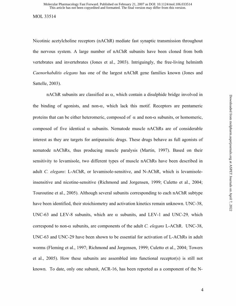
Table 1. Channel properties of nAChR channels from C. elegans muscle cells in culture
Agonist
pS Conc. (µM)
τ1 (µs)
τ2 (µs)
C1 (ms)
C2 (ms)
PD4251 (WT) 1
(n=6) 100 ± 20
280 ± 60
0.06 ± 0.01
180 ± 90
10
(n=13) 96 ± 23
320 ± 50
0.04 ± 0.02
40 ± 24
50
(n=8) 140 ± 30
240 ±20
0.05 ± 0.01
11 ± 6
ACh 38.7 ± 1.6
300 (n= 7)
80 ± 10 - 0.04 ± 0.02
9 ± 4
ACh + 10 µM DHβE
36.5 ± 1.7 50 (n=5)
170 ± 80 -
0.05 ± 0.02
12 ± 03
0.1 (n=6)
140 ± 30
600 ± 70
0.05 ± 0.01
895 ± 240
1 (n=5)
270 ± 10 - 0.08 ± 0.01
220 ± 40
10
(n=8) 150 ± 50
- 0.09 ± 0.01
24 ± 11
Lev 36.9 ± 0.8
100 (n=4)
120 ± 20 - 0.19 ± 0.07
5 ± 0.8
RB918 (acr-16) ACh 36.8 ± 1.3
10
(n=5) 120 ± 40 280 ± 20 0.06 ± 0.01 68 ± 20
Lev 36.2 ± 2.6
0.1 (n=6)
90 ± 20 480 ± 60 0.04 ± 0.01 560 ± 120
CB211 (lev-1) ACh 26.0 ± 2.0
10
(n=3) 490 ± 20 - 0.05 ± 0.02 170 ± 40
Lev 28.3 ± 1.2
1 (n=3)
390 ± 30 - 0.07 ± 0.01 280 ± 20
Single-channel recordings were performed from muscle cells in culture obtained from wild-
type and the specified mutant strains. ACh, ACh plus 10 µM DHβE or levamisole were
present in the pipette solution. The conductance, expressed in pS, was taken from the slope
of the current-voltage relationship (Fig. 1B) or from the current calculated at -100 mV. τ1
This article has not been copyedited and formatted. The final version may differ from this version.Molecular Pharmacology Fast Forward. Published on February 21, 2007 as DOI: 10.1124/mol.106.033514
at ASPE
T Journals on A
pril 7, 2022m
olpharm.aspetjournals.org
Dow
nloaded from

MOL 33514
35
and τ2 correspond to the open components of the open time distributions. C1 and C2 are the
closed components obtained from closed time histograms. n corresponds to the number of
recordings for each condition.
This article has not been copyedited and formatted. The final version may differ from this version.Molecular Pharmacology Fast Forward. Published on February 21, 2007 as DOI: 10.1124/mol.106.033514
at ASPE
T Journals on A
pril 7, 2022m
olpharm.aspetjournals.org
Dow
nloaded from

This article has not been copyedited and formatted. The final version may differ from this version.Molecular Pharmacology Fast Forward. Published on February 21, 2007 as DOI: 10.1124/mol.106.033514
at ASPE
T Journals on A
pril 7, 2022m
olpharm.aspetjournals.org
Dow
nloaded from

This article has not been copyedited and formatted. The final version may differ from this version.Molecular Pharmacology Fast Forward. Published on February 21, 2007 as DOI: 10.1124/mol.106.033514
at ASPE
T Journals on A
pril 7, 2022m
olpharm.aspetjournals.org
Dow
nloaded from

This article has not been copyedited and formatted. The final version may differ from this version.Molecular Pharmacology Fast Forward. Published on February 21, 2007 as DOI: 10.1124/mol.106.033514
at ASPE
T Journals on A
pril 7, 2022m
olpharm.aspetjournals.org
Dow
nloaded from

This article has not been copyedited and formatted. The final version may differ from this version.Molecular Pharmacology Fast Forward. Published on February 21, 2007 as DOI: 10.1124/mol.106.033514
at ASPE
T Journals on A
pril 7, 2022m
olpharm.aspetjournals.org
Dow
nloaded from

This article has not been copyedited and formatted. The final version may differ from this version.Molecular Pharmacology Fast Forward. Published on February 21, 2007 as DOI: 10.1124/mol.106.033514
at ASPE
T Journals on A
pril 7, 2022m
olpharm.aspetjournals.org
Dow
nloaded from

This article has not been copyedited and formatted. The final version may differ from this version.Molecular Pharmacology Fast Forward. Published on February 21, 2007 as DOI: 10.1124/mol.106.033514
at ASPE
T Journals on A
pril 7, 2022m
olpharm.aspetjournals.org
Dow
nloaded from


![Human a4b2 Nicotinic Acetylcholine Receptor as a Novel ......nicotine through the activation of nicotinic acetylcholine receptors (nAChRs) [22,23,24,25]. Previous studies indicate](https://img.dokumen.tips/doc/110x75/5f0f0a627e708231d442317c/human-a4b2-nicotinic-acetylcholine-receptor-as-a-novel-nicotine-through.jpg)