Embed Size (px)
Citation preview

www.wjpps.com Vol 8, Issue 8, 2019.
1615
Prabhavalkar et al. World Journal of Pharmacy and Pharmaceutical Sciences
EXPERIMENTAL ANIMAL MODELS FOR GASTROINTESTINAL
ULCER DISEASE
Mrunal Patila and Kedar S. Prabhavalkar
a,*
aDepartment of Pharmacology, SVKM’s Dr. Bhanuben Nanavati College of Pharmacy, Vile
Parle (W), Mumbai, India.
ABSTRACT
Gastric ulcer is one of the most serious gastrointestinal disease. Ulcers
are mainly caused by imbalance between the gastroduodenal mucosal
defensive factors versus aggressive factors. In gastric ulcer disease,
animal models have helped to understand the basic mechanisms
responsible for the formation of gastric ulcers and its treatment. The
limited number of animal models for the study of gastric ulcer has
hindered the process of research in gastrointestinal disorders.
Therefore it is important to review the available literature for animal
models of gastric ulcer. Available models include primates, rats, mice,
rabbits, cats, guinea pigs, ferrets and pigs. The main aim is to provide
scientific information about different animal models to induce gastric
ulcers as well as for the screening of antiulcer activity. In this review,
we summarize different experimental animal models used in clinical research during past few
decades to carry out antiulcerative activity of new agents as well as its underlying
mechanisms.
KEYWORDS: Gastric ulcer; animal models; NSAIDs; antiulcerative activity;
diethyldithiocarbamate; COX.
1.0 INTRODUCTION
Gastric ulcer is a common gastrointestinal disorder, which includes both gastric and duodenal
ulcers affecting many people throughout the world.[1]
It affects around 3%–10% of the global
population.[2]
About 10% of the world population is on high risk of developing gastric ulcer
at some point in their lifetime.[3]
Globally, an estimated 15 mortality was recorded per year
out of every 15,000 complications of gastric ulcer.[4]
It can be defined as a damage to the
WORLD JOURNAL OF PHARMACY AND PHARMACEUTICAL SCIENCES
SJIF Impact Factor 7.421
Volume 8, Issue 8, 1615-1634 Review Article ISSN 2278 – 4357
*Corresponding Author
Dr. Kedar S.
Prabhavalkar
Department of
Pharmacology, SVKM’s Dr.
Bhanuben Nanavati College
of Pharmacy, Vile Parle
(W), Mumbai, India.
Article Received on
17 June 2019,
Revised on 07 July 2019,
Accepted on 28 July 2019
DOI: 10.20959/wjpps20198-14539

www.wjpps.com Vol 8, Issue 8, 2019.
1616
Prabhavalkar et al. World Journal of Pharmacy and Pharmaceutical Sciences
mucosa that ruptures the muscle layer and forms an injury followed by inflammation.[2], [5]
Generally normal gastric mucosa is exposed to the offensive factors. This may result from an
imbalance between the defensive factors and the offensive factors present in the gastric
mucosal layer.[6]
The defensive factors like mucin, adequate blood flow, nitric oxide,
prostaglandin secretion, bicarbonate and growth factors and the offensive factors include
increased secretion of hydrochloric acid and pepsin, reactive oxygen species, improper
dietary habits, administration of non-steroidal anti-inflammatory drugs, consumption of
alcohol, stressful conditions and infection with Helicobacter pylori infection.[7], [8]
So the
imbalance between these offensive factors and the defensive factors is the main cause for the
pathogenesis of gastric ulcers.[9]
The gastric acid, reactive oxygen species (ROS), and
inflammatory cytokines are continuously in contact with gastric mucosa and can lead to
gastric tissue damage.[10]
Nowadays, the drug therapy for gastric ulcer is mainly based on the
reduction in acid secretion and the protection of gastric mucosa.[11]
The drugs which are
commonly used for the treatment of gastric ulcers consist of antacids, anticholinergics, proton
pump inhibitors and H2- receptor antagonists.[12], [13]
However, the current therapy is not
completely effective and continuous use of these drugs can produce side effects[14]
such as
hypersensitivity, hematopoietic changes, gynecomastia and arrhythmia.[15]
The search for a
new, effective and affordable ulcer treatment, reducing its side effects, current studies are
moving towards natural products.[16]
Hence for the screening of novel antiulcer agents,
suitable animal models are necessary, which can mimic the conditions and show resemblance
to that of the human disease state. Therefore it is important to review the available literature
for animal models of gastric ulcer to screen antiulcer agents. Researchers have used some
technologies to generate transgenic rats[17]
, cats[18]
, dogs[19]
, rabbits, pigs, sheep[20]
, goats,
cattle, chickens[21]
, zebrafish[22]
and non-human primates[23]
as animal models to study
different disease mechanisms.
The main aim is to provide scientific information about different animal models to induce
gastric ulcers as well as for the screening of antiulcer activity. Therefore in this review,
authors putforth different experimental animal models used in clinical research during past
few decades to carry out antiulcer activity of new agents as well as its underlying
mechanisms.
www.wjpps.com Vol 8, Issue 8, 2019.
1617
Prabhavalkar et al. World Journal of Pharmacy and Pharmaceutical Sciences
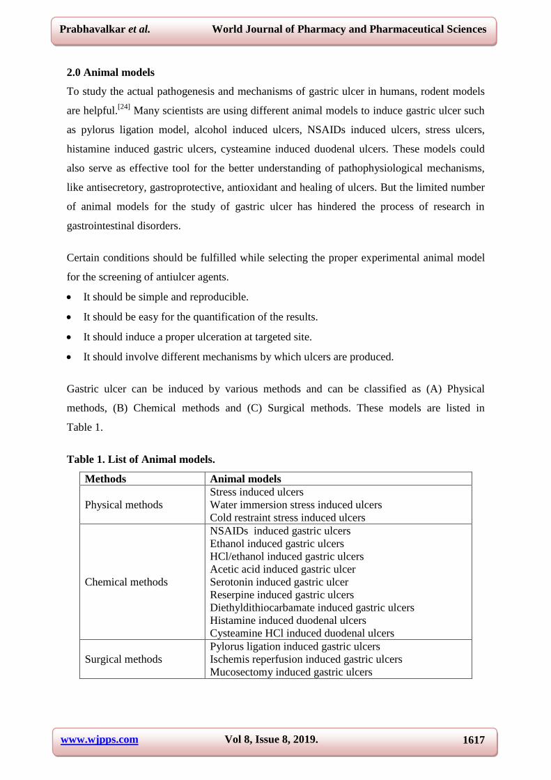
2.0 Animal models
To study the actual pathogenesis and mechanisms of gastric ulcer in humans, rodent models
are helpful.[24]
Many scientists are using different animal models to induce gastric ulcer such
as pylorus ligation model, alcohol induced ulcers, NSAIDs induced ulcers, stress ulcers,
histamine induced gastric ulcers, cysteamine induced duodenal ulcers. These models could
also serve as effective tool for the better understanding of pathophysiological mechanisms,
like antisecretory, gastroprotective, antioxidant and healing of ulcers. But the limited number
of animal models for the study of gastric ulcer has hindered the process of research in
gastrointestinal disorders.
Certain conditions should be fulfilled while selecting the proper experimental animal model
for the screening of antiulcer agents.
It should be simple and reproducible.
It should be easy for the quantification of the results.
It should induce a proper ulceration at targeted site.
It should involve different mechanisms by which ulcers are produced.
Gastric ulcer can be induced by various methods and can be classified as (A) Physical
methods, (B) Chemical methods and (C) Surgical methods. These models are listed in
Table 1.
Table 1. List of Animal models.
Methods Animal models
Physical methods
Stress induced ulcers
Water immersion stress induced ulcers
Cold restraint stress induced ulcers
Chemical methods
NSAIDs induced gastric ulcers
Ethanol induced gastric ulcers
HCl/ethanol induced gastric ulcers
Acetic acid induced gastric ulcer
Serotonin induced gastric ulcer
Reserpine induced gastric ulcers
Diethyldithiocarbamate induced gastric ulcers
Histamine induced duodenal ulcers
Cysteamine HCl induced duodenal ulcers
Surgical methods
Pylorus ligation induced gastric ulcers
Ischemis reperfusion induced gastric ulcers
Mucosectomy induced gastric ulcers

www.wjpps.com Vol 8, Issue 8, 2019.
1618
Prabhavalkar et al. World Journal of Pharmacy and Pharmaceutical Sciences
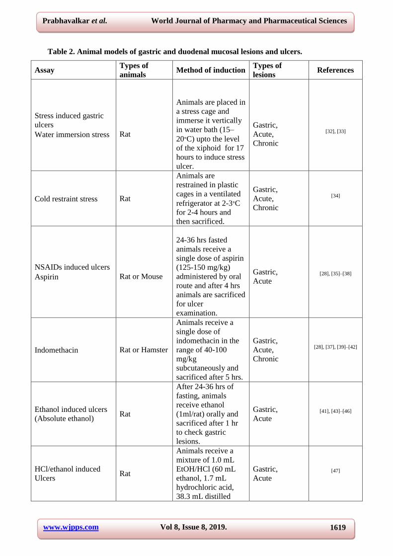
The principle along with their underlying mechanism of action is described below and a brief
procedure for the same is given in table 2.
2.1 Physical methods
Certain physical methods are used to induce gastric ulcers in experimental animals. Generally
physical methods are responsible for the production of oxidative stress and increases reactive
oxygen species in the stomch.
2.1.1 Water-immersion stress or cold-resistant stress induced gastric ulcers
Physical stress and psychological stress can cause gastric ulcers in humans. Hence various
stressors are responsible for the production of gastric ulcers in animal models.[25] This
model involves the restraint technique developed by Brodie and Hanson[26]
employed with
coupling of new method developed by Levine i.e. cold water immersion method which
induces gastric ulcers in a synergistic manner. Gastric ulcers induced by water-immersion
stress or cold water-restraint stress or cold-restraint stress in animals are completely resemble
to human ulcer condition by histopathogically.[27]
This model is widely used to study the
gastroprotective and healing effect of test agents, especially those with mucus enhancing and
cytoprotective properties, for gastric ulcers in rats. The pathophysiology behind stress-
induced gastric ulcers is complex and difficult to understand. Generally these ulcers are
produced due to the release of histamine, which resulting into increased acid secretion and
reduced mucus production[22]
and poor gastric blood flow.[28]
Excessive production of
reactive oxygen species and inhibition of prostaglandin synthesis also promote stress induced
ulcer formation.[29], [30]
Increased gastrointestinal motility produced by stress results into
formation of folds in the stomach[31]
which are more susceptible to damage gastric mucosa
when they come in contact with gastric acid. Increased vagal activity is also one of the factor
responsible for the production of stress-induced ulcers.[26]
As mucus plays a crucial role in
the protection of stomach wall and enhances healing of gastric ulcers, this model is suggested
for evaluating mucus enhancing and cytoprotective agents.
www.wjpps.com Vol 8, Issue 8, 2019.
1619
Prabhavalkar et al. World Journal of Pharmacy and Pharmaceutical Sciences
Table 2. Animal models of gastric and duodenal mucosal lesions and ulcers.
Assay Types of
animals Method of induction
Types of
lesions References
Stress induced gastric
ulcers
Water immersion stress
Rat
Animals are placed in
a stress cage and
immerse it vertically
in water bath (15–
20∘C) upto the level
of the xiphoid for 17
hours to induce stress
ulcer.
Gastric,
Acute,
Chronic
[32], [33]
Cold restraint stress Rat
Animals are
restrained in plastic
cages in a ventilated
refrigerator at 2-3∘C
for 2-4 hours and
then sacrificed.
Gastric,
Acute,
Chronic
[34]
NSAIDs induced ulcers
Aspirin
Rat or Mouse
24-36 hrs fasted
animals receive a
single dose of aspirin
(125-150 mg/kg)
administered by oral
route and after 4 hrs
animals are sacrificed
for ulcer
examination.
Gastric,
Acute
[28], [35]–[38]
Indomethacin Rat or Hamster
Animals receive a
single dose of
indomethacin in the
range of 40-100
mg/kg
subcutaneously and
sacrificed after 5 hrs.
Gastric,
Acute,
Chronic
[28], [37], [39]–[42]
Ethanol induced ulcers
(Absolute ethanol) Rat
After 24-36 hrs of
fasting, animals
receive ethanol
(1ml/rat) orally and
sacrificed after 1 hr
to check gastric
lesions.
Gastric,
Acute [41], [43]–[46]
HCl/ethanol induced
Ulcers Rat
Animals receive a
mixture of 1.0 mL
EtOH/HCl (60 mL
ethanol, 1.7 mL
hydrochloric acid,
38.3 mL distilled
Gastric,
Acute [47]

www.wjpps.com Vol 8, Issue 8, 2019.
1620
Prabhavalkar et al. World Journal of Pharmacy and Pharmaceutical Sciences
water) to induce
gastric ulcer.
Acetic acid induced
gastric ulcers Rat, Cat
After an acute
laparotomy, stomach
is exposed and 20%
acetic acid (0.03 mL)
is injected into the
sub-serosal layer of
the antrum at
multiple sites. After
24 hrs animals are
sacrificed.
Gastric,
Chronic [48]–[50]
Serotonin induced
ulcers Rat
After 30 min of drug
treatment, Serotonin
Creatinine Sulphate
(20-50 mg/kg) is
administered
subcutaneously and
after 6 hours animals
are sacrificed.
Gastric,
Acute [51], [52]
Reserpine induced
ulcers Rat
After 36 hrs of
fasting, reserpine (5–
8mg/kg) is
administered
intraperitoneally and
after 24 hrs animals
are sacrificed.
Gastric,
Acute [53]
Diethyldithiocarbamate
inducd ulcers Rat
In this model, acute
glandular lesions are
induced by
subcutaneous
injection of 1 mL of
diethyldithiocarbama
te (800mg/kg) in
saline followed by
oral dose of 1 mL of
0.1N HCl.
Gastric,
Acute [54], [55]
Histamine induced
ulcers Rat
Histamine phosphate
(40–100mg/kg) is
administered
subcutaneously and
after 2 hours animals
are sacrificed.
Gastric,
Duodenal,
Acute
[56], [57]
Cysteamine HCl
induced ulcers
Guinea pig,
Rat,
Mouse
A single dose of
cysteamine
hydrochloride
(400mg/kg p.o.) is
sufficient to induce
acute duodenal ulcers
in experimental rats.
Duodenal,
Acute [35], [36], [40]

www.wjpps.com Vol 8, Issue 8, 2019.
1621
Prabhavalkar et al. World Journal of Pharmacy and Pharmaceutical Sciences
Pylorus ligation induced
ulcers
Rat
After an acute
laparotomy, pyloric
end of animals
stomach is ligated for
4 hrs. Then animals
are sacrificed for
ulcerative lesions
examination.
Gastric,
Acute [38]
Ischemia reperfusion
induced ulcers Rats
A laparotomy is
carried out under
anesthesia, and the
superior mesenteric
artery (SMA) is
clamped with a
bulldog clip for 30
min (The ischemic
stage). After that,
bulldog clip is
removed to permit
reoxygenation of the
gastric tissue for 15
minutes (the
reperfusion stage).
Then animals are
sacrificed for
ulcerative lesions
examination.
Gastric,
Intestinal
Acute
[58], [59]
Mucosectomy induced
ulcers
Rabbits
Guinea pigs
After a median
laparotomy, 0.2 mL
of isotonic saline is
injected into the
submucosal layer of
the upper corpus. A
diameter of 7-10 mm
of the swollen
mucosal layer is
resected with
scissors. After the
procedure, animals
are placed to
individual cages for
recovery.
Gastric,
Acute [60]–[62]
2.2 Chemical methods
Various chemicals are used to induce ulcers because they directly ruptures the gastric mucosa
(Ethanol, Acetic acid) or some other mechanisms are also involved in it.

www.wjpps.com Vol 8, Issue 8, 2019.
1622
Prabhavalkar et al. World Journal of Pharmacy and Pharmaceutical Sciences
2.2.1 NSAID’s induced gastric ulcers
Gastric ulcers are known to be induced by excessive use of several Non-Steroidal Anti-
Inflammatory Drugs (NSAID’s) like aspirin, indomethacin, and ibuprofen.[63]
This is the most
common model used for inducing gastric ulcer. NSAID’s are responsible for the production
of gastric ulcers by inhibiting prostaglandin synthesis via cyclooxygenase (COX)
pathway.[64], [65]
Prostaglandins plays an important and protective role in the production of mucus and
secretion of bicarbonate, which maintains mucosal blood flow and regulates mucosal cell
integrity.[66]
Thus the suppression of production of prostaglandins by NSAIDs leads to the
gastric mucosal damage and gastric ulcers. The mechanism behind NSAIDs -induced gastric
ulceration includes blocking activity of the cyclooxygenase enzymes (COX-1 and COX-2),
by NSAIDs, which further leads to the decreased mucus and bicarbonate secretion, impaired
platelet aggregation, decreased mucosal blood flow, changes in microvascular structures
leading to epithelia damage.[65]
Increased reactive oxygen species (ROS) and lipid
peroxidation (LPO) are also responsible for gastric mucosal damage.[67]
NSAID’s specially
those are having chemically acidic nature, can exert direct cytotoxic effects on epithelial
cells, which disrupt surface active phospholipids on the mucosal surface thus making the
mucosa more susceptible to damage by luminal acid.[68]
Thus this model is commonly used
for the evaluation of antisecretory and cytoprotective agents. Specially, most commonly used
ulcerogen used for the induction of gastric ulcers are aspirin and indomethacin. A correct
route and an appropriate vehicle (e.g. water, 1% carboxymethyl cellulose) is selected for
administration of ulcerogen.
2.2.2 Ethanol induce gastric ulcers
Ethanol has been commonly used as a damaging agent to gastric mucosa for the induction of
gastric ulcers. Administration of absolute ethanol (>99%) has been used as a reproducible
method to induce gastric mucosal damage in experimental animals.[69]
The pathology behind
ethanol-induced gastric ulcer generally involves three main parameters: inflammatory
response, oxidative stress and apoptosis.
In the gastrointestinal tract, the motility of the esophagus, stomach, and gut as well as the
capacity of gut absorption can be severely affected by alcohol exposure. It can cause severe
mucosal damage and even carcinogenesis specially gastric cancer.[70]
Ethanol causes severe
gastric damage by producing several instabilities in the gastric mucosal layer[71]
such as a

www.wjpps.com Vol 8, Issue 8, 2019.
1623
Prabhavalkar et al. World Journal of Pharmacy and Pharmaceutical Sciences
decreased bicarbonate secretion, gastric blood flow and mucus production. Ethanol can
stimulate gastric acid secretion, resulting in microvascular injuries which facilitate vascular
permeability, through release of gastrin and histamine from sensitive nerve terminals present
in the gastric mucosa.[72]
Ethanol ruptures gastric mucosal integrity through exfoliation of
cells, which leads to increase in mucosal permeability and in somehow causes bleeding.[73],
[74] Intra-gastric administration of absolute ethanol results in severe gastric mucosal injury
characterized by disturbances in microcirculation, mast cell secretary products, inhibition of
prostaglandin synthesis, reduction in mucus production and reactive oxygen species.[75]
Neutrophil infiltration in the gastric mucosa is also responsible for the production of lesions
induced by absolute ethanol.[76]
Oxidative stress plays a significant role in alcohol-induced gastric mucosal damage.[77]
Ethanol is also known to increase cellular oxidative stress[78]
and produce changes in gastric
cell calcium levels[79]
which may lead to the pathogenesis of gastric mucosal injury.
However, it is not a suitable animal model to evaluate antisecretory activity and ulceration
dependent on acid secretion, because this model is independent of gastric acid secretion.
Ethanol directly increases the levels of free radicals that can alter the cell structure and
function[75]
and can also gives its direct toxic effects on the gastric mucosa resulting in
reduced bicarbonate secretion and gastric mucus production.[80]
Hence this model is more
preferable for evaluating the gastroprotective potential of test materials which has
cytoprotective as well as antioxidant activities.
2.2.3 HCl/ethanol induced gastric lesions
This model is considered to be an advanced model of an absolute ethanol induced ulcer
model. Instead of ethanol only, a mixture of HCl and ethanol is used to induce ulceration.
Direct necrotizing action of HCl/ethanol is the pathogenesis for gastric lesions on the gastric
mucosa. Hence the combination of ethanol with HCl leads to induction and progression of
gastric injury.[81]
2.2.4 Acetic acid-induced gastric ulcers
Chronicity of the gastric ulcer disease is one of the least understood aspects for researchers.
Takagi et al.,[82]
developed an animal model to induce chronic gastric ulcers by sub-mucosal
injection of acetic acid and he also reported healing process of ulcers for extended time
intervals after the ulcer formation. The experimental gastric ulcers induced by acetic acid are

www.wjpps.com Vol 8, Issue 8, 2019.
1624
Prabhavalkar et al. World Journal of Pharmacy and Pharmaceutical Sciences
considered as chronic ulcers due to its prolonged steadiness in gastric mucosa and
resemblance to human chronic ulcer both physiologically and histologically.
To overcome certain problems like adherence of ulcer to the adjacent organs such as the liver,
method given by Takagi, was modified and the one that is currently most commonly used is
developed by Okabe and Pfeiffer.[83]
This method consists of intraluminal application of
acetic acid solution. Thus acetic acid induced gastric ulcer model is most suitable for
evaluating antisecretory and cytoprotective activity of various test agents against chronic
gastric ulcers.[82], [84]
This model is easy and reliable to produce round and deep ulcers in the
stomach and duodenum of mice, rats, Mongolian gerbils, guinea pigs, cats, dogs, miniature
pigs, and monkeys.[49], [85]
2.2.5 Serotonin-induced gastric ulcers
Serotonin is a vasoconstrictor. It causes disturbance in gastric mucosal microcirculation and
reduces blood flow to gastric mucosa resulting in acute gastric mucosal damage which causes
gastric ulcers.[86]
2.2.6 Reserpine-Induced Peptic Ulcer
Reserpine is also used for induction of gastric ulcers by scientist. Reserpine causes
degranulation of mast cells resulting into release of histamine, mediated by cholinergic
system[87]
and this histamine is responsible for formation of gastric ulcer. Although this
model is dependent on gastric acid secretion, gastric hypermotility also plays an important
role in the induction of gastric mucosal lesions.[53]
2.2.7 Diethyldithiocarbamate induced gastric ulcers
This model is useful to assess the antioxidant activity of test drug which is mediates
gastroprotection by preventing gastric damage[54]
and also to evaluate the cytoprotective
potential of test agents. It is reported that antral lesions are induced by diethyldithiocarbamate
through the mobilization of superoxide and hydroxyl radicals. Superoxide radical and
hydroxyl radicals play a pathogenic role in the induction of this ulcer.[55]
2.2.8 Histamine-induced gastric ulcers
Release of histamine in stomach also causes gastric ulcers and hence histamine can be used
for inducing gastric ulcers in animals. On this basis histamine induced gastric ulcer model is
developed.[57]
Mast cells secretes histamine, which gets bind with receptors which are present

www.wjpps.com Vol 8, Issue 8, 2019.
1625
Prabhavalkar et al. World Journal of Pharmacy and Pharmaceutical Sciences
on the surface of parietal cells, resulting into activation of adenylate cyclase, which is
responsible for the conversion of ATP into c-AMP. This process of conversion of AMP to c-
AMP enhances gastric acid secretion from parietal cells. Histamine produces disturbances in
gastric mucosal layer subsequently cause severe damage in gastric mucosa.[88]
The mechanism by which histamine produces gastric ulcers is its potent acid stimulating and
vasodilatory effect in animals. Vasodilating capability of histamine causes increase in
vascular permeability.[89]
This model is commonly used for evaluation of antisecretory effect
of test drug which acts as H2 receptor antagonists.
2.2.9 Cysteamine-Induced Duodenal Ulcers
Selye and Szabo[32]
depicted the technique for acceptance of duodenal ulcers in rodents by
utilizing cysteamine HCl. Duodenal ulcers incited by cysteamine in rodents has been
generally utilized as a model of duodenal ulcer. Cysteamine prompted ulcer looks like
duodenal ulcer in human as for its area, histopathology and a few parts of pathophysiology
too. The genuine component associated with generation of ulcer by cysteamine has not been
completely known. But it is reported that Cysteamine is responsible for the excessive
secretion of gastric acid and inhibits the alkaline mucus secretion from Brunner’s glands in
the proximal duodenum resulting in the formation of duodenal ulcer. Cysteamine is also
responsible for excessive pepsin secretion in the gastric mucosa[90]
and consequently causes
decrease in the production in defensive factors like bicarbonate and mucus.[91]
From different
studies, it was reported that cysteamine also reduces somatostatin bioavailability and elevates
serum gastrin levels, which is associated with an increase in gastric acid secretion.[92]
Additionally certain transcription factors like early growth response factor-1, hypoxia-
inducible factor-1 and their target genes assumes a vital role in the pathogenesis of
cysteamine-induced duodenal ulcers.[57]
2.3 Surgical methods
By performing a surgical method we can induce gastric ulcers in experimental animals.
2.3.1 Pylorus-ligation induced peptic ulcer
Shay (1945) developed an animal model for the investigation of gastric secretory activity of a
drug by ligating the pylorus end of the stomach which causes accumulation of gastric acid in
the stomach and produces ulcers. This excessive gastric acid is responsible for auto digestion
of gastric mucosal layer which cause breakdown of the gastric mucosa. So obstruction in

www.wjpps.com Vol 8, Issue 8, 2019.
1626
Prabhavalkar et al. World Journal of Pharmacy and Pharmaceutical Sciences
pylorus causes mucosal digestion by an increase in acid pepsin pool. This model is
commonly used for the evaluation of cytoprotective and antisecretory effect of a drug which
increases secretion of mucus and reduce secretion of gastric acid, respectively.[57], [93]
2.3.2 Ischemia-Reperfusion induced gastric ulcer
Gastric mucosa is highly sensitive to ischemia or ischemic shock. Hence reperfusion
followed by ischemia causes formation of free radicals which is responsible for gastric
mucosal injury.[57]
This involves damage to the muscularis mucosac which is developed by
clamping the celiac artery. Intestinal injury is generally induced by 60 min clamping of the
superior mesenteric artery followed by 60 min reperfusion.[59]
2.3.3 Mucosectomy induced gastric ulcer
This is also called as mucosal resection method where gastric mucosa is exposed by median
laparotomy. Normal saline is injected into the submucosal layer of stomach and swollen
mucosal area is resected with scissors.[61]
Endoscopic resections have certain advantages over
conventional methods. They are more economical and less invasive.[62]
3.0 CONCLUSION
Actually it is not possible to understand the actual mechanism of any disease in human. No
one can predict which is the actual factor or mediator responsible for disease progression.
Without knowing this basic information, it is difficult to design treatment strategies for any
disease. In this review, we tried to cover several animal models of gastrointestinal ulcers that
are widely used in clinical research. Here we also reviewed available experimental animal
models of gastrointestinal ulcers for the evaluation new medicinal compounds having
potential antiulcerative as well as gastroprotective activity. In each model. we have discussed
the underlying mechanism behind the production of gastric ulcer. This will definitely help
scientists and investigators to select a suitable animal model of gastric ulcer for the evaluation
of antiulcerative activity of the test compound. Animal models also plays an important role in
pre-clinical research, specially for the identification of drug targets.
4.0 Abbreviations
ROS- Reactive Oxygen Species
NSAIDs- Non-Steroidal Anti Inflammatory Drugs
HCl- Hydrochloric Acid; SMA- Superior Mesenteric Artery
COX- cyclooxygenase

www.wjpps.com Vol 8, Issue 8, 2019.
1627
Prabhavalkar et al. World Journal of Pharmacy and Pharmaceutical Sciences
COX 1- Cyclooxygenase 1
COX 2- Cyclooxygenase 2
ATP- Adenosine triphosphate
c-AMP- Cyclic Adenosine monophosphate.
5.0 Conflict of interest
Authors have no conflict of interest.
6.0 ACKNOWLEDGEMENT
Authors are thankful to Dr. Lokesh kumar Bhatt, HOD Pharmacology department and Dr.
Munira Momin, Principal, SVKM’s Dr. Bhanuben Nanavati College of Pharmacy, Vile Parle,
Mumbai for their kind support.
REFERENCES
1. N. Rasheed et al., ―Differential response of A 68930 and sulpiride in stress-induced
gastric ulcers in rats,‖ Eur. J. Pharmacol., 2010; 643(1): 121–128.
2. J. M. Kirsch and C. Hirsch-Reilly, ―Peptic ulcer disease,‖ Acute Care Gen. Surg. Work.
Manag., 2017; 38(3): 159–164.
3. F. M. Snowden, ―Emerging and reemerging diseases: A historical perspective,‖ Immunol.
Rev., 2008; 225(1): 9–26.
4. H. M. A. Sidahmed et al., ―Phytomedicine Pyranocycloartobiloxanthone A , a novel
gastroprotective compound from Artocarpus obtusus Jarret , against ethanol-induced
acute gastric ulcer in vivo,‖ Eur. J. Integr. Med., 2013; 20(10): 834–843.
5. P. Aro et al., ―Peptic ulcer disease in a general adult population: The kalixanda study: A
random population-based study,‖ Am. J. Epidemiol., 2006; 163(11): 1025–1034.
6. A. Aslan et al., ―The inhibition of gastric mucosal lesion, oxidative stress and neutrophil-
infiltration in rats by the lichen constituent diffractaic acid,‖ Phytomedicine, 2005; 13(8):
584–590.
7. M. Lemos et al., ―Copaifera langsdorffii: Evaluation of potential gastroprotective of
extract and isolated compounds obtained from leaves,‖ Brazilian J. Pharmacogn., 2015;
25(3): 238–245.
8. K. Sowndhararajan and S. Chul, ―Protective effect of ethyl acetate fraction of Acacia
ferruginea DC. against ethanol-induced gastric ulcer in rats,‖ J. Ethnopharmacol., 2013;
148(1): 175–181.
9. M. K. Choudhary, S. H. Bodakhe, and S. K. Gupta, ―Assessment of the Antiulcer

www.wjpps.com Vol 8, Issue 8, 2019.
1628
Prabhavalkar et al. World Journal of Pharmacy and Pharmaceutical Sciences
Potential of Moringa oleifera Root-Bark Extract in Rats,‖ J. Acupunct. Meridian Stud.,
2013; 6(4): 214–220.
10. A. Bhattacharyya, R. Chattopadhyay, S. Mitra, and S. E. Crowe, ―Oxidative Stress: An
Essential Factor in the Pathogenesis of Gastrointestinal Mucosal Diseases,‖ Physiol. Rev.,
2014; 94(2): 329–354.
11. P. Malfertheiner, F. K. L. Chan, and K. E. L. Mccoll, ―Peptic ulcer disease,‖ Lancet,
2009; 374(9699): 1449–1461.
12. A. E. Bighetti et al., ―Antiulcerogenic activity of a crude hydroalcoholic extract and
coumarin isolated from Mikania laevigata Schultz Bip,‖ Phytomedicine, 2005; 12(1–2):
72–77.
13. V. Lakshmi et al., ―Gedunin and Photogedunin of Xylocarpus granatum show significant
anti-secretory effects and protect the gastric mucosa of peptic ulcer in rats,‖
Phytomedicine, 2010; 17(8–9): 569–574.
14. V. Bassi, O. Fattoruso, M. T. Polistina, and C. Santinelli, ― Graves’ disease shows a
significant increase in the Helicobacter pylori recurrence ,‖ Clin. Endocrinol. (Oxf).,
2014; 81(5): 784–785.
15. S. Chanda, Y. Baravalia, and M. Kaneria, ―Protective effect of Polyalthia longifolia var .
pendula leaves on ethanol and ethanol / HCl induced ulcer in rats and its antimicrobial
potency,‖ Asian Pac. J. Trop. Med., 2011; 4(9): 673–679.
16. Mohd. Akhtar, M. A. Ahmad, S. Sumbul, and Mohd. Asif, ―Role of phenolic compounds
in peptic ulcer: An overview,‖ J. Pharm. Bioallied Sci., 2011; 3(3): 361.
17. W. E. Filipiak and T. L. Saunders, ―Advances in transgenic rat production,‖ Transgenic
Res., 2006; 15(6): 673–686.
18. E. Poeschla, T. Otoi, P. Wongsrikeao, D. Saenz, and T. Rinkoski, ―Antiviral restriction
factor transgenesis in the domestic cat,‖ Nat. Methods, 2011; 8(10): 853–859.
19. S. G. Hong et al., ―Generation of red fluorescent protein transgenic dogs,‖ Genesis, 2009;
47(5): 314–322.
20. R. E. Hammer et al., ―Production of transgenic rabbits, sheep and pigs by
microinjection,‖ Nature, 1985; 315(6021): 680–683.
21. I. M. Mansuy and U. Suter, ―Special Review Series — Gene Manipulation and
Integrative Physiology Mouse genetics in cell biology,‖ Exp. Physiol., 2000; 85(6):
713–31.
22. S. Higashijima, Y. Hotta, and H. Okamoto, ―Visualization of cranial motor neurons in
live transgenic zebrafish expressing green fluorescent protein under the control of the

www.wjpps.com Vol 8, Issue 8, 2019.
1629
Prabhavalkar et al. World Journal of Pharmacy and Pharmaceutical Sciences
islet-1 promoter/enhancer.,‖ J. Neurosci., 2000; 20(1): 206–18.
23. [E. Sasaki et al., ―Generation of transgenic non-human primates with germline
transmission,‖ Nature, 2009; 459(7246): 523–527.
24. N. Choudhary, L. K. Bhatt, and K. S. Prabhavalkar, ―Experimental animal models for
rheumatoid arthritis,‖ Immunopharmacol. Immunotoxicol., 2018; 0(0): 1–8.
25. [N. Sezgin, N. Gürbüz, S. Demirbilek, İ. Gürses, and A. Karaman, ―Protective effect of
polyunsaturated phosphatidylcholine pretreatment on stress ulcer formation in rats,‖ J.
Pediatr. Surg., 2003; 39(1): 57–62.
26. D. A. Brodie and H. M. Hanson, ―A Study of the Factors Involved in the Production of
Gastric Ulcers by the Restraint Technique,‖ Gastroenterology, 1960; 38(3): 353–360.
27. P. C. Konturek et al., ―Pioglitazone, a specific ligand of peroxisome proliferator-activated
receptor-gamma, accelerates gastric ulcer healing in rat,‖ Eur. J. Pharmacol., 2003;
472(3): 213–220.
28. P. H. Guth, ―Gastric blood flow in restraint stress,‖ Am. J. Dig. Dis., 1972; 17(9):
807–813.
29. H. Kitagawa, M. Fujiwara, and Y. Osumi, ―Effects of water-immersion stress on gastric
secretion and mucosal blood flow in rats,‖ Gastroenterology, 1979; 77(2): 298–302.
30. P. Tamashiro Filho et al., ―Evaluation of antiulcer activity and mechanism of action of
methanol stem bark extract of Lafoensia pacari A. St.-Hil. (Lytraceae) in experimental
animals,‖ J. Ethnopharmacol., 2012; 144(3): 497–505.
31. M. N. Peters and C. T. Richardson, ―Stressful Life Events, Acid Hypersecretion, and
Ulcer Disease,‖ Gastroenterology, 1983; 84(1): 114–119.
32. H. Selye and S. Szabo, ―Experimental model for production of perforating duodenal
ulcers by cysteamine in the Rat,‖ Nature, 1973; 244(5416): 458–459.
33. S. Guo, Q. Gao, Q. Jiao, W. Hao, X. Gao, and J. M. Cao, ―Gastric mucosal damage in
water immersion stress: Mechanism and prevention with GHRP-6,‖ World J.
Gastroenterol., 2012; 18(24): 3145–3155.
34. M. A. Morsy, G. H. Heeba, S. A. Abdelwahab, and R. R. Rofaeil, ―Protective effects of
nebivolol against cold restraint stress-induced gastric ulcer in rats: Role of NO, HO-1,
and COX-1,2,‖ Nitric Oxide - Biol. Chem., 2012; 27(2): 117–122.
35. S. Szabo, ―Animal model of human disease. Cysteamine induced acute and chronic
duodenal ulcer in the rat,‖ Am J Pathol, 1978; 93(1): 273–276.
36. S. Szabo and C. H. Cho, ―From Cysteamine to MPTP: Structure-Activity Studies with
Duodenal Ulcerogens,‖ Toxicol. Pathol., 1988; 16(2): 205–211.
www.wjpps.com Vol 8, Issue 8, 2019.
1630
Prabhavalkar et al. World Journal of Pharmacy and Pharmaceutical Sciences
37. B. J. R. WHITTLE, ―Mechanisms Underlying Gastric Mucosal Damage Induced By
Indomethacin and Bile‐Salts, and the Actions of Prostaglandins,‖ Br. J. Pharmacol.,
1977; 60(3): 455–460.
38. Q. Li et al., ―Activity of Brucea javanica oil emulsion against gastric ulcers in rodents,‖
Asian J. Pharm. Sci., 2018; 13(3): 279–288.
39. J. Liang et al., ―Prophylactic efficacy of patchoulene epoxide against ethanol-induced
gastric ulcer in rats: Influence on oxidative stress, inflammation and apoptosis,‖ Chem.
Biol. Interact., 2018; 283, no. November 2017: 30–37.
40. S. Szabo and C. H. Cho, ―Animal models for studying the role of eicosanoids in peptic
ulcer disease,‖ Eicosanoids Gastrointest. Tract, 1988; b(2): 75–102.
41. S. Szabo and C. H. Cho, ―From Cysteamine to MPTP: Structure-Activity Studies with
Duodenal Ulcerogens,‖ Toxicol. Pathol., 2009; 16(2): 205–211.
42. J. Z. Wu et al., ―Protective role of β-patchoulene from Pogostemon cablin against
indomethacin-induced gastric ulcer in rats: Involvement of anti-inflammation and
angiogenesis,‖ Phytomedicine, 2018; 39, no. November 2017: 111–118.
43. S. Szabo and C. H. Cho, ―Animal models for studying the role of eicosanoids in peptic
ulcer disease,‖ Eicosanoids Gastrointest. Tract, 2011; b(2): 75–102.
44. J. Y. C. Chow, L. Ma, and C. H. Cho, ―Effect of cigarette smoke on ethanol-induced
gastric mucosal lesions: The role of nitric oxide and neutrophils,‖ Eur. J. Pharmacol.,
1998; 342(2–3): 253–260.
45. J. K. S. Ko and C. H. Cho, ―Adaptive gastric mucosal cytoprotection in rats: Different
modes of action by three mild irritants,‖ Digestion, 1996; 57(1): 54–59.
46. I. Boutemine et al., ―Gastro-protective , therapeutic and anti-inflammatory activities of
Pistacia lentiscus L. fatty oil against ethanol-induced gastric ulcers in rats,‖ J.
Ethnopharmacol., 2018; 224(January): 273–282.
47. P. Baiubon, P. Kunanusorn, P. Khonsung, N. Chiranthanut, A. Panthong, and C.
Rujjanawate, ―Gastroprotective activity of the rhizome ethanol extract of Zingiber
simaoense Y. Y. Qian in rats,‖ J. Ethnopharmacol., 2016; 194(July): 571–576.
48. Keijiro Takagi and Susumu Okabe, ―Studies of the mechanisms involved in the
production of stress and stress-atropine ulcers in rats,‖ Eur. J. Pharmacol., 1970; 10(3):
378–384.
49. S. Okabe, J. L. A. Roth, and C. J. Pfeiffer, ―Differential healing periods of the acetic acid
ulcer model in rats and cats,‖ Experientia, 1971; 27(2): 146–148.
50. S. K. Bhattamisra et al., ―Protective activity of geraniol against acetic acid and

www.wjpps.com Vol 8, Issue 8, 2019.
1631
Prabhavalkar et al. World Journal of Pharmacy and Pharmaceutical Sciences
Helicobacter pylori- induced gastric ulcers in rats,‖ J. Tradit. Complement. Med., 2018;
1–9.
51. I. H. M. MAIN and B. J. R. WHITTLE, ―Investigation of the Vasodilator and
Antisecretory Role of Prostaglandins in the Rat Gastric Mucosa By Use of Non‐Steroidal
Anti‐Inflammatory Drugs,‖ Br. J. Pharmacol., 1975; 53(2): 217–224.
52. S. Okabe, Y. Takata, K. Takeuchi, T. Naganuma, and K. Takagi, ―Effects of
carbenoxolone Na on acute and chronic gastric ulcer models in experimental animals,‖
Am. J. Dig. Dis., 1976; 21(8): 618–625.
53. M. B. Gupta, K. K. Tangri, and K. P. Bhargava, ―Mechanism of ulcerogenic activity of
reserpine in albino rats,‖ Eur. J. Pharmacol., 1974; 27(2): 269–271.
54. T. Takemoto et al., ―Role of active oxygen species in diethyldithiocarbamate-induced
gastric ulcer in the rat,‖ Experientia, 2005; 46(3): 281–283.
55. A. S. Salim, ―Protection against stress-induced acute gastric mucosal injury by free
radical scavengers,‖ Intensive Care Med., 1991; 17(8): 455–460.
56. M. E. Parsons, ―Histamine and the pathogenesis of duodenal ulcer disease,‖ Gut, 1985;
26(11): 1159–1164.
57. M. B. Adinortey, C. Ansah, I. Galyuon, and A. Nyarko, ―In Vivo Models Used for
Evaluation of Potential Antigastroduodenal Ulcer Agents ,‖ Ulcers, 2013; 2013: 1–12.
58. A. T. Salami, O. A. Odukanmi, O. F. Faniyan, T. P. Omayone, and S. B. Olaleye, ―Seeds
of Buchholzia coriacea in Diet Mitigate Ischemic Reperfusion–Induced Gastric
Ulceration in Experimental Rats,‖ J. Diet. Suppl., 2018; 15(6): 842–859.
59. A. Zeng et al., ―Gastrin attenuates ischemia-reperfusion-induced intestinal injury in rats,‖
Exp. Biol. Med., 2016; 241(8): 873–881.
60. A. Onen, Z. Kanay, C. Guzel, D. Kurt, and K. Ceylan, ―The effects of allopurinol on
stomach mucosal barrier of rats subjected to ischemia-reperfusion,‖ Turkish J. Med. Sci.,
2000; 30(5): 449–451.
61. J. H. Maeng, E. Lee, D. H. Lee, and S.-G. Yang, ―Rabbit gastric ulcer models:
comparison and evaluation of acetic acid-induced ulcer and mucosectomy-induced ulcer,‖
Lab. Anim. Res., 2013; 29(2): 96.
62. J. H. Maeng et al., ―Endoscopic application of EGF-chitosan hydrogel for precipitated
healing of GI peptic ulcers and mucosectomy-induced ulcers,‖ J. Mater. Sci. Mater. Med.,
2014; 25(2): 573–582.
63. K. D. Rainsford, ―The effects of 5-1ipoxygenase inhibitors and leukotriene antagonists on
the development of gastric lesions induced by nonsteroidal antiinflammatory drugs in
www.wjpps.com Vol 8, Issue 8, 2019.
1632
Prabhavalkar et al. World Journal of Pharmacy and Pharmaceutical Sciences
mice,‖ Agents Actions, 1987; 21: 316–319.
64. H. Matsui, O. Shimokawa, T. Kaneko, Y. Nagano, K. Rai, and I. Hyodo, ―The
pathophysiology of non-steroidal anti-inflammatory drug (NSAID)-induced mucosal
injuries in stomach and intestine,‖ J. Clin. Biochem. Nutr, 2011; 48(2): 107–111.
65. J. L. Wallace, W. McKnight, B. K. Reuter, and N. Vergnolle, ―NSAID-Induced gastric
damage in rats: Requirement for inhibition of both cyclooxygenase 1 and 2,‖
Gastroenterology, 2000; 119(3): 706–714.
66. [Hayllar and I. Bjarnason, ―NSAIDs, Cox-2 inhibitors, and the gut,‖ Lancet, 1995;
346(8982): 1105–1106.
67. B. J. R. Whittle, ―Gastrointestinal effects of nonsteroidal anti-inflammatory drugs,‖
Fundam. Clin. Pharmacol., 2003; 17(3): 301–313.
68. J. L. Wallace, ―Prostaglandins, NSAIDs, and Gastric Mucosal Protection: Why Doesn’t
the Stomach Digest Itself?,‖ Physiol. Rev., 2008; 88(4): 1547–1565.
69. C. Yu, X. T. Mei, Y. P. Zheng, and D. H. Xu, ―Gastroprotective effect of taurine zinc
solid dispersions against absolute ethanol-induced gastric lesions is mediated by
enhancement of antioxidant activity and endogenous PGE2production and attenuation of
NO production,‖ Eur. J. Pharmacol., 2014; 740: 329–336.
70. C. Bode and J. C. Bode, ―Alcohol’s role in gastrointestinal tract disorders.,‖ Alcohol
Health Res. World, 1997; 21(1): 76–83.
71. A. R. M. S. Brito et al., ―Antiulcerogenic action of ethanolic extract of the resin from
Virola surinamensis Warb. (Myristicaceae),‖ J. Ethnopharmacol., 2008; 122(2): 406–409.
72. A. B. Albino de Almeida et al., ―Antiulcerogenic effect and cytotoxic activity of semi-
synthetic crotonin obtained from Croton cajucara Benth.,‖ Eur. J. Pharmacol., 2003;
472(3): 205–212.
73. M. Guslandi, ―Effects of ethanol on the gastric mucosa,‖ Dig Dis, 1987; 5(1): 21–32.
74. G. G. Ortiz, G. Nisticò, B. Poeggeler, E. Sewerynek, D. Melchiorri, and R. J. Reiter, ―
Suppressive effect of melatonin administration on ethanol-induced gastroduodenal injury
in rats in vivo ,‖ Br. J. Pharmacol., 2006; 121(2): 264–270.
75. A. . Guseva et al., ―Antiulcer effects of amylin: a review,‖ Pathophysiology, 2003; 11(1):
1–6.
76. C. La Casa, I. Villegas, C. Alarcón De La Lastra, V. Motilva, and M. J. Martín Calero,
―Evidence for protective and antioxidant properties of rutin, a natural flavone, against
ethanol induced gastric lesions,‖ J. Ethnopharmacol., 2000; 71(1–2): 45–53.
77. P. Arda-Pirincci, S. Bolkent, and R. Yanardag, ―The role of zinc sulfate and

www.wjpps.com Vol 8, Issue 8, 2019.
1633
Prabhavalkar et al. World Journal of Pharmacy and Pharmaceutical Sciences
metallothionein in protection against ethanol-induced gastric damage in rats,‖ Dig. Dis.
Sci., 2006; 51(12): 2353–2360.
78. M. G. Repetto and S. F. Llesuy, ―Antioxidant properties of natural compounds used in
popular medicine for gastric ulcers,‖ Brazilian J. Med. Biol. Res., 2002; 35(5): 523–534.
79. S. H. Wong, C. H. Cho, and C. W. Ogle, ―CALCIUM AND ETHANOL-INDUCED
GASTRIC MUCOSAL DAMAGE IN RATS Department of Pharmacology , Faculty of
Medicine , University of Hong Kong , Hong Kong,‖ Pharmacol. Res., 1991; 23(1):
71–79.
80. E. Marhuenda, M. J. Martin, and C. D. La Alarcon Lastra, ―Antiulcerogenic activity of
aescine in different experimental models,‖ Phyther. Res., 1993; 7(1): 13–16.
81. P. J. Oates and J. P. Hakkinen, ―Studies on the mechanism of ethanol-induced gastric
damage in rats,‖ Gastroenterology, 1988; 94(1): 10–21.
82. K. Takagi, S. Okabe, and R. Saziki, ―A new method for the production of chronic gastric
ulcer in rats and the effect of several drugs on its healing.,‖ Jpn. J. Pharmacol., 1969;
19(3): 418–26.
83. S. Okabe and C. J. Pfeiffer, ―Chronicity of acetic acid ulcer in the rat stomach,‖ Am. J.
Dig. Dis., 1972; 17(7): 619–629.
84. S. Okabe, J. L. A. Roth, and C. J. Pfeiffer, ―A method for experimental, penetrating
gastric and duodenal ulcers in rats - Observations on normal healing,‖ Am. J. Dig. Dis.,
1971; 16(3): 277–284.
85. S. Okabe and K. Amagase, ―An Overview of Acetic Acid Ulcer Models<br>—The
History and State of the Art of Peptic Ulcer Research—,‖ Biol. Pharm. Bull.
Pharm. Bull., 2005; 28(8): 1321–1341.
86. S. Singh and D. K. Majumdar, ―Evaluation of the gastric antiulcer activity of fixed oil of
Ocimum sanctum (Holy Basil),‖ J. Ethnopharmacol., 1999; 65(1): 13–19.
87. S. Singh, ―Evaluation of gastric anti-ulcer activity of fixed oil of Ocimum basilicum Linn.
and its possible mechanism of action,‖ Indian J. Exp. Biol., 1999; 37(3): 253–257.
88. L. E. Sander et al., ―Selective expression of histamine receptors H1R, H2R, and H4R, but
not H3R, in the human intestinal tract,‖ Gut, 2006; 55(4): 498–504.
89. C. H. I. H. I. N. Cho and C. J. Pfeiffer, ―Gastrointestinal Ulceration in the Guinea Pig in
Response to Dimaprit , Histamine , and Ha- and H2-Blocking Agents,‖ Dig. Dis. Sci.,
1981; 26(4): 306–311.
90. H. Tamaki, Y. Onoda, and T. Kashida, ―Gastric Secretion and Duodenal Ulcer Formation
Induced By Cysteamine in Rats,‖ Jpn. J. Pharmacol., 2008; 28(4): 647–649.

www.wjpps.com Vol 8, Issue 8, 2019.
1634
Prabhavalkar et al. World Journal of Pharmacy and Pharmaceutical Sciences
91. D. Stiel, D. J. Murray, and T. J. Peters, ―Mucosal Enzyme Activities, with Special
Reference to Enzymes Implicated in Bicarbonate Secretion, in the Duodenum of Rats
with Cysteamine-Induced Ulcers,‖ Clin. Sci., 2015; 64(3): 341–347.
92. S. Szabo, X. Deng, T. Khomenko, G. D. McLaren, H. Ishikawa, and G. J. Anderson,
―Role of iron in the pathogenesis of cysteamine-induced duodenal ulceration in rats,‖ Am.
J. Physiol. Liver Physiol., 2009; 296(6): G1277–G1286.
93. F. A. Carone and R. E. Cooke, ―Effect of Potassium Deficiency on Gastric Secretion in
the Rat,‖ Am. J. Physiol. Content, 2017; 172(3): 684–688.