Embed Size (px)
Citation preview

Experiment 4
Cestodes

Taenia solium Taenia sagniata Echinococcus granulosus

Objectives and requirements :
To master the morphology of cestodes including adults,larve and eggs.
To study laboratory diagnostic methods of cestodes

CestodesThree Parts:Scolex:“head” of the
organism;has holdfast organs to keep the tapeworm in place.
Neck:area where new segments are created
Strobila:Series of reproductive organs in various stages of development

Taenia solium (pork tapeworm)
Adult: hermaphrodite ,flattened, ribbon-like, creamy white in color, measures about 2-4 m with 700-1000 proglottides, can be divided into scolex, neck and strobila.

The attachment organ, or scolex,has four large sucking with a rounded
rostellum containing a double row of hooks.
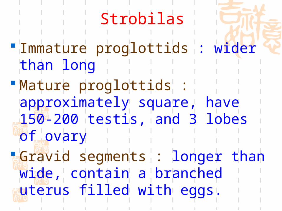
Strobilas
Immature proglottids : wider than long
Mature proglottids : approximately square, have 150-200 testis, and 3 lobes of ovary
Gravid segments : longer than wide, contain a branched uterus filled with eggs.
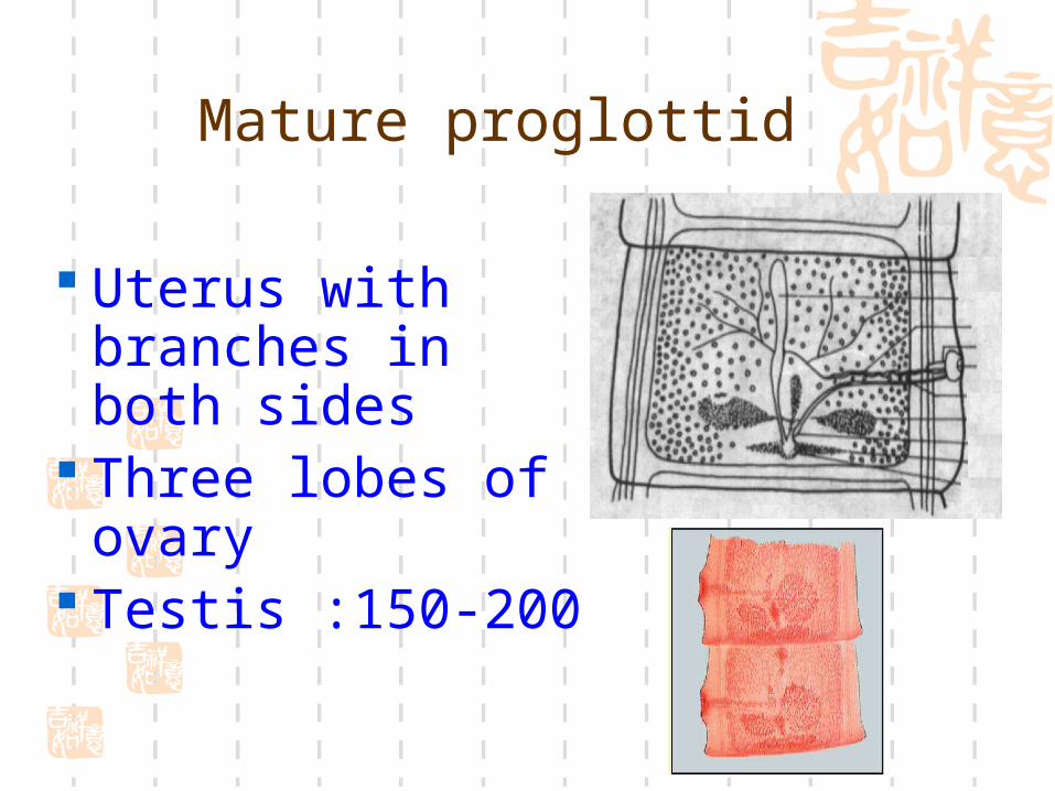
Uterus with branches in both sides
Three lobes of ovary
Testis :150-200
Mature proglottid

Gravid segment
The gravid segment contain a branched uterus filled with numerous eggs.
Identification to the species level is usually based on the number of lateral uterine branches should be counted where they come off the main central stem.Only one side is counted,and there are 7 to 13 lateral branches.
Ink-stained

Egg
Brown in color 31-43 µm in diameter Spherical with a thick, radially
striated embryophore Contain an oncosphere

10x40 Eggs in stool
Eggs

Cysticercus cellulosae
(stained with camine)Larval form of T.solium
ovoid and milkly-white bladder filled with fluids
Head invaginated in the bladder
the bladder worm measure 5mm long by 8-10mm wide.
Ingested by host ,the head will evaginate.

bean-pork

Cysticerci in brain

Cysticerci in heart
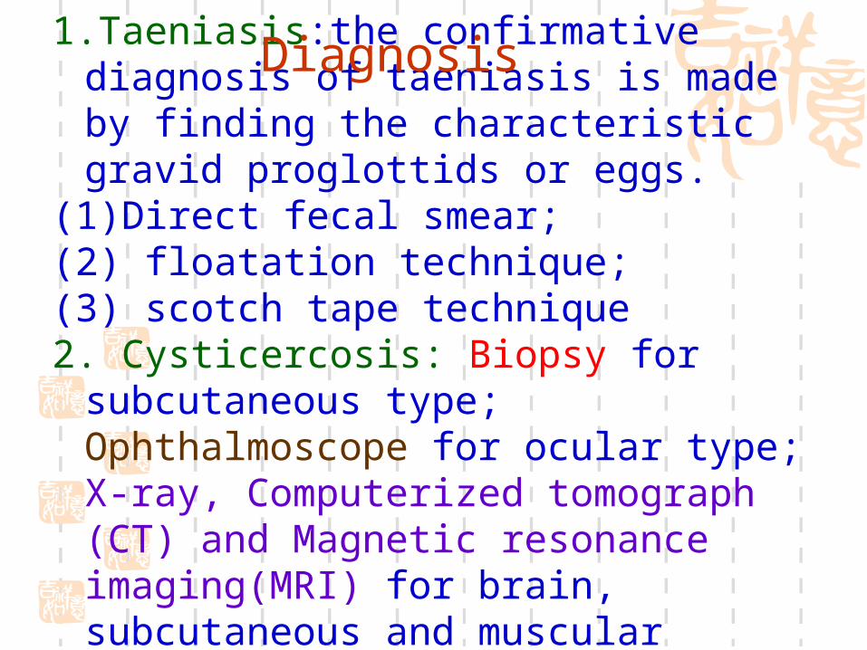
1.Taeniasis:the confirmative diagnosis of taeniasis is made by finding the characteristic gravid proglottids or eggs.
(1)Direct fecal smear; (2) floatation technique;(3) scotch tape technique2. Cysticercosis: Biopsy for subcutaneous
type; Ophthalmoscope for ocular type; X-ray, Computerized tomograph (CT) and Magnetic resonance imaging(MRI) for brain, subcutaneous and muscular types.
Diagnosis

3. Sometimes ,the diagnosis is confirmed by specific antibodies in the circulating blood.serum and CSF enzyme immunoassays and Western blot testing for specific anticysticercal antibodies have a sensitivity of 80%-95%.
Immunological tests are for reference only. (1)Intradermal test (2)Indirect hemaglutination (3)Enzyme-linked immunosorbent assay
(ELISA)
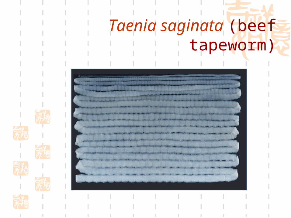
Taenia saginata (beef tapeworm)
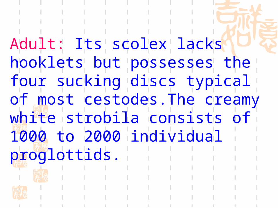
Adult: Its scolex lacks hooklets but possesses the four sucking discs typical of most cestodes.The creamy white strobila consists of 1000 to 2000 individual proglottids.

The terminal segments are longer than they are wide and contain a large uterus with 15 to 30 lateral branches;these characteristics are useful in differentiating them from those of the closely related pork tapeworm.

T. solium T. saginata
Differences between T. solium and T. saginata
2-4m 4-8m
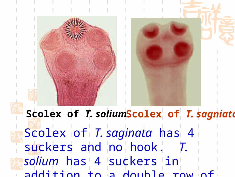
Scolex of T. solium Scolex of T. sagniata
Scolex of T. saginata has 4 suckers and no hook. T. solium has 4 suckers in addition to a double row of hooks.

Scolex of T. solium Scolex of T. sagniata
Under electron microscope

Mature proglottid of T. solium
Mature proglottid
of T. sagniata
3 lobes of ovary 2 lobes of ovary

Gravid segment of
T. solium
Gravid segment
of T. sagniata
T. saginata has 15 to 30 branches on each side ,while Taenia solium has 7 to 13 branches.

Egg of T.saginata(A) is same to T.solium’s(B) 31-43 µm in diameter, spherical, thick,
with radially striated shell. Inside each shell is an embryonated oncosphere with 6 hooks.
The egg in Figure B still has the primary membrane that surrounds eggs in the
proglottids.

Cysticercus bovis: is semitransparent,milk white in color.The bladder is filled with fluid and on side is seen a denser area that is the invaginated head equipped with four suckers but no hooklets.

Differences between T.solium and T.saginata
Adult T. solium T. saginata______________________________________________________________________________
Length 2-4 meters 4-8 metersNumber of segment 700 to 1000 1000-2000 thin and transparent thick but opaqueScolex 1mm in diameter with 2mm in diameter, with 4 suckers and hooklets 4 suckers but no hookletsMature proglottid 3 lobes of ovary 2 lobes of ovaryGravid proglottid 7-13 uterine lateral 15-30 uterine lateral branches each side branches each side
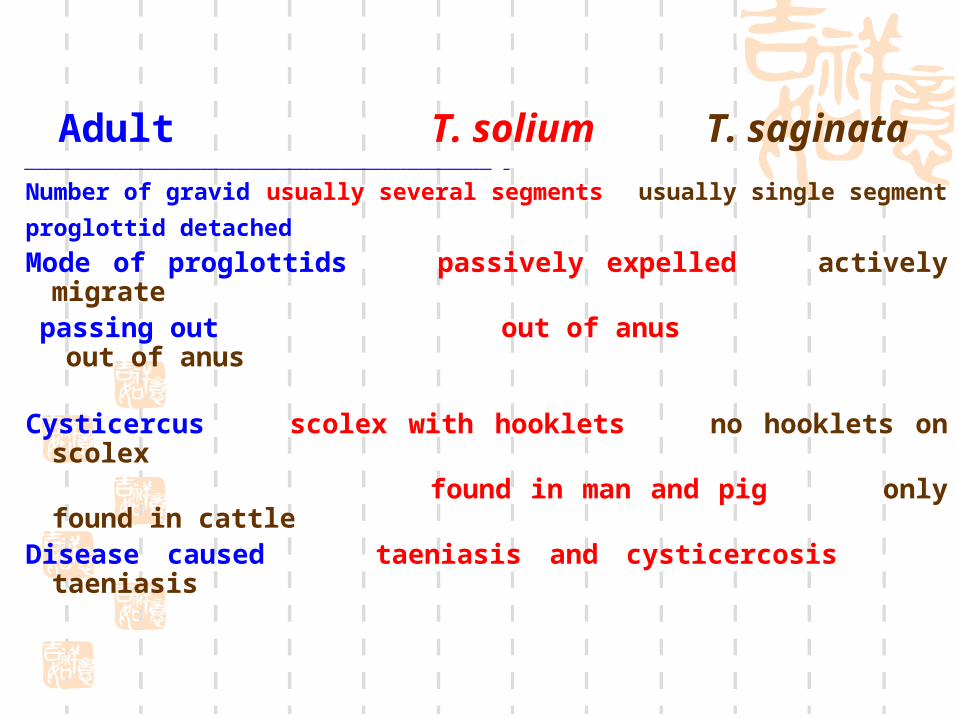
Adult T. solium T. saginata_____________________________________________________________________________ _
Number of gravid usually several segments usually single segment
proglottid detached Mode of proglottids passively expelled actively migrate passing out out of anus out of anus
Cysticercus scolex with hooklets no hooklets on scolex found in man and pig only found in cattleDisease caused taeniasis and cysticercosis taeniasis

Adult inhabits the small bowel of dogs,wolves and other canines.It is a smaller tapeworm of medical importance,ranges 2 to 7mm in length.It consists of a scolex,neck and three segments. The globular scolex,0.3mm in diameter,bears a prominent rostellum with a double crown of 24 to 40 large and small hooklets and 4 cuplike oval suckers.
Echinococcus granulosusmorphology

The scolex narrows posteriorly to form a slender neck. The first proglottid contains immature genital organs.The middle proglottid has fully developed male and female reproductive organs. The last or gravid proglottid consists principally of a median uterus with irregular lateral branches filled with eggs.The proglottid ,over 2mm long and 0.5 to 1mm wide,comprises about one-half the length of the worm. The genital pore is located in the poster half of proglottid.A gravid proglottid contains about 500 eggs.
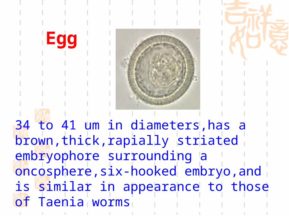
Egg
34 to 41 um in diameters,has a brown,thick,rapially striated embryophore surrounding a oncosphere,six-hooked embryo,and is similar in appearance to those of Taenia worms

Hydatid cyst
Protoscolex
Brood capsules
Daughter cysts
Granddaughter cysts
Laminated layer
Germinal layer
contents
Cyst wall
Hydatid fluid(囊液 )
hydatid sand
Hydatid Cyst:
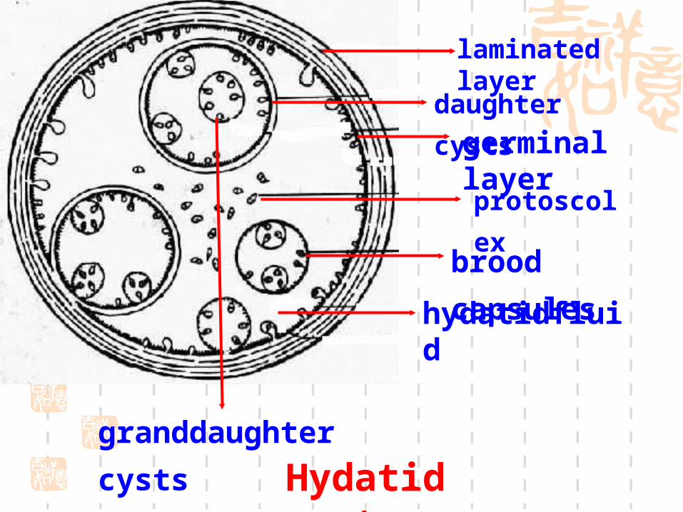
Hydatid cyst
daughter cysts
laminated layer
germinal layer
hydatidfluid
granddaughter cysts
protoscolex
brood capsules
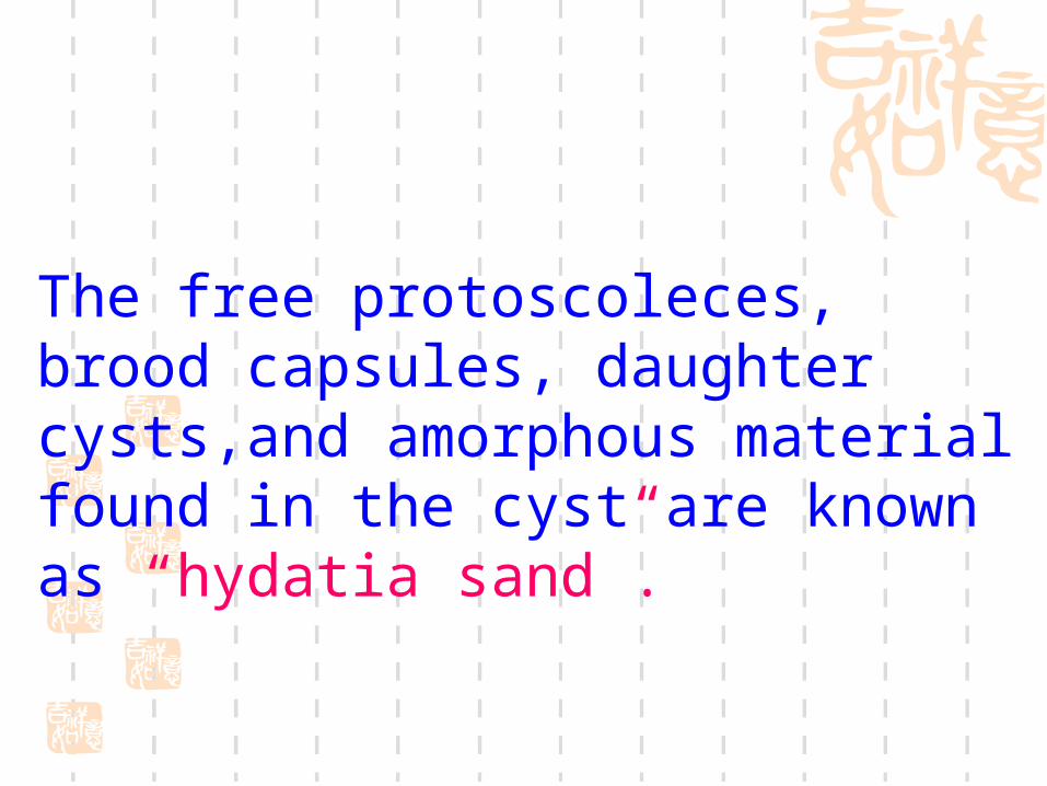
The free protoscoleces, brood capsules, daughter cysts,and amorphous material found in the cyst are known as “hydatia sand”.

Largest larval stage of all tapeworms
Hydatid cyst

Daughter cysts

Protoscolex
Notice the scolex armed with
suckers and hooklets.
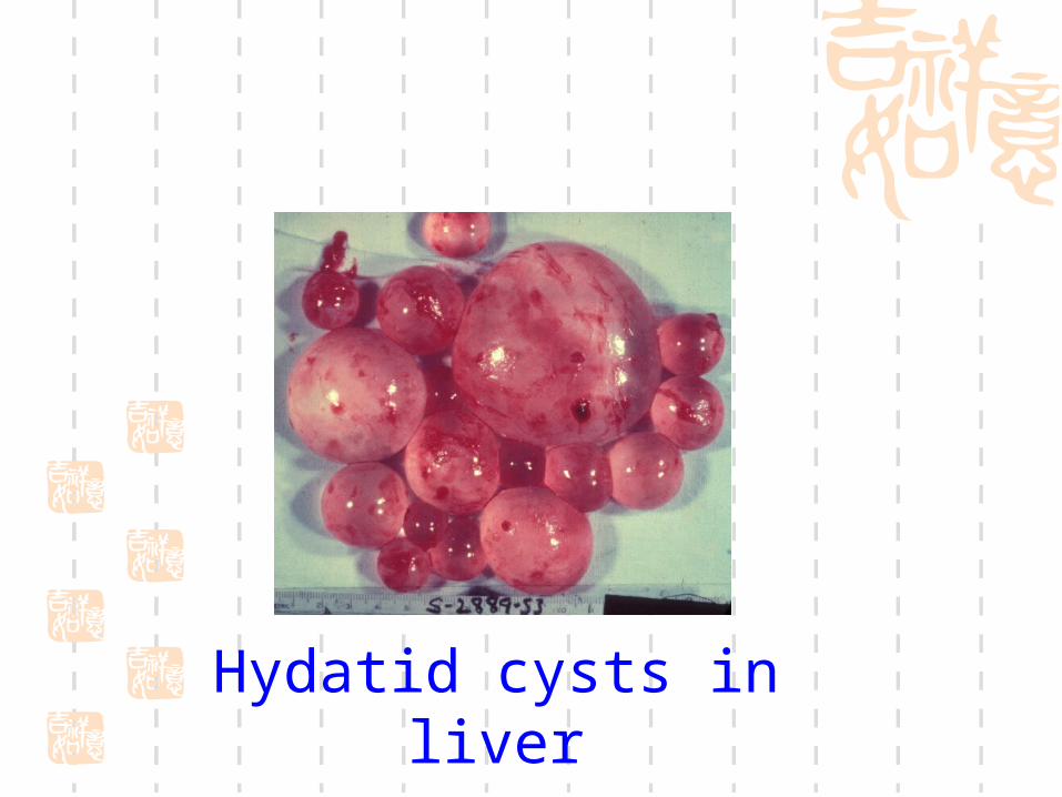
Hydatid cysts in liver

Presumptive diagnosis may be made by symptoms, signs and the history of living in pastoral areas. The clinical diagnosis depends on the detection of the indications of the hydatid cyst. The hydatid thrill, indicative of fluid, is a diagnostic sign. The exploratory puncture for obtaining fluid is contraindicated as a dangerous diagnostic procedure.
Diagnosis

1.B ultrasonography2.Computerized tomograph (CT) and magnetic
resonance (MR).3.X-ray for cysts in the lung and bone.4.Immunological tests are for reference only:(1)Casoni’s intradermal test.(2)Indirect hemagglutination(3)Enzyme-linked immunosorbent assay
(ELISA)

exercises
Draw the egg 、 scolex 、 gravid segment of T. solium.
Draw the scolex 、 gravid segment of T. sagniata.
Draw the hydatid sand (Protoscolex)of Echinococcus granulosus.

Protoscolex of Echinococcus granulosus
Gravid segment
of T. sagniata
Scolex of T. solium
Gravid segment of T. solium
Scolex of T. sagniata
egg of tapeworm