Embed Size (px)
Citation preview

RESEARCH ARTICLE Open Access
Exome screening to identify loss-of-function mutations in the rhesus macaquefor development of preclinical models ofhuman diseaseAdam S. Cornish, Robert M. Gibbs and Robert B. Norgren Jr.*
Abstract
Background: Exome sequencing has been utilized to identify genetic variants associated with disease in humans.Identification of loss-of-function mutations with exome sequencing in rhesus macaques (Macaca mulatta) could lead tovaluable animal models of genetic disease. Attempts have been made to identify variants in rhesus macaques by aligningexome data against the rheMac2 draft genome. However, such efforts have been impaired due to the incompletenessand annotation errors associated with rheMac2. We wished to determine whether aligning exome reads against our new,improved rhesus genome, MacaM, could be used to identify high impact, loss-of-function mutations in rhesus macaquesthat would be relevant to human disease.
Results: We compared alignments of exome reads from four rhesus macaques, the reference animal and three unrelatedanimals, against rheMac2 and MacaM. Substantially more reads aligned against MacaM than rheMac2. We followed theBroad Institute’s Best Practice guidelines for variant discovery which utilizes the Genome Analysis Toolkit to identify highimpact mutations. When rheMac2 was used as the reference genome, a large number of apparent false positives wereidentified. When MacaM was used as the reference genome, the number of false positives was greatly reduced. Afterexamining the variant analyses conducted with MacaM as reference genome, we identified two putative loss-of-functionmutations, in the heterozygous state, in genes related to human health. Sanger sequencing confirmed the presence ofthese mutations. We followed the transmission of one of these mutations (in the butyrylthiocholine gene) through threegenerations of rhesus macaques. Further, we demonstrated a functional decrease in butyrylthiocholinesterase activitysimilar to that observed in human heterozygotes with loss-of-function mutations in the same gene.
Conclusions: The new MacaM genome can be effectively utilized to identify loss-of-function mutations in rhesusmacaques without generating a high level of false positives. In some cases, heterozygotes may be immediately useful asmodels of human disease. For diseases where homozygous mutants are needed, directed breeding of loss-of-functionheterozygous animals could be used to create rhesus macaque models of human genetic disease. The approach wedescribe here could be applied to other mammals, but only if their genomes have been improved beyond draft status.
Keywords: Rhesus, Macaca mulatta, Macaque, Exome, Mutation, Loss-of-function, BChE, RNASEL, Null mutant, Cancer
* Correspondence: [email protected] of Genetics, Cell Biology and Anatomy, University of NebraskaMedical Center, Omaha 68198-5805, Nebraska
© 2016 Cornish et al. Open Access This article is distributed under the terms of the Creative Commons Attribution 4.0International License (http://creativecommons.org/licenses/by/4.0/), which permits unrestricted use, distribution, andreproduction in any medium, provided you give appropriate credit to the original author(s) and the source, provide a link tothe Creative Commons license, and indicate if changes were made. The Creative Commons Public Domain Dedication waiver(http://creativecommons.org/publicdomain/zero/1.0/) applies to the data made available in this article, unless otherwise stated.
Cornish et al. BMC Genomics (2016) 17:170 DOI 10.1186/s12864-016-2509-5
BackgroundThe study of genetic variants associated with disease isrevolutionizing biomedical research and promises tomake profound changes in clinical practice [1–4]. Onelimitation to current approaches has been the highcost of genome sequencing. However, next generationsequencing (NGS), which can focus on the “exome”(the sequences contained in exons), offers a less expen-sive method. Already, exome sequencing has been usedwith great success to identify genomic sequence variantsassociated with disease in humans [2, 4–6].Identifying a genetic variant associated with a human
disease is an important first step, but animal models thatmatch human phenotypes are necessary to develop effect-ive therapeutics. Although mouse models have been help-ful in understanding basic biology, their great evolutionarydistance and biological differences from humans can limittheir utility for preclinical studies [7]. Approximately 90 %of therapeutic drug candidates fail to advance during theclinical phases of pharmaceutical development [8]. Thelack of good animal models for efficacy testing is likelyone cause of this high failure rate.Rhesus macaques (Macaca mulatta) and other nonhu-
man primates are similar to humans in both physiologyand anatomy due to their close evolutionary relationship[9]. They have proven essential as models for infectiousdiseases such as AIDS/HIV and neurological and repro-ductive disorders [10–16].Rhesus macaques with genetic variations similar to
those that cause disease in humans might prove invalu-able as preclinical models. A rhesus macaque model ofHuntington’s disease has been created by inserting aHuntingtin gene with a pathological number of repeatsinto the genome of oocytes using lentiviral vectors [17].This gain-of-function mutation has been shown to resultin a phenotype similar to human Huntington’s patients[18]. Recently, the CRISPR/Cas9 system has been used togenetically modify the genome of a cynomolgus macaque(Macaca fascicularis) [19]. Although these approacheshave much promise, they require advanced in vitrofertilization facilities which are very limited for nonhumanprimates.Another potential approach to providing rhesus ma-
caques models of genetic disease would be to examine theexomes of “normal” rhesus macaques for high impactmutations. Each individual human has, on average, threeto five recessive mutations in genes related to Mendeliandisorders. This statistic was first obtained from studies ofconsanguineous marriages [20] and later confirmed withexome analyses [1]. A study that compared SNPs in thehippocampal RNA sequences of humans and rhesus ma-caques found that the number of damaging coding varia-tions was similar in the two species [21]. Assuming asimilar incidence of loss-of-function (LOF) mutations in
rhesus macaques as is found in humans, exome sequen-cing and analyses could be used to create a catalog of mu-tations in genes related to Mendelian disorders in largenumbers of animals. Heterozygotes may be immediatelyuseful. However, if homozygous individuals are required,heterozygotes with LOF mutations could be bred togetherto produce null mutant offspring [9].Exome analysis requires two important resources: ex-
ome capture probes that can capture genomic fragmentscontaining exons and a high quality reference genomeagainst which sequences can be aligned. For humansand mice, these resources exist. For rhesus macaques,no species-specific exome capture probe sets are avail-able. Fortunately, two previous studies have shown thathuman probes can be used to capture about 96 % of therhesus macaque coding exons with a minimum of 1Xcoverage [22, 23]. However, until recently, investigators in-terested in performing exome analyses in rhesus macaqueswere limited to using a draft rhesus genome, rheMac2,which contained many errors and was incomplete [24].Serious issues related to using this genome for exome ana-lyses have been reported [23]. We have recently completeda high quality reference genome for the rhesus macaque,MacaM [25]. Here, we show that alignments of exome se-quences with MacaM can be analyzed to identify LOF mu-tations in rhesus macaques related to human disease.
MethodsWe used two human exome capture kits to enrich forexons in four rhesus macaques: the TruSeq ExomeEnrichment Kit (Illumina) for animal 002 T-NHP andthe SureSelect XT HumanAllExon 50 Mb Kit (Agilent,G7544A) for animals 17573, ON12033 and ON22186.We sequenced exomic fragments with an Illumina Gen-
ome Analyzer IIx (for animal 002 T-NHP) and a HiSeq2000(for animals 17573, ON12033 and ON22186) and obtained101 bp paired-end reads for each exome generating a totalof 17.7 Gb of data. We deposited sequences in the Se-quence Read Archive at the National Center for Biotech-nology Information (NCBI) under accessions SRX144674(002 T-NHP), SRX115899 (animal 17573), SRX144808(animal ON12033) and SRX145282 (animal ON22186).We followed The Broad Institute’s Best Practices guide-
lines for discovering putative variants utilizing the Gen-ome Analysis Toolkit (GATK) [26] to identify LOFmutations. Briefly, we aligned exomic sequences from thefour rhesus macaques to the two different reference rhe-sus assemblies to be analyzed: rheMac2 (downloaded fromthe UCSC Genome Browser on June 24, 2014) andMacaM (version 7) using bwa mem (version 0.7.5a-r405).Preliminary analyses indicated that aligning exome readsagainst only chromosome files resulted in apparent mis-alignments of pseudogene reads against coding genes.When unplaced and unlocalized scaffolds were included
Cornish et al. BMC Genomics (2016) 17:170 Page 2 of 11

in the reference assembly, misalignments involvingcoding genes were reduced. All data reported in theresults for both rheMac2 and MacaM were collectedusing the latter approach. We processed the 8 align-ment files (four samples x two genomes) to remove PCRduplicates (using Picard Tools version 1.137), realignaround putative indels (using GATK version 3.4), and re-calibrate quality base scores (using GATK version 3.4).We used the Samtools (version 0.1.18) flagstat tool to ob-tain the “percent aligned” for each of these datasets(Table 1).We identified putative variants in the processed align-
ment files with the GATK HaplotypeCaller (version 3.4).We removed low quality variants using The BroadInstitute’s recommended filters [27]. For SNPs, we removedvariants that had a Depth (DP) < 5, QualByDepth (QD)score < 2.0, FisherStrand (FS) score > 60.0, RMSMapping-Quality (MQ) < 40.0, MappingQualityRankSum Test(MQRankSum) score < −12.5, or a ReadPosRankSumTest(ReadPosRankSum) score < −8.0. For insertions and dele-tions, we removed variants that had a DP < 5, QD< 2.0,FS > 200.0, or ReadPosRankSum < −20.0. We used snpEff(version 3.1 h) [28] to generate a tab-delimited file withvariant information to identify putative LOF mutations inboth genomes. We obtained the rheMac2 annotation filesfrom the NCBI ftp genome database on July 20, 2015. ForMacaM, we used annotation version 7.6 [25]. We focusedon mutations expected to cause complete LOF of protein,specifically premature stop codon and frameshift muta-tions. We used the Online Mendelian Inheritance in Mandatabase [29] to select genes known to be involved in dis-ease in humans. We manually inspected candidate muta-tions in these genes with the Integrated Genome Viewer(IGV) [30]. We did not further investigate variants that didnot seem likely to result in a high impact mutation. Codingvariant statistics were obtained using SnpEff followingfiltering and annotation.We designed PCR primers with Primer3 [31] to flank
target exons containing candidate mutations in twogenes, butyrylcholinesterase (BChE) and ribonuclease L(RNASEL). We included M13F (−20) sequence (GTAAAACGACGGCCAGT) and M13R sequence (GGAAA-CAGCTATGACCATG) on the 5' end of the primer se-quences to facilitate Sanger sequencing.
Primer sequences:
BChE (forward):GTAAAACGACGGCCAGTGGGGACAACAAATGCTTCATBChE (reverse):GGAAACAGCTATGACCATGTGGAACCCAAACACTGACCTRNASEL (forward):GTAAAACGACGGCCAGTGGGCCATATACTGCCTTGAARNASEL (reverse):GGAAACAGCTATGACCATGATTCTGCATGATGGGAGAGG
Integrated DNA Technologies (Coralville, Iowa) syn-thesized the primers. We performed PCR with theseprimers and genomic DNA from two animals from theOregon National Primate Research Center, ON12033(RNASEL) and ON22186 (BChE). Briefly, 200 ng of gen-omic DNA was used as the template for PCR using theAccuPrime Pfx Supermix Kit (Invitrogen) according tothe manufacturer’s instructions. We amplified targetedexons in a MJ Research PTC-100 thermocycler with thefollowing program:
Step 1. Denature at 95 °C for 5 minutes.Step 2. For 35 cycles: 95 °C for 15 seconds; 60 °C for30 seconds; 68 °C for 45 seconds.
We purified PCR products with the QIAquick PCRPurification Kit (Qiagen) and then sequenced the PCRproducts with the Sanger method. We manually inspectedtraces to identify putative mutations.We performed PCR for BChE on genomic DNA from
descendants of ON22186 including: ON22187, ON22188,ON22191, ON22192 and ON22193.To determine whether an identified LOF mutation in the
BChE gene (in the heterozygous state) resulted in a de-crease in BChE activity, a functional assay for this enzymewas performed. 4 ml of heparinized whole blood was drawnfrom two animals at the Oregon National Primate ResearchCenter. One animal, ON22193, had been identified with aLOF mutation (in the heterozygous state) in the BChE
Table 1 Percent of rhesus exome reads aligned against different assemblies
Sample ID Sample accession rheMac2assemblychr only
MacaMv7assemblychr only
rheMac2assemblychr + unplaced
MacaMv7assemblychr + unplaced
17573 SRX115899 94.28 % 97.03 % 96.74 % 97.57 %
ON12033 SRX144808 93.19 % 96.29 % 95.98 % 96.95 %
ON22186 SRX145282 92.62 % 96.21 % 95.84 % 96.95 %
002T-NHP SRX144674 92.09 % 95.12 % 94.87 % 95.84 %
Chr only alignments against only scaffolds placed on chromosomes; Chr + unplaced alignments against both placed and unplaced scaffolds
Cornish et al. BMC Genomics (2016) 17:170 Page 3 of 11
gene. Animal ON22197 was an unrelated cage mate. Bothanimals were female.The whole blood was transferred to sterile microfuge
tubes using aseptic conditions. 0.4 ml was placed in anonsterile tube for activity assays. Tubes were centri-fuged for 10 min to pellet the red blood cells. The redblood plasma was then transferred to new tubes.BChE activity was measured in 0.1 M potassium phos-
phate buffer pH 7.0 containing 0.5 mM dithiobisnitro-benzoic acid, and 1 mM butyrylthiocholine at 25 °C. Theincrease in absorbance was recorded at 412 nm and con-verted to μmoles butyrylthiocholine hydrolyzed per minusing the extinction coefficient 13,600 M−1 cm−1 [32].Units of activity were measured in μmoles per minute.Each plasma sample was assayed in triplicate.Two specific BChE inhibitors, 20 μM ethopropazine,
and 0.1 mM iso-OMPA were used to demonstrate thatthe measured activity was catalyzed by BChE. Plasmawas preincubated with 0.1 mM iso-OMPA for 30 minutesbefore the reaction was started by addition of 0.02 ml of0.2 M butyrylthiocholine.
Statement of ethical approvalMaterials used in these studies were from animal workperformed under Institutional Animal Care and UseCommittee approval from the University of NebraskaMedical Center and Oregon Health and Sciences Uni-versity. Animal welfare was maintained by followingNIH (Public Health Service, Office of Laboratory AnimalWelfare) and USDA guidelines by trained veterinary staffand researchers under Association for Assessment andAccreditation of Laboratory Animal Care certification,insuring standards for housing, health care, nutrition,environmental enrichment and psychological well-being.These met or exceeded those set forth in the Guide forthe Care and Use of Laboratory Animals from the Na-tional Research Council of the US National Academy ofSciences.
ResultsWe aligned exome capture reads from four rhesus ma-caques against rheMac2 and MacaM (Table 1). Substan-tially more reads aligned against MacaM than rheMac2when reads were aligned only against scaffolds placed onchromosomes. When unplaced scaffolds were used, thedifferences in alignment percentages between rheMac2and MacaM were greatly reduced.We used GATK HaplotypeCaller to identify potentially
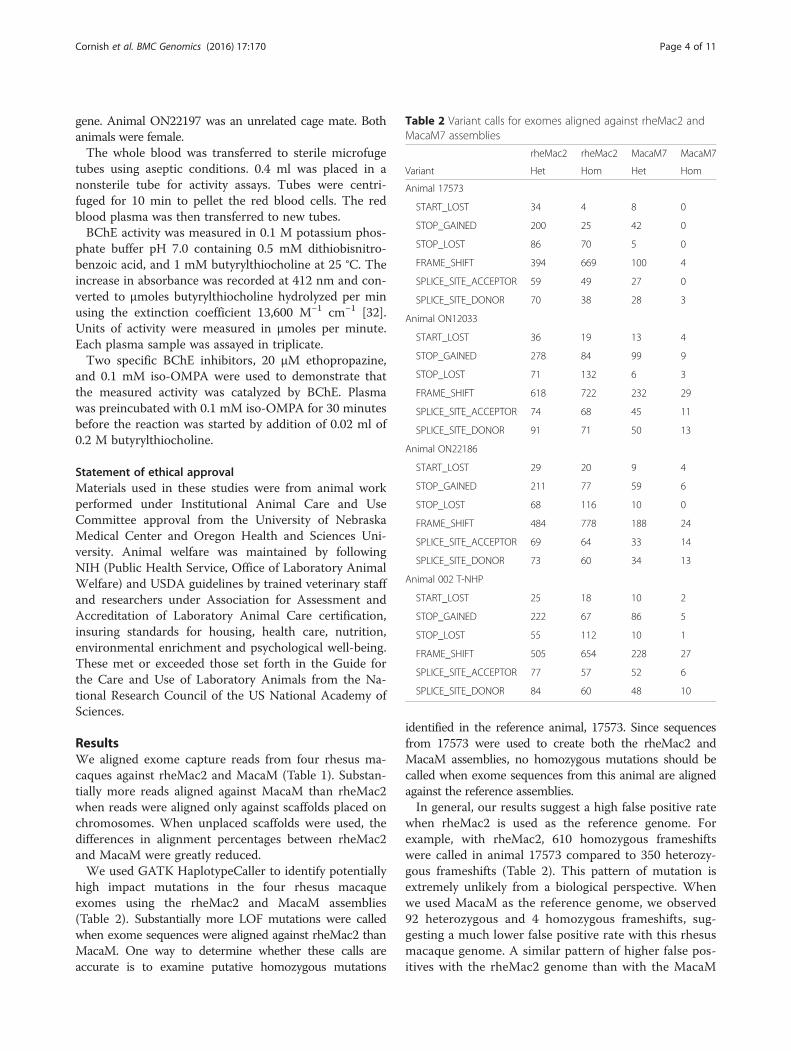
high impact mutations in the four rhesus macaqueexomes using the rheMac2 and MacaM assemblies(Table 2). Substantially more LOF mutations were calledwhen exome sequences were aligned against rheMac2 thanMacaM. One way to determine whether these calls areaccurate is to examine putative homozygous mutations
identified in the reference animal, 17573. Since sequencesfrom 17573 were used to create both the rheMac2 andMacaM assemblies, no homozygous mutations should becalled when exome sequences from this animal are alignedagainst the reference assemblies.In general, our results suggest a high false positive rate
when rheMac2 is used as the reference genome. Forexample, with rheMac2, 610 homozygous frameshiftswere called in animal 17573 compared to 350 heterozy-gous frameshifts (Table 2). This pattern of mutation isextremely unlikely from a biological perspective. Whenwe used MacaM as the reference genome, we observed92 heterozygous and 4 homozygous frameshifts, sug-gesting a much lower false positive rate with this rhesusmacaque genome. A similar pattern of higher false pos-itives with the rheMac2 genome than with the MacaM
Table 2 Variant calls for exomes aligned against rheMac2 andMacaM7 assemblies
rheMac2 rheMac2 MacaM7 MacaM7
Variant Het Hom Het Hom
Animal 17573
START_LOST 34 4 8 0
STOP_GAINED 200 25 42 0
STOP_LOST 86 70 5 0
FRAME_SHIFT 394 669 100 4
SPLICE_SITE_ACCEPTOR 59 49 27 0
SPLICE_SITE_DONOR 70 38 28 3
Animal ON12033
START_LOST 36 19 13 4
STOP_GAINED 278 84 99 9
STOP_LOST 71 132 6 3
FRAME_SHIFT 618 722 232 29
SPLICE_SITE_ACCEPTOR 74 68 45 11
SPLICE_SITE_DONOR 91 71 50 13
Animal ON22186
START_LOST 29 20 9 4
STOP_GAINED 211 77 59 6
STOP_LOST 68 116 10 0
FRAME_SHIFT 484 778 188 24
SPLICE_SITE_ACCEPTOR 69 64 33 14
SPLICE_SITE_DONOR 73 60 34 13
Animal 002 T-NHP
START_LOST 25 18 10 2
STOP_GAINED 222 67 86 5
STOP_LOST 55 112 10 1
FRAME_SHIFT 505 654 228 27
SPLICE_SITE_ACCEPTOR 77 57 52 6
SPLICE_SITE_DONOR 84 60 48 10
Cornish et al. BMC Genomics (2016) 17:170 Page 4 of 11

genome was also observed for other types of mutations(Table 2).Although there were many fewer false positives with
MacaM than rheMac2, it was still necessary to filter theoutput from SnpEff to focus on the candidate variantsmost likely to have a high impact. For example, apparentvariants that were found in multiple, unrelated animalswere considered unlikely to be high impact. We foundthat manual inspection of putative high impact muta-tions in IGV was helpful in understanding why this wasso. In some cases, snpEff reported variants which, whenconsidered in isolation, would cause high impact muta-tions, but when considered in context, did not cause asignificant change in the genome. For example, a changein one nucleotide reported to result in a premature stopcodon in the chloride channel accessory 4 (CLCA4) geneactually did not cause a premature stop codon becausean adjacent nucleotide change prevented a stop codonfrom being produced (Fig. 1a). In another case, a re-ported disruption of the splice donor site by the inser-tion of “TA” after the “G” in a splice donor site of theacetyl-CoA carboxylase alpha (ACACA) gene would infact would still result in an intact “GT” (Fig. 1b).Incorrect alignments also appeared to be responsible
for incorrect calls. In preliminary experiments, wealigned exome reads against chromosome files alone.We noted a significant numbers of “high impact” calls inregions with apparent very high polymorphism and alleleratios that were far from 1:1. We hypothesized that theexome capture kits had pulled down pseudogene frag-ments and that because pseudogenes had not been in-cluded in our chromosome assembly these fragmentswere aligned against coding genes similar in sequence topseudogenes thus creating false positive signals. To testthis hypothesis and attempt to decrease false positives,we redid our analyses, this time including unplaced scaf-folds in the alignment step. Our reasoning was thatpseudogene exome fragments would now align againstpseudogene loci in the unplaced scaffolds rather thanagainst protein coding genes thereby reducing the numberof false positives due to misalignments. In fact, we didobserve a dramatic reduction in such false positives afterincluding unplaced scaffolds for alignments [data notshown]. We also observed that there were many more ap-parent false positives due to misalignment in animal002T-NHP than the other three animals [data not shown].It may be that the TruSeq Exome Enrichment Kit, whichwas used only for this animal, contained probes whichwere more likely to hybridize to pseudogene fragmentsthan the the SureSelect XT HumanAllExon 50 Mb Kit,which was used for the other three animals.Exome analysis using MacaM as the reference identi-
fied many variants (Additional file 1: Table S1). Asexpected, mutations in noncoding regions were much
Fig. 1 Mutations identified as “high impact” by SnpEff but which likelyhave no impact. a. An A > T mutation at position 86408706 ofchromosome 1 (black arrow) was identified as a premature stop codonby SnpEff. If this mutation had happened in isolation, it would in factresult in p.Lys374* in the CLCA4 protein (bottom frame), as predictedby SnpEff. However, because there was also an A > G mutation in theadjacent nucleotide (blue arrow), the actual change would bep.Lys374Trp, a missense, not a LOF mutation. b. A “TA” insertion atposition 30707229 of chromosome 17 (black arrow) was identified as a“high impact” splice_donor_variant by SnpEff. In fact, this insertionwould leave a “GT” donor intact. It would simply replace one “T” foranother. It is also possible that HaplotypeCaller had difficulty with thealignments in this region due to “TATA” repeats. For both 1A and 1B,mutations are reported for animal 17573 using the MacaM genome.Figures are screenshots of alignments viewed with IGV.
Cornish et al. BMC Genomics (2016) 17:170 Page 5 of 11

more common than mutations in coding regions(especially nonsense mutations) (Table 3). As a per-cent of total SNPs located in the CDS, synonymous SNPsconstituted 64.6 to 65.7 % of the total, non-synonymousSNPs constituted 34.1 to 35.2 % of the total and nonsenseSNPs constituted 0.2 to 0.3 % of the total (Table 3). Wealso examined splice sites for exons containing codingsequence (CDS) and untranslated regions (UTR)(Additional file 2: Table S2). Mutations at splice siteswere rare for all exons suggesting that both types ofjunctions are under negative selection.We focused our analysis on two apparent high impact
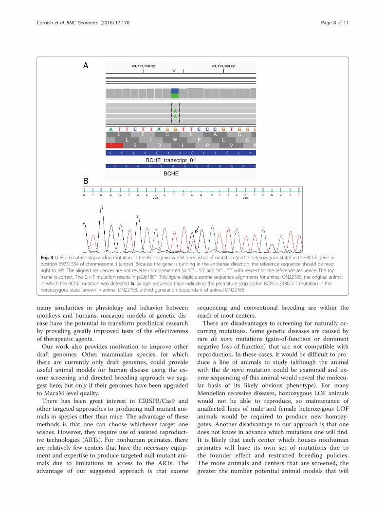
mutations in genes known to be related to human gen-etic disease, ribonuclease L (RNASEL) in animalON12033 (chr01: 184268143) and BChE in animalON22186 (chr03:69751554). Both mutations were anno-tated as “STOP-GAINED” in the heterozygous state bySnpEff with high confidence scores. The raw QUALscores for these two mutations were 2755.77 and1186.77, respectively. The mutation in the RNASEL gene(p.R552X) occurred in the fourth exon. This mutationwas visualized with IGV (Fig. 2a) and validated withSanger sequencing (Fig. 2b). The mutation in theBCHE gene (p.G180X) occurred in the second exon.This mutation was also visualized with IGV (Fig. 3a)and validated with Sanger sequencing (Fig. 3b). Wedetermined the transmission of BCHE in a family ofrhesus macaques using Sanger sequencing of exon 2(Fig. 4). This mutation was transmitted across threegenerations (Fig. 4).To determine whether the LOF mutation in BChE in the
heterozygous state decreased BChE activity in rhesus ma-caques as it would in humans, we collected blood from ani-mal ON22193 (a third generation carrier of the mutation)and animal ON22197 (an unrelated cage mate). Analysis ofthe serum of these two animals revealed that the heterozy-gous mutant had 30 % of the BChE activity of its unrelatedcage mate (Table 4).
DiscussionWe have previously demonstrated that MacaM yieldsbetter results than rheMac2 for mRNA-sequence expres-sion analysis [25, 33]. We now demonstrate that MacaMalso performs better than rheMac2 for exome analysis.More exome reads align against the MacaM chromo-some assembly than the rheMac2 assembly (Table 1).
This gap largely disappears when unplaced scaffolds areincluded for the alignments (Table 1) suggesting thatmore of the rhesus genome is represented in the MacaMchromosome assembly than in the rheMac2 chromo-some assembly. Further, MacaM has many fewer falsepositives than rheMac2 (Table 2). Although it is temptingto aggressively annotate as many genes as possible, for ex-ome studies the cost of incorrectly annotating genes isvery high. This is because an unacceptable number of falsepositive high impact mutations will result from these er-rors. This is likely in part due to the fact that the Gnomonautomated annotator used by NCBI will use intronic se-quence when an exon for a gene is missing from thechromosome assembly [24]. Since introns are often poorlyconserved, attempts to align sequence against spurious“exons” (actually intronic sequence) will frequently resultin apparent highly damaging mutations. The most time-intensive step in exome studies is filtering false-positives.Adding large numbers of false-positives as a result of in-correct gene annotations has made using the rhesus anno-tations for rheMac2 for exome studies in rhesus macaquesimpractical [23].The overall number of SNPs and percent nonsense
mutations that we observed for rhesus macaques aresimilar to those reported for humans [5]. The relativenumber of synonymous to non-synonymous SNPs,about 2 to 1, that we observed in rhesus macaques wasstrikingly similar to the ratio reported by Yuan et al. forrhesus macaque [21] but different from the approxi-mately 1 to 1 ratio found in humans [5]. Negative selec-tion is apparently acting in both species to limit thenumber of deleterious mutations.Our results suggest that the new MacaM genome can
be used with exome data to screen rhesus macaques fornaturally occurring LOF mutations in genes related tohuman disease. We describe LOF mutations, in the het-erozygous state, in two genes related to human health,RNASEL and BChE.LOF mutations in RNASEL have been linked to sus-
ceptibility to prostate cancer in humans [34, 35]. Individ-uals with LOF germline mutations in the heterozygousstate developed prostate tumors with complete loss offunction (loss of heterozygosity) [34, 35]. Rhesus ma-caque heterozygotes with LOF mutations in the RNA-SEL gene might serve as important animal models forprostate cancer.
Table 3 SNPs located in CDS
17573 ON12033 ON22186 002 T-NHP
Synonymous 15,689 (64.6 %) 30,566 (65.7 %) 23,145 (64.9 %) 25,066 (65.0 %)
Non-synonymous 8563 (35.2 %) 15,874 (34.1 %) 12,416 (34.8 %) 13,399 (34.7 %)
Nonsense 50 (0.2 %) 109 (0.2 %) 100 (0.3 %) 99 (0.3 %)
Total 24,302 46,549 35,661 38,564
Cornish et al. BMC Genomics (2016) 17:170 Page 6 of 11

Humans with mutations in the BChE gene that de-crease or eliminate expression of butyrylcholinesterasehave prolonged apnea in response to exposure to suc-cinylcholine or mivarcurium [36]. Further, treatmentwith BChE has been shown to be protective against anerve agent, Soman, in rhesus macaques [37] indicat-ing the possible importance of the levels of this en-zyme in the likelihood of surviving exposure to nerveagents. We demonstrated that a rhesus macaque witha LOF mutation in the BChE gene in the heterozy-gous state had decreased BChE activity compared toan unrelated cagemate. Rhesus macaques with LOFmutations in the BChE gene may serve as usefulmodels for pharmacogenomics and perhaps in theevaluation of the effects of nerve agents in individualswith varying levels of BChE activity.
Rare mutations are more likely to be present in theheterozygous state. For some diseases, the heterozygousmutants themselves would be useful. For Mendelianrecessive genetic diseases, heterozygotes could be bredtogether to increase the numbers of homozygote nullmutant rhesus macaques. In this way, nonhuman pri-mate models of LOF human genetic disease could beproduced. Given that there are tens of thousands of ma-caques at primate centers and at private facilities andthat every animal likely harbors one or more interestingmutations, models for many genetic diseases could likelybe created with a program of exome screening, genotyp-ing and directed breeding.The exome screening and directed breeding approach
suggested here could provide a much-needed alternativeto mouse models of human genetic disease. Given the
Fig. 2 LOF premature stop codon mutation in the RNASEL gene in animal ON12033. a. IGV screenshot of mutation (in the heterozygous state) inthe RNASEL gene in position 184268143 of chromosome 1 (arrow). The top frame is correct. The C > T mutation results in p.Arg552* in animalON12033. b. Sanger sequence trace indicating the premature stop codon RNASEL c.1654C > T mutation in the heterozygous state (arrow) inanimal ON12033
Cornish et al. BMC Genomics (2016) 17:170 Page 7 of 11
many similarities in physiology and behavior betweenmonkeys and humans, macaque models of genetic dis-ease have the potential to transform preclinical researchby providing greatly improved tests of the effectivenessof therapeutic agents.Our work also provides motivation to improve other
draft genomes. Other mammalian species, for whichthere are currently only draft genomes, could provideuseful animal models for human disease using the ex-ome screening and directed breeding approach we sug-gest here; but only if their genomes have been upgradedto MacaM level quality.There has been great interest in CRISPR/Cas9 and
other targeted approaches to producing null mutant ani-mals in species other than mice. The advantage of thesemethods is that one can choose whichever target onewishes. However, they require use of assisted reproduct-ive technologies (ARTs). For nonhuman primates, thereare relatively few centers that have the necessary equip-ment and expertise to produce targeted null mutant ani-mals due to limitations in access to the ARTs. Theadvantage of our suggested approach is that exome
sequencing and conventional breeding are within thereach of most centers.There are disadvantages to screening for naturally oc-
curring mutations. Some genetic diseases are caused byrare de novo mutations (gain-of-function or dominantnegative loss-of-function) that are not compatible withreproduction. In these cases, it would be difficult to pro-duce a line of animals to study (although the animalwith the de novo mutation could be examined and ex-ome sequencing of this animal would reveal the molecu-lar basis of its likely obvious phenotype). For manyMendelian recessive diseases, homozygous LOF animalswould not be able to reproduce, so maintenance ofunaffected lines of male and female heterozygous LOFanimals would be required to produce new homozy-gotes. Another disadvantage to our approach is that onedoes not know in advance which mutations one will find.It is likely that each center which houses nonhumanprimates will have its own set of mutations due tothe founder effect and restricted breeding policies.The more animals and centers that are screened, thegreater the number potential animal models that will
Fig. 3 LOF premature stop codon mutation in the BChE gene. a. IGV screenshot of mutation (in the heterozygous state) in the BChE gene inposition 69751554 of chromosome 3 (arrow). Because the gene is running in the antisense direction, the reference sequence should be readright to left. The aligned sequences are not reverse complemented so “C” = “G” and “A” = “T” with respect to the reference sequence. The topframe is correct. The G > T mutation results in p.Gly180*. This figure depicts exome sequence alignments for animal ON22186, the original animalin which the BChE mutation was detected. b. Sanger sequence trace indicating the premature stop codon BChE c.538G > T mutation in theheterozygous state (arrow) in animal ON22193, a third generation descendant of animal ON22186
Cornish et al. BMC Genomics (2016) 17:170 Page 8 of 11

be identified. In principal, if enough animals arescreened, eventually LOF mutations in every gene re-lated to disease in humans will be identified.In the current work, we focused on unequivocal loss-
of-function mutations (stop-gain, frameshifts, etc.).However, human genetic disease can be caused bysubtler mutations such as substitutions which resultin a change of a single amino acid (pathogenic mis-sense mutations). Although there are programs forestimating whether changes in amino acids are “dele-terious”, they generally require a database of proteinswith defined functional domains and/or a database ofproteins from multiple species. Although attempts touse programs such as Polyphen-2 [38] to identify“deleterious” missense mutations in rhesus macaqueshave been made [39], these programs were primarilyintended to be used with human data [38]. Such pro-grams often rely, in part, on search against proteinsderived from other mammals. However, we have re-ported that the draft rhesus macaque genome, andlikely all other draft genomes, is incomplete or incor-rect for approximately 50 % of all genes [24]. Hence,attempts to identify evolutionarily conserved regions withinmammals have been fraught with difficulty. This may be
one reason why the true impact of missense mutationsscored as “deleterious” (are they truly pathogenic?) can bedifficult to predict. As more mammalian genomes arebrought to the same level of quality as the MacaM genome,databases which include conserved regions among mam-mals are likely to improve, perhaps leading to predictiveprograms which identify “deleterious” mutations that aretruly pathogenic.In addition to evolutionary conservation, documented
association of a missense mutation with a negativephenotypic effect and variation among large numbers ofhumans is an invaluable source of information for deter-mining whether or not an amino acid change is likely tobe pathogenic. It is important to note that, due tospecies-specific differences in protein function, a variantwhich is pathogenic in humans is not necessarily patho-genic in rhesus macaques or other mammals. However,examination of large numbers of rhesus macaques forprotein variation will likely be a fruitful strategy to deter-mine which variants are likely to be pathogenic in thisspecies.
ConclusionsNGS sequences of rhesus DNA fragments captured withhuman exome kits can be can be aligned against thenew MacaM genome and the results analyzed accordingto GATK best practices to identify high impact variants.Identification of heterozygous LOF mutations combinedwith directed breeding could be used to create rhesusmacaque models of human genetic disease. This ispotentially an important step in advancing translationalresearch. This approach could also be applied to othermammalian species.
Availability of supporting dataThe exome sequence data sets supporting the results of thisarticle are available in the Sequence Read Archive reposi-tory under accessions SRX144674 [http://www.ncbi.nlm.-nih.gov/sra/SRX144674], SRX115899 [http://www.ncbi.nlm.nih.gov/sra/SRX115899], SRX144808 [http://www.ncbi.nlm.nih.gov/sra/SRX144808] and SRX145282 [http://www.ncbi.nlm.nih.gov/sra/SRX145282].
Additional files
Additional file 1: Table S1. List of effects of mutations in four rhesusmacaques (17573, ON12033, ON22186 and 002T-NHP) as annotated bySnpEff. SNV = single nucleotide variant; Indel = insertion or deletion. Effectsare as follows: Intergenic = in the intergenic region. Upstream = upstreamof a gene by at most 5kbp. Downstream = downstream of a gene by atmost 5kbp. 5' UTR = located in the 5' UTR. 3' UTR = located in the 3' UTR.Intronic = between two exons. Splice Acceptor = two bases before exonstart. Splice Donor = two bases after coding exon’s end. Splice Region = inthe putative (Lariat) branch point, located in the intron. Frameshift = indel inthe coding region that is not a multiple of 3. Inframe indel = indel in thecoding region that is a multiple of 3. Start Lost = start codon is mutated to
Table 4 Butyrlcholinesterase activity (units/ml) in a BChE LOFheterozygote (ON22193) and wild type cage mate (ON22197)
AnimalID
BChE activity -No inhibitor
BChE activity - 20 μMethopropazine
BChE activity -0.1 mM iso-OMPA
ON22193 1.38 0 0
ON22197 4.65 0 0
Fig. 4 BChE transmission across three generations of rhesusmacaques. * indicates that animal ON22193 was used for the BChEactivity assay. This is also the animal whose BChE mutation isdepicted in Fig. 3b.
Cornish et al. BMC Genomics (2016) 17:170 Page 9 of 11

non-start codon. Stop gained = non-stop codon is mutated to stop codon.Stop Lost = stop codon is mutated to non-stop codon. (DOCX 116 kb)
Additional file 2: Table S2. Number of variants at splice sites andpercent of total splice sites for CDS and UTR exons provided for four rhesusmacaques (17573, ON12033, ON22186 and 002T-NHP). (DOCX 11 kb)
AbbreviationsACACA: acetyl-CoA carboxylase alpha; BChE: butyrylcholinesterase;CLCA4: chloride channel accessory 4; LOF: loss-of-function; GATK: GenomeAnalysis Toolkit; IGV: Integrated Genome Viewer; NCBI: National Center forBiotechnology Information; NGS: next generation sequencing;RNASEL: ribonuclease L.
Competing interestsThe authors declare that they have no competing interests.
Authors’ contributionsAC participated in the design of the study, participated in the GATK bestpractices pipeline variant analysis and helped draft the manuscript. RGparticipated in the GATK best practices pipeline variant analysis. RBNconceived of the study, participated in the design of the study, analyzedvariant calls with IGV, designed the primers for PCR and Sanger validation,and drafted the manuscript. All authors have read and approve the finalversion of this manuscript.
AcknowledgmentsThis work was supported by a grant from the National Institutes of Health(R24RR017444) to RBN. We thank the Bioinformatics and Systems BiologyCore at the University of Nebraska Medical Center (UNMC) for providingcomputational resources. This core receives support from Nebraska ResearchInitiative (NRI) and NIH (2P20GM103427 and 5P30CA036727). We also thankDr. Alok Dhar at the University of Nebraska DNA Sequencing Core for hiswork in exome sequencing. We thank Dr. Betsy Ferguson for genomic DNAfor animals ON12033, ON22186, ON22186, ON22187, ON22188, ON22191,ON22192 and ON22193; Dr. Howard Fox for the genomic DNA from animal002 T-NHP; and Jerilyn Pecotte of the Southwest National Primate ResearchCenter for genomic DNA from the reference rhesus macaque (17573). Wealso thank Dr. Ferguson and her colleagues at the ONPRC for collectingblood from animals ON22193 and ON22197. We are very grateful to Dr.Oksana Lockridge for performing the BChE assay. We appreciate helpfulcomments from Dr. Etsuko Moriyama. We thank Dan Meehan for performingthe PCRs.
Received: 23 September 2015 Accepted: 22 February 2016
References1. Bell CJ, Dinwiddie DL, Miller NA, Hateley SL, Ganusova EE, Mudge J, et al.
Carrier testing for severe childhood recessive diseases by next-generationsequencing. Sci Transl Med. 2011;3:65ra4.
2. Gilissen C, Hoischen A, Brunner HG, Veltman JA. Unlocking Mendeliandisease using exome sequencing. Genome Biol. 2011;12:228.
3. Solomon BD, Pineda-Alvarez DE, Bear KA, Mullikin JC, Evans JP, ComparativeSequencing Program NISC. Applying genomic analysis to newbornscreening. Mol Syndromol. 2012;3:59–67.
4. Rabbani B, Tekin M, Mahdieh N. The promise of whole-exome sequencingin medical genetics. J Hum Genet. 2014;59:5–15.
5. Bamshad MJ, Ng SB, Bigham AW, Tabor HK, Emond MJ, Nickerson DA, et al.Exome sequencing as a tool for Mendelian disease gene discovery. Nat RevGenet. 2011;12:745–55.
6. MacArthur DG, Balasubramanian S, Frankish A, Huang N, Morris J, Walter K,et al. A systematic survey of loss-of-function variants in human protein-coding genes. Science. 2012;335:823–8.
7. Seok J, Warren HS, Cuenca AG, Mindrinos MN, Baker HV, Xu W, et al.Genomic responses in mouse models poorly mimic human inflammatorydiseases. Proc Natl Acad Sci U S A. 2013;110:3507–12.
8. Hay M, Thomas DW, Craighead JL, Economides C, Rosenthal J. Clinicaldevelopment success rates for investigational drugs. Nature Biotechnol.2014;32:40–51.
9. Norgren RB. Improving genome assemblies and annotations for nonhumanprimates. ILAR J. 2013;54:144–53.
10. Barr CS, Newman TK, Becker ML, Parker CC, Champoux M, Lesch KP, et al. Theutility of the non-human primate; model for studying gene by environmentinteractions in behavioral research. Genes Brain Behav. 2003;2:336–40.
11. Hewitson L. Primate models for assisted reproductive technologies.Reproduction. 2004;128:293–9.
12. Tachibana M, Sparman M, Sritanaudomchai H, Ma H, Clepper L, WoodwardJ, et al. Mitochondrial gene replacement in primate offspring andembryonic stem cells. Nature. 2009;461:367–72.
13. Messaoudi I, Estep R, Robinson B, Wong SW. Nonhuman primate models ofhuman immunology. Antioxid Redox Signal. 2011;14:261–73.
14. Vallender EJ, Miller GM. Nonhuman primate models in the genomic era: aparadigm shift. ILAR J. 2013;54:154–65.
15. Palermo RE, Tisoncik-Go J, Korth MJ, Katze MG. Old world monkeys and newAge science: the evolution of nonhuman primate systems virology. ILAR J.2013;54:166–80.
16. Phillips KA, Bales KL, Capitanio JP, Conley A, Czoty PW, ‘t Hart BA, et al. Whyprimate models matter. Am J Primatol. 2014;76:801–27.
17. Yang SH, Cheng PH, Banta H, Piotrowska-Nitsche K, Yang JJ, Cheng EC, et al.Towards a transgenic model of Huntington’s disease in a non-humanprimate. Nature. 2008;453:921–4.
18. Chan AW, Jiang J, Chen Y, Li C, Prucha MS, Hu Y, et al. Progressive cognitivedeficit, motor impairment and striatal pathology in a transgenic huntingtondisease monkey model from infancy to adulthood. PLoS One. 2015;10:e0122335.
19. Niu Y, Shen B, Cui Y, Chen Y, Wang J, Wang L, et al. Generation of gene-modified cynomolgus monkey via Cas9/RNA-mediated gene targeting inone-cell embryos. Cell. 2014;156:836–43.
20. Morton NE, Crow JF, Muller HJ. An estimate of the mutational damage inman from data on consanguineous marriages. Proc Natl Acad Sci U S A.1956;42:855–63.
21. Yuan Q, Zhou Z, Lindell SG, Higley JD, Ferguson B, Thompson RC, et al.The rhesus macaque is three times as diverse but more closelyequivalent in damaging coding variation as compared to the human.BMC Genet. 2012;13:52.
22. George RD, McVicker G, Diederich R, Ng SB, MacKenzie AP, Swanson WJ,et al. Trans genomic capture and sequencing of primate exomes revealsnew targets of positive selection. Genome Res. 2011;21:1686–94.
23. Vallender EJ. Expanding whole exome resequencing into non-humanprimates. Genome Biol. 2011;12:R87.
24. Zhang X, Goodsell J, Norgren RB. Limitations of the rhesus macaque draftgenome assembly and annotation. BMC Genomics. 2012;13:206.
25. Zimin AV, Cornish AS, Maudhoo MD, Gibbs RM, Zhang X, Pandey S, et al. Anew rhesus macaque assembly and annotation for next-generationsequencing analyses. Biol Direct. 2014;9:20.
26. McKenna A, Hanna M, Banks E, Sivachenko A, Cibulskis K, Kernytsky A, et al.The Genome Analysis Toolkit: a MapReduce framework for analyzing next-generation DNA sequencing data. Genome Res. 2010;20:1297–303.
27. Forums GATK. Broad Institute. 2015. http://gatkforums.broadinstitute.org/discussion/2806/howto-apply-hard-filters-to-a-call-set. Accessed 8August 2015.
28. Cingolani P, Platts A, le Wang L, Coon M, Nguyen T, Wang L, et al. Aprogram for annotating and predicting the effects of single nucleotidepolymorphisms, SnpEff: SNPs in the genome of Drosophila melanogasterstrain w1118; iso-2; iso-3. Fly (Austin). 2012;6:80–92.
29. McKusick-Nathans Institute of Genetic Medicine, Johns Hopkins University(Baltimore, MD), Online Mendelian Inheritance in Man, OMIM®. 2014. WorldWide Web URL: http://omim.org/.
30. Robinson JT, Thorvaldsdóttir H, Winckler W, Guttman M, Lander ES, Getz G,et al. Integrative genomics viewer. Nature Biotech. 2011;29:24–6.
31. Rozen S, Skaletsky HJ. 1998. Primer3. Code available at http://bioinfo.ut.ee/primer3/.
32. Ellman GL, Courtney KD, Andres Jr V, Feather-Stone RM. A new and rapidcolorimetric determination of acetylcholinesterase activity. BiochemPharmacol. 1961;7:88–95.
33. Sandler NG, Bosinger S, Estes J, Zhu R, Tharp G, Boritz E, et al. Type I IFNresponses in rhesus macaques prevent SIV transmission and slow diseaseprogression. Nature. 2014;511:601–5.
34. Carpten J, Nupponen N, Isaacs S, Sood R, Robbins C, Xu J, et al. Germlinemutations in the ribonuclease L gene in families showing linkage withHPC1. Nature Genet. 2002;30:181–4.
Cornish et al. BMC Genomics (2016) 17:170 Page 10 of 11
35. Rennert H, Bercovich D, Hubert A, Abeliovich D, Rozovsky U, Bar-Shira A,et al. A novel founder mutation in the RNASEL gene, 471delAAAG, isassociated with prostate cancer in Ashkenazi Jews. Am J Hum Genet. 2002;71:981–4.
36. Lockridge O. Review of human butyrylcholinesterase structure, function,genetic variants, history of use in the clinic, and potential therapeutic uses.Pharm & Therap. 2015;148:34–46.
37. Broomfield CA, Maxwell DM, Solana RP, Castro CA, Finger AV, Lenz DE.Protection by butyrylcholinesterase against organophosphorus poisoning innonhuman primates. J Pharmacol Exp Ther. 1991;259:633–8.
38. Adzhubei IA, Schmidt S, Peshkin L, Ramensky VE, Gerasimova A, Bork P,et al. A method and server for predicting damaging missense mutations.Nat Methods. 2010;7:248–9.
39. Fawcett GL, Raveendran M, Deiros DR, Chen D, Yu F, Harris RA, et al.Characterization of single-nucleotide variation in Indian-origin rhesusmacaques (Macaca mulatta). BMC Genomics. 2011;12:311.
• We accept pre-submission inquiries
• Our selector tool helps you to find the most relevant journal
• We provide round the clock customer support
• Convenient online submission
• Thorough peer review
• Inclusion in PubMed and all major indexing services
• Maximum visibility for your research
Submit your manuscript atwww.biomedcentral.com/submit
Submit your next manuscript to BioMed Central and we will help you at every step:
Cornish et al. BMC Genomics (2016) 17:170 Page 11 of 11