Embed Size (px)
Citation preview

DISEASES OF AQUATIC ORGANISMSDis Aquat Org
Vol. 48: 79–90, 2002 Published March 11
INTRODUCTION
Yellow head disease (YHD) in cultured black tigershrimp Penaeus monodon was first noticed in centralThailand by Limsuwan in 1991 (Limsuwan 1991). Thecausative agent was found to be an RNA-virus com-
monly known as yellow head virus (YHV) (Boonyarat-palin et al. 1993, Chantanachookin et al. 1993). On thebasis of morphology of negatively stained virions bytransmission electron microscopy (TEM) (Wongteera-supaya et al. 1995), RNA content (Wongteerasupaya etal. 1995) and sequence information, YHV is a positive-sense, single-stranded RNA virus most likely related tothe coronaviruses (Cowley et al. 1999, 2000a,b, Tang &Lightner 1999).
© Inter-Research 2002 · www.int-res.com
*Corresponding author. E-mail: [email protected]
Evidence for apoptosis correlated with mortality inthe giant black tiger shrimp Penaeus monodon
infected with yellow head virus
Kornnika Khanobdee1, Chumporn Soowannayan1, T. W. Flegel1, Sukathida Ubol2, Boonsirm Withyachumnarnkul1,*
1Centex Shrimp, and 2Department of Microbiology, Faculty of Science, Mahidol University, Rama 6 Rd., Bangkok 10400, Thailand
ABSTRACT: Histological, cytochemical and ultrastructural changes in giant black tiger shrimpPenaeus monodon were investigated at various time intervals after injection with yellow head virus(YHV). Hemocytes, lymphoid organs (LO) and gills were the main focus of the study. After injectionwith YHV, onset of mortality varied from 36 h onward. By normal hematoxylin and eosin staining, the3 tissues showed clear and increasing prevalence of nuclear condensation, pyknosis and karyor-rhexis from approximately 36 h post-injection (p.i.) until death, although pathology was evident in theLO as early as 12 h p.i. in some shrimp. By nuclear DNA staining with 4’,6-diamidino-2-phenylindole(DAPI) and by specific labeling of 3’-OH ends of nuclear DNA using a technique called terminaldeoxynucleotidyl transferase-mediated deoxy-UTP nick-end labeling (TUNEL), cells of the 3 tissuesshowed evidence of chromatin condensation and DNA fragmentation, respectively. Both are gen-erally considered to be characteristic of apoptosis. In addition to TUNEL labeling, evidence forDNA fragmentation was supported by the appearance of ~200 base pair DNA ladders at approxi-mately 48 h p.i. in hemocytes of YHV-infected but not uninfected shrimp. Transmission electronmicroscopy (TEM) of LO tissue revealed features of apoptosis in tissues of YHV-infected shrimp only.These included marginated, condensed and fragmented chromatin without concurrent cytoplasmicdamage. Histological, cytochemical, ultrastructural and biochemical data were consistent with thehypothesis that widespread and progressive apoptosis occurred in susceptible shrimp infected withYHV. Although no specific tests were carried out to determine whether this purported apoptosis wasthe cause of mortality, moribund shrimp had extensive deterioration of vital tissues such as thehemolymph, gills, heart and LO, suggesting that many essential bodily functions had been severelycompromised. This probably resulted in the gross signs of lethargy and weakness seen, and it isreasonable to suggest that further, progressive deterioration could have led to the collapse of vitalfunctions followed by death.
KEY WORDS: Yellow head virus · Yellow head disease · Penaeus monodon · Apoptosis
Resale or republication not permitted without written consent of the publisher
Dis Aquat Org 48: 79–90, 2002
YHV infects cells of both ectodermal and mesoder-mal origin (Boonyaratpalin et al. 1993, Chantana-chookin et al. 1993, Kasornchandra et al. 1995). ByTEM, it is a 150 to 170 nm by 40 to 50 nm rod-shapedvirus with a continuous fringe of spike-like projectionsradiating out approximately 11 nm from the outerenvelope (Wongteerasupaya et al. 1995). The lym-phoid organ (LO) of moribund shrimp suffering fromYHD usually has extensive abnormalities, includingmany degenerated cells in the stromal matrix of thetubules. These have condensed pyknotic and karyor-rhectic nuclei, and cytoplasmic basophilic inclusions(Chantanachookin et al. 1993, Lightner 1996). Othertissues, such as the interstitial tissues of the hepato-pancreas, connective tissue underlying the midgut,cardiac tissue, hematopoietic tissue, hemocytes andgill tissue, also had similar pathological features (Boon-yaratpalin et al. 1993, Lightner 1996).
Despite severe tissue damage by YHV, no inflamma-tory response has ever been detected in the tissues ofinfected shrimp (Flegel & Pasharawipas 1998). Thissuggests that cell death caused by YHV may be dueto apoptosis rather than necrosis. The occurrence ofapoptosis in shrimp or any other crustacean has notbeen substantiated, although there are some recentsuggestions in the literature that cell death by apop-tosis is associated with viral infections in shrimp(Flegel & Pahsarawipas 1998, Henderson & Stuck1999, Anggraeni & Owens 2000). Apoptosis is alsoinvolved in tissue and organ differentiation, as it isin other animals including a number of arthropods(Kerr et al. 1972).
The small amount of experimental work that hasbeen done so far concerning apoptosis in shrimp viralinfections has used white spot syndrome virus (WSSV)and has depended largely on use of terminal deoxy-nucleotidyl transferase (TdT)-mediated deoxy-UTPnick-end labeling (TUNEL) as an indication of DNAfragmentation (Henderson & Stuck 1999, Anggraeni &Owens 2000, Sahtout et al. 2001). In this study, we haveused several measures to determine whether there isevidence of apoptosis in shrimp infected with YHV.
MATERIALS AND METHODS
Grossly healthy Penaeus monodon, 10 to 15 g, werestocked in 6 concrete circular tanks of approximately2.0 m diameter containing 10‰ sea water at a 0.80 mdepth. Each tank contained 100 shrimp fed commercialfeed pellets at 3% body weight daily. Air stones wereused to ensure a dissolved oxygen level above 5 ppm.The shrimp were acclimated for 3 d before experi-ments were begun. They were then divided into 3groups of 2 tanks each, including an untreated control
group, a lobster hemolymph buffer (LHB)-injectedcontrol group and a YHV-injected group. LHB wasprepared using the method described by Paterson &Stewart (1974).
A crude extract of YHV was prepared from hemo-lymph of moribund shrimp from a natural YHD out-break in a culture pond in the following way. Hemo-lymph was withdrawn from the ventral hemolymphsinuses of the shrimp, diluted 1:100 with LHB and fil-tered through a sterile 0.45 µm membrane filter. Allprocesses were carried out under sterile conditions andthe diluted YHV crude extract was kept in aliquots at–80°C until used.
The LHB-injected and YHV-injected shrimp wereindividually injected with 0.1 ml LHB and YHVcrude extract, respectively, into the abdominal muscle.Twenty animals from each group, including the intactcontrol, were sampled at 0, 6, 12, 24, 36, 48 and 60 hfollowing the injection. From the sampled shrimp, LOs,gills and hemocytes were isolated immediately andprocessed for standard histology using hematoxylinand eosin (H&E) staining, for staining with 4’,6-diamidino-2-phenylindole (DAPI) and for labeling withTUNEL. DNA extracts from the hemocytes were alsoassayed by gel electrophoresis. In addition, a few othertissues were fixed and processed for standard histo-logy using H&E staining.
For DAPI staining, tissues fixed with Davidson’s fixa-tive were embedded in paraffin and 5 µm sectionswere prepared using standard procedures (Bell &Lightner 1988). For hemocytes, thin smears were pre-pared on clean glass slides. The specimens were thenwashed and flooded with DAPI solution (1 µg ml–1) andincubated for 15 min at 37°C inside a small humidifiedchamber covered with aluminum foil. After incubation,the specimens were washed once with methanol and 1drop of anti-fade solution (Vectashield, H-1000, VectorLaboratories Inc., Burlingame, California, USA) wasadded before mounting to examine for chromatincondensation and disintegration under a fluorescencemicroscope with a 340/380 nm excitation filter.
In the TUNEL procedure (Garvieli et al. 1992), tis-sues and hemocyte smears were processed as for DAPIstaining. The sectioned tissues were de-paraffinized,rehydrated, immersed in 4% paraformaldehyde inphosphate-buffered saline (PBS) for 15 min at roomtemperature, washed in PBS (2 × 5 min) and digestedwith 20 µg ml–1 Proteinase K at the volume of 100 µlper tissue section. The sections were allowed to reactwith the enzyme for 15 min at room temperaturebefore being washed with PBS. They were then post-fixed with 4% paraformaldehyde and washed in PBSat room temperature. Excess liquid was removed bytapping the slide and the tissue was covered with100 µl of equilibration buffer at room temperature for
80

Khanobdee et al.: Yellow head virus-infected Penaeus monodon
10 min. The tissue was then covered with 50 µl of theenzyme TdT diluted in incubation buffer (45 µl of equi-libration buffer, 5 µl of nucleotide mix and 1 µl of 25 Uµl–1 of TdT enzyme). Sections were covered with aplastic coverslip to ensure even distribution of thereagent and incubated at 37°C for 60 min inside ahumidified chamber to allow the tailing reaction tooccur. Exposure to light was prevented during this andfollowing steps by covering the chamber with alu-minum foil.
After the 60 min incubation, the reaction wasstopped by placing the slides in a coplin jar containing40 ml of 2 × saline sodium citrate (SSC) buffer for 15 minat room temperature. The slides were washed in freshPBS to remove unincorporated fluorescein-deoxy-UTP.The tissue was counterstained by immersing the slidesin 40 ml of a freshly prepared propidium iodide solu-tion (1 µg ml–1 in PBS) for 15 min at room temperature.The slides were then washed in de-ionized water (3 ×5 min) and 1 drop of anti-fade (Vectashield, H-1000)was added to the tissue before mounting. The slideswere immediately observed under a fluorescencemicroscope using a standard fluorescent filter set toview green fluorescence of the nuclei at 520 ± 20 nmfor a positive indication of DNA fragmentation and redfluorescence of propidium iodide at >620 nm for anegative indication of DNA fragmentation.
For hemolymph smears, the TUNEL assay was per-formed according to the manufacturer’s instructions(Promega, Madison, Wisconsin, USA), with some mod-ification to the permeabilization step. In this step, Tri-ton X-100 was used at 1.0% (v/v) in PBS on ice for5 min, followed by Proteinase K at 50 µg ml–1 for 15 minat room temperature. For positive controls, a smear ofnormal shrimp hemocytes treated with DNase I wasused, as recommended in the instruction manual. Thesamples were analyzed by fluorescence microscopy,by which apoptotic nuclei stained green and nuclei ofnormal cells stained red with the propidium iodidecounterstain.
Included in each preparation were positive and neg-ative control tissues. The positive controls comprisednormal tissues pre-treated with DNase I (Life Tech-nologies, Inc., Frederick, Maryland, USA) to digest thenuclear DNA into fragments before identical process-ing. The negative controls comprised normal tissuestreated identically but without addition of TdT en-zyme, except for hemocytes, where the TdT enzymewas added to the normal cell control preparation.
YHV-infected tissues were also studied by TEM. LOswere fixed in 2.5% glutaraldehyde, post-fixed in 1%OsO4, dehydrated in an acetone series and embeddedin Araldite-502 resin. Ultra-thin sections of silver-grayinterference (60 to 90 nm) were collected on coppergrids and stained with uranyl acetate and lead citrate
before viewing and photographing under a TEM(Hitachi H-300) at an acceleration voltage of 75 kV.
For gel electrophoresis, pooled hemolymph of 3shrimp from each group were used and total nucleicacid was extracted by the conventional phenol/chloro-form extraction method, as described by Sambrook etal. (1989), with some modification. Pooled hemolymph(approximately 4 ml group–1) was centrifuged at 400 × gat 4°C for 10 min and cell pellets were collected. Foreach pooled sample, 1 volume of lysis buffer (50 mMTris-HCl, pH 9.0, 100 mM EDTA, 50 mM NaC; and 2%sodium dodecyl sulfate) containing 20 µg ml–1 ofProteinase K was added in 2 times the amount of thepellet volume. The mixture was incubated at 65°C for1 h and then incubated further at room temperatureovernight. To remove digested proteins, an equalvolume of phenol was added before centrifugation at5000 × g and 4°C for 10 min. The supernatant wascollected and the step repeated once more beforeadding an equal volume of phenol:chloroform (1:1)followed by further centrifugation at 5000 × g, 4°C, for10 min; the supernatant containing nucleic acid wasthen collected and precipitated in 2 volumes of 100%ethanol at –20°C overnight. This nucleic fraction waswashed twice by centrifugation with 1 ml of 70%ethanol at 10 000 × g, 4°C, for 15 min and dried at roomtemperature. The DNA pellet was then dissolved in50 µl of Tris buffer, kept at 37°C for 30 min and thenstored at 4°C until used.
Agarose gel electrophoresis used 1.8% agarose gels.Markers comprised λDNA cut with EcoRI and HindIIIand a 123 base pair (bp) marker (ladder marker). Allsamples were mixed with loading buffer (10 mM EDTA,0.25% w/v bromophenol blue and 50% v/v glycerol)before loading. The gel was run at 50 V cm–1 for 80 minand stained with ethidium bromide. The results wereexamined on a UV transilluminator and photographed.
RESULTS
Light microscopy (LM) of gills, LO and hemocytesrevealed cells with nuclei that had pyknosis, karyor-rhexis and chromatin margination (Fig. 1), and withcytoplasm that had vacuolation; the prevalence ofthese cells progressively increased from 12 h post-infection (p.i.). The LO was the first organ attacked byYHV and had the highest degree of destruction, withmost of the pyknotic and karyorrhectic nuclei occur-ring in the LO tubules and very few in LO spheroids.Both the YHV-injected and uninjected shrimp hadthese spheroids among the LO tubules (Fig. 1, tA, tB)and these were classified as type A (+A), type B (+B)and type C (+C) based on the degree of vacuolizationas described by Hasson et al. (1999).
81

Dis Aquat Org 48: 79–90, 200282
Fig. 1. Light micrographs of normal and yellow head virus (YHV)-infected hemocytes, lymphoid organ (LO) and gills of Penaeusmonodon with hematoxylin and eosin (H&E) staining. These are arranged in a grid contrasting pairs of normal and YHV-infectedspecimens for 3 tissue types. Abnormal-shaped nuclei with pyknosis (p) and karyorrhexis (k) can be seen in all 3 YHV-infectedbut not in normal tissue types, except for LO spheroids. LO spheroid examples (type A [tA], type B [tB]) can be seen in the controlshrimp (tA, upper right, and tB, lower left part of the middle panels) and in YHV-infected shrimp (tB, right half of photomicro-graph), both containing relatively few karyorrhectic and pyknotic nuclei. Many can be seen in the LO tubules of the YHV-injected shrimp (left half of the photomicrograph). In the gills, some of the abnormal nuclei can be seen in the pillar epithelium
and some in the hemolymph vessels (probably hemocytes). All scale bars shown are in micrometers
Control
Hem
ocyt
esG
ills
Lyp
mp
hod
org
an
YHV-injected
Khanobdee et al.: Yellow head virus-infected Penaeus monodon
With DAPI staining, nuclei stand out since the stain isspecific for nuclear DNA. Typical pyknotic and karyor-rhectic nuclei were observed in the hemocytes, LO andgills of YHV-infected shrimp, whereas normal nucleiwere observed in the same tissues of control groups(Fig. 2). At the earliest, abnormal nuclei were observedat 12 h p.i. and they became more abundant thereafteruntil death.
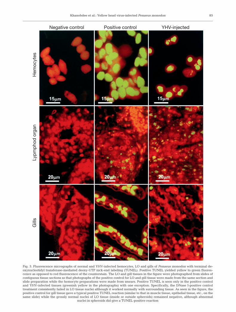
By TUNEL assay, gills and hemolymph from theuntreated and LHB-injected negative control shrimpreacted negatively except when treated with DNasebefore staining (i.e. positive control group) (Fig. 3).A curious phenomenon was the consistent absenceof TUNEL-stained nuclei in the DNase I-treated LOtissue, even though the cell nuclei of immediately sur-rounding tissue in the same slides gave a normal posi-tive reaction (Fig. 3). In spite of this, some TUNEL-positive nuclei were seen in LO spheroids in bothnormal and YHV-injected shrimp. Despite this anom-aly, the general reaction with surrounding tissue andcontrasts with YHV-injected and uninjected shrimpcould still be compared. In YHV-infected shrimp, theearliest positive TUNEL reaction (green fluorescence)was observed in only a few cells of the LO (outsidespheroids) at 12 h p.i., but stained nuclei became pro-gressively more prevalent thereafter in the LO, hemo-cytes, gills and other target tissues. The relative num-ber of positive-staining cells in the gills was generallyless than in the LO.
The total hemocyte numbers declined rapidly inYHV-infected shrimp and, from 30 h p.i. onward, theproportion of remaining hemocytes with karyorrhecticand pyknotic nuclei increased with time as detectedusing LM with H&E and DAPI staining and TUNEL(Fig. 4). In addition, DNA extracts from hemocytes ofYHV-injected shrimp prepared 48 h p.i. had ~200 bpladder patterns (Fig. 5) that were not present in un-injected or LHB-injected shrimp.
By TEM, LOs of the untreated and LHB-injectedshrimp contained normal nuclei with heterochromatinin peripheral patches and as a central mass (Fig. 6A).In YHV-injected shrimp, both normal and degenerateLO cells were observed. No YHV particles wereobserved before 12 h p.i. At this time (Fig. 6), nuclearchromatin of the infected cells became condensed andmarginated in sharply delineated masses abutting thenuclear envelope (Fig. 6B to D). The cytoplasm wasalso markedly condensed. In spite of these dramaticchanges, there was no TEM evidence of viral struc-tures inside these cells, although virions were occa-sionally seen in intercellular spaces. By 24 to 36 h p.i.,when viral structures including complete virions werepresent (Fig. 7), more abnormal features were detectedincluding some fragmentation of nuclear chromatin,disruption of presumed nucleolar structures and dense
nuclear condensation (Fig. 7A). Despite these rathersevere pathological changes in the nucleus, thenuclear envelope and cytoplasmic organelles such asmitochondria, ribosomes and rough endoplasmicreticulum (rER) remained relatively intact (Fig. 7A).
Virions in YHV-infected cells were more frequentlyobserved at 48 to 60 h p.i. (Fig. 8), as were condensednuclei (pyknotic nuclei) (Fig. 8A) and electron-dense,round structures surrounded by narrow electron-lucentspaces (Fig. 8B,C). On the basis of the pathologicalchanges observed during earlier stages of infection,the latter structures were considered to be fragmentednuclei surrounded by cytoplasmic residues (i.e. ‘apop-totic bodies’ arising from YHV-infected cells). Theseapoptotic bodies were frequently observed in clustersin intercellular spaces and, together with pyknoticnuclei, were often phagocytosed by neighboring cells(Fig. 8C), where they underwent degenerative changeswithin phagosomes.
DISCUSSION
In this study, evidence of apoptosis was found in thehemocytes, LO and gills of YHV-infected shrimp byLM using special nuclear stains to reveal chromatincondensation and DNA fragmentation. Further indica-tions of apoptosis were seen in the LO by TEM. Theultrastructural changes observed were similar to thosedescribed for early apoptosis, including nuclear chro-matin margination followed by partial and completechromatin condensation, nuclear budding or fragmen-tation and the appearance of nuclear fragments,termed apoptotic bodies, surrounded by an envelope(Kerr 1971, Kerr et al. 1972). The cytoplasm is alsomarkedly condensed. Despite severe pathologicalchanges in the nucleus, the nuclear envelope and cyto-plasmic organelles such as mitochondria, ribosomesand rER are preserved. In this study, all of these ultra-structural changes characteristic of apoptosis wereseen in YHV-infected shrimp cells.
We have no explanation as to why DNase I treatmentconsistently gave TUNEL-positive results in tissuessurrounding the LO but negative results in the LOexcept for some spheroid cells in both YHV-injectedand uninjected shrimp. Anggraeni & Owens (2000)have reported that TUNEL-positive nuclei are oftenseen in LO spheroids, where they may represent cellsundergoing apoptosis as a normal response to viralinfection. Indeed, it has been reported that grossly nor-mal shrimp may be infected with 1 or more viruseswithout gross signs of disease (Flegel 2001) and it ispossible that the spheroids arose from subclinical viralinfections present before YHV injection. However, wechecked tissue sections (not shown) for YHV using a
83

Dis Aquat Org 48: 79–90, 200284
Fig. 2. Fluorescence micrographs of hemocytes, LO and gills of normal and YHV-infected Penaeus monodon stained with 4’,6-diamidino-2-phenylindole (DAPI). Similar features can be seen as in Fig. 1 except that the nuclei are more clearly resolved andare in various stages of condensation. For the hemocytes, N: normal nuclei; 1: early condensation; 2: pyknotic; 3: karyorrhectic.
For the LO and gills, k: karyorrhexis; p: pyknosis. All scale bars shown are in micrometers
Control
Hem
ocyt
esG
ills
Lyp
mp
hod
org
an
YHV-injected
Khanobdee et al.: Yellow head virus-infected Penaeus monodon 85
Fig. 3. Fluorescence micrographs of normal and YHV-infected hemocytes, LO and gills of Penaeus monodon with terminal de-oxynucleotidyl transferase-mediated deoxy-UTP nick-end labeling (TUNEL). Positive TUNEL yielded yellow to green fluores-cence as opposed to red fluorescence of the counterstain. The LO and gill tissues in the figure were photographed from slides ofcontiguous tissue sections so that photographs of the positive control for LO and gill tissue were made from the same section andslide preparation while the hemocyte preparations were made from smears. Positive TUNEL is seen only in the positive controland YHV-infected tissues (greenish yellow in the photographs) with one exception. Specifically, the DNase I-positive controltreatment consistently failed in LO tissue nuclei although it worked normally with surrounding tissue. As seen in the figure, thepositive control for gill tissue gave a typical positive TUNEL reaction (similar to that in muscle tissue, epithelial tissue, etc., on thesame slide) while the grossly normal nuclei of LO tissue (inside or outside spheroids) remained negative, although abnormal
nuclei in spheroids did give a TUNEL-positive reaction
Negative control Positive controlH
emoc
ytes
Gill
sLy
pm
pho
d o
rgan
YHV-injected

Dis Aquat Org 48: 79–90, 2002
monoclonal antibody described by Sithigorngul et al.(2000) and we found that the tissues (including LOspheroids) were negative for YHV before the startof our experiments and that controls remained sothroughout. The results suggested that, even thoughour DNase I treatment failed, DNA fragmentation thathad occurred endogenously in the LO tissue (whateverthe cause) was still detectable by TUNEL assay. The
failure of DNase I to function in LO tissue requiresfurther investigation but may have resulted from thepresence of a tissue-specific inhibitor.
Under TEM, apoptotic bodies frequently occurred inclusters in intercellular spaces, where they were sub-sequently phagocytosed by neighboring cells. Thus,many of the cytoplasmic basophilic inclusion bodiescharacteristic of YHV infection with H&E staining(Boonyaratpalin et al. 1993, Chantanachookin et al.1993, Nash et al. 1995 ) are likely to be phagocytosedapoptotic bodies. As found previously (Flegel & Pash-arawipas 1998), there was no evidence of the type ofinflammatory response usually elicited by coagulativenecrosis (Kerr et al. 1972). In addition, the pathologicalchanges began in some LO cells as early as 12 h p.i.,before the appearance of virions in the cytoplasm, sug-gesting that viral assembly may not be a prerequisitefor them. Many previous studies have shown that viralcomponents are capable of inducing apoptosis (Everett& McFadden 1999). For example, the E2 structural pro-tein of Sinbis virus alone can induce apoptosis in theabsence of viral replication, as can the proteins Gp120or Gp41 of human immunodeficiency virus-1 (Everett& McFadden 1999). It is likely that the apoptoticshrimp cells we observed were infected with YHV andthat viral replication steps preceding the developmentof identifiable viral components had been initiated.Whether apoptosis can be initiated by viral proteinsalone will require further investigation.
Additional evidence of apoptosis was obtained fromthe ladder pattern of chromosomal DNA fragmentsseparated by intervals of 180 to 200 bp resolved by gelelectrophoresis of hemocyte DNA from YHV-infectedshrimp. This phenomenon is similar to that resultingfrom cutting by endonucleases specific for double-stranded DNA at inter-nucleosomal positions in com-plete chromatin, and it is considered to be a marker forapoptosis in other organisms (Arends et al. 1990). Theoccurrence of these DNA ladders in DNA extracts ofhemocytes from YHV-infected shrimp suggests thatdeath of hemocytes is mediated by endonucleolyticcleavage of their DNA, as in apoptotic cells. DNAcleavage has been reported to occur through Ca++-and Mg++-dependent or -independent endonucleasesthat cut chromosomal DNA at the linker regionbetween nucleosomes to give OH– at the 3’ ends of thecut DNA (see review by Sikorska & Walker 1998).In non-apoptotic cells, the enzyme caspase-activateddeoxyribonuclease (CAD) is present as an inactivecomplex controlled by its inhibitor (ICAD). During apop-tosis, ICAD is activated by caspase and this, in turn,frees CAD to function as an endonuclease (see reviewby Thornberry & Lazebnik 1998). The next logicalstep would be to identify these enzymes in apoptoticshrimp cells.
86
Fig. 4. Percentage of presumably apoptotic hemocytes inYHV-injected shrimp detected by 3 different methods (H&Estaining, DAPI staining and TUNEL) at different times post-injection (p.i.). Test positive nuclei were seen only from 30 hp.i. and numbers increased steadily thereafter. Each bar
represents the mean from 8 shrimp
Fig. 5. Agarose gel electrophoresis of a Penaeus monodon he-mocyte DNA extract prepared at 48 h p.i. Lane 1: markerλDNA cut with EcoRI and HindIII; lane 2: 123 base pairmarker (ladder marker); lane 3: hemocyte DNA from normalshrimp; lane 4: hemocyte DNA from YHV-infected shrimp;lane 5: 1:2 diluted solution of hemocyte DNA from YHV-infected shrimp. A DNA ladder pattern of approximately200 base pair interval can be clearly seen in lane 4. Similarbut fainter bands were observed in lane 5, but they are diffi-
cult to see in the black and white print
bp
21226
2027
1375
947
1 2 3 4 5

Khanobdee et al.: Yellow head virus-infected Penaeus monodon
Altogether, the data strongly suggest that cell deathin YHD results mainly from apoptosis and that highnumbers of apoptotic cells may be the primary cause ofdeath in YHV-infected shrimp. No specific tests wereundertaken to determine this. However, moribundshrimp have extensive deterioration of vital tissuessuch as the hemolymph, gills, heart and LO, suggest-ing that many essential bodily functions have beenseverely compromised. This probably results in gross
signs of lethargy and weakness and it is reasonable tosuggest that further progressive deterioration couldlead to the collapse of organ functions followed bydeath.
The results for YHV are similar to those reported forfield and laboratory infections with WSSV (Henderson& Stuck 1999, Sahtout et al. 2001), where there are alsoindications that shrimp mortality is correlated withhigh numbers of apoptotic cells. Thus, 2 very different
87
Fig. 6. Transmission electron photomicrograph of the LO of normal and YHV-infected Penaeus monodon at 12 h p.i. Cells from normalshrimp contained nuclei with heterochromatin distributed in peripheral patches and as a central mass (A). In the YHV-injected shrimp,normal cells dominated but a few had nuclear chromatin condensed and marginated in sharply delineated masses abutting the nuclearenvelope (B to D). The cytoplasm was also markedly condensed. There was no evidence of unenveloped or enveloped viral particles in
these or other cells. E: enveloped YHV particle; N: nucleus; ne: nuclear envelope. All scale bars shown are in micrometers
A
B
C D

Dis Aquat Org 48: 79–90, 2002
viruses (i.e. one DNA and the other RNA) appear tocause death in a similar way, and this suggests thatthere may similar underlying mechanisms in the hostreaction to both infections. This would be in keepingwith the viral accommodation theory (Flegel & Pasha-rawipas 1998, Flegel 2001), which proposes that apop-tosis is the general cause of shrimp death from viralinfections.
Previous studies have found that apoptosis is con-trolled by several gene products that interact directlywith components of highly conserved biochemicalpathways to regulate cell death (Everett & McFadden1999). Thus, by inference, death by apoptosis inPenaeus monodon infected by YHV may be, in part,genetically determined, and genetic factors mayexplain the phenomenon of tolerance reported to occurwith YHD (Pasharawipas et al. 1997) within approxi-mately 1.5 yr of its catastrophic appearance in southernThailand. By that time, most farmers were obtaininggood harvests in spite of the presence of YHV in theirponds, as revealed by TEM. If apoptosis is the cause of
death in YHV-infected shrimp, its induction must beinfluenced by both virus and host gene products piv-otal in determining the death or survival of infectedshrimp. It could be reasoned that shrimp would surviveYHV infection if they or their pathogens possessedactive genes that inhibit apoptosis. It has been pro-posed that tolerance develops in situations where anearly viral binding step results in specific memory thatsuppresses apoptosis upon subsequent or concurrentviral infection that might otherwise lead to death(Flegel & Pasharawipas 1998). If this hypothesis is cor-rect, it is possible that many carrier species with activeYHV infections have infected cells with large numbersof virions but possess genes that inhibit apoptosis andthus prevent death.
Studying viral induction or inhibition of apoptosis isan important tool for dissection of the cell death path-way, and such studies might give insights into possibletherapeutic interventions in situations where inhibitorymechanisms fail. Recently, a number of low molecularweight gene products have been used to inhibit or
88
Fig. 7. Transmission electron photomicrograph of the LO of YHV-infected Penaeus monodon at 24 to 36 h p.i. Cells with mar-ginated and condensed heterochromatin were seen (A), similar to those from 12 h p.i. except that they were more abundant andmore condensed (B), and they were found in the presence of unenveloped (UE) and enveloped (E) YHV particles. YHV virionswere frequently observed in the cytoplasm (E). Despite the rather severe pathological changes in the nucleus, the nuclearenvelope (ne) and cytoplasmic organelles such as mitochondria (mi), ribosomes and rough endoplasmic reticulum (rER) wererelatively intact (A) until late in the transformation (B). G: Golgi complex; N: nucleus. All scale bars shown are in micrometers
A B

Khanobdee et al.: Yellow head virus-infected Penaeus monodon
enhance this fundamental cellular process, and soapoptosis has now become amenable to pharmacologi-cal manipulation (Kinloch et al. 1999). Thus, further un-
derstanding of apoptosis in experimental and naturalYHV infections may lead to the development of feedadditives that could prevent or stem YHV disease.
89
Fig. 8. Transmission electron photomicrograph of the LO of YHV-infected Penaeus monodon at 40 to 48 h p.i. Many electron-dense pyknotic nuclei (A, D) and round structures surrounded by narrow electron-lucent spaces were observed (B, C). The latterwere frequently interpreted as ‘apoptotic bodies’ (a). They were frequently observed in clusters in intercellular spaces. They andpyknotic nuclei were often phagocytosed by neighboring cells and subsequently appeared to undergo degenerative changeswithin phagosomes. E: enveloped virions; N: nucleus; ne: nuclear envelope; UE: unenveloped virions. All scale bars shown are
in micrometers
DC
A
B

Dis Aquat Org 48: 79–90, 2002
Acknowledgements. The authors would like to thank An-chalee Pongsa-Asawapaiboon for assistance in preparing thefigures. Support for this investigation was through grantsfrom the Institution Strengthening Program, Faculty ofScience, Mahidol University, the Thai National Center forGenetic Engineering and Biotechnology, and the NationalInstitute of Fisheries, Arlington, Virginia, USA.
LITERATURE CITED
Anggraeni MS, Owens L (2000) The haemocytic origin of lym-phoid organ spheroid cells in the penaeid prawn Penaeusmonodon. Dis Aquat Org 40:85–92
Arends MJ, Moris RG, Wyllie AH (1990) Apoptosis the role ofendonuclease. Am J Pathol 136:593–608
Bell TA, Lightner DV (1988) A handbook of normal shrimphistology. World Aquaculture Society, Baton Rouge, LA
Boonyaratpalin S, Supamataya K, Kasornchandra J, Direk-busarakom S, Ekpanithanpong U, Chantanachookin C(1993) Non-occluded baculo-like virus the causative agentof yellow-head disease in the black tiger shrimp Penaeusmonodon. Fish Pathol 28:103–109
Chantanachookin C, Boonyaratpalin S, Kasornchandra J,Direkbusarakom S, Dkpanithapong U, Supamataya K,Sriurairatana S, Flegel TW (1993) Histology and ultra-structure reveal a new granulosis-like virus in Penaeusmonodon affected by yellow-head disease. Dis Aquat Org17:145–157
Cowley JA, Dimmock CM, Wongteerasypaya C, BoonsaengV, Panyim S, Walker PJ (1999) Yellow head virus fromThailand and gill associated-virus from Australia areclosely related but distinct prawn viruses. Dis Aquat Org36:153–157
Cowley JA, Dimmock CM, Spann KM, Walker PJ (2000a)Detection of Australian gill-associated virus (GAV) andlymphoid organ virus (LOV) of Penaeus monodon byRT-nested PCR. Dis Aquat Org 39:159–167
Cowley JA, Dimmock CM, Spann KM, Walker PJ (2000b) Gillassociated virus of Penaeus monodon prawns: an inverte-brate virus with ORF1a and ORF1b genes related to arteri-and coronaviruses. J Gen Virol 81:1473–1484
Everett H, McFadden G (1999) Apoptosis: an innate immuneresponse to virus infection. Trends Microbiol 7:160–165
Flegel TW (2001) The shrimp response to viral pathogens. In:Browdy CL, Jory DE (eds) The new wave. Proceedings ofthe special session on sustainable shrimp aquaculture,Aquaculture 2001. World Aquaculture Society, Boca Raton,LA, p 190–214
Flegel TM, Pasharawipas T (1998) Active viral accommoda-tion: a new concept for crustacean response to viralpathogens. In: Flegel TW (ed) Advances in shrimp bio-technology. National Center for Genetic Engineering andBiotechnology, Bangkok, p 245–250
Garvieli Y, Sherman Y, Ben-Sasson SA (1992) Identificationof programmed cell death in situ via specific labeling ofnuclear DNA fragmentation. J Cell Biol 119:493–501
Hasson KW, Lightner DV, Mohney LL, Redman RM, White B(1999) Role of lymphoid organ spheroids in chronic Taurasyndrome virus (TSV) infections in Penaeus vannamei. DisAquat Org 38:93–105
Henderson T, Stuck K (1999) Induction of apoptosis in re-sponse to white spot syndrome virus in the Pacific whiteshrimp, Penaeus vannamei. Abstract #67. AquacultureAmerica 1999, Tampa, Florida. World Aquaculture Soci-ety, Baton Rouge, LA
Kasornchandra J, Supamattaya K, Boonyaratpalin S (1995)Electron microscopic observations on the replication ofyellow-head baculovirus in the lymphoid organ ofPenaeus monodon. Asian Shrimp News 15:2–3
Kerr JFR (1971) Shrinkage necrosis: a distinct mode f cellulardeath. J Pathol 105:13–20
Kerr JFR, Wyllie AH, Currie AR (1972) Apoptosis: a basic bio-logical phenomenon with wide ranging implications intissue kinetics. Br J Cancer 26:239–257
Kinloch RA, Treheme JM, Furness LM, Hajimohamadreza I(1999) The pharmacology of apoptosis. Trends PharmacolSci 20:35–42
Lightner DV (1996) A handbook of shrimp pathology anddiagnostic procedures diseases of cultured penaeidshrimp. World Aquaculture Society, Baton Rouge, LA
Limsuwan C (1991) Handbook for cultivation of black tigerprawns. Tansetakit Co. Ltd, Bangkok
Nash GL, Akarajamorn A, Withyachumnarnkul B (1995)Histology and rapid haemocytic diagnosis of yellow-headdisease in Penaeus monodon. In: Shariff M, Arthur JR,Subasinghe RP (eds) Diseases in Asian aquaculture II.Fish Health Section, Asian Fisheries Society, Manila,p 89–98
Pasharawipas T, Flegel TW, Shiurairatana S, Morrison DJ(1997) Latent yellow-head infections in Penaeus monodonand implications regarding disease resistance or tolerance.In: Flegel TW, Menasveta P, Paisarnrat S (eds) Shrimpbiotechnology in Thailand. National Center of GeneticEngineering and Biotechnology, Bangkok, p 45–53
Paterson WD, Stewart JE (1974) In vitro phagocytosis byhemocytes of American lobster (Homorus americanus)J Fish Res Board Can 31:1051–1056
Sambrook J, Fritsch EF, Maniatis T (1989) Molecular cloning,a laboratory manual, 2nd edn. Cold Spring Harbor Labo-ratory Press, Cold Spring Harbor, NY, p 9.16–9.30
Sahtout AH, Hassan MD, Shariff M (2001) DNA fragmenta-tion, an indicator of apoptosis in cultured black tigershrimp Penaeus monodon infected with white spot syn-drome virus. Dis Aquat Org 44:155–159
Sikorska M, Walker PR (1998) Endonuclease activities andapoptosis In: Lockchin RA, Zakeri Z, Tilly JL (eds) Whencells die. Wiley-Liss, London, p 211–242
Sithigorngul P, Chauychuwong P, Sithigorngul W, LongyantS, Chaivisuthangkura P, Menasveta P (2000) Developmentof a monoclonal antibody specific to yellow head virus(YHV) from Penaeus monodon. Dis Aquat Org 42:27–34
Tang KFJ, Lightner DV (1999) A yellow head virus geneprobe: nucleotide sequence and application for in situhybridization. Dis Aquat Org 35:165–173
Thornberry NA, Lazebnik Y (1998) Caspases: enemies within.Science 281:1312–1316
Wongteerasupaya C, Sriurairatana S, Vickers JE, Akara-jamorn A, Boonsaeng V, Panyim S, Tassanakajon A, Tas-sanakajon A, Withyachumnarnkul B, Flegel TW (1995)Yellow-head virus of Penaeus monodon is an RNA virus.Dis Aquat Org 22:45–50
90
Editorial responsibility: Otto Kinne, Oldendorf/Luhe, Germany
Submitted: March 28, 2001; Accepted: September 4, 2001Proofs received from author(s): March 4, 2002


![MiR-5692a promotes proliferation and inhibits apoptosis by ... · tal carcinoma, HCC, and lung cancer [10-12]. As reported, HOXD8 is significantly correlated with proliferation, cell](https://img.dokumen.tips/doc/110x75/5fdac2267515ba2c375d3f5d/mir-5692a-promotes-proliferation-and-inhibits-apoptosis-by-tal-carcinoma-hcc.jpg)