Embed Size (px)
Citation preview
Every Day Endodontics for the General Practitioner
Randall Iwasiuk, DDS
Every Day Endodontics for the General Practitioner
• The purpose of this presentation is to provide a few tips for making endodontic treatment more predictable and enjoyable for the general practitioner that treats endodontic cases on a regular basis. These recommendations are based on observations made over 17 years of endodontic specialty practice.
• Learning objectives: I will attempt to cover all the questions that were submitted to me from you, the participants. They generally fall into the following categories.
• Diagnosis
• Case selection
• Instrument selection
• Cleaning and shaping
• Obturation
• Post treatment restoration
• Antibiotics
Diagnosis
• How do we know when endodontic treatment is necessary? Barring the obvious carious pulp exposure or fracture, we must pulp test.
• Our goal in pulp testing is to reproduce the patients chief complaint. ie: reproduce their symptoms via thermal, percussion, biting (in various excursions), and palpation. It is important to test as many teeth as necessary to isolate the tooth that is the source of the symptoms. For example: If you suspect tooth #19 is in a state of irreversible pulpitis and tooth #20 has had RCT clearly you would want to cold test #21 as well as #18. I won’t regurgitate how to pulp test and what the results of each test indicate. Every text book you pick up has plenty to say on this topic. I will say that there is an element of subjectivity to interpreting pulp testing that only experience will help you overcome. I have never performed RCT on a tooth that didn’t need it, but I’ve probably not performed RCT on many teeth that did.
• At this point I will jump to the specific questions that were submitted to me.
• I’ll go out on a limb here and assume that as part of the diagnostic work up adequate radiographs were taken of the area in question. This includes a BW as well as a mesially and distally angulated PA with a direct one with open contacts for posterior teeth.
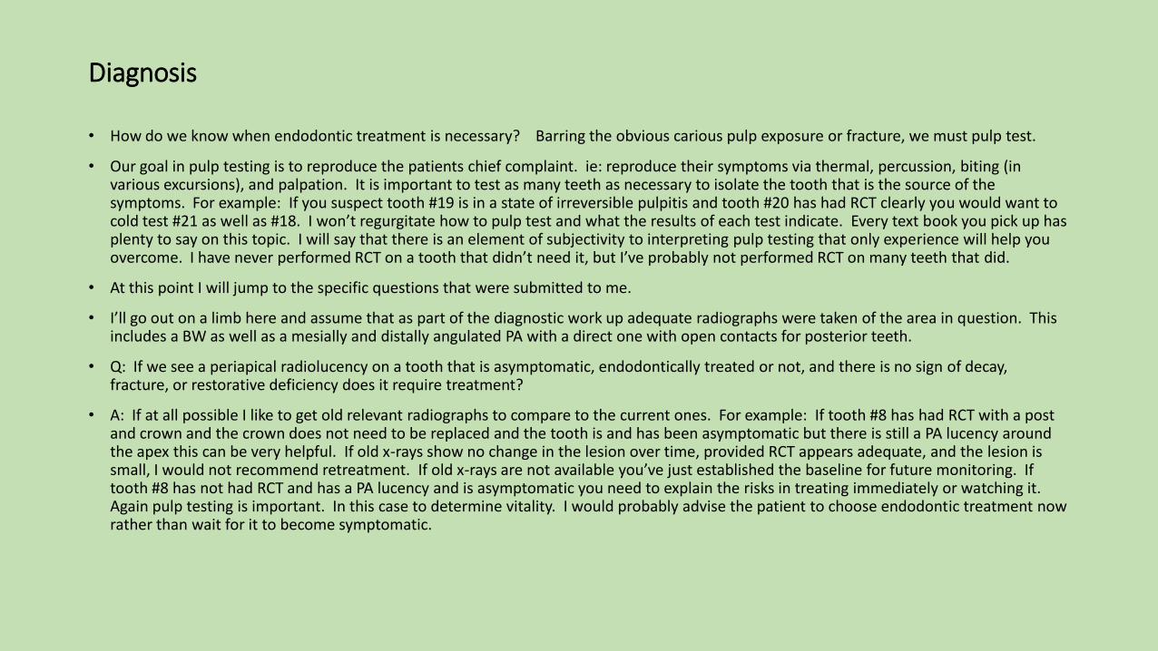
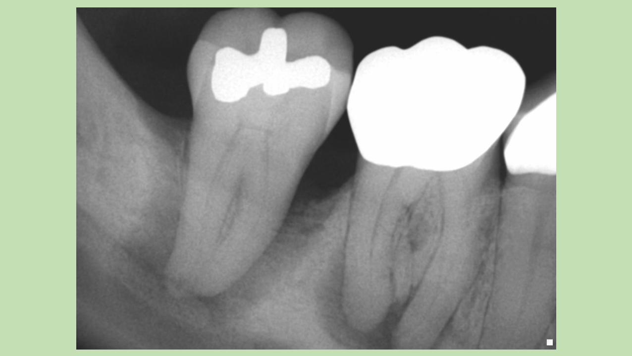
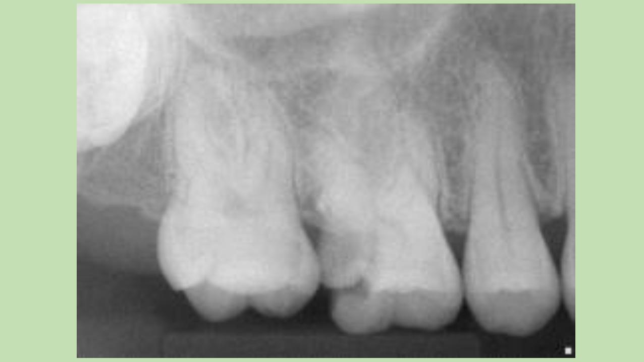
• Q: If we see a periapical radiolucency on a tooth that is asymptomatic, endodontically treated or not, and there is no sign of decay, fracture, or restorative deficiency does it require treatment?
• A: If at all possible I like to get old relevant radiographs to compare to the current ones. For example: If tooth #8 has had RCT with a post and crown and the crown does not need to be replaced and the tooth is and has been asymptomatic but there is still a PA lucency around the apex this can be very helpful. If old x-rays show no change in the lesion over time, provided RCT appears adequate, and the lesion is small, I would not recommend retreatment. If old x-rays are not available you’ve just established the baseline for future monitoring. If tooth #8 has not had RCT and has a PA lucency and is asymptomatic you need to explain the risks in treating immediately or watching it. Again pulp testing is important. In this case to determine vitality. I would probably advise the patient to choose endodontic treatment now rather than wait for it to become symptomatic.
Diagnosis cont.
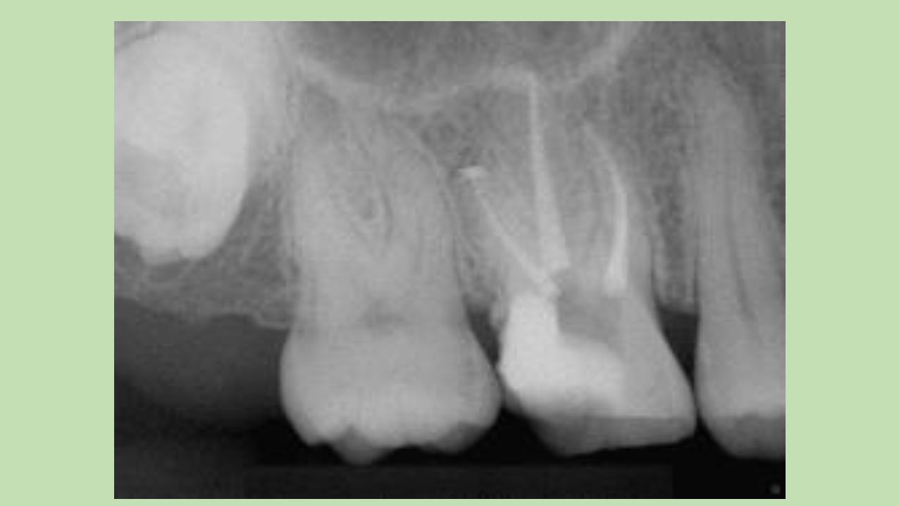
• How to look for fractures: I use a moist cotton roll, frac finder, transillumination, radiographic characteristics, and periodontal probing.
• Moist cotton roll and frac finder: look for excursive specific discomfort and/or pain on release when biting directly. Pain to biting without pain on release may be significant as well.
• Transillumination: a small bright light source will show a dark line blocking the transmission of light through the clinical crown when held directly against it. You’ll know it when you see it.
• Radiographic characteristics: The classic “J” lesion is very diagnostic especially when there is an extremely deep periodontal pocket associated with it. It will appear radiographically as a lucency traversing, say the distal side of the root from the crest of bone around the apex and partway up the mesial surface forming a letter J.
• Periodontal Probing: A very deep, narrow periodontal pocket appearing on any surface of the tooth. Beware, this may be the pathway of a draining sinus tract that didn’t form the more familiar lesion on the gingiva away from the tooth.
Diagnosis cont.
• Q: What is the difference between and endodontic abscess and a periodontal abscess?
• A: An endodontic abscess will test non-vital with an established sinus tract in the gingiva or through the sulcus. Beware, multi-rooted teeth may have a slight response to cold even if one or more roots are completely necrotic. I’ve seen teeth with draining sinus tracts exhibit a very delayed and slight response to cold. A tooth with a periodontal abscess will test vital even if it appears to have some elements of radiolucency.

Case Selection
• The first questions should be: What are the patient’s wishes? Are they committed to definitive restoration after RCT has been completed? What are the alternatives?
• The next question is: Is the tooth predictably restorable? Sometimes this can’t be answered until removal of decay and unsupported tooth structure.
• Is crown lengthening required? How will the removal and recontouring of crestal bone effect the adjacent teeth and periodontal status?
• Are there health conditions that may effect the predictability of healing?
• What challenges does the tooth present in performing predictable endodontic treatment? Long, curved, calcified, open apices, other “unusual” anatomy, retreatment, periodontal support.
• In my experience there is no such thing as a slam dunk, except in the rearview mirror. I am routinely put in the position of performing “routine” endodontic treatment when other factors severely limit the long term success restoratively or periodontaly.
Instrument selection
• Once your case has been documented, anesthetized and isolated with a rubber dam. Its time to choose your instruments.
• There is a wide array of instruments on the market today and I won’t pretend to say one is the best. Different ones work better for certain types of cases of course. I have found that for me the best approach is to learn to work with one or two systems thoroughly and stick with them.
• I like Tulsa Dental’s ProTapers in the new M wire format as well as the Vortex 15, 20, and 25 in the .02 taper. For anything bigger than a 60 I’m not obturating with gutta percha anyway so shape doesn’t matter.
• Hand files are still essential but I’ve found that I rarely use any larger than a 20. You should have available .06, .08 as well. I find that I use these sized hand files the most.
• You also need an apex locater. I like J. Morita’s root ZX, but then I’ve never tried the others. Some of them look pretty good and I’m sure they all work equally well.
• Apex locaters give you a faster, more accurate reading of canal length from a secure, repeatable reference point than even an x-ray.
• The trick to an accurate reading is using the largest file that fits gently to the apical foramen. Not to tight and not to loose. I always guestimate the canal length off an x-ray as well so I know within 2-3 mm what to expect. The pulp chamber must be dry. Keep in mind that large lateral canals can give you a false apex reading. That’s why the guestimate is helpful. If you can’t get an electronic reading you trust you can always take an x-ray.
• As for single file rotary systems. The only one I’ve tried is the Wave One by Tulsa Dental. It’s a progressive reciprocating file that requires a computerized hand piece control system to deliver the correct motion. My experience is that a relatively small number of cases lend themselves to a single file system.
Cleaning and Shaping
• This is where the rubber meets the road. Again you will find a selection of techniques promoted.
• I believe in the crown down technique with multiple recapitulations of the series of files each time reaching deeper and wider. Never force a file. Only use it to where it will passively go or with very gentle apical pressure. This applies to hand files as well as rotaries.
• Always use copious irrigation. I use full strength NaOCl. I routinely use two or three 12cc syringes of irrigant on a molar. Irrigating between each file used.
• We achieve and maintain apical patency. This is the point we are measuring with the apex locater, the apical foramen.
• Herb Schilder taught us to keep the apical foramen as small as practical. That means gauging the apical diameter with a hand file that neither slides through nor binds at the apical foramen.
• I’ve found that in my practice the vast majority of apical diameters are from size 25-40.
• I seldom use Gates Glidden burs. I just find I don’t need to open up the canal orifices that much.
• When I’m convinced that cleaning and shaping is complete, I dry the canal with a micro-capillary tip and do a final rinse with Qmix (Tulsa Dental). This is a solution of EDTA, chlorhexadine, and some proprietary wetting agent, and sonically agitate it for two minutes before a final Qmix rinse and cone fit.
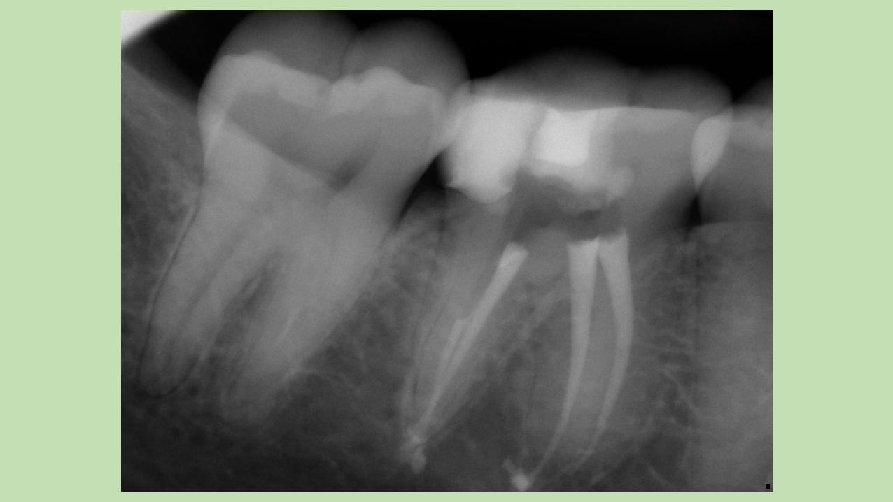
Obturation
• First of all I don’t condone the use of any carrier based obturation systems. The reason why is that they are very technique sensitive. If the cleaning and shaping is not adequate you will have difficulty properly fitting an obturator. Curved canals tend to strip off the gutta percha leaving the plastic carrier and sealer to complete the mission of the apical seal. Improperly fitted obturators result in short filled canals or over extended obturators
• Some of the carrier based systems are very difficult to remove if retreatment is necessary. This is especially true if the canals have been under shaped and the carriers trimmed apical the canal orifices.
• I’ll admit that my opinion is biased because I see only the cases that are failing.
• Cone Fitting
• A properly cleaned and shaped canal takes very little time to cone fit. Especially with the Pro-Taper file and gutta percha cone system.
• Once a canal is completely cleaned and shaped select the gutta percha cone that corresponds to the last file used to the apical foramen.
• The gutta percha cone should fit snugly to within 1mm of the apical foramen. This is verified with a radiograph.
• Tug back is that snug apical fit. You should be able to feel that the cone is binding at the apex and not along the shaft of the cone.
• Obturation
• I like to dry the canal with a luer lock capillary tip attached to a high volume hose. If that’s not practical, paper points work fine. Tulsa Dental has paper points that are color coded to the sizes of the Pro Taper rotary file system.
• After drying the canals place the sealer in the canal by buttering a dry appropriately sized paper point. Butter your gutta percha cone and gently and slowly place it in the canal until you feel the tug back when testing the seat. Until you develop confidence in cleaning and shaping and fitting cones it might be a good idea to take an x-ray with the gp cone and sealer to verify the position of the cone. (I seldom do that step anymore).
• Sealer
• I use BC sealer by Brasseler. It’s biocompatible, doesn’t shrink on setting, osteogenic, has a pH of +12 making it antimicrobial, and is in a pre-mixed syringe. I’ve seen one study that showed that traditional ZOE sealer actually promotes the growth of aspergillus, especially in posterior maxillary teeth that may be associated with the sinuses.
• Obturation Devices
• For obturation I use the SybronEndo Touch ‘n Heat 5004 or the System B.
• Lateral condensation. I did cold lateral condensation in dental school and for the first few years in private general practice. In 1994 I learned the above described warm vertical compaction technique. I haven’t used a spreader since. In my opinion there is no application of lateral condensation that out weighs the benefits of warm gp.
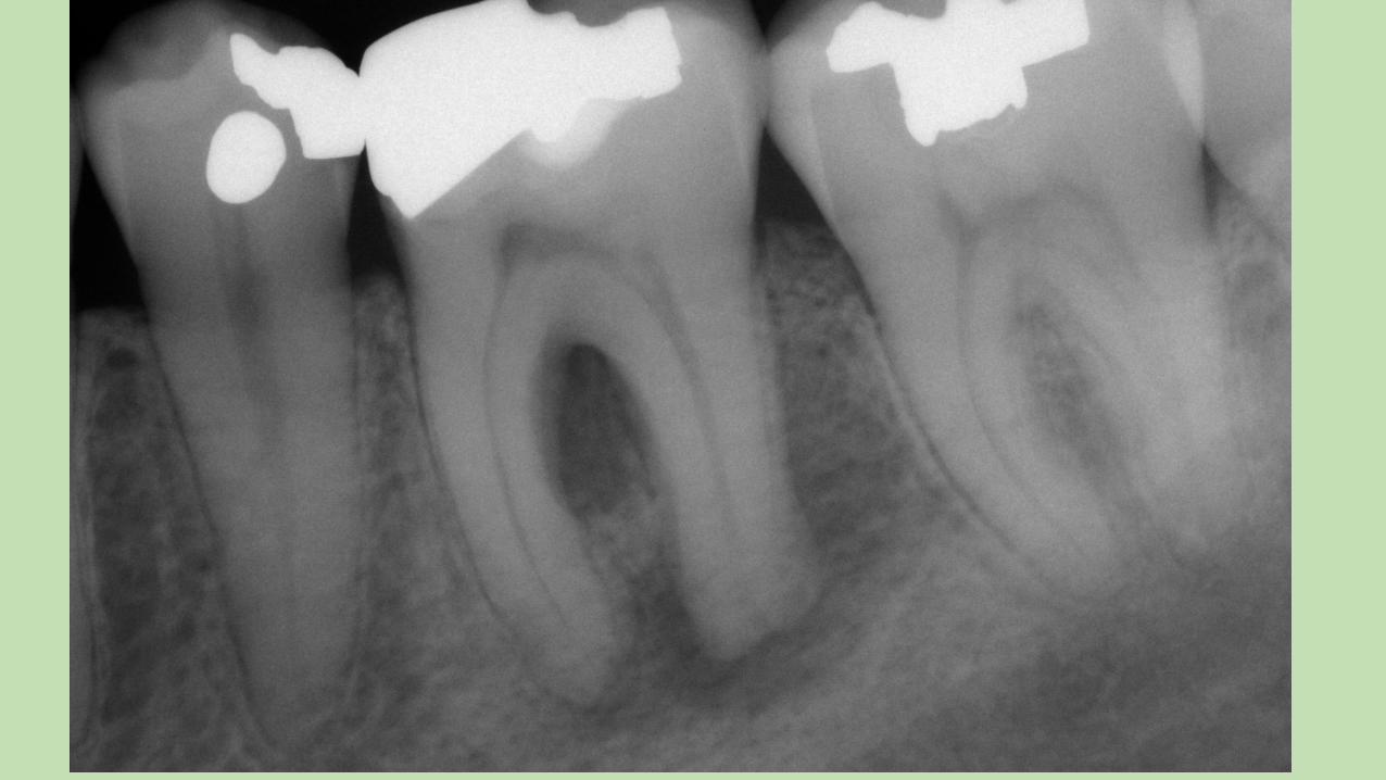
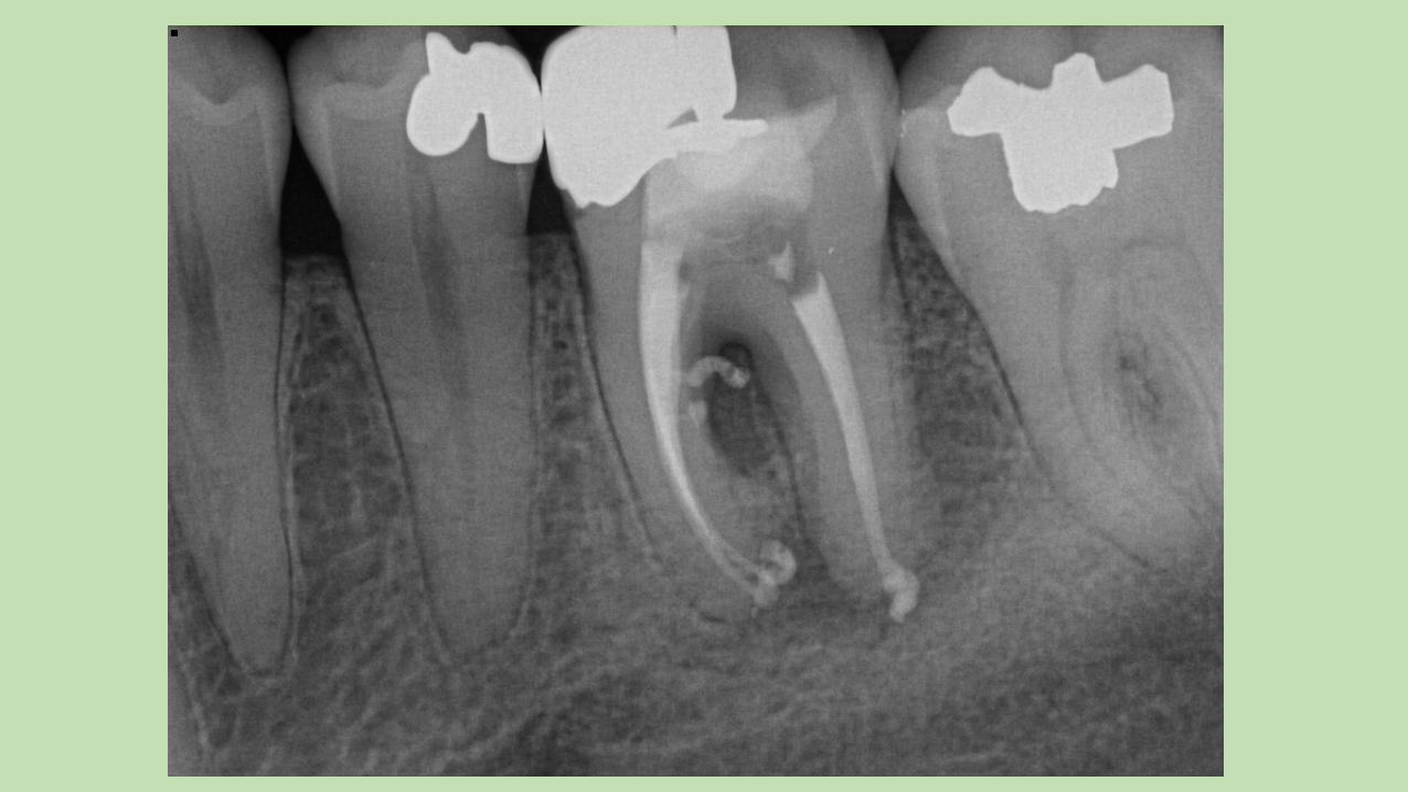
• Back filling the canals requires an Obtura gun. (Obtura Spartan). There are other ways to back fill the canal but they’re to tedious to even describe here.
• Q: Where do we end our fill?
• A: At the apical foramen. A sealer splash is biologically tolerated and will not impede healing. Sometimes a fill will appear short radiographically and a sealer puff will visually confirm that you’ve filled to the apical foramen. That’s another reason why I trust the apex locater more than a radiograph.
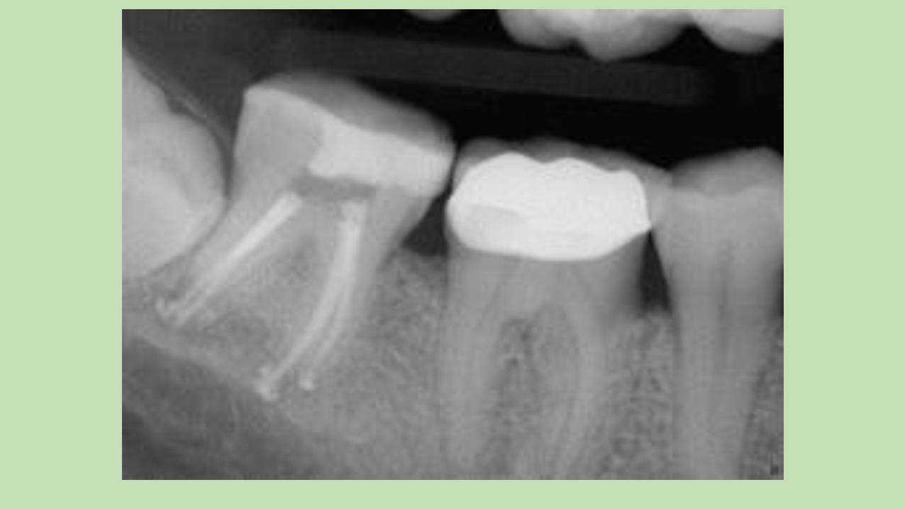
Post RCT restoration
• Some of our referrals request a core build-up or permanent access fill. We do this at the time of RCT completion whether it is single visit endo or whether it is on the second visit.
• In my opinion and those of many restorative dentists I work with, the indications for post placement are few. Those that request post spaces are honored. I don’t place posts.
• If you have 3mm or more of solid tooth structure on at least two opposing walls, posts are not indicated.
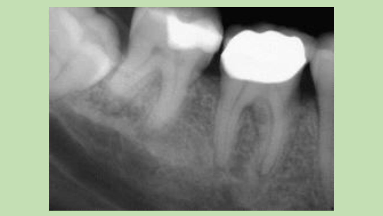
• Generally all molars and premolars should be crowned after RCT.
• Q: How soon should a crown be placed after RCT?
• A: Generally as soon as practical. Since you’ve taken the tooth out of occlusion (molars and premolars) and placed a solid core build-up your decision is guided by the size of the build-up and the patients overall occlusion pattern.
• Often incisors and canines can be restored without a crown. This of course depends on the amount of decay or fracturing a tooth sustained before endodontic treatment. Severe incisal wear may be an indication for full coronal protection.
Antibiotics
• Q: When should antibiotics be prescribed?
• A: If I was asked this question two years ago I would have said only if there is swelling, elevated temperature, and systemic signs of malaise. Over that past two years however I have seen a surge in the number of cases that have worsened even after treatment was initiated, along with an increase in cases that don’t quickly respond to a single antibiotic. So now I recommend antibiotics for any symptomatic tooth with a PA radiolucency (as opposed to a simple widened PLD space).
• It seems to be notable that antibiotic combinations are becoming more necessary as well. For example Clindamycin and Metronidazole.
• If the diagnosis is irreversible pulpitis antibiotics are not indicated.
• Q: When a tooth is symptomatic and has a PA radiolucency, can one just prescribe an antibiotic and watch?
• N: No. RCT is indicated.
• Q: Can one perform a pulpotomy when the patient has no definitive plan for subsequent RCT?
• A: I didn’t know where else to fit this question but my answer would be to perform a complete pulpectomy, in other words clean and shape the entire root canal system and fill it with CaOH. This would need to be refreshed every 8-12 weeks. But why would you want to do this?
Miscellaneous questions
• Q: Are retreats a good idea for general dentist to perform?
• A: In my opinion retreatment requires a surgical operating microscope and other specialized instruments to enhance to predictability of the outcome. At best the success rate is 65% to 80%, depending on who you believe. I would probably try and avoid them, especially molars.
• Q: Are there any tricks for dealing with convergent canals?
• A: Cone fit both of them, even if one cone is short of your working length. Obturate both canals simultaneously.
• Q: How do you look for 4th canals?
• A: The first step is to assume they are always there. Beyond that it is important to have an adequate access opening, a head lamp, and loupes with as much magnification as you can tolerate. I do everything from occlusal reduction to placing the core build-up or access filling through the surgical microscope so I can’t imagine having a high batting average without this advantage.
• Q: Are apicos a thing of the past?
• A: No. We are getting better and better at endodontic micro-surgery and there are a number of exciting advances in osteogenic and grafting materials that are making surgery more predictable in saving teeth.
• There were a number of questions about trauma and avulsions but I think that this is a large and complex area. I think the best thing is to enlist the aid of an endodontist or an oral surgeon in managing these demanding cases.


![NURSE PRACTITIONER: Lynne Day CLINICAL … Ageing + Care... · Page 1 of 24 Lynne DAY, ACNP: CPGs [October 2015] NURSE PRACTITIONER: Lynne Day CLINICAL PRACTICE GUIDELINES: October](https://img.dokumen.tips/doc/110x75/5aecbef47f8b9a36698ff31c/nurse-practitioner-lynne-day-clinical-ageing-carepage-1-of-24-lynne-day.jpg)