Embed Size (px)
Citation preview
ISSN: 2455-944X Int. J. Curr. Res. Biol. Med. (2018). 3(1): 10-21
10
INTERNATIONAL JOURNAL OF CURRENT RESEARCH INBIOLOGY AND MEDICINE
ISSN: 2455-944Xwww.darshanpublishers.com
DOI:10.22192/ijcrbm Volume 3, Issue 1 - 2018Original Research Article
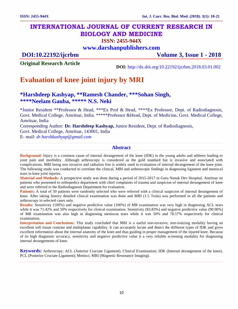
Evaluation of knee joint injury by MRI
*Harshdeep Kashyap, **Ramesh Chander, ***Sohan Singh,****Neelam Gauba, ***** N.S. Neki*Junior Resident **Professor & Head, ***Ex Prof & Head, ****Ex Professor, Dept. of Radiodiagnosis,Govt. Medical College, Amritsar, India. *****Professor &Head, Dept. of Medicine, Govt. Medical College,Amritsar, IndiaCorresponding Author: Dr. Harshdeep Kashyap, Junior Resident, Dept. of Radiodiagnosis,Govt. Medical College, Amritsar, 143001, IndiaE- mail: [email protected]
Abstract
Background: Injury is a common cause of internal derangement of the knee (IDK) in the young adults and athletes leading tojoint pain and morbidity. Although arthroscopy is considered as the gold standard but is invasive and associated withcomplications. MRI being non invasive and radiation free is widely used in evaluation of internal derangement of the knee joint.The following study was conducted to correlate the clinical, MRI and arthroscopic findings in diagnosing ligament and meniscaltears in knee joint injuries.Material and Methods: A prospective study was done during a period of 2015-2017 in Guru Nanak Dev Hospital, Amritsar onpatients who presented to orthopedics department with chief complaints of trauma and suspicion of internal derangement of kneeand were referred to the Radiodiagnosis Department for evaluation.Patients: A total of 50 patients were randomly selected who were referred with a clinical suspicion of internal derangement ofknee. After taking history detailed clinical examination was done and MRI (1.5 Tesla) was performed in all the patients andarthroscopy in selected cases only.Results: Sensitivity (100%) and negative predictive value (100%) of MR examination was very high in diagnosing ACL tearswhile it was 71.42% and 50% respectively for clinical examination. Sensitivity (83.83%) and negative predictive value (90.90%)of MR examination was also high in diagnosing meniscus tears while it was 50% and 78.57% respectively for clinicalexamination.Interpretation and Conclusions: This study concluded that MRI is a useful non-invasive, non-ionizing modality having anexcellent soft tissue contrast and multiplanar capability. It can accurately locate and detect the different types of IDK and givesexcellent information about the internal anatomy of the knee and thus guiding in proper management of the injured knee. Becauseof its high diagnostic accuracy, sensitivity and negative predictive value it a very reliable screening modality for diagnosinginternal derangements of knee.
Keywords: Arthroscopy; ACL (Anterior Cruciate Ligament), Clinical Examination; IDK (Internal derangement of the knee),PCL (Posterior Cruciate Ligament); Menisci; MRI (Magnetic Resonance Imaging).
DOI: http://dx.doi.org/10.22192/ijcrbm.2018.03.01.002
ISSN: 2455-944X Int. J. Curr. Res. Biol. Med. (2018). 3(1): 10-21
11
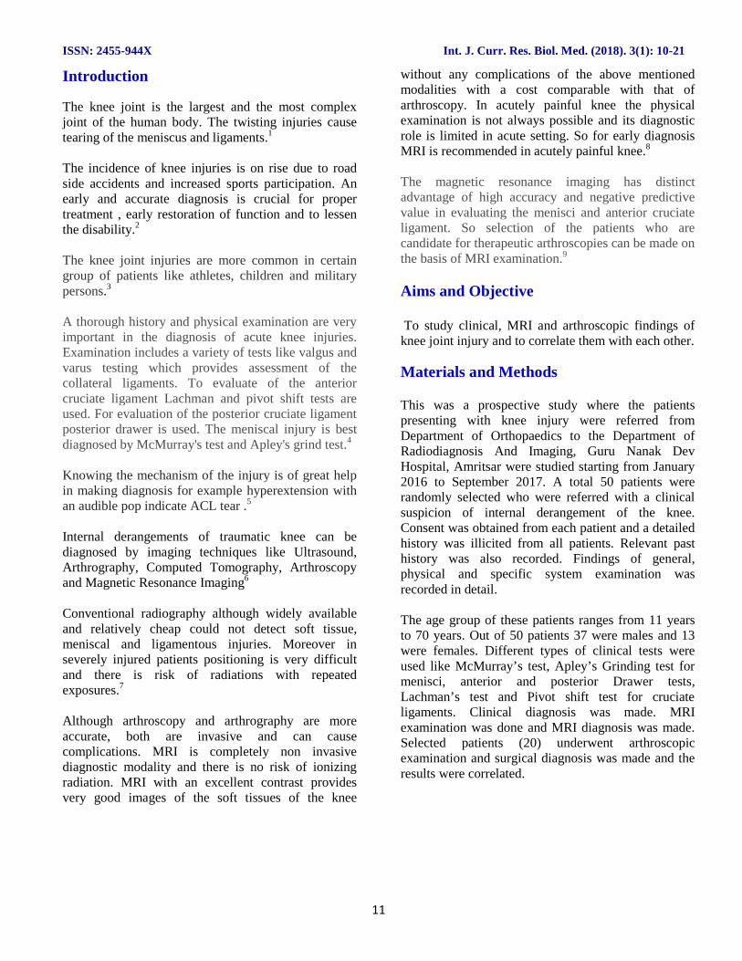
Introduction
The knee joint is the largest and the most complexjoint of the human body. The twisting injuries causetearing of the meniscus and ligaments.1
The incidence of knee injuries is on rise due to roadside accidents and increased sports participation. Anearly and accurate diagnosis is crucial for propertreatment , early restoration of function and to lessenthe disability.2
The knee joint injuries are more common in certaingroup of patients like athletes, children and militarypersons.3
A thorough history and physical examination are veryimportant in the diagnosis of acute knee injuries.Examination includes a variety of tests like valgus andvarus testing which provides assessment of thecollateral ligaments. To evaluate of the anteriorcruciate ligament Lachman and pivot shift tests areused. For evaluation of the posterior cruciate ligamentposterior drawer is used. The meniscal injury is bestdiagnosed by McMurray's test and Apley's grind test.4
Knowing the mechanism of the injury is of great helpin making diagnosis for example hyperextension withan audible pop indicate ACL tear .5
Internal derangements of traumatic knee can bediagnosed by imaging techniques like Ultrasound,Arthrography, Computed Tomography, Arthroscopyand Magnetic Resonance Imaging6
Conventional radiography although widely availableand relatively cheap could not detect soft tissue,meniscal and ligamentous injuries. Moreover inseverely injured patients positioning is very difficultand there is risk of radiations with repeatedexposures.7
Although arthroscopy and arthrography are moreaccurate, both are invasive and can causecomplications. MRI is completely non invasivediagnostic modality and there is no risk of ionizingradiation. MRI with an excellent contrast providesvery good images of the soft tissues of the knee
without any complications of the above mentionedmodalities with a cost comparable with that ofarthroscopy. In acutely painful knee the physicalexamination is not always possible and its diagnosticrole is limited in acute setting. So for early diagnosisMRI is recommended in acutely painful knee.8
The magnetic resonance imaging has distinctadvantage of high accuracy and negative predictivevalue in evaluating the menisci and anterior cruciateligament. So selection of the patients who arecandidate for therapeutic arthroscopies can be made onthe basis of MRI examination.9
Aims and Objective
To study clinical, MRI and arthroscopic findings ofknee joint injury and to correlate them with each other.
Materials and Methods
This was a prospective study where the patientspresenting with knee injury were referred fromDepartment of Orthopaedics to the Department ofRadiodiagnosis And Imaging, Guru Nanak DevHospital, Amritsar were studied starting from January2016 to September 2017. A total 50 patients wererandomly selected who were referred with a clinicalsuspicion of internal derangement of the knee.Consent was obtained from each patient and a detailedhistory was illicited from all patients. Relevant pasthistory was also recorded. Findings of general,physical and specific system examination wasrecorded in detail.
The age group of these patients ranges from 11 yearsto 70 years. Out of 50 patients 37 were males and 13were females. Different types of clinical tests wereused like McMurray’s test, Apley’s Grinding test formenisci, anterior and posterior Drawer tests,Lachman’s test and Pivot shift test for cruciateligaments. Clinical diagnosis was made. MRIexamination was done and MRI diagnosis was made.Selected patients (20) underwent arthroscopicexamination and surgical diagnosis was made and theresults were correlated.
ISSN: 2455-944X Int. J. Curr. Res. Biol. Med. (2018). 3(1): 10-21
12
Observations and Analysis
Table 1 Age wise distribution of the subjects
Age distribution(in years)
Number of subjectsN
Percentage
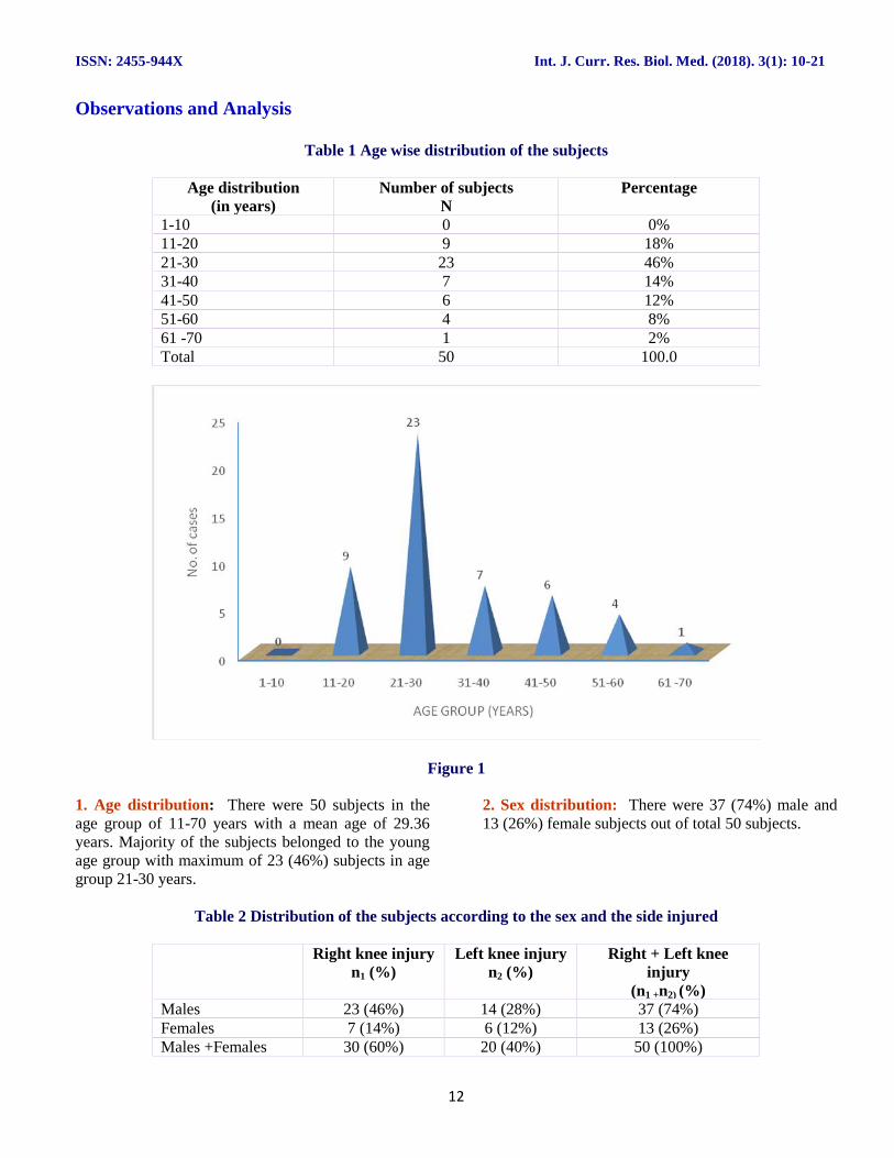
1-10 0 0%11-20 9 18%21-30 23 46%31-40 7 14%41-50 6 12%51-60 4 8%61 -70 1 2%Total 50 100.0
Figure 1
1. Age distribution: There were 50 subjects in theage group of 11-70 years with a mean age of 29.36years. Majority of the subjects belonged to the youngage group with maximum of 23 (46%) subjects in agegroup 21-30 years.
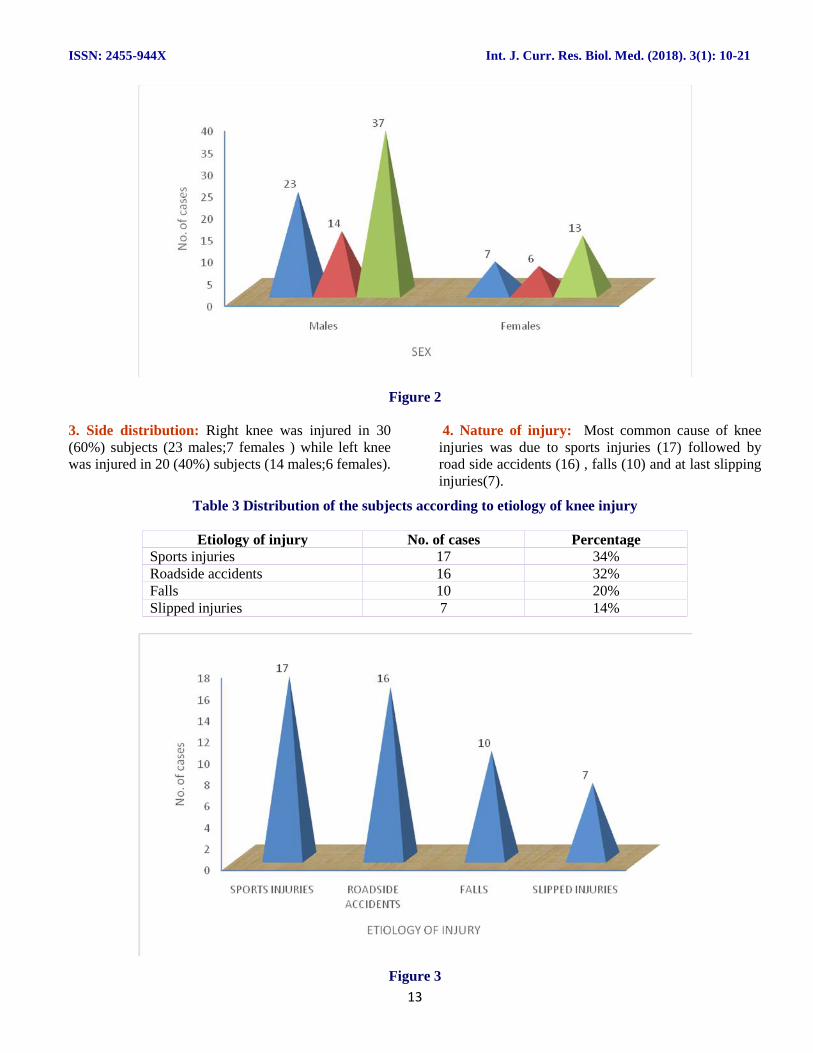
2. Sex distribution: There were 37 (74%) male and13 (26%) female subjects out of total 50 subjects.
Table 2 Distribution of the subjects according to the sex and the side injured
Right knee injuryn1 (%)
Left knee injuryn2 (%)
Right + Left kneeinjury
(n1 +n2) (%)Males 23 (46%) 14 (28%) 37 (74%)Females 7 (14%) 6 (12%) 13 (26%)Males +Females 30 (60%) 20 (40%) 50 (100%)
ISSN: 2455-944X Int. J. Curr. Res. Biol. Med. (2018). 3(1): 10-21
13
Figure 2
3. Side distribution: Right knee was injured in 30(60%) subjects (23 males;7 females ) while left kneewas injured in 20 (40%) subjects (14 males;6 females).
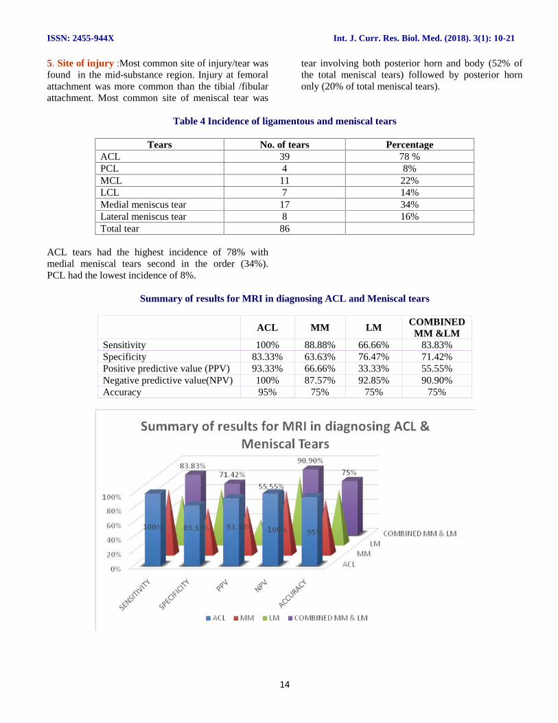
4. Nature of injury: Most common cause of kneeinjuries was due to sports injuries (17) followed byroad side accidents (16) , falls (10) and at last slippinginjuries(7).
Table 3 Distribution of the subjects according to etiology of knee injury
Etiology of injury No. of cases PercentageSports injuries 17 34%Roadside accidents 16 32%Falls 10 20%Slipped injuries 7 14%
Figure 3
ISSN: 2455-944X Int. J. Curr. Res. Biol. Med. (2018). 3(1): 10-21
14
5. Site of injury :Most common site of injury/tear wasfound in the mid-substance region. Injury at femoralattachment was more common than the tibial /fibularattachment. Most common site of meniscal tear was
tear involving both posterior horn and body (52% ofthe total meniscal tears) followed by posterior hornonly (20% of total meniscal tears).
Table 4 Incidence of ligamentous and meniscal tears
Tears No. of tears PercentageACL 39 78 %PCL 4 8%MCL 11 22%LCL 7 14%Medial meniscus tear 17 34%Lateral meniscus tear 8 16%Total tear 86
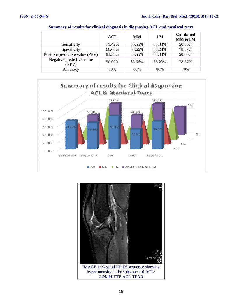
ACL tears had the highest incidence of 78% withmedial meniscal tears second in the order (34%).PCL had the lowest incidence of 8%.
Summary of results for MRI in diagnosing ACL and Meniscal tears
ACL MM LM COMBINEDMM &LM
Sensitivity 100% 88.88% 66.66% 83.83%Specificity 83.33% 63.63% 76.47% 71.42%Positive predictive value (PPV) 93.33% 66.66% 33.33% 55.55%Negative predictive value(NPV) 100% 87.57% 92.85% 90.90%Accuracy 95% 75% 75% 75%
ISSN: 2455-944X Int. J. Curr. Res. Biol. Med. (2018). 3(1): 10-21
15
Summary of results for clinical diagnosis in diagnosing ACL and meniscal tears
ACL MM LM CombinedMM &LM
Sensitivity 71.42% 55.55% 33.33% 50.00%Specificity 66.66% 63.66% 88.23% 78.57%
Positive predictive value (PPV) 83.33% 55.55% 33.33% 50.00%Negative predictive value
(NPV)50.00% 63.66% 88.23% 78.57%
Accuracy 70% 60% 80% 70%
IMAGE 1: Sagittal PD FS sequence showinghyperintensity in the substance of ACL:
COMPLETE ACL TEAR
A…M …
L…C…
0.00%
20.00%
40.00%
60.00%
80.00%
100.00%
S EN S IT IV ITY S PECIF ICITY PPV N PV ACCU R ACY
71.42%66.66%
83.33%
50.00%
70.00%
50.00%
78.57%
50.00%
78.57%70%
Sum m ary of results for C lin ical d iagnosingA CL & M eniscal Tears
ACL M M LM CO M B IN ED M M & LM
ISSN: 2455-944X Int. J. Curr. Res. Biol. Med. (2018). 3(1): 10-21
16
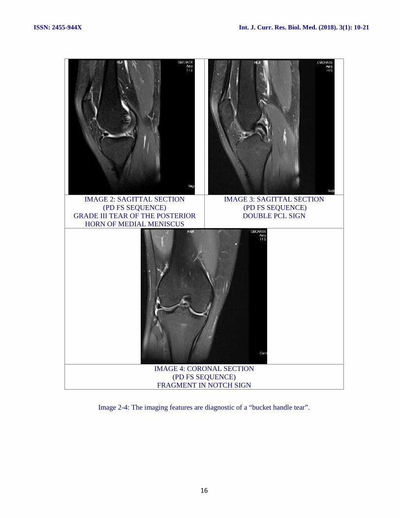
IMAGE 2: SAGITTAL SECTION(PD FS SEQUENCE)
GRADE III TEAR OF THE POSTERIORHORN OF MEDIAL MENISCUS
IMAGE 3: SAGITTAL SECTION(PD FS SEQUENCE)DOUBLE PCL SIGN
IMAGE 4: CORONAL SECTION(PD FS SEQUENCE)
FRAGMENT IN NOTCH SIGN
Image 2-4: The imaging features are diagnostic of a “bucket handle tear”.
ISSN: 2455-944X Int. J. Curr. Res. Biol. Med. (2018). 3(1): 10-21
17
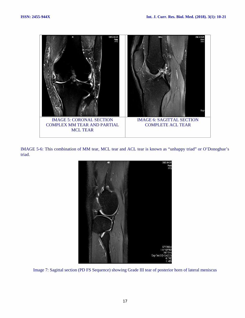
IMAGE 5: CORONAL SECTIONCOMPLEX MM TEAR AND PARTIAL
MCL TEAR
IMAGE 6: SAGITTAL SECTIONCOMPLETE ACL TEAR
IMAGE 5-6: This combination of MM tear, MCL tear and ACL tear is known as “unhappy triad” or O’Donoghue’striad.
Image 7: Sagittal section (PD FS Sequence) showing Grade III tear of posterior horn of lateral meniscus
ISSN: 2455-944X Int. J. Curr. Res. Biol. Med. (2018). 3(1): 10-21
18
Discussion
The maximum number of patients were young adults(78%) with a mean age of 29.36 years. Similar resultswere shown by Frobell et al and Chavadaki et al.11,6
The knee joint injuries was more common in males(74%) compared to females (26%). The incidence ofmeniscal and ligamentous injuries was higher in malesas compared to the females .Overall right knee (60%)was more commonly injured than the left knee (40%).Similar results were shown by Bari at al 12, Shahani etal13, Makhmalbaf H et al.14 , Frobell et al.11 andClayton et al.15
Knee injuries represent roughly 6% of all acuteinjuries treated in the emergency department of which27% to 48% were related to sports (Panigrahi et al andFrobell et al).2,11 The incidence of sports related kneeinjuries in previous studies matches with the incidenceshown in our study.
The most common sport causing knee injury wasfootball (47%).This was confirmed by a recent studydone by Kerr et.16, Kujala UM et al , Frobell et al andGilquist J et al.27,11,29
The incidence of ACL, PCL, medial and lateralmeniscus tears turned out to be 78%, 8%, 34% and16% respectively. In our study which corresponds tostudies done done by Bari et al12, Winters et al20 andAvcu S et al21.
The most common site of a ligament tear in our studywas the mid-substance (57.37%) and injury at femoralattachment (27.86%) was more common thantibial/fibular attachment (14.75%) corresponding withthe findings of Wing Hung et al and Singh et al.22,23
Grade III signal abnormality was seen in 17 medialmenisci (34%) and 8 lateral menisci (16%).There wasa preponderance of MM over LM in our study whichcorresponded with the study done by Bari et al.12
The ACL tears had incidence 78% followed bymeniscal tears at 50% (34% medial menisci and 16%lateral menisci).
In this prospective study of 50 subjects, we comparedthe findings of MR and clinical examination with thearthroscopic findings. We calculated sensitivity,specificity, PPV, NPV, and accuracy of clinical andMR examination in diagnosing ligament and meniscustears presuming arthroscopy to be gold standard.
In case of ACL tears, diagnostic accuracy for clinicalexamination (70%) was lower than MRI (95%).Sensitivity for diagnosing ACL tear by MR was 100%while it was 71.42% for clinical examination. So MRwas more sensitive in diagnosing ACL tears.
Sensitivity, NPV and accuracy of MR examination fordiagnosis of ACL tears turned out to 100%, 100% and95% respectively in our study. Similar results wereshown by Gujjar et al revealing sensitivity of 100%,NPV of 100% and accuracy of 90% respectively.25
The negative predictive value (NPV) of MR formeniscus examination was high (90.90%) while thepositive predictive value was low (55.55%) in thepresent study. Our results were similar to the results ofstudy done by Barronian et al in which the negativepredictive value and positive predictive values were91% and 65% respectively for menisci which againindicated that MRI is a good investigation formenisci.26 With a high negative predictive value, theMRI could be used as a negative diagnostic tool forthe meniscal injury that helps in preventingunnecessary surgery.
These results regarding the diagnostic accuracy ofmenisci had also been shown in the previous study byZairul-Nizam ZF et al who found accuracy of theclinical diagnosis of meniscus tears to be 46 to 65%compared with 80% to 84% for MRI. In our study,these values were 70% and 75% respectively.27 Thereason for this is unreliability of clinical examinationwhich had already been stated by some studies. Also,the clinical tests for detecting menisci are particularlyfallible. A study by Boeree NR et al had confirmed theunreliability of clinical diagnosis of meniscal andcruciate ligament.28
The results of different studies for evaluation of theinternal derangement of the knee with 1.5T MRI asfollows: 74- 96% sensitivity, 63-89% specificity and68-81% accuracy for diagnosis of the medial meniscaltear and 62-93% sensitivity, 88-95% specificity and77-86% accuracy for diagnosis of the lateral meniscaltear (Laoruengthana A et al).24
In the present study, the tear of medial meniscusyielded 88.88% sensitivity, 63.63% specificity and75% accuracy while the lateral meniscus yielded66.66% sensitivity, 76.47% specificity and 75%accuracy. So all the values of our study were withinthe range of previous studies done. In our study, wehad correlated the MRI finding with arthroscopy.
ISSN: 2455-944X Int. J. Curr. Res. Biol. Med. (2018). 3(1): 10-21
19
Studies Sensitivity SpecificityACL % MM% LM% ACL % MM% LM%
Ali Akhbar Esmaili et al29 78.30 75.00 66.60 95.70 94.70 86.20F. Rayan et al30 81.00 76.00 61.00 96.00 52.00 92.00Noha et al31 77.80 47.00 100 100 95.00 75.00Bari et al12 87.80 93.50 77.70 81.50 87.50 81.80Gupta et al32 100 90.00 100 50.00 70.00 95.00Our study 100 88.88 66.66 83.33 63.63 76.47
In terms of sensitivity and specificity our studyshowed results which were in agreement with theresults of the studies of recent publications asmentioned in the table above.
So in our prospective study we found that sensitivityand negative predictive value of MR examination fordiagnosing ACL and meniscal tears were quite high.Our study was in agreement with the findings ofPanigrahi R et al, Gujjar et al and Laoruengthana A etal.2,25,24
So with high sensitivity and negative predictive valueone can rely on MRI to avoid diagnostic arthroscopy.
Conclusion
MRI is a useful non-invasive, non-ionizing modalitywith a high diagnostic accuracy, high sensitivity andnegative predictive value which makes it a veryreliable screening tool for diagnosing internalderangements of the knee. MRI saves many kneesfrom unnecessary arthroscopies. MRI can also giveinformation about the structures like peripheral,inferior and intrasubstance tears and bony contusionwhich are not accessible on arthroscopy.
Source of funding: Nil
Conflict of interest: None declared
References
1. Khan HA, Ahad H, Sharma P, Bajaj P, Hassan N,Kamal Y. Correlation between magneticresonance imaging and arthroscopic findings inthe knee joint. Trauma Monthly2015;20(1):e18635.
2. Panigrahi R, Priyadarshi A, Palo N, Marandi H,Agrawalla DK, Biswal MR et al. Correlation ofClinical Examination, MRI and ArthroscopyFindings in Menisco-Cruciate Injuries of theKnee: A Prospective Diagnostic Study. ArchTrauma Res. 2017;6(1):e30364.
3. Swenson DM, Collins CL, Best TM, Flanigan DC,Fields SK, Comstock RD. Epidemiology of kneeinjuries among US high school athletes, 2005/06–2010/11. Medicine and Science in Sports andExercise 2013;45(3):462.
4. Smith BW, Green GA. Acute knee injuries: Part I.History and physical examination. AmericanFamily Physician 1995;51(3):615-21.
5. Mendelsohn CL, Paiement GD. physicalexamination of the knee Primary Care: Clinics inOffice Practice 1996;23(2):321-8.
6. Chavadaki RH, Paramban RU, Gurram S.Magnetic resonance imaging of the internallyderanged knee joint-a prospective study. Journalof Evolution of Medical and Dental Sciences2013;2(42):8186-207.
7. Mustonen AO, Koskinen SK, Kiuru MJ. Acuteknee trauma: analysis of multidetector computedtomography findings and comparison withconventional radiography. Acta Radiologica2005;46(8):866-74.
8. Polly Jr DW, Callaghan JJ, Sikes RA, McCabeJM, McMahon KE, Savory CG. The accuracy ofselective magnetic resonance imaging comparedwith the findings of arthroscopy of the knee. JBJS.1988;70(2):192-8.
9. Crotty JM, Monu JU, Pope Jr TL. MagneticResonance Imaging of the MusculoskeletalSystem: Part 4. The Knee. Clinical Orthopaedicsand Related Research 1996;330:288-303.
ISSN: 2455-944X Int. J. Curr. Res. Biol. Med. (2018). 3(1): 10-21
20
10. Berg HF, Vermeulen M, Algra PR, Boonman-deWinter LJ. Direct access to magnetic resonanceimaging improved orthopaedic knee referrals inthe Netherlands. Family Practice 2016;33(5):482-7.
11. Frobell RB, Lohmander LS, Roos HP. Acuterotational trauma to the knee: poor agreementbetween clinical assessment and magneticresonance imaging findings. Scandinavian Journalof Medicine & Science in Sports 2007;17(2):109-14
12. Bari AA, Kashikar SV, Lakhkar BN, Ahsan MS.Evaluation of MRI versus arthroscopy in anteriorcruciate ligament and meniscal injuries. Journal ofclinical and diagnostic research: JCDR.2014;8(12):RC14.
13. Shahani MA, Sah RK, Khan RA, Awais SM.Arthroscopic determination of accuracy of clinicalexamination in injuries with internal derangementof knee. Annals of King Edward MedicalUniversity 2015;21(3):168.
14. Makhmalbaf H, Moradi A, Ebrahimzadeh MH,Mazloumi SM, Ebrahimi H, Seyf P. Comparingknee clinical examination and MRI findings witharthroscopy results in meniscus ruptures among100 patients admitted at Qaem hospital from 2010to 2012. J Am Sci. 2013;9(10):344-9.
15. Clayton RA, Court-Brown CM. The epidemiologyof musculoskeletal tendinous and ligamentousinjuries. Injury 2008;39(12):1338-44.
16. Kerr ZY, Marshall SW, Dompier TP, Corlette J,Klossner DA, Gilchrist J. College sports-relatedinjuries—United States, 2009-10 through 2013-14academic years. Morb Mortal Wkly Rep.2015;64(48):1330-6.
17. Kujala UM, Taimela S, Antti-Poika I, Orava S,Tuominen R, Myllynen P. Acute injuries insoccer, ice hockey, volleyball, basketball, judo,and karate: analysis of national registry data. BMJ.1995;311(7018):1465-8.
18. Naraghi AM, White LM. Imaging of AthleticInjuries of Knee Ligaments and Menisci: SportsImaging Series. Radiology 2016;281(1):23-40.
19. Gillquist J, Hagberg G, Oretorp N. Arthroscopy inacute injuries of the knee joint. Acta OrthopaedicaScandinavica 1977;48(2):190-6.
20. Winters K, Tregonning R. Reliability of magneticresonance imaging of the traumatic knee asdetermined by arthroscopy. The New ZealandMedical Journal (Online). 2005 Feb 11;118(1209).
21. Avcu S, Altun E, Akpinar I, Bulut MD, Eresov K,Biren T. Knee joint examinations by magneticresonance imaging: The correlation of pathology,age, and sex. North American Journal of MedicalSciences 2010;2(4):202.
22. Ng WH, Griffith JF, Hung EH, Paunipagar B, LawBK, Yung PS. Imaging of the anterior cruciateligament. World Journal of Orthopedics2011;2(8):75
23. Singh JP, Garg L, Shrimali R, Setia V, Gupta V.MR Imaging of knee with arthroscopic correlationin twisting injuries. Indian Journal of Radiologyand Imaging 2004;14(1):33.
24. Laoruengthana A, Jarusriwanna A. Sensitivity andspecificity of magnetic resonance imaging forknee injury and clinical application for theNaresuan University Hospital. J Med Assoc Thai.2012;95(Suppl 10):S151-7.
25. Gujjar R, Bansal RP, Gotecha LK, Kollu R. MRIversus clinical examination for the diagnosis ofmeniscal and ligamentous injuries ofknee.International Archives of IntegratedMedicine 2015 May;2(5):43-7.
26. Barronian AD, Zoltan JD, Bucon KA. Magneticresonance imaging of the knee: correlation witharthroscopy. Arthroscopy: The Journal ofArthroscopic & Related Surgery 1989;5(3):187-91.
27. Zairul-Nizam ZF, Hyzan MY, Gobinder S, RazakMA. The role of preoperative magnetic resonanceimaging in internal derangement of the knee. TheMedical Journal of Malaysia 2000;55(4):433-8.
28. Boeree NR, Watkinson AF, Ackroyd CE, JohnsonC. Magnetic resonance imaging of meniscal andcruciate injuries of the knee. Bone & Joint Journal1991;73(3):452-7.
29. Jah AE, Keyhani S, Zarei R, Moghaddam AK.Accuracy of MRI in comparison with clinical andarthroscopic findings in ligamentous and meniscalinjuries of the knee. Acta Orthop Belg.2005;71(2):189-96.
30. Rayan F, Bhonsle S, Shukla DD. Clinical, MRI,and arthroscopic correlation in meniscal andanterior cruciate ligament injuries. InternationalOrthopaedics 2009;33(1):129-32.
31. Behairy NH, Dorgham MA, Khaled SA. Accuracyof routine magnetic resonance imaging inmeniscal and ligamentous injuries of the knee:comparison with arthroscopy. InternationalOrthopaedics 2009;33(4): 961-7.
ISSN: 2455-944X Int. J. Curr. Res. Biol. Med. (2018). 3(1): 10-21
21
32. Gupta K, Guleria M, Sandhu P, Galhotra R.Correlation of clinical, MRI and arthroscopicfindings in diagnosing meniscus and ligamentinjuries at knee joint: A prospective study. Journalof Orthopaedics and Allied Sciences 2013;1(1):2.
Access this Article in Online
Website:www.darshanpublishers.com
Subject:Medical Sciences
Quick ResponseCode
How to cite this article:Harshdeep Kashyap, Ramesh Chander, Sohan Singh, Neelam Gauba, N.S. Neki. (2018). Evaluation of kneejoint injury by MRI. Int. J. Curr. Res. Biol. Med. 3(1): 10-21.DOI: http://dx.doi.org/10.22192/ijcrbm.2018.03.01.002