Embed Size (px)
Citation preview

MQP-BIO-DSA-0861
EVALUATION OF INTRAOSSEOUS INFUSION OF
LIPOSOMAL OXYGEN IN RABBIT MODELS
OF HYPOXEMIA
A Major Qualifying Project Report
Submitted to the Faculty of the
WORCESTER POLYTECHNIC INSTITUTE
in partial fulfillment of the requirements for the
Degree of Bachelor of Science
in
Biology and Biotechnology
by
_________________________
James Reese
April 28, 2011
APPROVED:
_________________________ _________________________
John Kheir, M.D. David Adams, Ph.D.
Dept of Cardiology Biology and Biotechnology
Children’s Hospital Boston WPI Project Advisor
Major Advisor

2
ABSTRACT
The intravenous administration of a lipid-based oxygen suspension has been
shown to raise blood oxygen saturations to survivable levels in models of asphyxia.
However, emergency situations often render intravenous access impossible, so the
intraosseous (IO) route is sometimes used to obtain vascular access. This study examined
the pharmacokinetics of infusion of a lipid-based oxygen suspension administered
through an IO line. IO infusion raised the blood oxygen content of hypoxemic animals
in a manner similar to IV infusion.

3
TABLE OF CONTENTS
Signature Page ………………………………….……………………………………. 1
Abstract ………………………………………….…………………………………… 2
Table of Contents ……………………………………….……………………….…… 3
Acknowledgements ………………………………………………………………….. 4
Background ………………………………………………………………………….. 5
Project Purpose ………………………………………………………………………. 19
Methods ……………………………………………………………………………… 20
Results ……………………………………………………………………………….. 24
Discussion …………………………………………………………………………… 36
Bibliography ………………………………………………………………………… 40

4
ACKNOWLEDGEMENTS
I would like to thank Dr. John Kheir for the opportunities and guidance he has
given me over the past year. I would also like to thank Katie Mullen, Laurie Scharp and
Craig Smallwood for all the help before, during, and after the surgeries. Lastly, I would
like to thank Professor Dave Adams for advising this project and allowing me the
freedom to work in a field I have a true interest in pursuing.

5
BACKGROUND
Hypoxemia
Hypoxemia is clinically defined as a condition in which a patient’s blood oxygen
content has fallen below a healthy level. This condition is often diagnosed using arterial
oxyhemoglobin saturations (SpO2), arterial oxygen partial pressure (PO2) or a
combination of both. In healthy individuals breathing atmospheric oxygen at sea level,
normal levels are around 110 mmHg PO2 and 99% SpO2 (Gootman et al., 1963).
Hypoxemia is recognized as a dip below 90 mmHg PO2 or 90% SpO2 for an extended
period of time (Bowton et al., 1994).
Hypoxemia can be caused by a number of conditions. Acute respiratory distress
syndrome, chronic obstructive pulmonary disorder, pulmonary edema, and atelectasis
(alveolar collapse) are all known to cause hypoxemia in patients (Bone, 1980; Miller et
al., 1984; Suchyta et al., 1991; Piantadosi and Schwartz, 2004). Alveolar-capillary block
syndrome, a condition that thickens the alveolar walls and compromises the diffusion of
oxygen, also causes arterial hypoxemia in patients (Finley et al., 1962). Pulmonary
embolism causes hypoperfusion or total ischemia of the lungs, preventing adequate blood
oxygenation (Wilson et al., 1971). Anatomical anomalies can also lead to low blood
oxygenation. Many congenital heart defects cause right-to-left shunting, the mixing of
oxygenated and deoxygenated blood (Sukumalchantra et al., 1970). A patent foramen
ovale, a hole in the atrial septum, allows pre- and post-pulmonary flow to mix.
Transposition of the Great Arteries, an anatomic translocation of the aorta and pulmonary

6
artery, causes profound hypoxemia in patients (Petit et al., 2009). There has even been a
documented case of partial occlusion of the right pulmonary artery by a mediastinal
tumor that led to severe hypoxemia (Takeda et al., 1999).
Dangers of Hypoxemia
The biological need for a constant supply of oxygen is rooted in the production of
ATP, the basic source of energy for cells. ATP is made through three steps – glycolysis,
the Krebs cycle and oxidative phosphorylation – together referred to as cellular
respiration. Oxidative phosphorylation operates through the electron transport chain
where oxygen acts as the final electron acceptor. The final reduction of oxygen
completes the chain, and allows for the production of ATP via ATP synthase. In the
absence of oxygen, however, the electron transport chain cannot function. Oxidative
phosphorylation and the Krebs cycle come to a complete stop. In the absence of the
electron transport chain, glycolysis is the only source of ATP. This process, called
anaerobic respiration, is far less productive than full cellular respiration, producing only 2
ATP molecules per molecule of glucose, as opposed to 38 ATP in the case of aerobic
respiration. In addition, the pyruvic acid that normally enters the Krebs cycle is instead
reduced to lactic acid (Hole et al., 2009).
In rat models, a 50% reduction in ATP levels in the brain occurs shortly after the
arterial PO2 drops below 30 mmHg. This drop in energy corresponds with an almost
immediate impairment of brain function. Neurons utilize anaerobic respiration as a
source of energy, causing a rapid accumulation of lactic acid. The continuous flow of
blood carrying glucose with low oxygen provides additional fuel for anaerobic respiration

7
and lactic acidosis. This acidotic condition triggers and accelerates further injury and
insults to neuronal tissue. This condition of low oxygen delivery is known as ischemia,
and is a final common pathway ending in cell death (Hass, 1983).
A higher occurrence of periventricular leukomalacia (PVL), a type of white-
matter necrosis in brain tissue, has been linked to a lower average PO2. Newborns with
D-transposition of the great arteries (d-TGA), a congenital heart defect that transposes the
aorta and the pulmonary artery, are characteristically hypoxemic due to the recirculation
of desaturated systemic venous blood into the systemic arteries. Petit et al. (2009) in a
study of preoperative neonates with TGA, discovered a negative association between
blood oxygen tension and the occurrence of PVL. Among preoperative patients, those
showing signs of PVL had a significantly lower mean PO2 than those without PVL.
Additionally, the longer the patient spent in the hypoxemic state (time to palliative
measures taken), the higher the probability of developing PVL (Petit et al., 2009). PVL
results in significant decrement in quality of life, including motor dysfunction, in these
children.
Gootman et al. (1963) conducted a study of pediatric and neonatal patients with
cyanotic congenital heart defects. Researchers found a link between low blood oxygen
saturation (and partial pressure) and a build-up of lactic acid. This excess lactic acid
acted to bring the patients overall blood pH to acidic levels, under 7.2 (Gootman et al.,
1963). Lactic acidosis often results from poor tissue oxygenation due to diminished
oxygen delivery, a condition frequently present in hypoxemic patients (Mizock and
Faulk, 1992).
8
Hypoxemia and resulting acidosis has also been linked to the development of
cardiac arrhythmias. Ayres and Grace (1969) reported on nine cases involving patients
admitted with symptoms indicative of acute myocardial infarction or respiratory distress.
During hospitalization, the patients displayed varying degrees of hypoxemia, acidosis,
and alkalosis. Analysis of the data revealed that traditional anti-arrhythmic treatments
were ineffective on the patients. Rather, the arrhythmias only returned to normal sinus
rhythm when the hypoxemia, acidosis or alkalosis was corrected. Arrhythmic events
seen in patients often occurred when mechanical administration of oxygen was disrupted,
and were corrected shortly after arterial PO2 was raised (Ayres and Grace, 1969).
Aside from these specific anomalies, hypoxemia has been linked to survival. An
in-hospital study conducted in 1994 correlate hypoxemic events with patient outcome. A
group of 100 patients admitted to the hospital with varying diagnoses were followed with
continuous pulse oximetry monitoring. The study stratified patients into three different
groups of hypoxemia – SpO2 <90%, <85%, and <80% – all lasting for periods of five
minutes or more. APACHE (Acute Physiology and Chronic Health Evaluation) scores
were calculated on all patients as a means of incorporating the severity of the diagnosis
into the results. After hospital discharge, patients were contacted within four to seven
months for a follow-up to determine long-term outcome (Bowton et al., 1994). Of the
100 patients studied, 26 were found to have hypoxemic episodes <90% SpO2, 16 patients
below 85% SpO2, and 9 patients below 80% SpO2. 74 patients experienced no prolonged
periods of hypoxemia. The study revealed a striking increase in mortality among the 26
patients experiencing hypoxemia. Of the patients contacted, 32% of those with <90%
SpO2 died within the follow up period. This was significantly higher than the 7%

9
mortality rate among non-hypoxemic patients (p=0.0003). Even when adjusted for
severity of illness, the hypoxemic group still exhibited a significantly higher mortality
rate. Furthermore, the follow-up study revealed that mortality increased with the severity
of the hypoxemic events experienced. 31% of those with <85% SpO2, and 44% of those
with <80% SpO2, died in the months following hospital discharge. Additionally, those
patients who spent longer times at lower saturations had a lower survival rate. The study
ultimately revealed a correlation, though not definitively causal, between decreased
arterial oxygenation and lowered survival rates. Patient mortality could not be explained
by the severity of respective illnesses: those with severe injuries or illnesses were
affected to a similar degree as those with non-critical diagnoses (Bowton et al., 1994).
Treatments of Hypoxemia
The treatment of hypoxemia is limited. Many hypoxemic patients are treated with
supplied oxygen, either through a nasal tube or a face mask. In more critical events,
patients may be intubated and placed on mechanical ventilation (Ayres and Grace, 1969).
A specific aspect of mechanical ventilation, positive end-expiratory pressure (PEEP), is
used to optimize oxygenation. PEEP provides a positive pressure throughout the
expiratory phase, preventing alveolar collapse (Bone, 1980). This requires an intact
airway and lung unit to oxygenate the blood. A treatment of refractory hypoxemia
known as extra-corporeal membrane oxygenation (ECMO) involves the ex vivo
oxygenation of the blood. A patient’s great vessels are connected to a perfusion pump
which circulates the blood through a series of membranes exposed to oxygen gas which
act as artificial alveoli. This method has proven to be highly effective in patients with

10
ARDS. However, the risks of this technique are great, including intracranial bleeding,
renal injury, hemolysis and worsening of the inflammatory cascade make this a
suboptimal method of emergency oxygenation. More importantly, the time, expertise and
resources for the institution of this invasive therapy make it available only in a limited
number of tertiary care centers. Even in centers with rapid response ECMO teams,
ECMO may take more than an hour to institute during which irreversible organ injury can
take place (Suchyta et al., 1991). The limitations of the modern treatments have led some
physicians to seek an alternative way to increase the blood oxygen content of hypoxemic
patients in an emergency setting.
Lipid-based Oxygen Suspension
Kheir et al. (2010) of Children’s Hospital Boston and Harvard Medical School
has recently developed a lipid-based suspension that acts as a vehicle for oxygen gas.
Rather than endotracheal or extra-corporeal administration of oxygen, the suspension was
initially designed to deliver oxygen gas directly into the bloodstream. In 2010, the
suspension was studied in vivo with rabbit models of asphyxia. Rabbits were
anesthetized and paralyzed, allowing researchers full control over the breathing pattern of
the animal. The animals were stabilized with mechanical ventilation on 21% inspired
oxygen. After stabilization, the endotracheal tube was completely occluded, resulting in
no mechanical or spontaneous respiration. Experimental animals received an intravenous
infusion of the lipid-based oxygen suspension at a rate matching normal oxygen
consumption, and control animals received an intravenous infusion of oxygenated
intravenous fluid. Asphyxia was maintained for 15 minutes. Arrested animals were
11
resuscitated using advanced critical life support protocol (Kheir et al., 2010). The mean
time to arrest in control models was 6.7 minutes. All control animals exhibited a rapid
decline in arterial PO2 and blood oxygen saturation. Control animals also became
critically hypotensive prior to arrest, adding to the oxygen delivery deficit. In contrast,
8/10 animals receiving the lipid-based oxygen suspension survived to the 15-minute
endpoint with no arrest. These animals maintained arterial PO2 and blood oxygen
saturation at survivable levels. Experimental animals also initially displayed a slight
increase in mean arterial blood pressure followed by a return to baseline tension (Kheir et
al., 2010).
The results of this experiment reveal that an intravenous lipid-based oxygen
suspension can act as a temporary supplement to inspired air. A therapy capable of
supplying a patient with oxygen outside of traditional mechanical ventilation and ECMO
carries profound implications in the world of critical care and emergency medicine.
Given the dangers of hypoxemia and the conditions it is associated with, administration
of intravenous oxygen could provide the rapid increase in arterial PO2 and blood oxygen
saturation needed for survival in critical situations.
Venous Drug Delivery
The administration of drugs in emergency situations has been done through
numerous routes. Percutaneous peripheral intravenous access (PPIV), central venous
catheterization (CVC), and surgical catheterizations into major veins all rely on direct
access to the circulatory system (Brunette and Fisher, 1988). Because of this direct
access, PPIV has been used to quickly administer therapies to patients in critical and
12
emergency situations. However, PPIV relies on the ability of personnel to place the
catheter into the vein percutaneously. But in many cases, conditions make accurate and
rapid insertion of the catheter difficult. Burns, edema, and massive limb trauma make
insertion of PPIV more difficult (Rosetti et al., 1985; Blumberg et al., 2008).
Additionally, peripheral veins can be difficult to locate in obese patients and in children
(Rosetti et al. 1985; Paxton et al., 2009). Dehydration, poor hemodynamics, and previous
IV drug abuse can make insertion of the IV difficult and time consuming (Paxton et al.,
2009). Peripheral veins will often collapse in cases of shock and cardiac arrest, rendering
them useless for PPIV (Tocantins and O’Neill, 1951; Rosetti et al. 1984; Rosetti et al.
1985; Orlowski et al. 1989; Blumberg et al. 2008). Furthermore, placement of a CVC is
recognized as a high-risk procedure in many patients (Rosetti et al., 1985). Surgical
catheterizations are often used as a last-resort technique, involve additional trauma to the
patient, and are time-consuming, making them less suited for emergency situations
(Paxton et al., 2009).
Endotracheal Drug Delivery
Endotracheal drug administration is also used in some applications. This method
of drug delivery can be convenient, especially if the patient is already intubated. It is,
however, limited in its uses. Only a handful of drugs can be administered via the airway,
thus restricting endotracheal therapies in emergency settings (Brunette and Fischer,
1988).
13
History of Intraosseous Infusion
Intraosseous infusion (IO) uses the medullary canal of long bones to introduce
drugs into the circulatory system. IO infusion involves the placement of a rigid needle
through the periosteum and into the red marrow of a patient. Drinker et al. (1922) first
documented the use of IO infusion in mammals. The study showed that perfusion
through a catheter inserted into the tibial red marrow of a dog was a feasible way to
deliver fluids to the circulatory system. Drinker reported that the canal functions as a
rigid “noncollapsable vein” with a direct connection the rest of the circulation (Drinker et
al., 1922). In 1940, L.M. Tocantins and O’Neill published the first study of IO infusion
conducted on human patients. 17 infusions were conducted on 14 patients, with a success
rate of 94% (Tocantins and O’Neill, 1951). Further studies pushed IO infusion into
general recognition, and it was adopted by battlefield medics during World War II as a
quick and efficient way of establishing vascular access. Yet with the coming of more
sophisticated PPIV apparati and techniques, IO infusion fell by the wayside in the late
1940s (Orlowski et al., 1989; Blumberg et al., 2008).
IO Infusion Physiology
The physical structure of the long bones allows intraosseous infusion of drugs and
fluids. Bones, although composed of a dense and compact cellular matrix, are highly
vascular. The basic unit of bone structure, the osteon, is built around an inner canal that
contains arterial and venous blood flow. The Haversian canals and their vessels are
interconnected by vessels running through Volkmann canals. The heads of long bones
contain red marrow, which serves as generative tissue for red and white blood cells (Hole

14
et al., 2009). This area has a high perfusion rate – necessary to introduce new cells into
circulation – and is the target for insertion of an IO line (Leidel et al., 2010). Several
bones have been utilized in both experimental and clinical settings. The sternal
manubrium, tibia, femur, humerus, radius and clavicle have all been successfully infused.
Preferred sites for penetration in pediatric patients and adult patients are the anteromedial
face of the tibia and the sternum, respectively (Rosetti et al., 1985; Blumberg et al.,
2008).
Contraindications of IO Infusion
As with all methods of drug administration, there are contraindications of IO
infusion. Placing an IO line into a fractured or damaged bone results in perfusion of the
drug through the fracture rather than into circulation. Bones that have previously been
used for IO infusion are avoided for the same reason. Percutaneous infection in the area
of insertion serves as a contraindication, as it may introduce infection to the periosteum
or marrow (Blumberg et al., 2008; Paxton et al., 2009). In addition to these factors,
patients also suffering from bone disorders such as osteoporosis should not receive IO
infusions (Blumberg et al., 2008). In these cases, trauma inflicted to the bone during the
placement of the needle could far outweigh the benefits of vascular access.
Complications of IO Infusion
Even under normal circumstances, IO infusion carries risks. Despite the trauma
inflicted on the periosteum and compact bone upon penetration, risks associated with
intraosseous infusion are few and for the most part benign. Main concerns lie in the

15
development of infection as a result of aseptic administration. Percutaneous infection or
improper sterilization of the skin could introduce infectious agents to the marrow.
Subcutaneous abscesses, osteomyelitis and skin necrosis can all result from IO infusion
(Rosetti et al., 1985; Christensen et al., 1991; Rosovsky et al., 1994 Paxton et al., 2009;
Leidel et al., 2010). Although these conditions are possible, they are unlikely to occur in
a majority of patients. In 1985, Rosetti et al. compiled data from 30 individual studies of
human IO infusions conducted between 1942 and 1977. The collection profiled 4,270
patients. Of these patients, it was found that only 27, less than 0.6% of the group,
developed osteomyelitis. It was also noted that in these cases, a high percentage of them
occurred in patients with extended periods of infusion or previously existing infections.
Subcutaneous abscesses formed in only 0.09% of the patients (Rosetti et al., 1985).
The development of fat or bone marrow emboli has been proposed as a possible
risk in IO infusion. Since these emboli would be in venous circulation, they have the
potential to disrupt blood flow in the pulmonary capillary beds (Rosetti et al., 1985;
Orlowski et al., 1989). However, a study of 30 dogs and two pediatric patients
examining the frequency and implications of emboli formation in IO infusion contradicts
these concerns. Researchers found that regardless of what drug was infused, fat and bone
marrow emboli were discovered in lung tissue. The study concluded that 89 to 100% of
all IO infusions would lead to fat or marrow emboli. Arterial blood gas analysis revealed
that despite the high prevalence of emboli, alveolar perfusion was not compromised and
the emboli were not clinically relevant (Orlowski et al., 1989).
Other possible complications include compartment syndrome resulting from
subcutaneous or subperiosteal infiltration, bone fracture due to the penetrating force of

16
the needle and epiphyseal plate disruption (Rosetti et al., 1985; Paxton et al., 2009). The
latter, however, can be avoided by correct use of anatomical landmarks (Rosetti et al.,
1985). As with all methods of vascular access, dislodgement of the catheter and
extravasation, improper insertion and bleeding are concerns (Paxton et al., 2009; Leidel
et al., 2010).
Overcoming the Downfalls of Traditional Vascular Access
Paxton et al. (2003) conducted a two-phase study comparing the efficiency of the
traditional methods of intravascular access, PPIV and CVC infusion, against IO infusion.
The study was conducted in the emergency department of a level I trauma center with an
annual volume of 92,000 emergency patients. Phase I of the study involved 62 patients
subjected to conventional PPIV access. In the event of PPIV failure, CVC was
attempted. One patient received immediate CVC due to poor peripheral vein conditions,
and four received CVC after failed PPIV. Phase II of the study subjected 29 patients to
proximal humeral IO infusion. One patient received a subsequent infusion two weeks
after the initial, resulting in an n = 30. Endpoints included time from beginning of
catheter placement to proper flow and failure attempts of catheterization (Paxton et al.,
2009). The data revealed a striking difference between the times required to administer
PPIV or CVC versus IO lines. In phase I, the mean time to peripheral intravenous access
was 3.6 minutes over a range of 1-16 minutes (SD = 3.7). On average, each successful
PPIV placement required 1.5 attempts. For the five patients that received CVC, mean
time to access was 15.6 minutes over a range of 11-25 minutes (SD=6.7). CVC required
an average of 2.4 attempts per successful placement. In contrast, times to proximal

17
humeral IO insertion were much faster. Over the 29 patients (30 infusions) of phase II,
the mean time to placement was 1.5 minutes over a much tighter range of 1-6 minutes
(SD=1.1). IO insertion was significantly faster that PPIV (p < 0.001) and CVC
(p=0.0056). In addition, IO infusion had a first attempt success rate of 80.6% versus
73.7% for PPIV and 20.0% for CVC (Paxton et al., 2009).
A similar study was conducted on strictly pediatric patients under the age of two
years. 33 infants in cardiac arrest were studied. The time between arrival in the
emergency department and intubation and administration of endotracheal drugs was
measured. Time to the application of each kind of intravascular access was also
recorded. Intravascular methods included PPIV, CVC, surgical catheterization and IO
infusion. PPIV was found to have a faster mean time to administration of 3.0 +/- 2.0
minutes versus 4.7 +/- 1.49 minutes for IO infusion. However, IO infusion had a success
rate of 83% and was the initial access route in 75% of patients, which were the highest of
all intravascular methods. Despite PPIV having a faster time to administration, IO
infusion proved to be a suitable alternative to patients with difficult access (Brunette and
Fischer, 1988).
Industry Standards
IO infusion’s ability to complement direct venous infusions and to nearly usurp
endotracheal drug administration has prompted the medical industry to re-adopt its use in
clinical situations. In the 1980s, the American Heart Association (AHA) adopted IO
infusion into its pediatric advanced life support guidelines (Blumberg et al., 2008).
Modern AHA pediatric guidelines recommend it for all pediatric patients. Furthermore,

18
the AHA also recommends that IO infusion be used in adults as the primary substitute to
tradition IV access in its advanced cardiac life support guidelines (Kleinman et al., 2010;
Neumar et al., 2010).

19
PROJECT PURPOSE
Although the intravenous administration of a lipid-based oxygen suspension has
previously been shown to raise blood oxygen saturations to survivable levels in models of
asphyxia, emergency situations often render intravenous access impossible, so an
intraosseous (IO) route is sometimes used to ensure vascular access. The purpose of this
study is to determine the effectiveness of an intraosseous infusion of the lipid-based
oxygen suspension in rabbit models of hypoxemia. We hypothesized that the suspension
infused through the intraosseous route would raise blood oxygen content in a manner
comparable to the intravenous route.

20
METHODS
Rabbits
Three female New Zealand white rabbits of approximately 4.5 kg were studied.
All animals were housing in the Animal Resources at Children’s Hospital (ARCH) for a
three-day acclimation period prior to each procedure.
Surgery
Prior to each procedure, the animal was induced and sedated using Glycopyrollate
(10 micrograms/kg) and Ketamine (10 mg/kg), both intramuscularly. 0.1 mg/kg
Midazolam was given via a percutaneous IV line placed in an ear vein. The animal was
then intubated and ventilated at 21% FiO2. Upon presence of EtCO2 (expired CO2), the
animal was sedated. A Fentanyl bolus, between 25-50 micrograms/kg titrated
specifically to each animal was administered via an ear vein. The animal also received a
continuous Fentanyl infusion of 50-100 micrograms/kg/hr throughout the entire
procedure. The animal was then paralyzed using Pancuronium 0.1 mg/kg IV. Paralysis
of the animal allowed for full control of respiration.
After the animal had been properly sedated and securely intubated, the abdomen,
neck, and groin area were fully shaved. A pulse oximetry probe was fixed to the ear, paw
or tongue. An esophageal probe was inserted to measure heart rate, body temperature
and EKG. Following this, placement of arterial and venous lines via surgical
catheterizations began. A 22-gauge catheter was placed in both the left and right femoral
arteries. The left femoral artery catheter was connected to an extension and a three-way

21
stopcock, both primed with normal saline. A saline flush was administered to ensure
proper flow. This line was used specifically for the drawing of arterial blood gas
samples. The right femoral artery catheter was connected to a three-way stopcock, held
permanently in the open position. Through this stopcock, and into the femoral artery, an
Oxford Optronix fiber optic PO2 probe was fed for continuous oxygen tension
measurement. The auxiliary port of the three-way stopcock was used to transduce
systolic and diastolic blood pressure. A 4 French Cordis sheath was inserted into a
femoral vein for intravenous administration of the lipid-based oxygen suspension.
Using a 15 gauge Life/form® intraosseous (IO) infusion needle, an intraosseous line was
placed in the animal’s humerus. Because of the large gauge of the needle and small size
of the bone, the needle was inserted into the proximal epiphysis of the humerus,
longitudinally, running parallel with the medullary canal. An extension set and stopcock
were attached to the IO needle to prevent movement of the needle. A 10 ml saline flush
was administered to ensure proper placement and flow.
Following line placements, the FiO2 was decreased from 21% to 11%. This
caused alveolar hypoxia and reduced the overall blood oxygen tension and saturation of
the animal. The animal was allowed to reach a stable blood oxygen tension and
saturation, and an arterial blood gas was drawn.
After the animal had stabilized at 11% FiO2, preparation for the infusion began.
A 140ml syringe of the lipid-based oxygen suspension was fixed to the 4-French Cordis
sheath in the right femoral vein. With the stopcock closed to the vein, the line was
primed with the lipid-based oxygen suspension. At this time, a baseline arterial blood gas
was drawn to determine starting-point measurements. A blood gas was drawn every 30

22
seconds throughout the infusion. The stopcock attached to the 4-French Cordis sheath
was opened to venous flow, and the suspension was hand infused. The infusion
continued for 1:45 minutes. Following a 1:15 minute observation period, the FiO2 was
increased to 21% for a 10-minute recovery period.
After the 10-minute recovery period, the FiO2 was again reduced to 11% as
described earlier. Another baseline arterial blood gas was drawn. One 140ml syringe of
the lipid-based oxygen suspension was attached to the stopcock leading to the IO line in
the humerus of the animal. The lipid-based oxygen suspension was hand-infused for a
period of 1:45 minutes. At the end of the infusion, the animal was given a recovery
period as described above. Infusion alternated between IV and IO, with a recovery
period following each one, so that two of each type of infusion was recorded.
Following the end of the final infusion, the animal was terminated using FatalPlus
(pentobarbital sodium).
Autopsy
After death, the animal underwent a sternectomy to reveal the heart, lungs and
great vessels. The heart and lungs were excised. The heart was dissected along the
transverse plane to expose the atria and ventricles. The heart and samples of both lungs
were placed into formalin for preservation. Tissue samples were also taken from the
liver. The humerus of the rabbit was then excised and longitudinally bisected to reveal
the bone marrow. This entire procedure was documented with photographs.
Endpoints included systolic, diastolic and mean arterial blood pressure,
temperature, heart rate and pulse oximetry, as recorded on a SurgiVet Monitor.

23
Continuous arterial blood oxygen tension was measured via the fiber optic probe in the
right femoral artery. Lung mechanics were also recorded. Arterial blood gas
measurements were used to corroborate blood oxygen tension and saturation. All data
was recorded on a one-second time resolution, and blood gasses were drawn every
minute throughout the infusions.

24
RESULTS
This project investigated the effectiveness of infusing a lipid-based oxygen
suspension intraosseously compared to intravenous administration.
Three rabbits underwent infusions during this study. Animal #1 was successfully
prepped for surgery and intravenous infusion began. No increase in arterial oxygen
saturation was seen, even as the infusion surpassed 1:30 minutes. Suspensions
manufactured in a different manner than usual were utilized for this experiment, and were
more viscous than usual. The infusion was continued beyond the 1:45 minute endpoint
with no increase in oxygen tension. The animal then became hypotensive and
increasingly hypoxemic. The infusion was stopped, and the rabbit underwent a failed
resuscitation. Autopsy revealed that the lipid-based oxygen suspension had backed up in
the inferior vena cava, forming a massive embolus and preventing blood flow. The death
of this animal was attributed to a different manufacturing of the suspension.
The second animal subject also experienced difficulties during the operation.
Researchers experienced difficulty placing the venous and femoral catheters resulting in a
significant loss of blood volume prior to the beginning or any infusions. Both the left and
right femoral veins were perforated, and catheterization of these vessels was deemed
impossible. An attempt to catheterize the right jugular resulted in perforation of the
jugular vein. It was decided that further attempts at venous catheterization would
jeopardize the immediate survival of the animal, and, as is often the case in hemorrhaging
patients, that intraosseous line insertion was the only remaining solution. An
intraosseous line was placed in the proximal humerus running parallell to the diaphysis.
25
A total of 40 ml of normal saline was flushed through this line, both to ensure proper
placement and to restore volume lost due to line placement.
A total of three intraosseous infusions were administered to animal #2, each
lasting 1:45 minutes. The animal survived the first two infusions with no hemodynamic
perturbations. A 0.1 mg bolus of epinephrine was given after each infusion to treat
hypotension in the animal, possibly due to the acidosis found on blood gases. During the
final infusion, the animal again showed signs of hypotension. End tidal CO2 levels
dropped to zero, indicting that the animal had become entirely anaerobic despite the
administration of oxygen. FiO2 was increased to 21% with little effect, and the animal
was terminated 10 minutes after the final infusion.
Upon necropsy of the animal, upwards of 20 ml of the suspension was found to
have infiltrated the fascia surrounding the triceps (Fig.1). Examination of the thoracic
cavity revealed no embolus to the lungs. A transverse dissection of the heart revealed
evidence of subendocardial ischemia in the left ventricle (Fig.2).
Data collected from animal #2 was recorded on a 1-second time resolution from
the SurgiVet monitor and the Oxford Optronix PO2 probe. Veltilation data from the
DATEX, however, was recorded on a 10-second time resolution. Accordingly, plots of
SurgiVet and PO2 probe data are far smoother than plots of DATEX data.
The third animal did not experience any noteworthy problems during preparation.
The first intravenous infusion was successful, and the animal was recovered on 21%
FiO2. The second infusion was adminstered intraosseously through a line placed in the
right proximal humerus. Towards the end of the infusion, suspension was seen emerging
from the puncture site of the intraosseous line. After another period of stabilization, the

26
rabbit was given another intravenous infusion. This infusion was also successful,
however the animal became severly hypotensive and end-tidal CO2 levels began to drop
shortly after. 0.4 mg epinephrine was adminstered, and CPR was performed for 5
minutes before the animal was declared dead.
Fig.1 Images of infiltration (top) and extravasation (bottom) of the lipid-based
oxygen suspension from the intraosseous lines placed in the proximal humerus
of animals #2 and #3, respectively.
Autopsy of the animal revealed bruising of the lungs due to the trauma of CPR.
No other cardiopulmonary abnormalites were seen. A transverse dissection of the heart
again revealed signs of subendocardial ischemia (Fig. 2). Both the left and right humerus
were excised from the animal and bisected longitudinally. No signs of marrow
disruption, gas emboli or lipid deposits were seen in either the infused or uninfused
humeri (Fig. 3).

27
Fig. 2 Transverse dissections of the hearts collected during autopsy of
animals #2 (top) and #3 (bottom). Both exhibit signs of subendocardial
ischemia inside the left ventricle. They appear as darker areas, similar
to bruising, on the inner walls of the ventricles.
Fig. 3 The left humerus (top, uninfused) and right humerus (bottom,
infused) bisected longitudinally. No gas emboli or lipid deposits can be
seen. The white areas seen in the marrow of the bottom humerus is
glare from surgical lamps.
The data collected from animal #3 was collected on a 1 second interval on the
SurgiVet monitor and a 5-second interval on the probe and DATEX. Arterial blood
pressure measurements were disrupted by an occlusion in the catheter that prevented
transduction. This occlusion was later found to be due to the PO2 probe being forced too
far into the catheter. Instead of transducing from the same stopcock as the probe, the

28
blood pressure measurements were taken from the stopcock used for arterial blood gas
sample collection. Because the flow to the transducers was disrupted for drawing
samples, reliable blood pressure data from animal #3 was not measured. On both animals
#2 and #3, pulse oximetry was never reliably obtained.
In total, animal #2 received 3 intraosseous infusions, and animal #3 received 2
intravenous and one intraosseous infusions. Thus, 4 successful intraosseous infusions
and 2 successful intravenous infusions were completed for the study.
The intravenous infusions served as comparators for this experiment as the gold-
standard for drug adsorption. Both intravenous infusions on animal #2 began when the
animal was desaturated to a mean PO2 of 31.76 mmHg. An increase in arterial tension
was seen beginning at 00:30 seconds into the infusion. The oxygen tension increased at a
steady rate throughout the entire infusing period, ending at a mean tension of 57.74
mmHg. The arterial tension continued to rise for approximately 00:45 seconds post
infusion. Over the infusion, the ventilator was set at an FiO2 of 12%, however measured
inspiratory oxygen fraction fluctuated between 9.92 – 11.73%. FeO2, the fraction of
oxygen exhaled by the animal, increased over the period of the infusion, even surpassing
the inspired oxygen fraction for a short period of time, a finding present only when
pulmonary arterial blood (normally the most desaturated blood in the body) is at a higher
partial pressure of oxygen than inspired air (Fig. 4).

29
Fig. 4 The arterial oxygen tension measured using the invasive fiberoptic probe placed
in the femoral artery is plotted over the inspired and expired oxygen percentage for the
intravenous infusions (n=2). PO2 is plotted on the left y-axis and Fe/FiO2 is plotted on
the right y-axis. Vertical dashed lines denote the start and end of the 1:45 minute
infusion.
The intraosseous infusions began at a mean PO2 of 37.23 mmHG. PO2 increased
at a gradual rate, and peaked in the middle of the infusion period. The infusion ended
with mean arterial oxygen tension of 44.49 mmHG. The arterial oxygen tension began to
decrease approximately 00:20 seconds before the end of the infusion, and continued to
decrease after the end of infusion. An increase in arterial oxygen tension occurred
shortly after infusion. Measured FiO2 levels fluctuated between 11.44 – 11.89%. The
FeO2 of the animal increased steadily throughout the infusion, and peaked just after the
endpoint. The FeO2 values did not surpass FiO2 values at any point (Fig. 5).

30
Fig. 5 Arterial oxygen tension measured using the femoral fiber optic probe plotted with
the inspired and expired oxygen percentage measured during the intraossoues infusions
(n=4). PO2 is plotted on the left y-axis and Fe/FiO2 is plotted on the right y-axis. Vertical
dashed lines denote the start and end of the 1:45 minute infusion.
When directly compared to one another, the mean PO2 curves of the intravenous
and intraosseous infusions both show an increase in arterial oxygen tension. Linear
regression analysis revealed that the rate of increase in arterial oxygen tension in the
intravenous group was almost three times greater than that of the intraosseous group
(0.3034±0.009502 vs. 0.1198±0.01612, p<0.0001) over the course of the infusion (Fig.
6).

31
Fig. 6 The arterial oxygen tensions of both the intravenous and intraosseous infusions
plotted on the same field along with corresponding regression lines. Dashed lines
represent 95% confidence values of the regression line.
As a supplement to the constant arterial oxygen tension measurements taken by
the fiberoptic probe, arterial oxyhemoglobin saturation was also measured. These
measurements were taken from arterial blood samples drawn at infusion start and every
30 seconds during the infusion and the immediate post infusion period.
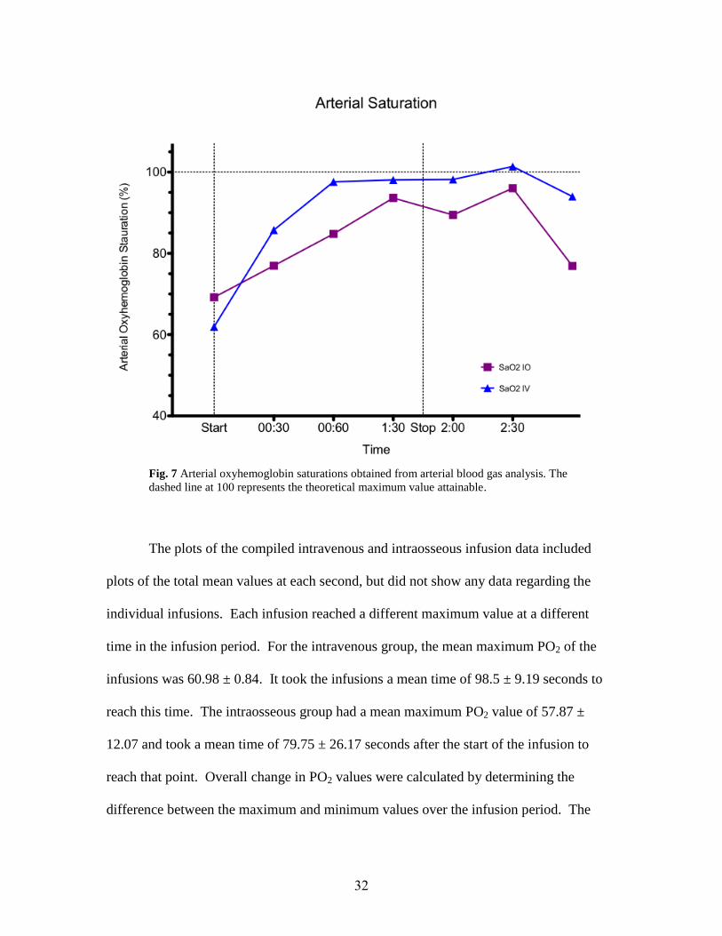
The SaO2 values of the intravenous and intraosseous infusions show an increase
over the infusion period. Linear regression analysis reveals that there is no statistical
significance between the two lines (p=0.253), though the slopes of both lines were
significantly non-zero. These results do, however, corroborate the rises in arterial tension
seen in the animals (Fig. 7).
32
Fig. 7 Arterial oxyhemoglobin saturations obtained from arterial blood gas analysis. The
dashed line at 100 represents the theoretical maximum value attainable.
The plots of the compiled intravenous and intraosseous infusion data included
plots of the total mean values at each second, but did not show any data regarding the
individual infusions. Each infusion reached a different maximum value at a different
time in the infusion period. For the intravenous group, the mean maximum PO2 of the
infusions was 60.98 ± 0.84. It took the infusions a mean time of 98.5 ± 9.19 seconds to
reach this time. The intraosseous group had a mean maximum PO2 value of 57.87 ±
12.07 and took a mean time of 79.75 ± 26.17 seconds after the start of the infusion to
reach that point. Overall change in PO2 values were calculated by determining the
difference between the maximum and minimum values over the infusion period. The

33
intravenous group displayed a higher mean change in PO2 of 32.95 ± 1.80 than that of the
intraosseous group (23.12 ± 11.70) (Table 1).
Mean Maximum PO2 Mean Change in PO2 Mean Time to Max
IV 60.98 ± 0.84 32.95 ± 1.80 98.5 ± 9.19
IO 57.87 ± 12.07 23.12 ± 11.70 79.75 ± 26.17
Table 1 Calculated mean values of the intravenous and intraosseous infusions with
standard deviation included. Standard deviation values are considerably higher in the
intraosseous group.
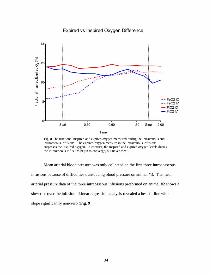
The mean plots of the Fe/FiO2 values collected from intravenous and intraosseous
infusions, although plotted separately in Figures 4 & 5, are easier to compare to one
another when shown on the same field. The mean FeO2 of the intravenous infusions
increases to surpass the FiO2. Although the FeO2 of the intraosseous infusions rises over
the infusion, it never surpasses the values of the FiO2. Over the period of the infusion,
the FeO2 lines were not found to be significantly different (p= 0.1736) (Fig. 8).
34
Fig. 8 The fractional inspired and expired oxygen measured during the intravenous and
intraosseous infusions. The expired oxygen measure in the intravenous infusions
surpasses the inspired oxygen. In contrast, the inspired and expired oxygen levels during
the intraosseous infusions begin to converge, but never meet.
Mean arterial blood pressure was only collected on the first three introaosseous
infusions because of difficulties transducing blood pressure on animal #3. The mean
arterial pressure data of the three intraosseous infusions preformed on animal #2 shows a
slow rise over the infusion. Linear regression analysis revealed a best-fit line with a
slope significantly non-zero (Fig. 9).

35
Fig. 9 Mean arterial pressure measured from the first three intraosseous infusions.
Included is a linear regression analysis of the mean values, plotted in blue, with 95% CI
(black dashed-line).

36
DISCUSSION
This study has many limitations, the most important of which is the low sample
number and variations imposed upon manufacturing of the microparticle suspensions.
The study suggests that infusion of a lipid-based oxygen suspension through the
intraosseous route can increase the oxygen content of the blood of a hypoxemic patient.
The compiled data collected from intraosseous infusions depicted a steady rise of arterial
oxygen tension while the animal was held at a constant FiO2. The only possible source of
the increase in tension over that time frame was the lipid-based oxygen suspension
delivered via the IO line. The rise in oxyhemoglobin saturations measured throughout
the IO infusions verifies that the animals were receiving supplemental oxygen over the
period of infusion, and signifies an important rise in circulating oxygen content. Further
confirmation of oxygen delivery can be found in the increase in FeO2 seen over the
infusion. A rapid increase in expired oxygen under constant inspired oxygen is a
physiologic anomaly; mammals do not produce oxygen as waste. Therefore, this increase
in FeO2 indicates the rapid oxygenation of venous blood after the start of infusion. The
blood passing through the pulmonary circulation had a higher oxygen tension than
alveolar gas, causing back-diffusion of oxygen into the alveoli.
The linear regression analysis preformed on oxygen tension during the
intravenous and intraosseous infusions revealed that intravenous infusion increases blood
oxygen tension at a higher rate than intraosseous infusion (Fig. 6). The slope of the
intravenous line was almost three times greater than that of the intraosseous line, and the
difference in the slope values was found to be significant. However, because the

37
infusions were all hand injections, these differences may represent differences in infusion
rates alone, as intraosseous infusions required subjectively more force to infuse than did
intravenous injections. Further studies which include quantification of injection rates are
needed to accurately comment on the kinetics of absorption.
The oxyhemoglobin saturations collected from both the intravenous and
intraosseous studies were not significantly different, suggesting that intraosseous infusion
acts to resaturate hemoglobin in a manner similar to the intravenous route established by
Kheir et al. in 2010 (Fig. 7). This, in fact, is a more relevant variable for oxygen
delivery, as it indicates oxygen content absorbed and carried rather than the surrounding
oxygen tension. Calculated mean peak PO2 values of the individual infusions (Table 1)
suggest that both the infusion routes achieve a similar level of oxygen saturation (IV:
60.98 ± 0.84, IO: 57.87 ± 12.07). A large gap between the overall change in PO2 would
suggest that intravenous infusion carries the most impact (IV: 32.95 ± 1.80, IO: 23.12 ±
11.70), however the large standard deviation of the intraosseous value brings this
conclusion into question as well. Linear regression analysis of the FeO2 values from both
infusion groups suggested that the increases seen in expired oxygen content were not
significantly different, consistent with our hypothesis that there is not a substantial
difference in oxygen delivery between these two groups (Fig. 8). The mean arterial
pressure shows a steady rise over the period of infusion (Fig. 9). Unfortunately, mean
arterial pressure was only reliably transduced on animal#2. Therefore, with only one data
group, no legitimate conclusions can be drawn.

38
As addressed by Paxton et al. (2009), dislodgement of the intraosseous needle
from the bone was a problem encountered in both animals #2 and #3. One instance led to
infiltration of approximately 20 ml of the suspension into surrounding tissue, and the
other involved percutaneous leakage from the puncture site (Fig. 2). In both cases, the
complications were likely due to a pressure buildup within the bone. The high rate of
infusion needed to compensate for the animals’ oxygen intakes could feasibly create
pressure that would push the needle out of the marrow. A different, more secure
intraosseous line could provide the needed stability throughout infusion.
The subendocardial ischemia seen in the hearts of both animals #2 and #3 could
potentially be related to hypotension, however there is no way to determine which
specific instance caused the damage (Fig. 3). Additional studies could potentially reveal
whether this ischemia is present in all infused patients or is an iatrogenic insult incurred
due to the infusion. The humeri that were bisected showed no signs of lipid deposits
from the suspension (Fig.1). Furthermore, no oxygen emboli or visible disruption of the
integrity of the marrow was seen.
Future experiments are needed to draw sound conclusions regarding the
hemodynamics of intraosseous infusion of the suspension. There are not enough tests
subjects and not enough data points to be confident in any accessory conclusions. To
ensure proper data collection, future experiments should correctly align the time
resolutions of all recording devices. Also, all means of recording data, such as the pulse
oximeter and blood pressure transducers, should be functioning soundly. Proper data
collection of future experiments will allow for more reliable conclusions to be drawn.

39
To better profile the hemodynamics of intraosseous infusion of the suspension, a
radioopaque suspension could be infused into an animal. Fluoroscopic images taken at a
set time interval could reveal the pathway of the suspension from the humerus to the
heart and the approximate time it takes to reach arterial circulation. Histological analysis
of tissue samples taken from the necropsy could reveal any underlying damages to the
heart, brain, lungs or bone marrow that may be overlooked or invisible to the human eye.
Although many of the results were clouded by a low number of data points, the
experiment still fulfilled its overall purpose; it has provided evidence that intraosseous
infusion of a lipid-based oxygen suspension increases the oxygen content of the blood.
This serves as a pilot study carrying proof of concept, allowing for funding of future
experiments of intraosseous infusion.

40
BIBLIOGRAPHY
Ayres, S., & Grace, W. (1969). Inappropriate Ventilation and Hypoxemia as Causes of
Cardiac Arrhythmias. The American Journal of Medicine, 46, 495-505.
Blumberg, S., Gorn, M., & Crain, E. (2008). Intraosseous Infusion: A review of Methods
and Novel Devices. Pediatric Emergency Care, 24(1), 50-56.
Bone, R. (1980). Treatment of Severe Hypoxemia due to the Adult Respiratory Distress
Syndrome. Archives of Internal Medicine, 140, 85-89.
Bowton, D., Scuderi, P., & Haponik, E. (1994). The Incidence an Effect on Outcome of
Hypoxemia in Hospitalized Medical Patients. The American Journal of
Medicine, 97, 38-46.
Brunette, D., & Fischer, R. (1988). Intravascular Access in Pediatric Cardiac Arrest.
American Journal of Emergency Medicine, 6(6), 577-579.
Christensen, D., Vernon, D., & Jr., W. B. et al. (1991). Skin necrosis complicating
intraosseous infusion. Pediatric Emergency Care, 7(5), 289-290.
Drinker, C., Drinker, K., & Lund, C. (1922). The Circulation in the Mammalian Bone-
Marrow. The American Journal of Physiology, 62(1), 1-92.
Finley, T., Swenson, E., & Comroe J. et al. (1962). The Cause of Arterial Hypoxemia at
Rest in Patients with "Alveolar-Capillary Block Syndrome". The Journal of
Clinical Investigation, 41(3), 618-622.
Gootman, N., Scarpelli, E., & Rudolph, A. (1963). Metabolic Acidosis in Children With
Severe Cyanotic Congenital Heart Disease. Pediatrics, 31(2), 251-254.
Hass, W. (1983). The Cerebral Ischemic Cascade. Neurological Clinics, 1(1), 345-353.
Hole, J. W., Shier, D., & Butler, J. et al. (2009). Hole's essentials of human anatomy &
physiology (10th ed.). Boston: McGraw-Hill/McGraw-Hill Higher Education.
Kheir, J., Walsh, B., & Scharp, L. et al. (2010). Intravenous Oxygen Gas-Filled Abstract
6: Liposomes Prevent Death From Asphyxia. Circulation, 122(A6).
Kleinman, M., Chameides, L., & Schexnayder, S. et al. (2010). Care Guidelines for
Cardiopulmonary Resuscitation and Emergency Cardiovascular Part 14:
Pediatric Advanced Life Support: 2010 American Heart Association.
Circulation, 122, 876-908.
Leidel, B., Kirchoff, C., & Braunstein, V. et al. (2010). Comparison of two intraosseous

41
access devices in adult patients under resuscitation in the emergency department:
A prospective, randomized study. Resuscitation, 81, 994-999.
Miller, W., Heard, J., & Unger, K. et al. (1984). Enlarged pulmonary arteriovenous
vessels in COPD. Another possible mechanism of hypoxemia. Chest, 86, 704-
706.
Mizock, B., & Falk, J. (1992). Lactic acidosis in critical illness. Critical Care Medicine,
20(1), 80-93.
Neumar, R., Otto, C., & Link, M. et al. (2010). Cardiovascular Care Association
Guidelines for Cardiopulmonary Resuscitation and Emergency Part 8: Adult
Advanced Cardiovascular Life Support: 2010 American Heart. Circulation, 122,
729-767.
Orlowski, J., Julius, C., & Petras, R. et al. (1989). The Safety of Intraosseous Infusions:
Risks of Fat and Bone Marrow Emboli to the Lungs. Annals of Emergency
Medicine, 18(10), 1062-1067.
Paxton, J., Kunth, T., & Klausner, H. (2009). Proximal Humerus Intraosseous Infusion: A
Preferred Emergency Venous Access. The Journal of Trauma, 67(3), 606-611.
Petit, C., Rome, J., & Wernovsky, G. et al. (2009). Preoperative Brain Injury in
Transposition of the Great Arteries Is Associated With Oxygenation and Time to
Surgery, Not Balloon Artial Septostomy. Circulation, 119, 709-716.
Piantadosi, C., & Schwartz, D. (2004). The Acute Respiratory Distress Syndrome.
Annals of Internal Medicine, 141, 460-470.
Rosetti, V., Thompson, B., & Aprahamian, C. et al. (1984). Difficulty and Delay in
Intravascular Access in Pediatric Patients. Annals of Emergency Medicine, 13(5),
406.
Rosetti, V., Thopmson, B., & Miller, J. et al. (1985). Intraosseous Infusion: An
Alternative Route of Pediatric Intravascular Access. Annals of Emergency
Medicine, 14(9), 885-888.
Rosovsky, M., FitzPatrick, M., & Goldfarb, C. et al. (1994). Bilaterla osteomyelitis due
to intraosseous infusion: case report and review of the English-language
literature. Pediatric Radiology, 24, 72-73.
Suchyta, M., Clemmer, T., & Orme, J. et al. (1991). Increased survival of ARDS patients
with severe hypoxemia (ECMO criteria). Chest, 99, 951-955.
Sukumalchantra, Y., Danzig, R., & Levey, S. et al. (1970). The Mechanism of Arterial
Hypoxemia in Acute Myocardial Infarction. Circulation, 41, 641-650.

42
Takeda, S., Miyoshi, S., & Omori, K. et al. (1999). Surgical Rescue for Life-Threatening
Hypoxemia Caused by a Mediastinal Tumor. Annals of Thoracic Surgery, 68,
2324-2326.
Tocantins, L., & O'Neill, J. (1951). Infusion of Blood and Other Fluids into the
Circulation Via the Bone Marrow. The American Journal of Medicine, 11(5),
571.
Wilson, J., Pierce, A., & Johnson, R. et al. (1971). Hypoxemia in Pulmonary Embolism, a
Clinical Study. The Journal of Clinical Investigation, 50, 481-491.


![6c-Ciarallo-Liposomal Bupivacaine.ppt [Last saved by user] · anesthetics, including lidocaine, ropivacaine, mepivacaine, or bupivacaine HCl ... • Slow infusion of liposomal bupivacaine](https://img.dokumen.tips/doc/110x75/5cbb91b588c99345128bd95b/6c-ciarallo-liposomal-last-saved-by-user-anesthetics-including-lidocaine.jpg)