Embed Size (px)
Citation preview

RESEARCH ARTICLE Open Access
Evaluation of human cytomegalovirusantigen expression in invasive breastcarcinoma in a population of IranianpatientsFereshteh Mohammadizadeh and Fatemeh Mahmudi*
Abstract
Background: The role of human cytomegalovirus (HCMV) in the development of breast carcinoma is questionable.The aim of this study was to evaluate the expression of the immediate early antigen (IE) of HCMV in breast carcinomaand its association with some clinicopathologic factors in a population of Iranian patients.
Methods: Formalin-fixed and paraffin-embedded tissue blocks from the pathology laboratories of the Azahra andShahid Beheshti hospitals, Isfahan, Iran, from 2013 to 2016, were used in the study. We used immunohistochemistryand real-time PCR to detect the IE-antigen of HCMV in breast carcinoma, normal tissue adjacent to carcinoma, andnormal tissue from mammoplasty specimens.
Results: A total of 96 samples were evaluated: 70 invasive breast carcinoma of different histologic subtypes and 26mammoplasty normal breast tissues. All the samples were negative for IE-antigen expression. No relationship was seenbetween breast cancer and HCMV in this study.
Conclusions: The results of this study failed to show any relationship between HCMV and the development of breastcarcinoma.
Keywords: Breast cancer, Human cytomegalovirus, Immediate early antigen, Iran
BackgroundBreast carcinoma is the second-most-common cause ofcancer-related death among women worldwide. How-ever, breast cancer-related death has decreased in recentyears due to early detection and treatment [1]. Breastcancer is the most common type of cancer among Iran-ian women, according to official statistics from the can-cer registry [2].Some of the documented risk factors of breast carcin-
oma include early age of menarche, late age of meno-pause, a positive family history of breast cancer,hormone replacement therapy, age, sex, and nulliparity[3–8]. At present, the etiologic role of viral infections insome human cancers—for example, the role of hepatitisB and C viruses in hepatocellular carcinoma, and the
role of the Epstein–Barr virus (EBV) in nasopharyngealcarcinoma—has been confirmed [9, 10]. Although sev-eral studies led by molecular and protein technologieshave detected some viruses, such as the human papillo-mavirus (HPV) [11, 12] and the EBV [13–15] in breastcancer specimens, findings regarding the human cyto-megalovirus’s (HCMV) role in breast cancer is contro-versial. It has been posed that breast carcinoma may beassociated with late exposure to a common virus [16].Breast cancer incidence has been shown to be low incountries where HCMV seroconversion occurs in child-hood, and most adults are HCMV seropositive [14]. Onthe other hand, breast cancer is most prevalent in thosecountries where there is late exposure to common vi-ruses [14].HCMV is a member of the herpes virus family, which
can remain latent lifelong after the primary infectionand shows recurring activation [7]. It has some gene
* Correspondence: [email protected] of Pathology, School of Medicine, Isfahan University of MedicalSciences, Isfahan, Iran
© The Author(s). 2017 Open Access This article is distributed under the terms of the Creative Commons Attribution 4.0International License (http://creativecommons.org/licenses/by/4.0/), which permits unrestricted use, distribution, andreproduction in any medium, provided you give appropriate credit to the original author(s) and the source, provide a link tothe Creative Commons license, and indicate if changes were made. The Creative Commons Public Domain Dedication waiver(http://creativecommons.org/publicdomain/zero/1.0/) applies to the data made available in this article, unless otherwise stated.
Mohammadizadeh and Mahmudi Infectious Agents and Cancer (2017) 12:39 DOI 10.1186/s13027-017-0148-3

products with dysregulatory effects on the proliferativecell cycle, resulting in a block of the apoptotic pathways,tumor suppressor proteins dysfunction, and DNA muta-tion [17, 18]. Consequently, the cells infected by CMVare prone to DNA instability and neoplastic transform-ation [19]. HCMV has been found in the milk of in-fected women [3]. So, the virus may have the potentialto spread to the adjacent breast epithelial cells [3].Moreover, HCMV has been found to infect the macro-phages and induce an atypical phenotype of M1/M2macrophages. This phenotype of macrophage releasescytokines, which are involved in cancer initiation or pro-motion [3].Since the glandular epithelium of breast tissue can act
as a reservoir of latent CMV infection [7], the aim of thisstudy is the evaluation of the probable role of HCMV inthe development of breast cancer in a population ofIranian patients, and the possible association betweenthe presence of the virus in carcinoma tissue and someprognostic clinicopathologic parameters of the cancer.
MethodsFormalin-fixed and paraffin-embedded breast tissueblocks archived in the pathology laboratory of theAzahra and Shahid Beheshti hospitals, Isfahan, Iran,from 2013 to 2016, were used. All of them were previ-ously evaluated for at least ER, PR, and Her2/neu IHCmarkers. The ethics and scientific research committee ofthe Isfahan University of Medical Sciences approved theprotocol. We had two groups of specimens. The firstgroup included invasive breast carcinoma tissue (astumor) and adjacent non-neoplastic normal breast tissue(as the tumor control); the second group included nor-mal breast tissue from reduction mammoplasty speci-mens (as the normal control). The total sample numberwas 96, including 70 neoplastic (tumor specimens) andadjacent non-neoplastic breast tissue specimens (tumorcontrol), and 26 normal breast tissue specimens (normalcontrol).Four micron sections were prepared from the tissue
blocks and stained by the immunohistochemistrymethod (IHC) for immediate early antigen (IE) ofHCMV. The used antibody was a monoclonal mouseantibody, subclass IgG1, kappa, clone CCH2 (DakoCompany, Denmark).The IHC was carried out as follows: Sections were in-
cubated in an oven at 37 °C for 48 h and then dewaxatedby the use of 100% xylol. Rehydration was done by aseries of decreasing concentrations of ethanol (100%,85%, and 75%) and distilled water. The sections wererinsed in a 10% phosphate-buffered saline (PBS) solu-tion. Next, they were incubated in 10% H2O2 andmethanol (1:9) for 30 min to prevent endogenous perox-idase activity. They were rinsed again in a 10% PBS
solution and then incubated in a citrate-buffered solu-tion (PH = 6.1) in a microwave (power at 800) for14 min. After rinsing in 100% PBS, blocking of the en-dogenous non-specific bindings was done by adding ablocking serum (for 30 min), and drying subsequently.In this step, a primary monoclonal antibody (anti-IEwith 1:200 ratio) was added, and the samples were incu-bated at room temperature for 30 min. The sampleswere then rinsed in a PBS solution and a secondarybroad-spectrum antibody was added (30 min). Horserad-ish peroxidase-streptavidin and diaminobenzidine (DAB)were added for 30 min and 10 min, respectively. Thesamples were rinsed in 10% PBS, then dehydrated by in-creasing concentrations of alcohol (75%, 85%, and100%), and finally counterstained with hematoxylin, andmounted.Two established CMV positive tissue specimens—an
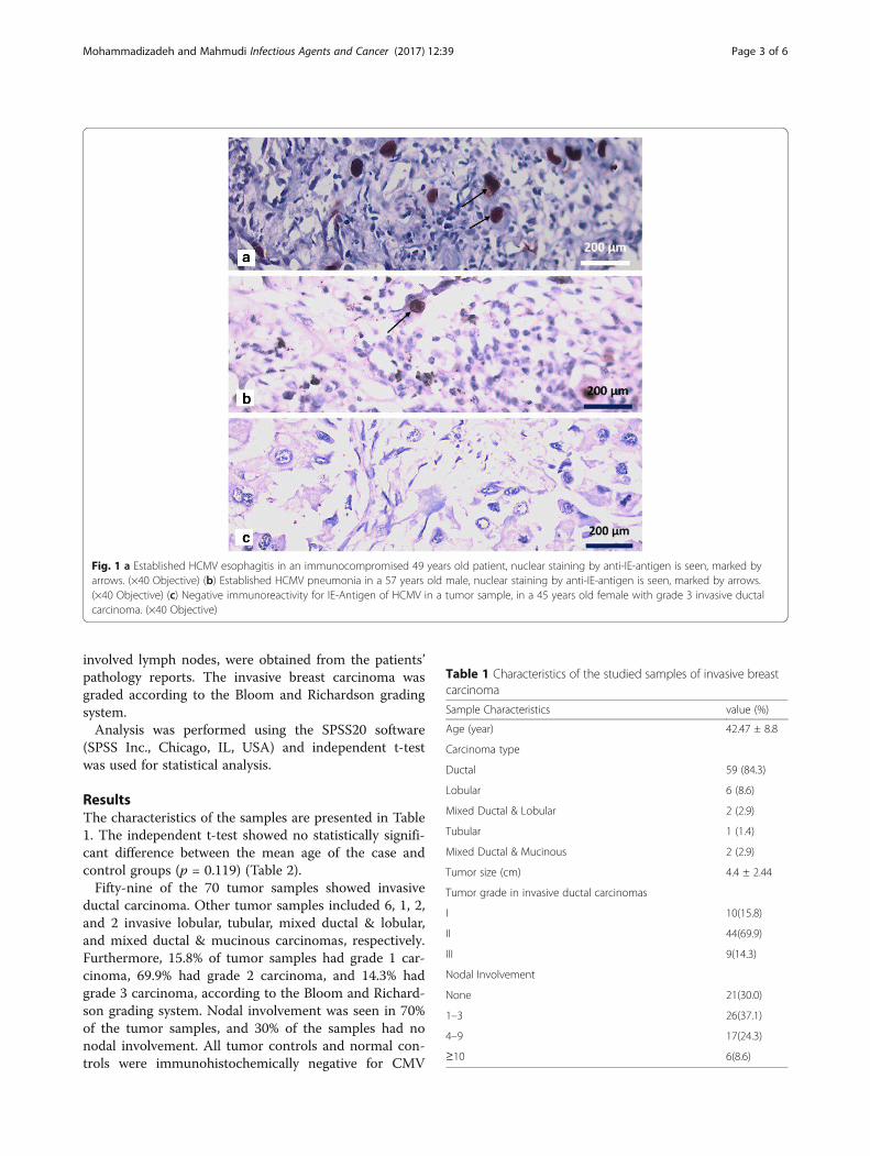
esophageal biopsy and a lung biopsy—were also stainedconcurrently as positive controls Fig. 1a and b .Finally, two pathologists assessed the IHC-stained
slides using a two-headed light microscope.Nuclear staining with the CMV monoclonal antibody
was considered as a positive result, according to the fol-lowing grading system [7]:
Negative: 0Grade I: < 25% positive cellsGrade II: 25–49% positive cellsGrade III: 50–75% positive cellsGrade IV: > 75% positive cells
In order to confirm the IHC results, the specimens withpositive or equivocal IHC findings were reassessed byreal-time PCR. DNA was purified from the tissue blocksusing a QIAamp DNA FFPE kit (Qiagen) according to themanufacturer’s instructions. In brief, the process of theDNA purification was as follows: 5–10-μm sections fromthe paraffin-embedded tissue blocks were prepared.Dewaxation was done by xylene and tissue lysis by pro-teinase K. Then the sections were incubated at 90 °C todeactivate the proteinase K. DNA binding to the mem-brane was done, the contaminations were washed, and theDNA was eluted from the membrane.Finally, real-time PCR was performed with a Corbett
Rotor-Gene 6000 analyzer, using a GeneProof CMVPCR kit (the GeneProof CMV PCR kit primer andprobes are proprietary and unpublished) to amplify theexon 4 of the IE antigen. Amplification was done with a10 μl isolated nucleic acid sample and 30 μl Master Mix,as follows: 37 °C/2 min and 95 °C/10 min, and 45 cycles:95 °C/5 s and 60 °C/40 s, and 72 °C/20 s. All PCR reac-tions were performed in duplicate.Other necessary information, including age, tumor
size, histologic subtype, histologic grade, and number of
Mohammadizadeh and Mahmudi Infectious Agents and Cancer (2017) 12:39 Page 2 of 6
involved lymph nodes, were obtained from the patients’pathology reports. The invasive breast carcinoma wasgraded according to the Bloom and Richardson gradingsystem.Analysis was performed using the SPSS20 software
(SPSS Inc., Chicago, IL, USA) and independent t-testwas used for statistical analysis.
ResultsThe characteristics of the samples are presented in Table1. The independent t-test showed no statistically signifi-cant difference between the mean age of the case andcontrol groups (p = 0.119) (Table 2).Fifty-nine of the 70 tumor samples showed invasive
ductal carcinoma. Other tumor samples included 6, 1, 2,and 2 invasive lobular, tubular, mixed ductal & lobular,and mixed ductal & mucinous carcinomas, respectively.Furthermore, 15.8% of tumor samples had grade 1 car-cinoma, 69.9% had grade 2 carcinoma, and 14.3% hadgrade 3 carcinoma, according to the Bloom and Richard-son grading system. Nodal involvement was seen in 70%of the tumor samples, and 30% of the samples had nonodal involvement. All tumor controls and normal con-trols were immunohistochemically negative for CMV
Fig. 1 a Established HCMV esophagitis in an immunocompromised 49 years old patient, nuclear staining by anti-IE-antigen is seen, marked byarrows. (×40 Objective) (b) Established HCMV pneumonia in a 57 years old male, nuclear staining by anti-IE-antigen is seen, marked by arrows.(×40 Objective) (c) Negative immunoreactivity for IE-Antigen of HCMV in a tumor sample, in a 45 years old female with grade 3 invasive ductalcarcinoma. (×40 Objective)
Table 1 Characteristics of the studied samples of invasive breastcarcinoma
Sample Characteristics value (%)
Age (year) 42.47 ± 8.8
Carcinoma type
Ductal 59 (84.3)
Lobular 6 (8.6)
Mixed Ductal & Lobular 2 (2.9)
Tubular 1 (1.4)
Mixed Ductal & Mucinous 2 (2.9)
Tumor size (cm) 4.4 ± 2.44
Tumor grade in invasive ductal carcinomas
I 10(15.8)
II 44(69.9)
III 9(14.3)
Nodal Involvement
None 21(30.0)
1–3 26(37.1)
4–9 17(24.3)
≥10 6(8.6)
Mohammadizadeh and Mahmudi Infectious Agents and Cancer (2017) 12:39 Page 3 of 6
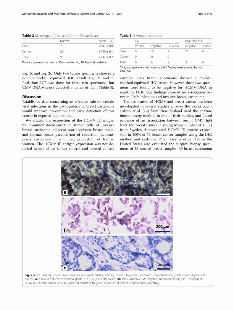
Fig. 1c and Fig. 2c. Only two tumor specimens showed adouble-checked equivocal IHC result Fig. 2a and b.Real-time PCR was done for these two specimens, butCMV DNA was not detected in either of them (Table 3).
DiscussionEstablished data concerning an effective role for certainviral infections in the pathogenesis of breast carcinomawould improve prevention and early detection of thiscancer in exposed populations.We studied the expression of the HCMV IE antigen
by immunohistochemistry in tumor cells of invasivebreast carcinoma, adjacent non-neoplastic breast tissue,and normal breast parenchyma of reduction mammo-plasty specimens in a limited population of Iranianwomen. The HCMV IE antigen expression was not de-tected in any of the tumor control and normal control
samples. Two tumor specimens showed a double-checked equivocal IHC result. However, these two speci-mens were found to be negative for HCMV DNA inreal-time PCR. Our findings showed no association be-tween CMV infection and invasive breast carcinoma.The association of HCMV and breast cancer has been
investigated in several studies all over the world. Rich-ardson et al. [14] from New Zealand used the enzymeimmunoassay method in one of their studies, and foundevidence of an association between serum CMV IgGlevel and breast cancer in young women. Taher et al. [7]from Sweden demonstrated HCMV IE protein expres-sion in 100% of 73 breast cancer samples using the IHCmethod and real-time PCR. Harkins et al. [19] in theUnited States also evaluated the surgical biopsy speci-mens of 38 normal breast samples, 39 breast carcinoma
Table 2 Mean Age of Case and Control Group (year)
Number Mean ± SD
Case 70 42.47 ± 8.86
Control 26 39.50 ± 6.14
Total 96 41.47 ± 8.28
Data are presented as mean ± SD or number (%), SD Standard deviation
Fig. 2 a & b Two equivocal tumor Samples with weak nuclear staining, marked by arrows: invasive ductal carcinoma, grade 2 in a 34 years oldpatient (a) & invasive ductal carcinoma, grade 2 in a 52 years old patient (b). (×40 Objective) (c) Negative immunoreactivity for IE-Antigen ofHCMV in a tumor sample, in a 44 years old female with grade 1 invasive ductal carcinoma. (×40 Objective)
Table 3 IE-Antigen expression
IHC Real time PCR
Positive Negative Equivocal Negative Positive
Case 0 68 2 2a 0
Control 0 26 0 - -
Total 0 94 2 2 0aOnly two specimens with equivocal IHC finding were assessed by realtime PCR.
Mohammadizadeh and Mahmudi Infectious Agents and Cancer (2017) 12:39 Page 4 of 6

samples, and paired normal breast tissue from 21 breastcancer patients, and demonstrated a higher expressionof HCMV antigens in breast cancer compared to normalbreast epithelium (97% versus 63%).Another study by Cox et al. [20] in New Zealand
showed that the elevation of the serum HCMV IgG pri-oritizes breast cancer development in a nested case-control study. El-Shinawi et al. [21] also reported a sig-nificant association between HCMV and breast cancer,with higher serum levels of HCMV IgG in 82% of 28 pa-tients with inflammatory breast carcinoma compared to65% of 49 patients with non-inflammatory breast carcin-oma. Karimi et al. [22] from Iran also detected HCMVDNA in 26 of the 50 samples (58%) of invasive breastcarcinoma by using the nested-PCR method in Sanandajcity, Kurdistan Province. Perhaps the different preva-lence of HCMV in various geographical areas in Iranmay justify the different findings of the Karimi et al.study and ours. Moreover, the different fixation times oftissue samples in various centers, the specificity and sen-sitivity of the antibody used for the IHC, the variableprevalence of CMV infection in different countries, andtime of exposure to the virus in life (early or late) mayjustify some differences in the research findings.On the other hand, several studies have failed to show
any relationship between HCMV and breast cancer.Utrera-Barillas et al. [23] evaluated 27 breast cancer speci-mens and 20 fibroadenoma samples by quantitative PCRand reported no significant association between HCMVand breast cancer development. Richardson et al. [24] alsoevaluated the CMV IgG levels in plasma (by the enzymeimmunoassay method) and checked HCMV DNA in 70tumor samples using the quantitative PCR method, andfound no relationship between HCMV and breast cancer,which went against their previous studies. They suggestedsome possibilities for these contrasts, such as the limita-tion of molecular methods and an absence of virus aftercarcinogenesis, a so-called “hit and run” oncogenesis [24].Antonsson et al. [11] also failed to detect CMV in breastcancer specimens with the quantitative PCR method in 54Australian breast cancer tumor specimens.Sample size, primer designing in PCR based studies,
and site of tissue sampling may justify another part ofthe controversial findings of these studies [25].
ConclusionOur results failed to show any relationship betweenHCMV and breast cancer development. It appears thatHCMV does not play an important role in breast cancercarcinogenesis in the Iranian population.Due to the controversial findings concerning the rela-
tionship between HCMV and breast cancer developmentin several studies in different countries, further studiesin this field are mandatory.
AbbreviationsEBV: Epstein–Barr virus; HCMV: Human cytomegalovirus; HPV: HumanPapillomavirus; IE: immediate early antigen; PCR: Polymerase chain reaction
AcknowledgementsThe authors would like to thank Javad Harati and Parya Mahmudi for theirsupport at various stages of preparing the manuscript.
FundingThis paper is derived from a specialty thesis No. 390385 in Isfahan Universityof Medical Sciences, Isfahan, Iran. The university vice chancellery for researchhas approved and financially supplied the study.
Availability of data and materialsThe data and materials are available in the main manuscript.
Authors’ contributionsFMZ and FM designed the study. FMZ and FM carried out data collectionand wrote the draft.FM carried out the initial screening of breast cancersamples. FMZ contributed to the interpretation of findings. All authors haveapproved the final manuscript for publication.
Ethics approval and consent to participateNot applicable.
Consent for publicationNot applicable.
Competing interestsThe authors declare that they have no competing interests.
Publisher’s NoteSpringer Nature remains neutral with regard to jurisdictional claims inpublished maps and institutional affiliations.
Received: 20 April 2017 Accepted: 20 June 2017
References1. Jemal A, Bray F, Center MM, Ferlay J, Ward E, Forman D. Global cancer
statistics. CA Cancer J Clin. 2011;61(2):69–90.2. Sadjadi A, Nouraie M, Ghorbani A, Alimohammadian M, Malekzadeh R.
Epidemiology of breast cancer in the Islamic Republic of Iran. first resultsfrom a population-based cancer registry. 2009;15(6):1426–31.
3. Herbein G, Kumar A. The oncogenic potential of human cytomegalovirusand breast cancer. Front Oncol. 2013;4:230.
4. Ban KA, Godellas CV. Epidemiology of breast cancer. Surg Oncol Clin N Am.2014;23(3):409–22.
5. Mohammadizadeh F, Zarean M, Abbasi M. Association of Epstein-Barr viruswith invasive breast carcinoma and its impact on well-knownclinicopathologic parameters in Iranian women. Advanced biomedicalresearch. 2014;3:141.
6. Joshi D, Quadri M, Gangane N, Joshi R, Gangane N. Association of EpsteinBarr virus infection (EBV) with breast cancer in rural Indian women. PLoSOne. 2009;4(12):e8180.
7. Taher C, de Boniface J, Mohammad A-A, Religa P, Hartman J, Yaiw K-C, et al.High prevalence of human cytomegalovirus proteins and nucleic acids inprimary breast cancer and metastatic sentinel lymph nodes. PLoS One.2013;8(2):e56795.
8. Anders CK, Carey LA. Biology, metastatic patterns, and treatment of patientswith triple-negative breast cancer. Clinical breast cancer. 2009;9:73–81.
9. Hsu C-R, Lu T-M, Chin LW, Yang C-C. Possible DNA viral factors of humanbreast cancer. Cancers. 2010;2(2):498–512.
10. Kogevinas M, Pearce N, Susser M, Boffetta P. Infection with hepatitis B and Cviruses, social class and cancer. IARC Sci Publ. 1997;138:319–24.
11. Antonsson A, Bialasiewicz S, Rockett RJ, Jacob K, Bennett IC, Sloots TP.Exploring the prevalence of ten polyomaviruses and two herpes viruses inbreast cancer. PLoS One. 2012;7(8):e39842.
12. Heng B, Glenn W, Ye Y, Tran B, Delprado W, Lutze-Mann L, et al. Humanpapilloma virus is associated with breast cancer. Br J Cancer. 2009;101(8):1345–50.
Mohammadizadeh and Mahmudi Infectious Agents and Cancer (2017) 12:39 Page 5 of 6

13. Grinstein S, Preciado MV, Gattuso P, Chabay PA, Warren WH, De Matteo E, etal. Demonstration of Epstein-Barr virus in carcinomas of various sites. CancerRes. 2002;62(17):4876–8.
14. Richardson A, Cox B, McCredie M, Dite G, Chang J, Gertig D, et al.Cytomegalovirus, Epstein–Barr virus and risk of breast cancer before age 40years: a case–control study. Br J Cancer. 2004;90(11):2149–52.
15. Hachana M, Amara K, Ziadi S, Romdhane E, Gacem RB, Trimeche M.Investigation of Epstein–Barr virus in breast carcinomas in Tunisia.Pathology-Research and Practice. 2011;207(11):695–700.
16. Richardson A. Is breast cancer caused by late exposure to a common virus?Med Hypotheses. 1997;48(6):491–7.
17. Söderberg-Nauclér C. Does cytomegalovirus play a causative role in thedevelopment of various inflammatory diseases and cancer? J Intern Med.2006;259(3):219–46.
18. Basta S, Bennink JR. A survival game of hide and seek: cytomegalovirusesand MHC class I antigen presentation pathways. Viral Immunol. 2003;16(3):231–42.
19. Harkins LE, Matlaf LA, Soroceanu L, Klemm K, Britt WJ, Wang W, et al.Detection of human cytomegalovirus in normal and neoplastic breastepithelium. Herpesviridae. 2010;1(1):8.
20. Cox B, Richardson A, Graham P, Gislefoss R, Jellum E, Rollag H. Breast cancer,cytomegalovirus and Epstein–Barr virus: a nested case–control study. Br JCancer. 2010;102(11):1665–9.
21. El-Shinawi M, Mohamed HT, El-Ghonaimy EA, Tantawy M, Younis A,Schneider RJ, et al. Human cytomegalovirus infection enhances NF-κB/p65signaling in inflammatory breast cancer patients. PLoS One. 2013;8(2):e55755.
22. Karimi M, Hosseini SZ, Nikkhoo B, Soleimani-Mohammadi F. Relativefrequency of Cytomegalovirus (CMV) in tissue samples of women withbreast cancer in Sanandaj. Iran International Journal of Bioassays. 2016;5(3):4907–11.
23. Utrera-Barillas D, Valdez-Salazar H-A, Gómez-Rangel D, Alvarado-Cabrero I,Aguilera P, Gómez-Delgado A, et al. Is human cytomegalovirus associatedwith breast cancer progression? Infectious agents and cancer. 2013;8(1):12.
24. Richardson AK, Currie MJ, Robinson BA, Morrin H, Phung Y, Pearson JF, et al.Cytomegalovirus and Epstein-Barr virus in breast cancer. PLoS One. 2015;10(2):e0118989.
25. Lazzeroni M, Serrano D. Potential use of vaccines in the primary preventionof breast cancer in high-risk patients. Breast Care. 2012;7(4):281–7.
• We accept pre-submission inquiries
• Our selector tool helps you to find the most relevant journal
• We provide round the clock customer support
• Convenient online submission
• Thorough peer review
• Inclusion in PubMed and all major indexing services
• Maximum visibility for your research
Submit your manuscript atwww.biomedcentral.com/submit
Submit your next manuscript to BioMed Central and we will help you at every step:
Mohammadizadeh and Mahmudi Infectious Agents and Cancer (2017) 12:39 Page 6 of 6