Embed Size (px)
Citation preview

Limited Model Antigen Expression by Transgenic Fungi InducesDisparate Fates during Differentiation of Adoptively TransferredT Cell Receptor Transgenic CD4� T Cells: Robust Activation andProliferation with Weak Effector Function during Recall
Marcel Wüthrich,a Karen Ersland,a John C. Pick-Jacobs,a Benjamin H. Gern,a Christopher A. Frye,a Thomas D. Sullivan,a
Meghan B. Brennan,a Hanna I. Filutowicz,a Kevin O’Brien,c Keegan D. Korthauer,d Stacey Schultz-Cherry,c and Bruce S. Kleina,b,c
Departments of Pediatrics,a Internal Medicine,b Medical Microbiology and Immunology,c and Biostatistics and Medical Informatics,d University of Wisconsin MedicalSchool, University of Wisconsin Hospital and Clinics, Madison, Wisconsin, USA
CD4� T cells are the key players of vaccine resistance to fungi. The generation of effective T cell-based vaccines requires an un-derstanding of how to induce and maintain CD4� T cells and memory. The kinetics of fungal antigen (Ag)-specific CD4� T cellmemory development has not been studied due to the lack of any known protective epitopes and clonally restricted T cell subsetswith complementary T cell receptors (TCRs). Here, we investigated the expansion and function of CD4� T cell memory aftervaccination with transgenic (Tg) Blastomyces dermatitidis yeasts that display a model Ag, E�-mCherry (E�-mCh). We reportthat Tg yeast led to E� display on Ag-presenting cells and induced robust activation, proliferation, and expansion of adoptivelytransferred TEa cells in an Ag-specific manner. Despite robust priming by E�-mCh yeast, antifungal TEa cells recruited and pro-duced cytokines weakly during a recall response to the lung. The addition of exogenous E�-red fluorescent protein (RFP) to theE�-mCh yeast boosted the number of cytokine-producing TEa cells that migrated to the lung. Thus, model epitope expressionon yeast enables the interrogation of Ag presentation to CD4� T cells and primes Ag-specific T cell activation, proliferation, andexpansion. However, the limited availability of model Ag expressed by Tg fungi during T cell priming blunts the downstreamgeneration of effector and memory T cells.
Diseases due to fungi represent a growing public health prob-lem that demand new treatments and methods of vaccine
prevention (8). The rational design of vaccines against fungi re-quires an understanding of the elements of antifungal immunity.Cellular immunity is pivotal in acquired resistance to fungal in-fections and is organized into clonal populations of antigen (Ag)-specific CD4� T cells (8, 30, 40). The ability to track, enumerate,and characterize Ag-specific T cells precisely requires knowledgeof the Ag peptide. With such information, peptide-major histo-compatibility complex (MHC) tetramers and T cell receptor(TCR) transgenic (Tg) mice have been used to track and enumer-ate Ag-specific T cells ex vivo to circumvent in vitro expansion ordistortion of immune responses.
Reagents are available to precisely study T cell immunity withmodel agents such as lymphocytic choriomeningitis virus and Lis-teria (9, 22), but the study of most other pathogens is not readilyapproachable with these high-resolution methods. For the sys-temic dimorphic fungi, no T cell Ag epitopes have been elucidatedto provide the tools to address this gap in knowledge. To bridgethis gap, we engineered heterologous Ag and epitopes into a vac-cine strain of a pathogenic fungus to let us induce, track, quantify,characterize, and functionally analyze adoptively transferred TCRTg T cells specific for the foreign Ag in vaccinated animals.
Blastomycosis is a systemic infection due to the dimorphic fun-gus Blastomyces dermatitidis. We have created a live attenuatedvaccine against lethal experimental infection (38). The vaccineinduces sterilizing immunity that is mediated by CD4� T cells,although the protective antigen remains unknown. Still, thismodel and the potent activity of these CD4� T cells offer thechance to elucidate the requirements for inducing and maintain-
ing antifungal CD4� T cells by vaccination. As a surrogate meansto study the in vivo activation, proliferation, and maintenance ofBlastomyces-specific CD4� T cells, we expressed model epitopeson the vaccine B. dermatitidis yeast using BAD1, an abundantsurface protein, as a carrier. Yeast surface Ag display is thought tobe one feature that promotes the generation of antifungal immuneresponses.
In other nonfungal models, the availability of Ag and the num-ber of naïve T cell precursors in a host can affect the priming anddevelopment of CD4� effector and memory T cells (1, 27). How-ever, little is known about the identity, cellular distribution, andexpression levels of fungal T cell epitopes and how these factorsinfluence the development of antifungal immunity. We reportthat expressing a model epitope such as E� peptide on vaccineyeast induced the activation and proliferation of correspondingnaïve, adoptively transferred TCR Tg TEa cells. We describe theexperimental system and our results enabling the tracking of fun-gal Ag presentation to CD4� T cells and the corresponding Ag-specific T cell response during their earliest stages of activation,proliferation, and expansion. Interestingly, these antifungal T
Received 2 May 2011 Returned for modification 22 May 2011Accepted 15 November 2011
Published ahead of print 28 November 2011
Editor: G. S. Deepe, Jr.
Address correspondence to Marcel Wüthrich, [email protected].
Copyright © 2012, American Society for Microbiology. All Rights Reserved.
doi:10.1128/IAI.05326-11
0019-9567/12/$12.00 Infection and Immunity p. 787–797 iai.asm.org 787

cells ultimately failed to differentiate into potent effector cells andmigrate to lung upon rechallenge. We propose that this functionaldeficit of antifungal TEa cells is likely due to an insufficient Agthreshold reached by the vaccine since addition of exogenous E�peptide corrected the deficit.
MATERIALS AND METHODSMouse strains. Inbred strains of C57BL/6 mice (males 7 to 8 weeks old atthe time of these experiments) and the T lymphocyte-specific Thy1.1allele-carrying congenic C57BL/B6 strain B6.PL-Thy1a/Cy (stock no.000406) (12) were obtained from Jackson laboratories, Bar Harbor, ME.Two male TEa Tg mice of the C57BL/6J (B6; I-Ab, I-E�) background (13,15) expressing the Thy1.2 allele were generously provided by A. Y. Ruden-sky at the Howard Hughes Medical Institute, University of Washington.The Tg TCR in TEa mouse lymphocytes recognizes a peptide representingresidues 52 to 68 of the I-E� chain (E� peptide) bound to class II I-Ab
molecules. TEa Tg mice expressing the Thy1.1 allele were produced at theUniversity of Wisconsin by backcrossing the original TEa males two timesto wild-type B6 females expressing the congenic Thy1.1 marker andscreened for the Thy1 allele and transgene. To identify the transgene-positive progeny, lymphocytes from peripheral blood were stained withphycoerythrin (PE)-Cy7-labeled anti-CD4, fluorescein isothiocyanate(FITC)-labeled anti-V�2, and PE-labeled anti-V�6 antibodies (BD Phar-mingen) and analyzed by fluorescence-activated cell sorter (FACS) anal-ysis as previously described (15). Blastomyces-specific TCR Tg strain 1807mice were generated in the Klein lab and described elsewhere (37, 42, 43).Strain 1807 mice were backcrossed to congenic Thy1.1-positive(Thy1.1�) mice as described above. Mice were housed and cared for ac-cording to guidelines of the University of Wisconsin Animal Care Com-mittee, who approved all aspects of this work.
Fungi and growth conditions. Strains used were ATCC 26199 (Amer-ican Type Culture Collection) (16), a wild-type virulent strain, and theisogenic, attenuated mutant lacking BAD1, designated strain 55 (6). Iso-lates of B. dermatitidis were maintained as yeast on Middlebrook 7H10agar with oleic acid-albumin complex (Sigma Chemical Co., St. Louis,MO) at 39°C.
Engineering yeast expressing the model antigenic epitopes E� and2W1S. Yeast cells expressing either E� or 2W1S Ag epitopes were gener-ated by Agrobacterium-mediated transformation using a binary plasmidexpressing a three-way fusion of the epitope, the red fluorescent protein(RFP) mCherrry (mCh) (32), and a truncated version of BAD1. Tostreamline the generation of chimeric genes expressing different epitopes,an Agrobacterium binary plasmid that contains unique cloning sites andthe ccdB gene inserted into the NcoI site in the truncated BAD1 gene wasconstructed. The ccdB gene aids in the cloning of sequences of choice bypreventing Escherichia coli clones harboring nonrecombinant, parentalplasmids from growing (3). Primers were obtained from Integrated DNATechnologies (Coralville, IA). PCR for cloning was done using Elongase(Invitrogen, Carlsbad, CA). The starting plasmid was a derivative ofpBAD1-6H, containing the truncated tandem repeat sequence (�TR20)of BAD1 (5), called p�5-6H. The ccdB gene was PCR amplified from anInvitrogen Gateway cassette-containing plasmid using primers that addedNcoI and AscI sites at the 5= end of the forward primer, tdsP182 (GCGCCCATGGCGCGCCTGGCCGGCCTACTAAAAGCCAGATAACAGT), andNcoI and SwaI at the 5= end of the reverse primer, tdsP181 (GCGCCCATGGGATTTAAATCGGCCGGCCAGTCGTTCGGCTTCATCT). Fol-lowing PCR, the 800-bp fragment was digested with NcoI and ligated withp�5-6H plasmid DNA that had also been digested with NcoI, and theligated DNA was electroporated into an E. coli strain permissive forgrowth with the ccdB gene, DB3.1 (35). Transformants were screened forthe presence of the insert and correct orientation. In addition, the clonedplasmids were tested for a functional ccdB gene by electroporating theminto the nonpermissive strain DH10� and finding that transformantswere severely restricted in growth. The �TR20-ccdB chimeric gene wasexcised with XbaI (filled in) and EcoRI and moved into the Agrobacterium
binary vector pCTK4 (28) at the HindIII (filled in) and EcoRI sites, cre-ating plasmid pCTS33. A fragment containing the E� epitope region ofpTrc-E�-RFP (encoding amino acids 52 to 68 and 6 flanking amino acidsof the E� protein [19, 31] and the mCherry RFP gene [32]) was generatedusing splicing overlap (SOE) PCR (23) with E� forward primer tdsP202containing an incorporated AscI site at its 5= end (CGCGGGCGCGCCGAAGAATTTGCAAAGTT), SOE fusion E�-mCherry (E�-mCh) reverseprimer tdsP216 (AACCTGGATGTCATGGAGGTGAGCAAGGGCGAGGAG), SOE fusion E�-mCherry forward primer tdsP217 (CTCCTCGCCCTTGCTCACCTCCATGACATCCAGGTT), and the mCherry reverseprimer tdsP218 with a SmaI site at its 5= end (CGCGCCCGGGCCTTGTACAGCTCGTCCAT). The fused fragment was digested with AscI andSmaI and ligated with pCTS33, which had been digested with AscI andSwaI, and the DNA was electroporated into E. coli strain DH10�. Tocreate a 2W1S epitope-mCherry fusion, a 93-nucleotide (nt) oligomericprimer that contains sequence encoding the 14-amino-acid 2W1S epitope(29) flanked by 2 amino acids that match those flanking the comparablesequence of the E� epitope and two glycine residue spacers was generated(composite amino acid sequence, GGSFEAWGALANWAVDSANLGG).In addition, this primer includes a 5= end AscI site and 3= end sequence forpriming at the start of the mCherry sequence (tdsP615, GCGCGGCGCGCCGGCGGTAGCTTTGAGGCTTGGGGTGCACTGGCTAATTGGGCTGTGGACAGCGCTAACCTGGGCGGTGTGAGCAAGGGCGAG). PrimertdsP615 was used with tdsP618 (GGGCCTTGTACAGCTCGTCCAT) toamplify a cloned mCherry sequence and, in so doing, add the 2W1Sepitope sequence. The fragment was digested with AscI, which cuts at the5= end of the fragment (the 3= end was left blunt), and ligated to AscI- andSwaI-digested pCTS33. For both E� and 2W1S, transformants werescreened for plasmids with the new insert and sequenced to find thosewithout PCR-induced mutations and to confirm that the E�-mCherry or2W1S-mCherry fragment had been inserted to create a single translationalfusion in the �TR20 protein backbone. The plasmids were electroporatedinto Agrobacterium tumefaciens strain LBA1100 harboring the Ti plasmidpAL1100 (2, 10). Confirmed A. tumefaciens strains were used to transformyeast-phase cells of B. dermatitidis strain ATCC 26199 and strain 55, theBAD1-knockout derivative of ATCC 26199 (5, 35).
Screening and characterization of Tg yeast. Transformants of strain55 were initially screened for production of the fusion protein using anoverlay assay to detect secreted BAD1 (6) and subsequently analyzed byWestern blotting with anti-BAD1 antibodies on yeast cell extracts to con-firm that the fusion protein is the predicted size (5) (data not shown).Positive yeast cell lines were also screened for red fluorescence microscop-ically on an Olympus BX60 fluorescence microscope (Center Valley, PA)and by FACS analysis using the dichroic mirror at 595LP and the PE-TexasRed band-pass filter (6). Finally, the strains showing high expression ofthe Tg protein were also noted to have visibly pink/red colonies whenplated on 7H10 medium, and this characteristic was used to ensure thatyeast cells used for in vivo and in vitro experiments were expressing the E�-or 2W1S-mCherry-�TR20 (truncated BAD1) fusion protein at high lev-els. To identify transformants with the highest transgene expression, westained them with 1 �g of anti-BAD1 monoclonal antibody (MAb) DD5-CB4 (44) and goat anti-mouse IgG FITC-conjugated secondary antibody(1:200 dilution; Sigma) for 30 min and analyzed the yeast for maximalfluorescence in the FITC (BAD1) and PE-Texas Red (mCh) channels byFACS analysis. Tg ATCC 26199 yeast cells were identified by using the redcolor as an indication for transgene expression as described above.
Preparation of E�-RFP from E. coli. The generation of recombinantE. coli expressing the E�-RFP fusion protein containing the E� and DsRedsequences, kindly provided by Marc Jenkins (Minneapolis, MN), was de-scribed elsewhere (19). Protein production was induced with 1 mMisopropyl-�-D-thiogalactopyranoside overnight, and the E�-RFP fusionprotein was purified from the bacterial lysate using either a chitin-beadaffinity column (New England BioLabs) or an Ni2� resin His-Bind col-umn (Novagen).
Wüthrich et al.
788 iai.asm.org Infection and Immunity

Generation and use of Y-Ae antibody. The Y-Ae hybridoma was gen-erously provided by Marc Jenkins (University of Minnesota) (19). Mono-clonal antibody from ascites was ammonium sulfate precipitated, purifiedon protein A/G agarose according to the manufacturer’s specifications forthe isolation and purification of IgG (product 21001; Pierce Chemical,Rockford, IL), and quantified by measuring the optical density at 280 nm.The MAb was biotinylated using an EZ-Link N-hydroysulfosuccinimidelong chain (sulfo-NHS-LC) kit (Thermo Fisher Scientific) according tothe manufacturer’s protocol. To detect E� peptide-MHC class II (MHC-II) displayed in bone marrow-derived dendritic cells (BM-DCs), Y-Aeantibody was used at 50 to 100 �g per sample. Y-Ae staining was per-formed with a staining buffer of phosphate-buffered saline (PBS) contain-ing 1% bovine serum albumin plus 2 mM EDTA.
Recombinant VSV-SED- and 2W1S-expressing Listeria monocyto-genes. The recombinant vesicular stomatitis virus (VSV) containing agene cassette encoding the ovalbumin-derived peptide SIINFEKL, the E�peptide (amino acid residues 52 to 68), and DsRed (VSV-SED) was kindlyprovided by Leo Lefrancois (Storrs, CT) (4). The recombinant Listeriamonocytogenes strain expressing the 2W1S peptide was kindly provided byMarc Jenkins (11).
Vaccinations with soluble E�-RFP, 2W1S peptide plus LPS, E�-mCh- and 2W1S-mCh-expressing yeast, and VSV-SED- and 2W1S-expressing L. monocytogenes. Unless otherwise stated, C57BL/6 micewere vaccinated subcutaneously (s.c.) at two sites, dorsally and at the baseof the tail, using one injection of the following formulations, doses, androutes of delivery: recombinant yeast expressing E�-mCh, 2W1S-mCh, orBAD1-null attenuated yeast was injected either live s.c. or in a heat-killedform intravenously (i.v.) using a dose range of 105 to 107 yeast per mouse.Soluble E�-RFP from 0.8 �g to 100 �g recombinant protein was injecteds.c. alone or as an emulsion with incomplete Freund’s adjuvant (IFA). The2W1S peptide (EAWGALANWAVDSA) was purchased from Biosynthe-sis, Lewisville, TX, and injected i.v. as a mixture containing 50 �g 2W1Speptide and 5 �g lipopolysaccharide (LPS; Sigma). VSV-SED was injectedeither i.v. using a dose of 105 PFU (4) or s.c. at a range of 105 to 108 PFUrecombinant L. monocytogenes expressing 2W1S peptide, which was in-jected i.v. using a range of 10 � 106 to 100 � 106 live bacteria.
Adoptive transfer of Tg CD4� T cells and surface staining. Single-cell suspensions of splenocytes from TEa or 1807 mice (Thy1.1� back-ground) were labeled with 5- and 6-carboxyfluorescein diacetate succin-imidyl ester (CFSE; Molecular Probes) as described previously (33), andvarious numbers of labeled cells were injected i.v. into wild-type congenicThy1.2� C57BL/6 male recipients. At various times after Ag injection,single-cell suspensions from draining inguinal and brachial lymph nodesof recipient mice were stained with monoclonal antibodies directedagainst the following surface markers; CD4, CD8, Thy1.1, CD44, CD62L,and B220 (as a dump marker). MAbs were obtained from BD Pharmingen(San Diego, CA) and eBioscience (San Diego, CA), and cytometry datawere gathered with an LSRII flow cytometer (BD Biosciences, San Jose).Data were analyzed by using FlowJo software (Tree Star, Ashland, OR).The number of TEa and 1807 CD4� T cells in a lymph node was calculatedby multiplying the percentage of Thy1.1� CD4� cells by the number ofviable cells determined by trypan blue dye exclusion.
BrdU incorporation. On the day of challenge and daily thereafter,mice were given 2 mg of 5=-bromo-2-deoxyuridine (BrdU) in PBS byintraperitoneal (i.p.) injection. Four days later, the mice were sacrificedand T cells from the lung and mediastinal lymph node (MLN) werestained for surface markers and stained for BrdU incorporation using anFITC BrdU flow kit from BD Pharmingen, San Diego, CA.
Intracellular cytokine staining. Lung and draining lymph node cellswere obtained as described previously (39). An aliquot of isolated cells wasstained for surface CD4 and Thy1.1 to determine the percentage of trans-ferred TCR Tg cells. The numbers of Tg cells per lung or lymph nodes werederived by multiplying the percentage of cells by the total number of cellsper organ isolated. The rest of the cells were stimulated with anti-CD3 andanti-CD28 MAbs. After the 4- to 6-h stimulation, cells were stained for
surface markers, fixed, permeabilized with a Cytofix/Cytoperm kit (BDPharmingen), and stained with anticytokine antibodies as previously de-scribed (41, 45).
Peptide–MHC-II tetramer-based enrichment to detect 2W1S-specific endogenous T cells. The spleen and inguinal and brachial lymphnodes were harvested for each mouse analyzed. A single-cell suspensionwas prepared and enriched with PE-conjugated 2W1S tetramer as de-scribed previously (26, 27). The resulting enriched fractions were resus-pended in 0.1 ml of sorter buffer, and a small volume was removed for cellcounting, while the rest of the sample was stained with a cocktail of thefollowing fluorescently labeled antibodies: CD3-FITC, 2W1S tetramer-PE, CD4-peridinin chlorophyll protein, CD44 Alexa 700, CD8 V780, andbiotinylated B220, CD11c, and CD11b-Strep V450 (as a dump channel).The entire sample was collected on an LSRII flow cytometer and analyzedwith FlowJo software using the gating strategy described previously (26,27). The percentage of tetramer-positive events was multiplied by the totalnumber of cells in the enriched fraction to calculate the total number oftetramer-positive cells in the organs harvested.
Cytokine protein measurements. Cell culture supernatants were gen-erated in 24-well plates in 1 ml containing 2 � 105 TEa, OT-II, or naïve CD4�
T cells and BM-DCs (1:1 ratio), 10 �M E� peptide, or 105 E�-RFP or TR20yeast (38). Supernatants were collected after 48 h of coculture. Granulocyte-macrophage colony-stimulating factor (GM-CSF) and interleukin-2 (IL-2)(R&D System, Minneapolis, MN) were measured by enzyme-linked immu-nosorbent assay (ELISA) according to manufacturer specifications (detectionlimits, 0.05 ng/ml and 0.02 ng/ml, respectively).
Experimental infection. At 4 to 5 weeks postvaccination, mice wereinfected intratracheally (i.t.) with 2 � 103 to 2 � 104 wild-type strainATCC 26199 or E�-mCh-expressing ATCC 26199 yeast cells as describedpreviously (38). At day 4 postinfection, coinciding with the peak of T cellinflux (39, 40), the mice were sacrificed and lung T cells were analyzed byFACS analysis.
Statistical analysis. The number and percentage of activated, prolif-erating, or cytokine-producing T cells and differences in numbers of CFUwere analyzed using the Wilcoxon rank test for nonparametric data (14)or the t test when data were normally distributed. Contractions of T cells(number of T cells at the burst of expansion versus during the memoryphase) were analyzed by the two-way analysis of variance test. A P value of�0.05 is considered statistically significant.
RESULTSGeneration and characterization of transgenic B. dermatitidisyeast. To enumerate, characterize, and track Ag-specific T cells,we expressed the model epitope E� (for which TCR Tg TEa miceare available) fused to mCherry (E�-mCh) in the vaccine strain ofB. dermatitidis. E�-mCh protein is a recombinant 32-kDa chime-ric fusion protein consisting of amino acids 46 to 74 of the I-Ed�MHC-II subunit at the N terminus and the red fluorescent proteinmCherry at the C terminus (32). We hypothesized that chimericE�-mCh protein expressed on a backbone of the signal sequenceand 10 copies of the BAD1 tandem repeat (Fig. 1A) would bedelivered to the cell surface and stain the yeast red. We assessed Tgyeast by microscopy and FACS analysis. E�-mCh yeast stained red(Fig. 1B and C) and expressed BAD1 at levels comparable to TR20control yeast, whereas BAD1-null yeast failed to stain red or ex-press BAD1 (Fig. 1C). Thus, our Tg yeast displayed chimeric E�-mCh-BAD1 in and on the yeast cell.
We tested whether chimeric E�-mCh-BAD1 on yeast is pro-cessed and presented by antigen-presenting cells (APCs). Whenthe Tg yeast was added to bone marrow-derived DCs in vitro, thefungi were processed and the pE�–I-Ab complexes were displayedon the DC surface and detected by FACS analysis with Y-Ae MAb(Fig. 2A). The findings with E�-mCh yeast were comparable to
Monitoring Induction of Antifungal T Cells In Vivo
February 2012 Volume 80 Number 2 iai.asm.org 789

FIG 1 Tg yeast of B. dermatitidis. (A) Intact BAD1 (top) with 30 copies of the tandem repeat (white box) and �TR20 (2nd row), a derivative with 20 repeatsdeleted (5). We used BAD1 in �TR20, driven by its own promoter, as a fusion partner to make chimeric proteins with either the E� or the 2W1S epitope fusedto the RFP mCherry (25) and inserted in frame at the start of the C terminus (dark blue box; see Materials and Methods). The N-terminal BAD1 sequence (lightblue) encodes a signal sequence for export, and the C terminus is needed for binding to the yeast cell wall. The antigenic peptides (green or orange box) with theirflanking sequences (black) are shown. The construct name is on the left, the total amino acid length is given on the right, and the amino acid sequence of theepitope is given below in green or orange. The underlined amino acids are those that differ in the comparable sequence of E� and 2W1S. Map domain lengthsare not to scale. (B) E�-mCh yeasts fluoresce red inside and on the cell wall (arrow). (C) BAD1-null (strain 55 [6]), �TR20, and E�-mCh-expressing yeasts werestained and analyzed for BAD1 expression and red fluorescence by FACS analysis. The mean fluorescent intensity (MFI) for BAD1 expression on E�-mCh yeastand TR20 yeast was 58 and 33 times higher, respectively, than that on BAD1-null yeast. The red fluorescence of E�-mCh yeast was 25 times higher than that ofTR20 and BAD1-null yeast, as measured by determination of the mean fluorescent intensity. The histogram depicts BAD1 expression (FITC) and red fluores-cence (PE-Texas Red) for BAD1-null (red), �TR20 (blue), and E�-mCh (green) yeasts.
Wüthrich et al.
790 iai.asm.org Infection and Immunity

FIG 2 Yeast E� peptide is processed and presented by DCs and stimulates TEa T cells in vitro and in vivo. (A) Bone marrow-derived DCs were cultured inmedium alone, 30 �M E� peptide, or 2 � 105 yeast cells (1:1 ratio). After 24 h, DCs were stained with anti-CD11b and Y-Ae MAbs and analyzed. Blue denotesY-Ae display by DCs cultured with medium alone or �TR20 yeast. Red denotes Y-Ae staining by DCs cultured with E� peptide or E�-mCh yeast. (B) A TEa Tcell line (2 � 105 cells/well) or naïve TEa T cells (106 cells/well) were cultured with DCs (1:1 ratio) and the stimuli shown for 48 h. Supernatants were tested forcytokine by ELISA for IL-2 and GM-CSF as standard measures of T cell activation. OT-II cells were from a T cell line (2 � 105 cells/well). (C) Thy1.2� wild-typemice got 5 � 106 Thy1.1� TEa cells i.v. and were vaccinated with E�-RFP yeast or 50 �g E�-RFP s.c. Four days later, nodes were taken and Thy1.1� T cells wereassayed for proliferation (CFSE) and activation (CD44) by FACS analysis. The gates and percentages indicate the percentage of CFSElow TEa cells that express highlevels of CD44. The data are representative of 4 mice/group, and the experiment was repeated three times.
Monitoring Induction of Antifungal T Cells In Vivo
February 2012 Volume 80 Number 2 iai.asm.org 791

results seen with a lysate of E. coli cells that express recombinantE�-RFP-DsRed (25) or with His-tagged E�-RFP purified from E.coli (data not shown). Importantly, E�-mCh yeast coculturedwith DC in vitro triggered activation and cytokine production innaïve and primed TEa cells when the T cells were added to cellculture wells (Fig. 2B). Thus, the DCs process and present Ag in Tgvaccine yeast and display model E� peptide that can be detectedwithin MHC-II on the DC surface by Y-Ae MAb, and these pE�–I-Ab complexes trigger Ag-specific T cells in vitro.
In view of these in vitro results, we undertook in vivo studiesto explore activation, expansion, and tracking of TEa T cellsafter administration of E�-mCh yeast vaccine (Fig. 2C). Here,we adoptively transferred 5 � 106 Thy1.1 TEa cells into Thy1.2congenic mice 2 h before vaccination. Four days later, T cellsfrom draining lymph nodes and spleen were analyzed. Controlsincluded the TR20 strain lacking the transgene (�TR20; nega-tive control) and the purified E�-RFP (positive control). Vac-cination with E�-mCh yeast specifically activated and ex-panded TEa T cells in the node and spleen. About 68 to 70% ofthe Thy1.1 TEa cells expressed the activation marker CD44 andproliferated (defined by loss of CFSE) (Fig. 2C). The resultswere similar after vaccination with the positive control, E�-RFP. In contrast, vaccination with the negative-control yeastexpressing only the �TR20-BAD1 fusion partner (lacking E�-mCh) failed to activate TEa T cells.
Impact of precursor frequency and vaccine dose on T cellexpansion and activation. The number of transferred Tg T cellsand amount of available Ag can impact expansion and activationof naïve precursor cells (1, 17). To determine the optimal condi-tions for T cell activation and expansion, we determined by criss-cross titration the frequency of naïve TEa precursors with the vac-cine dose of recombinant yeast and protein. Decreasing numbersof TEa cells were transferred into recipients on the day of vaccina-tion, and the expansion of these cells was assessed in the draininglymph nodes 4 days after vaccination (peak of expansion; data notshown) using a dose range of 105 to 107 vaccine yeast or 0.8 �g to100 �g of E�-RFP. Expansion and activation of TEa cells wereassessed by calculating the number of total CFSElow and CD44�
Thy1.1� cells. Independent of the transferred frequency, �80%TEa precursors became activated (data not shown) and expandedin a vaccine dose-dependent and Ag-specific manner (Fig. 3A andB). TEa cells showed significantly greater activation and expan-sion in mice vaccinated with 106 or 107 E�-mCh yeast than micevaccinated with 105 E�-mCh yeast, TR20 yeast, or no vaccine.Even though TEa cell activation and expansion were the greatestwith the highest vaccine dose, they approached a plateau with adose of 106 Tg yeast. The precursor frequency of 5 � 105 TEa cellsyielded the greatest T cell activation and expansion.
At a high precursor frequency, the amount of Ag may be lim-iting, thus reducing T cell activation and expansion. However, at
FIG 3 Impact of T cell precursor frequency and dose of vaccine Ag on expansion of adoptively transferred TEa cells in draining lymph nodes. (A) Crisscrosstitration of TEa cells and vaccine dose. TEa cells were labeled with CFSE and transferred into Thy1.2� congenic recipients at cell frequencies starting at 5 � 106
per recipient and decreasing by 10-fold. Thy1.1� CD4� cells were examined by cell surface staining and FACS analysis 4 days after vaccination with various dosesof yeast (107, 106, and 105 E�-mCh yeast) (left) or amounts of recombinant E�-RFP from 0.8 �g to 100 �g (right). White numbers, n-fold increase in the numberof TEa cells versus that in the corresponding �TR20-vaccinated group. (B) The number, percentage, and expansion of TEa cells that proliferated (CFSElo) weredetermined by FACS analysis. White numbers, n-fold increase in the number of TEa cells versus that in the corresponding group vaccinated with �TR20. The datarepresent averages � SEMs of 4 mice per group from one representative experiment. The experiment was repeated twice with similar results. �, P � 0.05 versusunvaccinated- and �TR20-vaccinated groups; ��, P � 0.05 versus corresponding 105 E�-mCh yeast-vaccinated group.
Wüthrich et al.
792 iai.asm.org Infection and Immunity
lower precursor frequencies, in the absence of E� Ag (e.g., theno-vaccine group), the number of TEa cells approaches the limitof detection, making the calculations for T cell expansion lessaccurate. Thus, we also calculated the ratio of total expanded TEacells in mice that were vaccinated with the same dose and regimenbut received log dilutions of TEa cell precursors. We calculatedthese ratios only for the two highest vaccine doses (106 and 107
yeast) since they drove significant expansion. The differences inTEa numbers were 5- to 6-fold less in E�-mCh yeast-vaccinatedmice that received 5 � 106 precursors than in those vaccinatedwith 5 � 105 precursors, indicating that TEa cells expanded rela-tively more at the lower precursor frequency (data not shown); incontrast, the relative expansion of TEa cells in mice that received5 � 105 versus 5 � 104 precursors was nearly 10-fold, indicatingthat the lower precursor frequency did not yield increased prolif-eration. Thus, adoptive transfer of 1 � 105 to 5 � 105 TEa cellsyielded maximal expansion, whereas a plateau was reached withtransfer at higher frequencies.
Vaccination with E�-RFP at �4 �g yielded maximal TEa cellactivation and expansion, whereas a dose of 0.8 �g was limiting.Similar to yeast vaccination, TEa cells expanded most when theywere transferred at a frequency of 5 � 105 precursors, likely for the
reasons noted above. On the basis of these findings, in subsequentexperiments, we chose to transfer 1 � 105 to 5 � 105 TEa cellsunless otherwise stated, and we vaccinated mice with 106 E�-mChyeast or 20 �g of E�-RFP because those doses induced maximalproliferation and activation at this precursor frequency.
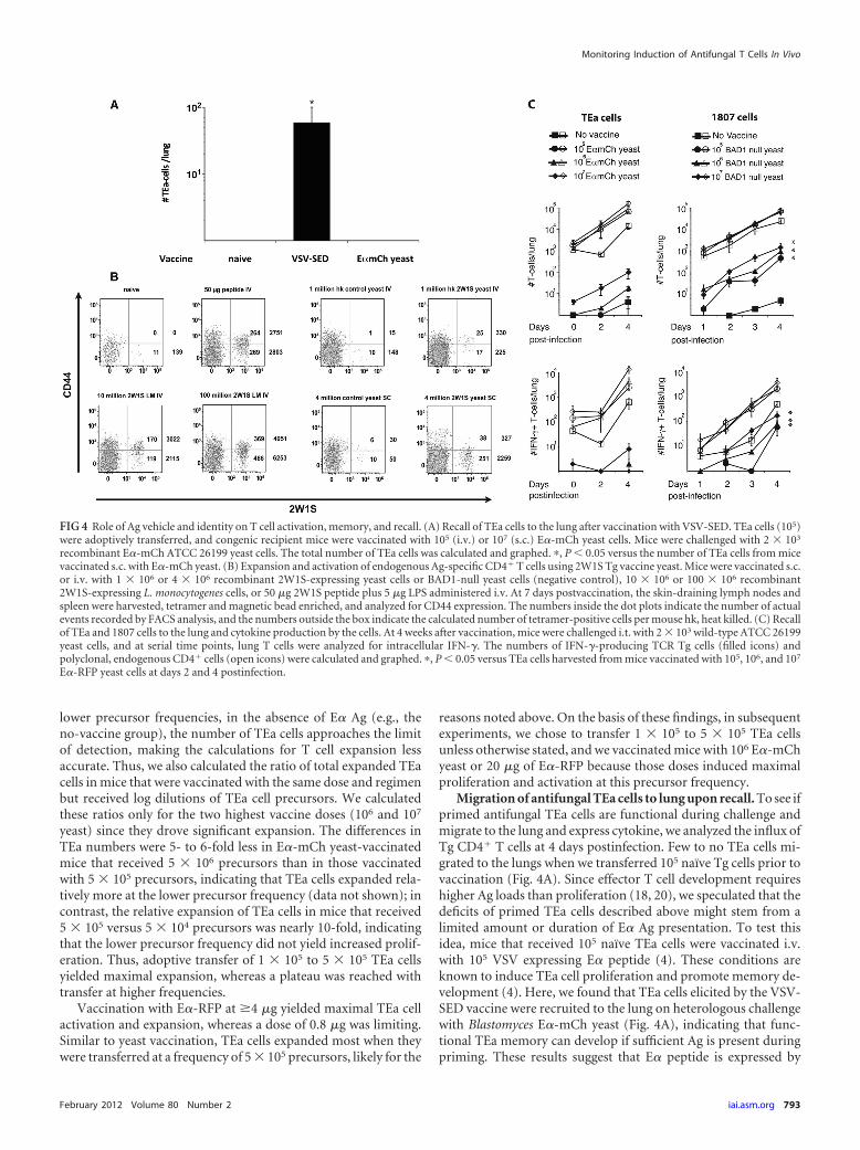
Migration of antifungal TEa cells to lung upon recall. To see ifprimed antifungal TEa cells are functional during challenge andmigrate to the lung and express cytokine, we analyzed the influx ofTg CD4� T cells at 4 days postinfection. Few to no TEa cells mi-grated to the lungs when we transferred 105 naïve Tg cells prior tovaccination (Fig. 4A). Since effector T cell development requireshigher Ag loads than proliferation (18, 20), we speculated that thedeficits of primed TEa cells described above might stem from alimited amount or duration of E� Ag presentation. To test thisidea, mice that received 105 naïve TEa cells were vaccinated i.v.with 105 VSV expressing E� peptide (4). These conditions areknown to induce TEa cell proliferation and promote memory de-velopment (4). Here, we found that TEa cells elicited by the VSV-SED vaccine were recruited to the lung on heterologous challengewith Blastomyces E�-mCh yeast (Fig. 4A), indicating that func-tional TEa memory can develop if sufficient Ag is present duringpriming. These results suggest that E� peptide is expressed by
FIG 4 Role of Ag vehicle and identity on T cell activation, memory, and recall. (A) Recall of TEa cells to the lung after vaccination with VSV-SED. TEa cells (105)were adoptively transferred, and congenic recipient mice were vaccinated with 105 (i.v.) or 107 (s.c.) E�-mCh yeast cells. Mice were challenged with 2 � 103
recombinant E�-mCh ATCC 26199 yeast cells. The total number of TEa cells was calculated and graphed. �, P � 0.05 versus the number of TEa cells from micevaccinated s.c. with E�-mCh yeast. (B) Expansion and activation of endogenous Ag-specific CD4� T cells using 2W1S Tg vaccine yeast. Mice were vaccinated s.c.or i.v. with 1 � 106 or 4 � 106 recombinant 2W1S-expressing yeast cells or BAD1-null yeast cells (negative control), 10 � 106 or 100 � 106 recombinant2W1S-expressing L. monocytogenes cells, or 50 �g 2W1S peptide plus 5 �g LPS administered i.v. At 7 days postvaccination, the skin-draining lymph nodes andspleen were harvested, tetramer and magnetic bead enriched, and analyzed for CD44 expression. The numbers inside the dot plots indicate the number of actualevents recorded by FACS analysis, and the numbers outside the box indicate the calculated number of tetramer-positive cells per mouse hk, heat killed. (C) Recallof TEa and 1807 cells to the lung and cytokine production by the cells. At 4 weeks after vaccination, mice were challenged i.t. with 2 � 103 wild-type ATCC 26199yeast cells, and at serial time points, lung T cells were analyzed for intracellular IFN-�. The numbers of IFN-�-producing TCR Tg cells (filled icons) andpolyclonal, endogenous CD4� cells (open icons) were calculated and graphed. �, P � 0.05 versus TEa cells harvested from mice vaccinated with 105, 106, and 107
E�-RFP yeast cells at days 2 and 4 postinfection.
Monitoring Induction of Antifungal T Cells In Vivo
February 2012 Volume 80 Number 2 iai.asm.org 793

recombinant yeast, but perhaps in amounts too small to promotethe development of functional memory. Nevertheless, the amountof E� peptide expressed by wild-type E�-mCh yeast upon lungrechallenge was sufficient to recruit T cells initially primed byVSV-SED but not E�-mCh yeast. Thus, it is possible that theamount of E� peptide expressed by Blastomyces Tg yeast limitedthe initial priming but not the recruitment of already primed TEacells during the recall response.
Expansion and activation of endogenous Ag-specific CD4� Tcells by recombinant yeast. The preceding studies required adop-tive transfer of relatively large numbers of Ag-specific T cells fromTCR Tg mice. In that approach, T cells may receive less intensestimulation when present in large numbers due to intraclonalcompetition and the limiting amounts of Ag available per naïve Tcell precursor (1, 4, 17, 24). To see if poor T cell recall to the lungand function may be due to the limiting amount of recombinantAg expressed by Blastomyces yeast, we investigated the activationand expansion of an endogenous Ag-specific CD4� T cell. Weengineered yeast that expresses 2W1S peptide (29). Tetramer-based enrichment recently showed the pool size of naïve 2W1S–I-Ab-specific CD4� T cells in C57BL/6 mice to be 190 precursors inthe spleen and lymph nodes (27). An i.v. injection of 2W1S pep-tide plus LPS increased the population of 2W1S–I-Ab-specific Tcells 300-fold to a peak of �80,000 cells by day 6 (27).
To see if 2W1S-expressing yeast induced proliferation and ac-tivation of corresponding endogenous Ag-specific CD4� T cells,we vaccinated mice s.c. with live yeast or i.v. with heat-killed yeastthat expresses surface 2W1S. As positive controls, mice were vac-cinated with 50 �g 2W1S peptide plus LPS- or 2W1S-expressing L.monocytogenes. The spleen and draining nodes were harvested andtetramer enriched, and the number and activation of 2W1Stetramer-positive cells were analyzed (26, 27). Vaccination with2W1S peptide plus LPS or recombinant L. monocytogenes in-creased the numbers of total and CD44hi tetramer-positive cells by40- and 80-fold, respectively, compared to those for naïve mice(Fig. 4B). In contrast, s.c. or i.v. vaccination with yeast expressing2W1S increased the number of tetramer-positive cells by 32- and3.4-fold compared to that for control vaccine yeast. Remarkably,only 15% � 2% of the tetramer-positive cells showed increasedsurface expression of CD44 after s.c. vaccination with recombi-nant yeast, suggesting that the primed cells had not been fullyactivated. Although Ag-specific T cells expanded less in mice vac-cinated i.v. with recombinant yeast, about half of the tetramer-positive cells expressed elevated CD44. Similarly, about half of thetetramer-positive cells in the groups vaccinated with peptide orrecombinant L. monocytogenes exhibited an activated phenotype(CD44hi). Thus, 2W1S yeast did modestly expand endogenousAg-specific CD4� T cells, but the activation state of these T cellswas surprisingly low, as measured by CD44 expression.
Migration of vaccine-induced 1807 cells into lung upon chal-lenge. The preceding studies with Tg yeasts expressing E� and2W1S suggest that low antigen expression may have limited thedevelopment of effector T cells. To investigate whether endoge-nous fungal antigens prime naïve CD4� T cells more efficientlythan the model antigens, we adoptively transferred naïve B.dermatitidis-specific 1807 cells into mice prior to vaccination withBAD1-null yeasts that do not express model antigens (6, 37).Comparable numbers of polyclonal CD4� T cells migrated intothe lungs after challenge and expressed gamma interferon (IFN-�)in mice that had received adoptively transferred TEa or 1807 cells
and were vaccinated with E�-mCh yeast or BAD1-null yeast (Fig.4C). In contrast, 10-fold fewer antifungal TEa cells than 1807 cellsmigrated into the lung and produced cytokine. Thus, the limitedrecall response by model Ag-specific TEa cells was not observedwith endogenous yeast Ag-specific 1807 cells.
Effect of Ag dose and duration on recall and function ofprimed TEa cells. To further explore the impact of Ag limitationon the recall response to the lung, we took a third approach. Weregulated the dose of Ag by providing supplemental E� proteinwith E�-mCh yeast vaccine, and we prolonged the duration of Agdelivery by emulsifying E�-RFP in IFA. E�-RFP emulsified in IFAinduced the maximal generation of effector/memory cells in thedraining lymph nodes at 5 weeks postvaccination (Fig. 5A). Theaddition of E�-RFP alone or with IFA to mice vaccinated withE�-mCh yeast also increased the number of effector TEa cells inthe draining nodes. However, the lung recall response was weakafter vaccination with E�-RFP plus IFA, similar to that after vac-cination with E�-mCh yeast (Fig. 5B). The addition of exogenousE�-RFP alone or in conjunction with IFA to the E�-mCh vaccinesignificantly enhanced the numbers of IFN-�- and IL-17-producing TEa cells upon lung recall. These results indicate thatAg dose and duration influence the quantity and quality of the TEarecall response and illustrate that the amount of model Ag ex-pressed by the recombinant vaccine is likely insufficient to drivean efficient recall of TEa effector cells into the lung. However,vaccination with E�-RFP alone was insufficient to induce maxi-mal responses, and Tg yeast in conjunction with high Ag loadsdelivered by E�-RFP led to maximal recruitment of TEa effectorsto the lung.
To investigate whether a lack of secondary expansion uponlung rechallenge contributes to the reduced numbers of TEa cellsin mice vaccinated with E�-mCh yeast, we measured TEa cellproliferation in the lung and MLN at day 4 postinfection. In allthree groups of vaccinated mice (mice vaccinated with TR20, E�-mCh, and E�-mCh–E�-RFP–IFA), �90% of the CD44�
CD62Llow TEa cells in the lung and MLN underwent proliferation,as measured by BrdU staining (Fig. 5C). Similarly, �90% of acti-vated 1807 cells from vaccinated mice underwent secondary pro-liferation in the lung and MLN upon rechallenge. Since BrdUincorporation does not indicate the number of cell divisions, wecannot exclude the possibility that T cells expanded to differentdegrees among the vaccine groups. Interestingly, we observed aninverse relationship between the number of proliferated and acti-vated T cells in the lung versus the MLN among the differentgroups of mice. Mice vaccinated with E�-mCh–E�-RFP–IFA orBAD1-null yeast showed efficient recruitment and/or secondaryexpansion of TEa and 1807 cells into the lung but poor secondaryexpansion into the MLN. On the other hand, poor recruitment ofT cells into the lungs of E�-mCh yeast-vaccinated mice or unvac-cinated mice was accompanied by increased secondary expansionin the MLN. In summary, recruitment and/or secondary expan-sion is the determining factor for the number of TEa and 1807 cellsfound in the lung of rechallenged mice.
DISCUSSION
An understanding of the factors that influence Ag presentation,priming of Ag-specific T cells, and memory T cell development isessential for the rational design of vaccines. In work describedhere, we sought to establish a heterologous adoptive transfer sys-tem to quantitatively and qualitatively monitor the development
Wüthrich et al.
794 iai.asm.org Infection and Immunity

FIG 5 Influence of Ag dose and duration on TEa cell activation, memory, and recall. (A) Effector/memory TEa cells in skin-draining lymph nodes at 6 weekspostvaccination. TEa cells (106) were adoptively transferred, and recipient mice were vaccinated with 107 E�-mCh yeast cells alone, E�-mCh yeast cells mixedwith 100 �g E� emulsified with IFA, or E�-mCh yeast cells plus 100 �g E�-RFP and IFA. Six weeks later, the skin-draining lymph nodes were harvested and TEacells were analyzed by FACS analysis. The data represent averages � SEMs of 4 to 6 mice per group from one representative experiment. The experiment wasrepeated twice with similar results. �, P � 0.05 versus �TR20-vaccinated group, ��, P � 0.05 versus all other groups. (B) Recall of TEa cells into the lung at 4 dayspostinfection. Mice received TEa cells and were vaccinated as described for panel A. At 5 weeks postvaccination, the mice were challenged with 2 � 104
E�-mCh-expressing wild-type (ATCC 26199) yeast cells. The number of cytokine-producing TEa cells was determined by FACS analysis. The data representaverages � SEMs of 6 to 8 mice per group from two independent experiments. �, P � 0.05 versus �TR20 group; ��, P � 0.05 versus E�-mCh yeast andE�-RFP–IFA groups; ���, P � 0.05 versus E�-mCh group. (C) Proliferation of T cells in the lung and MLN postchallenge. Mice were vaccinated as described forpanel A and challenged with 2 � 104 E�-mCh-expressing wild-type yeast cells. On the day of challenge and daily thereafter, mice received BrdU i.p. At 4 dayspostinfection, the mice were sacrificed and the number of BrdU-positive T cells was determined by FACS analysis. The data represent averages � SEMs of 4 to5 mice per group from one representative out of two independent experiments. �, P � 0.05 versus number of BrdU-positive TEa cells in groups vaccinated with�TR20 or E�-mCh yeast; ��, P � 0.05 versus number of BrdU-positive cells in the unvaccinated group.
Monitoring Induction of Antifungal T Cells In Vivo
February 2012 Volume 80 Number 2 iai.asm.org 795

of fungus-specific CD4� T cell memory. To establish a system tostudy antifungal CD4� T cell development in vivo, we performedextensive testing of the variables that influence T cell phenotypesand function. It is now recognized that the input frequency ofnaïve TCR Tg precursors and the availability of Ag are the mostimportant factors that dictate the fate of T cell development. Ahigh level of initial TCR Tg precursor has frequently led to anintrinsic defect in the T cells and resulted in reduced division,altered kinetics of expansion, reduced cytokine production, andthe lack of a detectable memory population (4, 17, 21, 24). In thosestudies, T cell competition could be overcome by adding more Ag(7, 34) or Ag-bearing APCs (36). Below, we discuss our resultsobtained using a heterologous, adoptive transfer system to studyantifungal CD4� T cell immunity. We analyzed selected stages ofthe antifungal T cell response, including activation, proliferation,differentiation, memory, and migration to the lung upon a recallchallenge.
We found that E�-mCh expressed on vaccine yeasts did notimpair their ability to confer resistance and that transgene expres-sion on wild-type yeast did not impair their virulence during re-challenge (data not shown). E�-mCh on yeast was processed andpresented on APCs, and the peptide triggered the activation andproliferation of corresponding TEa cells in an Ag-specific manner.TEa cells expanded maximally using an input precursor frequencyof 5 � 105 Tg cells and a vaccine dose of 106 to 107 yeast cells.Transfer of �5 � 103 TEa precursors did not yield a detectablenumber of primed TEa cells in the skin-draining lymph nodes atthe burst of expansion (at day 7 postvaccination). Fungus-specificTEa cells expanded a maximum of 1,000- to 3,000-fold using aprecursor frequency of 5 � 105 naïve cells and less than 1,000-foldusing higher and lower precursor frequencies (Fig. 3A). It is con-ceivable that the expansion of 5 � 104 TEa precursors might havebeen higher than we calculated since the number of TEa cells in theunvaccinated group was near the detection limit, and therefore,the calculations of the n-fold expansion factor might not havebeen as precise as in the higher-precursor-frequency groups.Thus, 5 � 104 to 5 � 105 TEa precursors had to be transferred toyield a measurable pool of primed TEa cells at the peak T cellresponse in the skin-draining lymph nodes.
Antifungal TEa cells primed with E�-mCh yeast alone failed toefficiently acquire memory and migrate to the lung upon chal-lenge. In contrast, adoptive transfer of comparable numbers (1 �105 to 5 � 105) of 1807 cells that recognize an endogenous fungalantigen led to the induction of memory and recruitment of effec-tor cytokine-producing 1807 cells into the lung (Fig. 4C). Wehypothesize that the amount of E� peptide expressed by vaccineyeast and/or the duration of Ag presentation was sufficient to in-duce proliferation, but not differentiation and memory develop-ment. This idea is compatible with earlier reports indicating thatthe signaling threshold for proliferation of naïve T cells is lowerthan the threshold necessary to drive the differentiation of mem-ory cells (4). Ag limitation characterized by the amount of Agavailable per T cell precursor can be the result of transferring toomany initial precursor T cells into recipient mice or limited avail-ability of the cognate Ag. However, we found that reducing theprecursor frequency to 104 naïve TEa cells and enriching the cellswith magnetic beads (17) at the time of harvest did not yield suf-ficient numbers of trackable TEa cells (data not shown). Rather,we found that delivery of recombinant VSV-SED vaccine restoredTEa functions, indicating that the vehicle (virus versus fungus)
and/or the delivery route (i.v. versus s.c.) critically impacts thelevels of Ag available per naïve T cell precursor. Interestingly,VSV-SED-primed TEa cells were efficiently recruited to the lungin response to rechallenge with E�-mCh yeast. These results sug-gest that the antigen threshold for recruiting already primed TEacells is lower than it is for TEa cells initially primed naïve duringvaccination.
To verify that poor TEa recall resulted from limited Ag expres-sion by recombinant E�-mCh vaccine yeast, we added exogenousE�-RFP to the vaccine yeast. The addition of E�-RFP alone orwith IFA significantly boosted the number of cytokine-producingTEa cells that migrated to the lung and expanded upon challenge,whereas administration of just E�-RFP plus IFA (without yeast)yielded poor recall responses and secondary expansion in the lung.These results are consistent with our hypothesis that the amountof Ag expressed by E�-mCh yeast alone during vaccination lim-ited the quality and quantity of functional memory TEa cells.However, because vaccine yeast added to E�-RFP enhanced lungrecall, our results also highlight the fact that the yeast is a potentadjuvant for inducing functional effector T cells.
We used an additional strategy to exclude the possibilities thatthe high frequency of transferred cells or the identity of the modelAg may have skewed our results. Here, we controlled for themeans of Ag expression (yeast) while changing the antigen andusing tetramer staining to track endogenous T cells. We engi-neered yeasts that express the 2W1S peptide, an epitope that isrecognized by the most frequent population of endogenous Ag-specific CD4� T cells known to date (27). The expansion andactivation of 2W1S tetramer-positive T cells in response to vacci-nation with recombinant yeast were modest compared to those inresponse to vaccination with recombinant L. monocytogenes infec-tion, and the population of primed (CD44�) 2W1S-specific T cellswas too small to evaluate memory and recall functions. This resultsupports our interpretation that the amount of yeast-displayedmodel Ag was limiting and did not exceed the necessary threshold.
We conclude that the heterologous adoptive transfer systemdescribed here is a powerful method to study the earliest events ofAg presentation to antifungal CD4� T cells. We showed that Y-AeMAb nicely detected yeast-derived E�–pMHC-II complexes onDCs. These levels of Ag display corresponded with robust T cellactivation, proliferation, and expansion of antifungal CD4� TEacells during the first week after vaccination. However, the down-stream effector, memory, and recall functions of TEa cells wereblunted by insufficient Ag display on the vaccine yeast. Thus, thisTEa Tg system is ideally suited to studying fungal Ag presentationand early T cell priming, whereas T cell effector and memory de-velopment may instead be studied with the autologous TCR Tg1807 cells described recently (37).
ACKNOWLEDGMENTS
We thank Roger Tsien from the University of California at San Diego forprovided the clone sequence for the mCherry gene.
This work was supported by NIH grants R21 AI076700 (to M.W.) andR01 AI40996 (to B.S.K.).
REFERENCES1. Badovinac VP, Haring JS, Harty JT. 2007. Initial T cell receptor trans-
genic cell precursor frequency dictates critical aspects of the CD8(�) T cellresponse to infection. Immunity 26:827– 841.
2. Beijersbergen A, Dulk-Ras AD, Schilperoort RA, Hooykaas PJ. 1992.
Wüthrich et al.
796 iai.asm.org Infection and Immunity

Conjugative transfer by the virulence system of Agrobacterium tumefa-ciens. Science 256:1324 –1327.
3. Bernard P, Couturier M. 1992. Cell killing by the F plasmid CcdB proteininvolves poisoning of DNA-topoisomerase II complexes. J. Mol. Biol. 226:735–745.
4. Blair DA, Lefrancois L. 2007. Increased competition for antigen duringpriming negatively impacts the generation of memory CD4 T cells. Proc.Natl. Acad. Sci. U. S. A. 104:15045–15050.
5. Brandhorst TT, Gauthier GM, Stein RA, Klein BS. 2005. Calciumbinding by the essential virulence factor BAD-1 of Blastomyces dermati-tidis. J. Biol. Chem. 280:42156 – 42163.
6. Brandhorst TT, Wüthrich M, Warner T, Klein B. 1999. Targeted genedisruption reveals an adhesin indispensable for pathogenicity of Blasto-myces dermatitidis. J. Exp. Med. 189:1207–1216.
7. Catron DM, Rusch LK, Hataye J, Itano AA, Jenkins MK. 2006. CD4�T cells that enter the draining lymph nodes after antigen injection partic-ipate in the primary response and become central-memory cells. J. Exp.Med. 203:1045–1054.
8. Cutler JE, Deepe GS, Jr, Klein BS. 2007. Advances in combating fungaldiseases: vaccines on the threshold. Nat. Rev. Microbiol. 5:13–28.
9. De Boer RJ, Homann D, Perelson AS. 2003. Different dynamics ofCD4� and CD8� T cell responses during and after acute lymphocyticchoriomeningitis virus infection. J. Immunol. 171:3928 –3935.
10. den Dulk-Ras A, Hooykaas PJ. 1995. Electroporation of Agrobacteriumtumefaciens. Methods Mol. Biol. 55:63–72.
11. Ertelt JM, et al. 2009. Selective priming and expansion of antigen-specificFoxp3-CD4� T cells during Listeria monocytogenes infection. J. Immu-nol. 182:3032–3038.
12. Fabien N, Bergerot I, Maguer-Satta V, Orgiazzi J, Thivolet C. 1995.Pancreatic lymph nodes are early targets of T cells during adoptive transferof diabetes in NOD mice. J. Autoimmun. 8:323–334.
13. Firpo EJ, et al. 2002. Antigen-specific dose-dependent system for thestudy of an inheritable and reversible phenotype in mouse CD4� T cells.Immunology 107:480 – 488.
14. Fisher LD, van Belle G. 1993. Biostatistics: a methodology for the healthsciences, p 611– 613. John Wiley & Sons, New York, NY.
15. Grubin CE, Kovats S, deRoos P, Rudensky AY. 1997. Deficient positiveselection of CD4 T cells in mice displaying altered repertoires of MHCclass II-bound self-peptides. Immunity 7:197–208.
16. Harvey RP, Schmid ES, Carrington CC, Stevens DA. 1978. Mousemodel of pulmonary blastomycosis: utility, simplicity, and quantitativeparameters. Am. Rev. Respir. Dis. 117:695–703.
17. Hataye J, Moon JJ, Khoruts A, Reilly C, Jenkins MK. 2006. Naive andmemory CD4� T cell survival controlled by clonal abundance. Science312:114 –116.
18. Iezzi G, Karjalainen K, Lanzavecchia A. 1998. The duration of antigenicstimulation determines the fate of naive and effector T cells. Immunity8:89 –95.
19. Itano AA, et al. 2003. Distinct dendritic cell populations sequentiallypresent antigen to CD4 T cells and stimulate different aspects of cell-mediated immunity. Immunity 19:47–57.
20. Jelley-Gibbs DM, Lepak NM, Yen M, Swain SL. 2000. Two distinct stagesin the transition from naive CD4 T cells to effectors, early antigen-dependent and late cytokine-driven expansion and differentiation. J. Im-munol. 165:5017–5026.
21. Kemp RA, Powell TJ, Dwyer DW, Dutton RW. 2004. Cutting edge:regulation of CD8� T cell effector population size. J. Immunol. 173:2923–2927.
22. Lara-Tejero M, Pamer EG. 2004. T cell responses to Listeria monocyto-genes. Curr. Opin. Microbiol. 7:45–50.
23. Lefebvre B, Formstecher P, Lefebvre P. 1995. Improvement of the genesplicing overlap (SOE) method. Biotechniques 19:186 –188.
24. Marzo AL, et al. 2005. Initial T cell frequency dictates memory CD8� Tcell lineage commitment. Nat. Immunol. 6:793–799.
25. Matz MV, et al. 1999. Fluorescent proteins from nonbioluminescentAnthozoa species. Nat. Biotechnol. 17:969 –973.
26. Moon JJ, et al. 2009. Tracking epitope-specific T cells. Nat. Protoc.4:565–581.
27. Moon JJ, et al. 2007. Naive CD4(�) T cell frequency varies for differentepitopes and predicts repertoire diversity and response magnitude. Im-munity 27:203–213.
28. Nemecek JC, Wüthrich M, Klein BS. 2006. Global control of dimor-phism and virulence in fungi. Science 312:583–588.
29. Rees W, et al. 1999. An inverse relationship between T cell receptoraffinity and antigen dose during CD4(�) T cell responses in vivo and invitro. Proc. Natl. Acad. Sci. U. S. A. 96:9781–9786.
30. Romani L. 2004. Immunity to fungal infections. Nat. Rev. Immunol.4:1–23.
31. Rudensky A, Rath S, Preston-Hurlburt P, Murphy DB, Janeway CA, Jr.1991. On the complexity of self. Nature 353:660 – 662.
32. Shaner NC, et al. 2004. Improved monomeric red, orange and yellowfluorescent proteins derived from Discosoma sp. red fluorescent protein.Nat. Biotechnol. 22:1567–1572.
33. Singh A, Wuthrich M, Klein B, Suresh M. 2007. Indirect regulation ofCD4 T-cell responses by tumor necrosis factor receptors in an acute viralinfection. J. Virol. 81:6502– 6512.
34. Smith AL, Wikstrom ME, Fazekas de St Groth B. 2000. Visualizing T cellcompetition for peptide/MHC complexes: a specific mechanism to mini-mize the effect of precursor frequency. Immunity 13:783–794.
35. Sullivan TD, Rooney PJ, Klein BS. 2002. Agrobacterium tumefaciensintegrates transfer DNA into single chromosomal sites of dimorphic fungiand yields homokaryotic progeny from multinucleate yeast. Eukaryot.Cell 1:895–905.
36. Willis RA, Kappler JW, Marrack PC. 2006. CD8 T cell competition fordendritic cells in vivo is an early event in activation. Proc. Natl. Acad. Sci.U. S. A. 103:12063–12068.
37. Wüthrich M, Ersland K, Galles KJ, Sullivan TD, Klein BS. Fungi subvertvaccine T-cell priming at the respiratory mucosa by inducing MMP2 andretarding CCL7-mediated influx of inflammatory monocytes. Immunity,in press.
38. Wüthrich M, Filutowicz HI, Klein BS. 2000. Mutation of the WI-1 geneyields an attenuated Blastomyces dermatitidis strain that induces hostresistance. J. Clin. Invest. 106:1381–1389.
39. Wüthrich M, Filutowicz HI, Warner T, Deepe GS, Jr, Klein BS. 2003.Vaccine immunity to pathogenic fungi overcomes the requirement forCD4 help in exogenous antigen presentation to CD8� T cells: implica-tions for vaccine development in immune-deficient hosts. J. Exp. Med.197:1405–1416.
40. Wüthrich M, Filutowicz HI, Warner T, Klein BS. 2002. Requisite ele-ments in vaccine immunity to Blastomyces dermatitidis: plasticity uncov-ers vaccine potential in immune-deficient hosts. J. Immunol. 169:6969 –6976.
41. Wüthrich M, Fisette PL, Filutowicz HI, Klein BS. 2006. Differentialrequirements of T cell subsets for CD40 costimulation in immunity toBlastomyces dermatitidis. J. Immunol. 176:5538 –5547.
42. Wüthrich M, et al. 2011. Vaccine-induced protection against 3 systemicmycoses endemic to North America requires Th17 cells in mice. J. Clin.Invest. 121:554 –568.
43. Wüthrich M, et al. 2011. A TCR transgenic mouse reactive with multiplesystemic dimorphic fungi. J. Immunol. 187:1421–1431.
44. Wüthrich M, Klein BS. 2000. Investigation of anti-WI-1 adhesinantibody-mediated protection in experimental pulmonary blastomycosis.J. Infect. Dis. 181:1720 –1728.
45. Wüthrich M, Warner T, Klein BS. 2005. IL-12 Is required for inductionbut not maintenance of protective, memory responses to Blastomycesdermatitidis: implications for vaccine development in immune-deficienthosts. J. Immunol. 175:5288 –5297.
Monitoring Induction of Antifungal T Cells In Vivo
February 2012 Volume 80 Number 2 iai.asm.org 797

![Belowground environmental effects of transgenic crops: a ... · phytic fungi (nor on earthworms, nematodes or protozoa) [19]. Small or no changes in culturable microflora were detected](https://img.dokumen.tips/doc/110x75/6051652101c7875ad445dbdf/belowground-environmental-effects-of-transgenic-crops-a-phytic-fungi-nor-on.jpg)