Embed Size (px)
Citation preview

University of Texas at El PasoDigitalCommons@UTEP
Open Access Theses & Dissertations
2019-01-01
Evaluation Of CTLA-4 Blockage Therapy WithMetronomic Chemotherapy For The Treatment OfPreclinical Breast CancerKarla ParraUniversity of Texas at El Paso
Follow this and additional works at: https://digitalcommons.utep.edu/open_etdPart of the Biology Commons
This is brought to you for free and open access by DigitalCommons@UTEP. It has been accepted for inclusion in Open Access Theses & Dissertationsby an authorized administrator of DigitalCommons@UTEP. For more information, please contact [email protected].
Recommended CitationParra, Karla, "Evaluation Of CTLA-4 Blockage Therapy With Metronomic Chemotherapy For The Treatment Of Preclinical BreastCancer" (2019). Open Access Theses & Dissertations. 2005.https://digitalcommons.utep.edu/open_etd/2005

EVALUATION OF CTLA-4 BLOCKAGE THERAPY WITH METRONOMIC
CHEMOTHERAPY FOR THE TREATMENT OF PRECLINICAL BREAST
CANCER
KARLA PARRA
Doctoral Program in Biological Sciences
APPROVED:
Giulio Francia, Ph.D., Chair
Renato Aguilera, Ph.D.
Charles Spencer, Ph.D. Siddhartha Das, Ph.D. Russell R. Chianelli, Ph.D. Anita Quintana, Ph.D.
Stephen Crites, Ph.D Dean of the Graduate School

Copyright ©
by
Karla Parra
2019

EVALUATION OF CTLA-4 BLOCKAGE THERAPY WITH METRONOMIC
CHEMOTHERAPY FOR THE TREATMENT OF PRECLINICAL BREAST
CANCER
by
KARLA PARRA, B.S
DISSERTATION
Presented to the Faculty of the Graduate School of
The University of Texas at El Paso
in Partial Fulfillment
of the Requirements
for the Degree of
DOCTOR OF PHILOSOPHY
Department of Biological Sciences
THE UNIVERSITY OF TEXAS AT EL PASO
August 2019

iv
Abstract
Background Although there are reports that metronomic cyclophosphamide can be immune
stimulating, the impact of its combination with anti-CTLA-4 immunotherapy for the treatment of
cancer remains to be evaluated.
Methods Murine EMT-6/P breast cancer, or its cisplatin or cyclophosphamide (CTX) resistant
variants, or CT-26 colon, were implanted into Balb/c mice. Established tumors were monitored for
relative growth following treatment with anti-CTLA-4 antibody alone or in combination with; a)
metronomic CTX (ldCTX; 20mg/kg/day), b) Bolus (150mg/kg) plus ldCTX, or c) sequential
treatment with gemcitabine (160mg/kg every 3 days).
Results EMT-6/P tumors responded to anti-CTLA-4 therapy, but this response was less effective
when combined with Bolus plus ldCTX. Anti-CTLA-4 could be effectively combined with either
ldCTX (without a bolus), or with regimens of either sequential or concomitant gemcitabine,
including in orthotopic EMT-6 tumors, and independently of the schedule of drug administration.
Tumor responses were confirmed with CT-26 tumors but were less pronounced in drug resistant
EMT-6/CTX or EMT-6/DDP tumor models than in the parent tumor. A number of tumor bearing
mice developed spontaneous metastases under continuous therapy. The majority of cured mice
rejected tumor re-challenges.
Conclusion Metronomic CTX can be combined with anti-CTLA-4 therapy, but this therapy is
impaired by concomitant bolus CTX. Sequential therapy of anti-CTLA-4 followed by gemcitabine
is effective in chemotherapy naïve tumors, although tumor relapses can occur, in some cases
accompanied by the development of spontaneous metastases.

v
Table of Contents
Abstract ................................................................................................................... iv
Table of Contents .................................................................................................... v
List of Figures ......................................................................................................... vi
List of Illustrations ................................................................................................ vii
Chapter 1: Introduction…………………………………………………………….1
1.1 Breast Cancer and Standard Therapies .................................................. 1
1.1.2 Immunotherapy ...................................................................................... 5
1.1.3 Materials and Methods .......................................................................... 8
1.1.4 Results ................................................................................................. 10
1.1.5 Discussion ............................................................................................ 33
Chapter 2: Alternative Therapies: Low Dose Chemotherapy ............................... 38
2.1 Low-dose Chemotherapy: Angiogenesis and the Immune System ..... 39
2.1.1 Mechanisms of resistance to metronomic chemotherapy .................... 40
2.1.2 Mechanisms of resistance to metronomic: Endothelial Resistance ..... 44
2.1.3 Mechanisms of resistance to metronomic: Host-driven resistance…. 45
2.1.4 Overcoming Resistance to Metronomic Chemotherapy ...................... 46
2.1.5 Metronomic Chemotherapy versus Maximum Tolerated Dose .......... 47
Chapter 3: Identification of New Anti-cancer Drugs for Combination Therapies 49
3.1.1 Introduction ......................................................................................... 49
3.1.2 Materials and Methods ........................................................................ 50
3.1.3 Results and Discussion ........................................................................ 54
Chapter 4: Summary .............................................................................................. 58
References ............................................................................................................. 61
Appendix ............................................................................................................... 76
Supplementary Figure 1 ............................................................................... 76
Supplementary Figure 2 and 3 ...................................................................... 77
Vita… .................................................................................................................... 80
vi
List of Figures
Figure 1A-C. Impact of Bolus plus low-dose CTX combined with anti-CTLA-4 therapy on the growth of EMT-6/P tumors. .......................................................................................................... 13 ....................................................................................................................................................... 14 Figure 1D. Impact of the different therapies as assessed by analysis of event-free survival (Kaplan-Meier analysis), where duration of event-free survival is defined as time to primary tumor progression beyond 1,200 mm3 or >15% weight loss. .................................................................. 14 Figure 2. Effective combination of chemotherapy with anti-CTLA-4 therapy for the inhibition of the growth of EMT-6/P tumors. .................................................................................................... 18 ....................................................................................................................................................... 21 Figure 3. Combination of chemotherapy with anti-CTLA-4 therapy for the inhibition of the growth of EMT-6/CTX tumors, including gemcitabine administration (as indicated by the green and orange arrows). .............................................................................................................................. 23 ....................................................................................................................................................... 25 Figure 4. Combination of chemotherapy with anti-CTLA-4 therapy for the inhibition of the growth of EMT-6/DDP tumors. ................................................................................................................. 26 ....................................................................................................................................................... 29 Figure 5. Anti-CTLA-4 therapy plus gemcitabine inhibits orthotopic EMT-6 tumors. ................ 29 ....................................................................................................................................................... 30 Figure 6. Impact of anti-CTLA-4 therapies on CT-26 colon tumors. ........................................... 30 ....................................................................................................................................................... 31 Figure 7. Evaluation of different anti-CTLA-4 combination therapies on EMT-6/P tumor growth. ....................................................................................................................................................... 31 ....................................................................................................................................................... 32 Figure 8. Analysis of CD31-positive staining in EMT-6 tumor treated with anti-CTLA-4 therapies. ....................................................................................................................................................... 32 ....................................................................................................................................................... 53 Figure 9. Identified HITS from PremiumSet and DIVERSet libraries tested on LM2-4-luc cells. ....................................................................................................................................................... 53 ....................................................................................................................................................... 55 ....................................................................................................................................................... 55 Figure 10. CC50 for compound 5935953. ...................................................................................... 55 ....................................................................................................................................................... 56 Figure 11. Compound 5935953 as first line therapy with sequential PTX. .................................. 56 Figure 11. Cell cycle profile for compound 5935953. .................................................................. 57 ....................................................................................................................................................... 59 Figure 12. Expression of proinflammatory immune response chemokines. ................................. 59 ....................................................................................................................................................... 76 Supplementary Figure 1. Examples of spontaneous metastasis under continuous therapy, from subcutaneously implanted EMT-6 tumor models .......................................................................... 76 Supplementary Figure 2 and 3. Evaluation of CT-26 tumor with CTLA-4 therapy. ..................... 77

vii
List of Illustrations
Illustration 1: Mechanisms of Resistance to Metronomic Chemotherapy. ................................... 42 Illustration 2: DNS Assay workflow adapted from Lema et al., 2011. ......................................... 52

1
Chapter 1: Introduction
1.1 Breast Cancer and Standard Therapies
Cancer is a disease that causes uncontrolled growth of mutated cells that spread through
the body. Breast cancer originates in either the lobules (i.e., milk production glands) or ducts (i.e.,
tubes carrying milk) of the breast tissue. Breast cancer progresses from an early stage to a more
advanced stage by forming a lump or tumor made up of mutated cells, where the cells can enter
the nearby blood vessels and spread to a secondary site (a process termed metastasis). It is
projected that by 2018, there will be 266,120 new cases of invasive breast cancer and 63,960 cases
of in situ breast lesions diagnosed in women in the United States1. In Texas alone, by 2018 there
will be 18,260 new cases of invasive and in situ breast cancer combined. The predicted death rate
for women diagnosed with breast cancer in Texas is 120.2 per every 100,000 cases reported for
2018. From the 18,260 new cases of invasive and in situ breast cancer combined in Texas, 2,880
will result in death1.
The vast majority of breast cancers are sporadic in origin and are not restricted to women
only, breast cancer can also develop in men, though men have a relatively lower risk (1 in 1,000)
of getting the disease. The most common symptom of breast cancer is the appearance of a lump in
the breast tissue. Other symptoms of breast cancer include inverted nipples, swelling, skin
irritation, redness, and unusual nipple discharge. However, early breast cancer disease has no
symptoms, therefore early detection via mammography screening is vital. Oncologists look for
calcifications (i.e., white spots of calcium deposits) in the mammography screening that indicate
a change in the breast tissue. Not all calcifications are an indication of breast cancer, some are due
to changes caused by age or injuries (e.g., macrocalcifications). Smaller calcium deposits, called
2
microcalcifications, need to be checked via a biopsy to discard the possibility of breast cancer.
Treatment for breast cancer often involves either surgical removal of the tumor (lumpectomy) or
surgical removal of the breast (mastectomy) along with a chemotherapy or radiation regimen
depending on the subtype of breast cancer.
There are different molecular subtypes (e.g. luminal A, luminal B, Her-2 positive, basal,
and normal like) of breast cancer each with a distinct morphology and clinical implication2. For
example, HER-2 positive breast cancer is a subtype of breast cancer that has an overexpression of
the human epidermal growth factor receptor 2 (HER2) protein on cells. Mutation of the HER2
gene causes an overexpression of the HER2 protein therefore promoting the growth of cells at
uncontrollable rates. This uncontrolled growth rate is the major driver of tumor development
making HER2 an oncogene, or a gene that can cause cancer. HER2 overexpression has been
observed in 20% of the breast cancers diagnosed in women, making it a crucial therapeutic target3.
Targeted therapies against HER2 protein include humanized monoclonal antibodies (e.g.,
trastuzumab, pertuzumab) and tyrosine kinase inhibitors (e.g., lapatinib). Monoclonal antibodies
(mAb) prevent the dimerization (outside of the cell) of HER2 receptors with other HER family
members thus halting the continual expansion of cells. On the other hand, lapatinib is a drug that
targets the tyrosine kinase domain of the HER2 (as well as HER1) receptor inside the cell thus
halting HER activation.
Other subtypes of breast cancer require the use of chemotherapeutic regimens to halt the
progression of the tumor. Chemotherapy regimens may be given either before (i.e., neoadjuvant
chemotherapy) or after (i.e., adjuvant chemotherapy) the surgical removal of the tumor. A typical
chemotherapy regimen consists in administering drugs at maximum tolerable doses (MTD) in a
specific number of cycles over a set period of time with breaks in between each dose to allow for

3
recovery. For example, cyclophosphamide (a DNA alkylating agent) in combination with
docetaxel (a drug that prevents microtubule disassembly) is administered as a standard adjuvant
chemotherapy for HER2 negative breast cancers4.
Cyclophosphamide (CTX) was synthesized as a chemotherapeutic agent derived from
nitrogen mustards - it alkylates DNA (via intermediates that are generated by the liver) thus
damaging DNA. The general mechanism and structure of nitrogen mustard compounds is that you
have a nitrogen in the center and two chloroethyl groups attached to it. These chloroethyl groups
are the main characteristic of nitrogen mustard compounds. In CTX you have a phosphorus atom
double bonded to oxygen and a 6-member ring. Nitrogen mustard will attack guanine nucleotides
in the DNA due to a series of events. The carbon from the ethyl group (in the nitrogen mustard
group) binds to the double bond in the nitrogen center forming a positive charge (carbonium ion).
This forms a very unstable 3-member ring, termed immonium ion and cleaves off the chloride
atom (forming a chloride anion). The carbonium ion then attacks guanine organic base at the N7
position by nicking electrons. This transfers the positive charge from the carbonium ion to the
nitrogen (N7) at the guanine organic base. An alkyl group has now been attached to the organic
base, resulting in alkylating DNA. This process can keep going resulting in interstrand or
intrastrand alkylating DNA. CTX requires enzymatic activation by hepatic cytochrome P-450 to
form 4-hydroxycyclophosphamide, which is then able to diffuse within the cell as
aldophosphamide and 4-hydroxycyclophosphamide (non-toxic metabolites). Aldophosphamide is
later activated as a toxic metabolite and cleaved into nitrogen mustard resulting in interstrand and
intrastrand DNA crosslinking. This crosslink is thought to result in the cytotoxic properties of
CTX- which is primarily given in combination with other chemotherapeutic agents rather than as
a monotherapy5.

4
In the event that breast cancer transitions to metastatic disease (e.g., spread to distant sites)
due to relapse or late stage diagnosis, different therapies are administered. Gemcitabine has been
extensively studied and used an anticancer drug for the treatment of metastatic pancreatic cancer,
non-small cell lung cancer, bladder and breast cancer6–8. Gemcitabine is a cytidine analogue that
requires cellular uptake by human equilibrative nucleoside transporters (hENT) and intracellular
pohosporylation by deoxycytidine kinase (dCK) and thymidine kinase 2 (TK2) in order to be
converted into its two active metabolites, gemcitabine di- and triphosphate (dFdCDP, and
dFdCTP)9. Integration of dFdCTP into DNA is considered to be the main method by which
gemcitabine causes cell apoptosis. Furthermore, dFdCDP metabolite inhibits ribonucleotide
reductase (RR) leading to a more efficient phosphorylation of gemcitabine. This enhancement of
the overall inhibitory activities on cell growth is termed self-potentiation.
hENT1 transports gemcitabine into the cell; cells lacking this transporter are resistant to
gemcitabine therapy10. The presence of NTs are a key factor for cell growth inhibition when
treating with gemcitabine. Gemcitabine is then phosphorylated by dCK (deoxycytidine kinase) and
TK2 (thymidine kinase 2) to produce gemcitabine monophosphate (dFdCMP). Gemcitabine is
inactivated within the cell by deoxycytidine deaminase (dCDA) and converted into
difluorodeoxyuridine monophosphate (dFdUMP) and subsequently to difluorodeoxyuridine
(dFdU) [Heinemann]. These substrates are either excreted out of the cell due to the decreased
affinity and competitive binding of deoxycytidine11, or are kept as dFdCMP. If dFdCMP is not
exported outside the cell, then it is converted to its two active metabolites; dFdCDP, and dFdCTP.
After dephosphorylation, gemcitabine is transformed to gemcitabine triphosphate (dFdCTP) and
starts accumulating in the cell. dFdCTP outcompetes deoxycytidine triphosphate (dCTP) to be
incorporated into the DNA12. After incorporation of dFdCTP on the end of the elongating DNA

5
strand, an additional deoxynucleotide is added thus preventing DNA polymerases to continue. This
mechanism (commonly termed as masked termination) prevents detection by the proofreading
enzymes13. Despite the advances in therapy, breast cancer remains the second leading cause of
cancer death among women. There is a current need to develop new therapies or combination of
therapies to treat breast cancer. Several therapies available that are not precisely against breast
cancer but have shown significant overall survival against other cancers, such as melanoma may
have the potential to reduce breast cancer mortality.
1.1.2 IMMUNOTHERAPY
In 1984, Dr. Steven Rosenberg of the National Cancer Institute treated a woman diagnosed
with metastatic melanoma with interlukin-2 (IL-2) infusion therapy. IL-2 helps different T-cell
populations to proliferate and activate the immune system. Linda Taylor became the first patient
to successfully respond to an immunotherapy. The FDA approved IL-2 therapy in 1992 and 1998
for the treatment of metastatic renal cancer and melanoma. On the same note, in 1996, Jim
Allison’s team reported that by blocking an immune system checkpoint, cytotoxic T lymphocyte
antigen-4 (CTLA-4), the immune system was able to re-ignite an attack against tumors. The New
England Journal of Medicine issued a publication revealing the effectiveness of ipilimumab
therapy, a humanized antibody against CTLA-4, that delayed tumor progression and prolonged the
lives of patients with metastatic melanoma. Immunotherapy has opened up new avenues for
combination therapies. Nivolumab, an antibody targeting programmed cell death protein (PD-1)
expressed on T-cells was approve in combination with ipilimumab for the treatment of metastatic,
non-resectable melanoma in 2015 (DiGiulio S, 2015).

6
CTLA-4, is a receptor that restricts T-cell response after co-stimulatory activation. The
activation of T-cells and requires two processes, (1) presentation of antigen and, (2) activation of
co-stimulatory signals14. Antigen presenting cells (APCs) process foreign or abnormal antigens,
and they present on their cell surface for display through a protein known as major
histocompatibility complex class II (MHC-II). Helper T-cells (TH) express specific T-cell receptors
(TCR) on their surface that are able to recognize and engage the specific antigenic peptide bound
to MHC-II. When engaged with MHC-II, TH cells send co-stimulation signals through CD28 to
induce activation of cytotoxic T cells (Tc) by binding to APCs via B7-1 and B7-2 receptors in
addition to the TCR. CD28 is the most extensively studied co-stimulatory receptor on T-cells15–17.
When TCR is bound to MHC-II and CD28 is bound to B7-1 (or B7-2), an immune response is
stimulated inducing the proliferation of T-cells (due to IL-2 secretion). Preventing overstimulation
of the immune system requires the synthetization and activation of CTLA-4 on activated T cells.
Expression of CTLA-4 varies depending on the T-cell population – transient expression on CD8
T-cells but constitutive on T-regulatory (TReg) cells18. CTLA-4 checkpoint re-establishes
homeostasis after activation of immune responses, which some tumors use for their advantage to
resist immune-mediated killing. CTLA-4 is approximately 20% homologous to CD28 as they share
similar structures and are 27% identical in mice and 31% in human19. Brunet et al, reported that
the CTLA-4 receptor is a competing co-stimulatory inhibiting signal that binds to the same ligands
(with higher avidity) as CD28. Blocking CTLA-4 receptor with antibodies restores T-cell
expansion and proliferation by dephosphorylating TCR signals (e.g., CD3) thus inducing a
heightened immune response20.
The overall survival of patients diagnosed with breast cancer still depends on how far the
cancer has spread to other organs. Patients who respond to immunotherapy (e.g., checkpoint
7
blockade therapy) seem to have a long-term benefit that can be further enhanced with the
combination of traditional treatment modalities such as chemotherapy. Immunotherapy’s success
is mainly due to the known immunogenicity of melanoma and pancreatic tumors. Breast cancer
has been reported to be not immunogenic therefore suggesting no response to immunotherapy.
However, the proposed preclinical study in this thesis and other clinical studies now suggest that
immunotherapy in combination with chemotherapy has improved the outcome for breast cancer
patients. Recently, atezolizumab – an anti-programmed death-ligand 1 antibody (PD-L1) was
approved in combination with paclitaxel for the treatment of metastatic triple-negative breast
cancer in patients whose tumors express PD-L1. This thesis reports the effective combination of
an anti- cytotoxic T lymphocyte antigen-4 (CTLA-4) antibody in combination with gemcitabine
and/ or cyclophosphamide for the treatment of breast cancer in a preclinical mouse model.
In 2010, fourteen years after the report of CTLA-4 blockade causing tumor responses in
preclinical models by blocking the immune suppressive functions of the CTLA-4 protein, the anti-
CTLA-4 antibody ipilimumab was approved by the FDA for the treatment of non-resectable or
metastatic melanoma. This approval was a pivotal event for cancer immunotherapy21,22, a field now
enriched by additional targets such as PD-1, PD-L1, and LAG-321–23. Despite these successes, there
remain several hurdles to be overcome in the quest for optimal anti-CTLA-4 based therapy
regimens, including minimizing the likelihood of the development of autoimmune toxicity21,24,25,
or devising means to overcome the evolution of drug resistance to therapy26. Currently, there is a
growing interest to improve anti-CTLA-4 therapy by exploiting the immunostimulating properties
of some conventional chemotherapeutics27,28. In this study, we tested whether continuous low-dose
(metronomic) chemotherapy, in this case cyclophosphamide (CTX), which has been reported to
act in part by boosting the immune system17,27,29,30, could be effectively combined with CTLA-4

8
antibody therapy for the treatment of breast cancer in a preclinical model. Surprisingly, we found
that our previously designed protocol31, consisting of bolus (high-dose) CTX injection combined
with oral low-dose CTX, actually hindered the anti-tumor efficacy of anti-CTLA-4 therapy.
Conversely, we noted that metronomic CTX (without an upfront bolus) can enhance anti-CTLA-
4 therapy.
Furthermore, even more impressive tumor responses were obtained using a sequential
regimen of CTLA-4 blockade followed by a previously described32 metronomic gemcitabine
chemotherapy (160mg/kg, every 3 days), irrespective of whether it was evaluated on the parent
EMT6/P tumor or on variants selected for resistance to cisplatin or to CTX. We also noted that
acquired drug resistance (at least in a subset of mice) was observed with all therapies evaluated in
this study, as was the emergence of spontaneous metastases. Our results contribute to our
understanding of the preclinical benefits of chemotherapy regimens in combination with CTLA-4
blockade33–35. They also serve as cautionary notes in that some regimens (e.g., high-dose CTX)
may hinder the beneficial anti-tumor effects of CTLA-4 blockade-based therapies, and that the use
of chemotherapy naïve tumor models36.
1.1.3 MATERIALS AND METHODS Drug Preparation: Gemcitabine Hydrochloride was purchased from Selleck Chemicals
(Houston, TX) and made up in sterile phosphate buffered saline (PBS) immediately prior to i.p.
administration. CTX was purchased from Sigma and made up in PBS prior to i.p. injection or prior
to its addition to the mice’s drinking water. Metronomic low-dose CTX (ldCTX) was administered
at an estimated 20mg/kg/day as previously described (Man et al., 2002). Some regimens (termed
B+ldCTX) included an upfront Bolus dose of CTX, administered on day 1 as a 150mg/kg i.p.
injection of CTX 31,32.

9
Anti-CTLA-4 Antibody Preparation: Anti-mouse CD152 (CTLA-4), FG purified clone
9H10, purchased from Ebioscience (San Diego, CA), was diluted in PBS immediately prior to i.p.
injection. Mice were administered 100µg of the antibody on day 1 of treatment, followed by a
35µg injection on day 6.
Cell lines: Murine EMT-6/P mammary carcinoma cells (ER+/PR+/HER2+, B. Teicher –
personal communication, and as previously reported37,38, and the CTX resistant EMT-6/CTX and
cisplatin resistant EMT-6/DDP variants, were a gift from Beverly Teicher, and they were grown
in RPMI supplemented with 10% fetal bovine serum and 2mM L-glutamine. Cells were grown in
a humidified incubator at 37C and 5% CO2.
In vivo tumor growth assessment: Six-week-old female Balb/c mice were purchased from
Harlan (Indianapolis, IN). Mice were allowed to acclimatize for 2 weeks before implantation of
tumor cells. To prepare cells for injection, subconfluent plates were harvested with 1% trypsin-
EDTA, and cells were then washed and resuspended in RPMI at 2 million cells per ml. Two
hundred thousand EMT-6 cells were injected subcutaneously into the flank of the mice (for CT26
cells, 1 million cells per mouse were implanted). Mice were monitored twice weekly for
fluctuations in body weight, and for tumor growth, as measured by Vernier calipers, and tumor
volume was calculated by the formula (length x width2)/2. Institutional guidelines were followed
to determine when the experimental end points were reached. Results were also plotted as event-
free survival (Kaplan-Meier analysis) over time, where duration of event-free survival is defined
as time to primary tumor progression beyond 1,200 mm3 or >15% weight loss, as per our previous
study39. Primary tumor fragments, or established lung metastases, were isolated from selected
euthanized mice, and used to derive cell cultures as previously described40,41. The orthotopic
implant of EMT-6 and EMT-6DDP cells was carried out as previously described 40,41; one hundred
10
thousand cells in 50 microlitres were implanted in the inguinal mammary fat pad of mice. All in
vivo procedures and experiments were performed with the approval of the UTEP IACUC (IACUC
reference #: A-201201-1).
Immunohistochemical Analysis: Paraffin embedded EMT-6 tumor sections were cut to 5
micron thickness and stained for anti-CD31 (Abcam 28364) used at a dilution of 1:400, using an
antigen retrieval of citrate buffer pH6. Secondary antibody was goat anti-rabbit at a dilution of
1:200, using DAB for detection of positive staining, and counter stained with Hematoxylin for
contrast.
Statistical Analysis: The analysis of variance among groups (ANOVA), followed by the Student-
Newman-Keuls test, was used to assess the statistical differences of data in vivo. Tumor therapy
results are reported as mean ± SD. Survival curves were plotted by the method of Kaplan and
Meier and were tested for survival differences using the log-rank test. The level of significance
was set at P<0.05.
1.1.4 RESULTS Anti-CTLA-4 therapy combined with bolus plus low-dose CTX: To evaluate whether
metronomic CTX can be effectively combined with anti-CTLA-4 treatment, we tested a
combination regimen on subcutaneously implanted EMT-6/P tumors (Fig. 1A and 1B). For the
chemotherapy component, we sought to use a B+ldCTX protocol consisting of a Bolus CTX (given
i.p. on day 1) plus low-dose CTX (20mg/kg/day, p.o.). Our choice was guided by our previous
study (Shaked et al., 2005) in which the B+ldCTX protocol was shown to more effectively inhibit
tumor growth than the sole low-dose CTX in the EMT6/P tumor, as well as in other tumor models.
Mice (n=32) bearing EMT-6/P tumors were treated with saline (control), B+ld CTX, anti-CTLA-
4 antibody, or with B+ldCTX plus anti-CTLA-4 antibody. Figure 1B shows the resulting impact
11
of the therapies on tumor growth. Thus, control treated tumors grew rapidly, the B+ldCTX
treatment slowed down tumor growth, whereas anti-CTLA-4 antibody treatment caused tumor
regressions over a 20 day period – followed by tumor relapses in the subsequent 15 days.
Surprisingly, the B+ldCTX plus anti-CTLA-4 combination therapy did not produce tumor
regressions and, furthermore, it produced a tumor growth rate that was only marginally slower
than was observed with B+ldCTX alone. Thus, B+ldCTX significantly hinders the efficacy of
antiCTLA-4 therapy in the EMT6/P tumor model. This was an unexpected finding, as we had
recently reported that B+ldCTX could be effectively combined with an anti-VEGFR2 antibody41,
or with metronomic oral gemcitabine (LY2334737)40. We also noted that by day 22 the CTLA-4
antibody monotherapy resulted in complete tumor regression in 2 mice, one of which then began
to show tumor regrowth a few days later. All anti-CTLA-4 treated tumors shrank after the therapy
began, although tumor relapses were eventually observed in 6 out of 7 mice in this group.

12
#
# p<.05 vs. CTLA-4 #
# #
#
#
#
#
# # #
* p<.05 vs. Control
*
* * * *
1A.
1B.
1C

13
Figure 1A-C. Impact of Bolus plus low-dose CTX combined with anti-CTLA-4 therapy on the growth of EMT-6/P tumors.
A) Schematic of evaluation of anti-CTLA-4 therapies with metronomic chemotherapy. EMT-6/P breast tumors, or CTX resistant (EMT-6/CTX) or DDP resistant (EMT-6/DDP) tumors were treated with anti-CTLA-4 antibody (administered on days 1 and 6). Chemotherapy regimens included low-dose metronomic CTX (ldCTX), Bolus plus ldCTX, or gemcitabine. Confirmatory studies were carried out with the murine CT-26 colon tumor. B) Murine EMT-6/P cells were implanted s.c. in female Balb/c mice. Therapies began when tumors were 50mm3; the mice received control (n=8) saline (i.p.), anti-CTLA-4 (n=7), Bolus plus low-dose CTX (n=10), or the combination (n=7) of anti-CTLA-4 together with Bolus plus low-dose CTX. *P<0.05 vs. control, #P<0.05 vs. CTLA-4 (mean values ±SD). C) Mouse weights, as a measure of toxicity of the different treatments. D) Impact of the different therapies as assessed by analysis of event-free survival (Kaplan-Meier analysis), where duration of event-free survival is defined as time to primary tumor progression beyond 1,200 mm3 or >15% weight loss. Significant event-free survival was observed with anti-CTLA-4 therapy, but this benefit was reduced by the addition of Bolus + low dose CTX. The sole survivor, by day 46, in the anti-CTLA-4 therapy group was still alive and tumor free at day 400 after tumor cell injection. *P<.05 was taken as statistical indication of difference vs. controls and between treated groups.
Therefore, anti-CTLA-4 therapy is effective in the EMT-6/P tumor model, but its
therapeutic efficacy is significantly hampered by concurrent B+ldCTX treatment. To assess the
relative toxicity of the therapies, we monitored body weights of the mice in the course of the
experiment (as per our previous studies32,39. Figure 1C shows that the treatments that included a
B+ldCTX component produced a short-term weight loss (as previously reported Shaked et al.,
2005), followed by a gain in weight by the treated mice. We also plotted the tumor responses as a
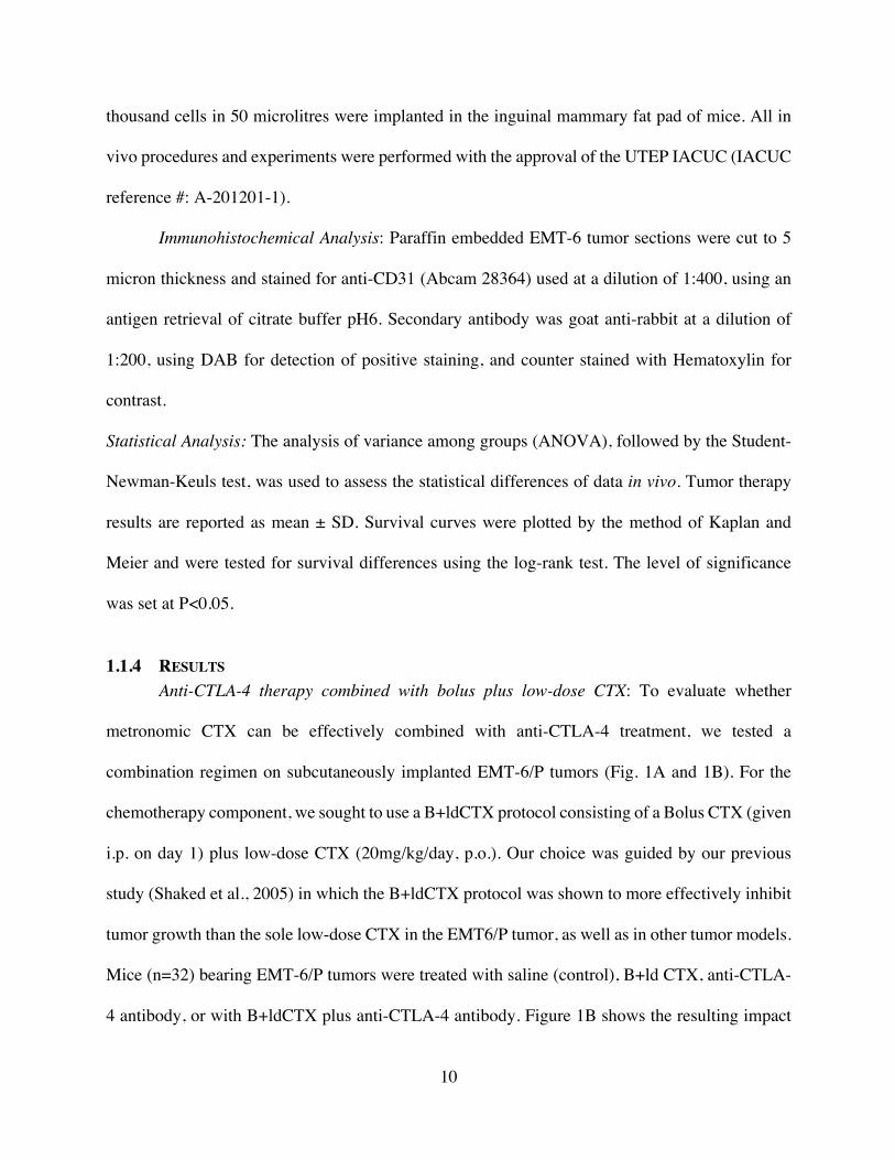
Kaplan-Meier plot (Fig. 1D), which shows that time to 50% event-free survival of CTLA-4
antibody treatment was 38 days, a significant increase compared to 22 days for the control group
(P= 0.0011 CTLA-4 vs control). B+ldCTX had no significant impact on survival. Fig. 1D also
shows that one anti-CTLA-4 antibody treated mouse, which had been bearing a palpable tumor in
the first 2 weeks of this experiment, showed a tumor regression and remained tumor-free for the
whole follow-up period. This mouse was still tumor-free 400 days later.
14
Figure 1D. Impact of the different therapies as assessed by analysis of event-free survival (Kaplan-Meier analysis), where duration of event-free survival is defined as time to primary tumor progression beyond 1,200 mm3 or >15% weight loss.
Significant event-free survival was observed with anti-CTLA-4 therapy, but this benefit was reduced by the addition of Bolus + low dose CTX. The sole survivor, by day 46, in the anti-CTLA-4 therapy group was still alive and tumor free at day 400 after tumor cell injection. *P<.05 was taken as statistical indication of difference vs. controls and between treated groups.
Anti-CTLA-4 therapy combined with low-dose CTX, or with sequential gemcitabine
therapy: We next decided to test whether we could incorporate other chemotherapy regimens,
either in combination with or subsequent to the anti-CTLA-4 administration. We reasoned that
since high-dose CTX can be immunosuppressive5, the bolus CTX dose might impair the immune
stimulating impact of the CTLA-4 blockade – thus negating its therapeutic benefit. Therefore, we
either had to separate time of the dosing of chemotherapy from that of the anti-CTLA-4 antibody
or omit the bolus CTX component. We chose to evaluate two additional strategies. One was to
combine anti-CTLA-4 administration with low-dose CTX (i.e., without a bolus). This choice was
based on reports that (in contrast to the high-dose CTX regimens) low-dose CTX can stimulate the
1D.

15
immune system27,29,30. Furthermore, in our previous studies, low-dose CTX effectively inhibited
tumor growth without producing any obvious toxicity40,42. The second strategy involved the
incorporation of a metronomic regimen of 160mg/kg gemcitabine given every 3 days, since we
recently observed that it can produce remarkable tumor responses in a preclinical breast cancer
model32. However, to avoid the possibility that gemcitabine might impair the therapeutic effect of
the CTLA-4 blockade, we decided to administer the two drugs sequentially (i.e., after tumors
began to regress following the anti-CTLA-4 treatment).
To evaluate these alternative therapies, we implanted EMT-6/P tumors into 43 mice, which
were subsequently divided into 7 groups. Therapies began when all mice had established tumors
and when the average tumor volume was approximately 50mm3. We noted no or minimal impact
of tumor growth, compared to controls, in the groups treated with ldCTX (Fig. 2A). We also noted
that anti-CTLA-4 therapy led to a significant (initial) tumor regression in the first 2 weeks after
therapy started, followed by tumor relapses. The combination of anti-CTLA-4 plus ldCTX (both
co-administered from day 6 onwards) produced a greater inhibition of tumor growth that was
observed with the anti-CTLA-4 monotherapy, although the results did not reach a statistical
significance. The group treated with a single dose of gemcitabine unexpectedly showed weight
loss in the days following treatment, and therefore this treatment was interrupted; the treatment
caused initial inhibition of tumor growth, followed by tumor re-growth (Fig 2A), and a recovery
of mouse body weight (Fig 2B). One group (treated with anti-CTLA-4 then Gem) initially received
anti-CTLA-4 antibody, which again produced a tumor regression that lasted 2 weeks, followed by
a tumor relapse (Fig. 2A). For this group, as soon as tumors started to relapse (i.e., around day 21),
the second line therapy of gemcitabine was administered. In this group, we did not observe any
weight loss after gemcitabine injection, and therefore gemcitabine treatment was continued. At the

16
onset of the second-line gemcitabine therapy on day 21, all mice in this group had visible tumors
and an average tumor volume of 145mm3. Gemcitabine administration caused tumors to regress
again, and by day 36 only two out of the six mice had any palpable tumors. These two mice
eventually showed tumor re-growths, whilst under continuous gemcitabine therapy (Fig 2C). The
remaining 4 mice showed significant (P<0.05) and complete tumor regression and are still tumor-
free over 400 days later.
With regard to the relative toxicity of the tested therapies, other than the single gemcitabine
treatment group as noted above, no significant changes in mouse weight were observed compared
to controls (Fig. 2B). As noted in the subsequent studies (detailed below), we infrequently
observed toxicity following gemcitabine injection, and this was typically resolved by allowing the
mice a break from the treatment, as in our previous study43. No toxicity was observed in the CTLA-
4 then gemcitabine group after the gemcitabine treatment started, and treatment continued for
another 30 days without producing any obvious toxicity. We also performed a Kaplan-Meier
analysis (Fig. 2C), which showed the observed event-free survival in 4 out of 6 mice in the CTLA-
4 then gemcitabine group. We also noted long term event-free survival (>400 days) in 1 out of 6
mice for both the CTLA-4 monotherapy group and for the CTLA-4 plus ldCTX group. With
regards to the CTLA-4 then gemcitabine treated group, we note that the 2 mice with visible tumors
after day 36 eventually showed tumor relapses, and that one of these mice developed advanced
lung metastases (whilst under gemcitabine therapy), and was sacrificed on day 107
(Supplementary Figure 1 – see appendix). These results show that although the anti-CTLA-4 then
gemcitabine therapy is highly effective, tumor drug resistance eventually can develop in a subset
of mice, and that metastatic disease (to the lungs) can develop, under therapy, in this tumor model.

17
# p<.05 vs. CTLA-4 then gem * p<.05 vs. Control
* * * * * * * * * * *
* * *
#
#
#
2A.
2B.

18
Figure 2. Effective combination of chemotherapy with anti-CTLA-4 therapy for the inhibition of the growth of EMT-6/P tumors.
A) Murine EMT-6/P cells were implanted s.c. in female Balb/c mice. Therapies began when tumors were 50mm3; the mice received control saline (i.p.; n=7), anti-CTLA-4 (n=6), Bolus plus low-dose CTX (n=7), a single dose of gemcitabine (160mg/kg; n=5), low-dose CTX (ldCTX; n=6), or the combination of anti-CTLA-4 plus ldCTX (n=6). One additional group received anti-CTLA-4 therapy as a first line treatment and then (when the tumors began to relapse around day 21) a second line therapy consisting of gemcitabine (160mg/kg every 3 days, i.p.; n=6), starting on day 21. *P<0.05 vs. control, #P<0.05 vs. CTLA-4 then gem (mean values ±SD). B) Mouse weights, as a measure of toxicity of the different treatments. C) Kaplan-Meier plot of event-free survival, where duration of event-free survival is defined as time to primary tumor progression beyond 1,200 mm3 or >15% weight loss. *P<.05 was taken as statistical indication of difference vs. controls and between treated groups. Significant event-free survival was observed with anti-CTLA-4 therapy, and this benefit could be improved by the combination of anti-CTLA-4 therapy plus metronomic CTX, or by the sequential regimen using a first line of anti-CTLA-4 followed by gemcitabine chemotherapy on relapsing tumors.
2C.

19
Anti-CTLA-4 based therapies in EMT-6 tumors resistant to Cyclophosphamide (CTX): To
evaluate the impact of selected CTLA-4 based therapies on drug resistant tumors, we implanted
50 mice with the EMT-6/CTX tumor, a population previously selected for in vivo resistance to
CTX44. We noted a similar relative response to the different therapies as we had noted with the
parental EMT-6/P tumor, although therapeutic benefit was reduced in the EMT-6/CTX drug
resistant model (Fig. 3A). The initial administration of gemcitabine, either on its own or following
CTLA-4 therapy, did not produce any immediate toxicity, and was continued. Only after more
than 5 cycles of gemcitabine did we see some drop in mouse weights (particularly with
gemcitabine alone), but this was readily resolved by adopting our previously reported strategy43 of
giving the mice short breaks in therapy (as shown by the arrows in Figures 3A/B). Gemcitabine
monotherapy initially produced a significant (P<0.05) impact on tumor growth, but eventually all
mice developed drug resistance (whilst under continuous therapy) as shown in Fig. 3C. Similarly,
CTLA-4 therapy followed by gemcitabine treatment resulted in some tumors initially regressing,
although in a number of mice drug resistance later developed under continuous gemcitabine
therapy. 40% of mice in the CTLA-4 followed by gemcitabine therapy showed cures, whereas
others developed drug resistance by day 80. The mice that did not develop resistance by day 80
were alive past day 200 – with no sign of tumor growth after cessation of therapy. Thus, although
in drug resistant tumors (i.e., EMT-6-CTX) the CTLA-4 based therapies are less effective and drug
resistance can readily emerge, we still noted a number of “cures” with the CTLA-4 based therapies.
Furthermore, for those tumors that showed the development of drug resistance, CTLA-4 followed
by gemcitabine therapy was the most effective regimen in delaying tumor re-growth although not
statistically significant if compared to gemcitabine alone (with the exception of the volume
measurement at day 50). Instead, Fig. 3C shows that CTLA-4 then gemcitabine treatment produced

20
a significantly longer survival compared to controls (P= 0.0002) and other treatments such as
B+ldCTX (P= 0.0003) or gemcitabine alone (P= 0.0236). In contrast, metronomic CTX plus anti-
CTLA-4 did not produce a significantly different response when compared to anti-CTLA-4
therapy alone.
Anti-CTLA-4 based therapies in EMT-6 tumors resistant to Cisplatin: Our results with the
EMT-6/CTX model suggested that although metronomic CTX can be combined with CTLA-4
blockade, this regimen is not effective in tumors resistant to CTX. To further explore how drug
resistant tumors respond to CTLA-4 based therapies, we evaluated their impact on the EMT-
6/DDP model, a variant selected for resistance to cisplatin treatment in vivo44. Fig 4A shows that
in this drug resistant tumor the CTLA-4 plus ldCTX had a greater anti-tumor effect than CTLA-4,
although the difference was not statistically significant. The gemcitabine monotherapy was
initially very effective, and did not produce toxicity, although all the mice eventually had tumors
that became resistant to this therapy. The greatest anti-tumor effect was obtained with CTLA-4
followed by gemcitabine, and in this case, we also did not see significant toxicity with the
gemcitabine therapy (Figures 4A/B). Moreover, also in terms of survival this combination
determined a significant (P= 0.0018) benefit if compared to controls or CTLA-4 alone (P= 0.0010)
(Figure 4C). In this aggressive EMT-6/DDP model, we did not observe any CTLA-4 blockade
induced tumor regression, but only a growth delay. Consequently, the second line gemcitabine
therapy started on day 19, when the data showed that tumors were no longer growth delayed. These
results confirm the effectiveness of combining some chemotherapy regimens with anti-CTLA-4
therapy, but they also indicate that the use of chemotherapy naïve tumors (e.g. EMT-6/P) may
produce overly optimistic results on the therapeutic benefit of some combination therapies.

21
gemcitabine gem
* p<.05 vs. Control
#
# p<.05 vs. CTLA-4 then gem
#
* * *
* * *
3A.

22
gemcitabine gem
* p<.05 vs. Control
#
# p<.05 vs. CTLA-4 then gem
#
* * *
* * *
3B.

23
Figure 3. Combination of chemotherapy with anti-CTLA-4 therapy for the inhibition of the growth of EMT-6/CTX tumors, including gemcitabine administration (as indicated by the green and orange arrows).
Murine EMT-6/CTX cells were implanted s.c. in female Balb/c mice. Therapies began when tumors were 50mm3; the mice received control saline (i.p.; n=8), anti-CTLA-4 (n=7), gemcitabine (160mg/kg every 3 days, i.p.; n=5), Bolus plus low-dose CTX (n=8), metronomic CTX (n=7), or the combination of anti-CTLA-4 plus metronomic CTX (n=8). One additional group received anti-CTLA-4 therapy as a first line treatment and then a second line therapy consisting of gemcitabine (160mg/kg every 3 days, i.p.; n=7). *P<0.05 vs. control (on days 13, 15, and 17, P<0.05 for gemcitabine, CTLA-4+ldCTX and B+ldCTX; whereas for CTLA-4 and for CTLA-4 then gem, P<0.05 on days 15
3C.

24
and 17), #P<0.05 vs. CTLA-4 then gem (mean values ±SD). B) Mouse weights, as a measure of toxicity of the different treatments (θ indicates significant toxicity caused by gemcitabine treatment). C) Kaplan-Meier plot of event-free survival, where duration of event-free survival is defined as time to primary tumor progression beyond 1,200 mm3 or >15% weight loss. *P<.05 was taken as statistical indication of difference vs. controls and between treated groups. Significant event-free survival was observed with anti-CTLA-4 therapy, and this benefit could be improved by the sequential regimen using a first line of anti-CTLA-4 followed by gemcitabine chemotherapy on relapsing tumors. No significant difference was observed between antiCTLA-4 therapy and combination of anti-CTLA-4 therapy plus metronomic CTX.

25
+ +
* p<.05 vs. Control
* * * * * * * * *
* * *
4A.
4B.
4A.
4C.

26
Figure 4. Combination of chemotherapy with anti-CTLA-4 therapy for the inhibition of the growth of EMT-6/DDP tumors.
A) Murine EMT-6/DDP cells were implanted s.c. in female Balb/c mice. Therapies began when tumors were 50mm3; the mice received control saline (i.p.; n=7), anti-CTLA-4 (n=7), gemcitabine (160mg/kg every 3 days, i.p.; n=6), low-dose CTX (ldCTX; n=7), or the combination of anti-CTLA-4 plus ldCTX (n=7). One additional group received anti-CTLA-4 therapy as a first line treatment and then a second line therapy consisting of gemcitabine (160mg/kg every 3 days, i.p.; n=7). *P<0.05 vs. control (mean values ±SD). B) Mouse weights, as a measure of toxicity of the different treatments. C) Kaplan-Meier plot of event-free survival, where duration of event-free survival is defined as time to primary tumor progression beyond 1,200 mm3 or >15% weight loss. *P<.05 was taken as statistical indication of difference vs. controls and between treated groups. Event-free survival was observed with anti-CTLA-4 therapy, and this benefit could be improved or by the sequential regimen using a first line of antiCTLA-4 followed by gemcitabine chemotherapy on relapsing tumors (note that, for this group, two green + signs indicate two mice with large tumors sacrificed around day 60, revealing a subgroup of mice with tumors that were very responsive to therapy).
CTLA-4 therapies on orthotopically implanted tumors: Our results raised a number of
questions: 1) Are the tumor responses also observable in orthotopic tumors?, 2) can the results be
replicated in a tumor model other than the EMT-6, and 3) is metronomic chemotherapy more
effective when given in conjunction with anti-CTLA-4, or should it be given sequentially? To
answer the first question, we implanted EMT-6/P and EMT-6/DDP in the inguinal mammary fat
pad, as previously described40,41, and then evaluated our most effective therapy (i.e., anti-CTLA4
with sequential gemcitabine). Anti-CTLA-4 produced a tumor growth delay in both models (Fig.
5A and 5B), and the sequential addition of gemcitabine produced an initial tumor regression. Due
to the rapid growth of these orthotopic tumors, it was difficult to compare gemcitabine alone with
anti-CTLA followed by gemcitabine, since the latter sequential treatment started with larger
tumors (i.e., 400mm3) following the end of CTLA-4 administration (i.e., around day 16 in both Fig
5A and 5B). Nonetheless, in spite of this challenge, the response obtained with the EMT-6/P and
EMT-6/DDP models was consistent with our results with these tumors when grown
subcutaneously (Figs. 3 and 5). Thus, EMT-6/P respond to CTLA-4 with sequential gemcitabine
therapy, but the drug resistant EMT-6/DDP are less responsive to this regimen.

27
Evaluation of therapies in CT-26 tumors: Our preliminary studies with the murine CT26
colon tumor (Suppl. Figs. 2 and 3 – see appendix) showed that it responded to CTLA-4 therapies,
and to gemcitabine. We therefore implanted CT-26 cells s.c. in Balb/c mice and evaluated our most
effective combination therapy from our experiments with the EMT-6/P tumor (Fig. 2). As shown
in Fig. 6, while CTLA-4 or gemcitabine monotherapies inhibited CT-26 tumor growth, the
administration of CTLA-4 followed by metronomic gemcitabine led to tumor regression.
Evaluation of combination versus sequential treatment: To test whether gemcitabine is
more effective than CTX as a drug partner for CTLA-4 therapies, and whether sequential
chemotherapy is better than combination therapy, we implanted the highly responsive EMT-6/P
tumor and evaluated different CTLA-4 based therapies. As shown in Fig. 7, anti-CTLA-4 is more
effective when it is combined with gemcitabine - given either sequentially, or concomitantly - than
when it is combined with metronomic CTX (Fig. 7). Therefore, our data suggest that the drug
partner for CTLA-4 (i.e., gemcitabine) is more critical for effective anti-tumor response than is the
schedule by which the drugs are given.
Intratumoral CD31 staining and Assessment of immune memory: To evaluate the impact
of the different therapies on intratumoral blood vessel distribution (as a relative measure of
angiogenic activity within treated tumors), we reassessed the data in Figures 1 and 2, and noted
that maximal therapeutic response was noted 9-12 days after treatment started. This, taking note
of the data in Fig. 2, we implanted EMT-6 tumors s.c., and then administered the different therapies
(using 5-7 mice per group, following the same regimens shown in Figure 2) when tumors reached
an average size of 200mm3. Thereafter tumors were measured daily, and the mice were sacrificed,
and the tumors excised after 7-12 days of therapy. At this termination point, tumors were 400-
500mm3 in size. Excised tumors were paraffin embedded and then evaluated for CD31 staining

28
(Fig. 8A). Analysis of the results (Fig. 8B) showed that, as expected, low-dose CTX caused a
relative reduction in intratumoral CD31 staining compared to controls - but no significant
differences in staining were observed in tumors that received CTLA-4 based therapies. In a number
of our experiments (shown in Figures 2-5), a few mice were cured by antiCTLA-4 or combination
therapies, and a number of mice survived for more than 500 days (some of the mice died of old
age, after 400 or more days, without evidence of tumor regrowth). These results provided us with
a very small pool of animals that had been cured of the implanted tumor, and these were used in
tumor re-challenge experiments, where each mouse was given a second s.c. injection of originally
implanted tumor cell line. Our results show that in 11/14 cases, the mice rejected the re-implanted
tumor (injected between 60-500 days after the first tumor implant), suggesting that CTLA-4
combination therapies can produce tumor responses that are accompanied by the establishment of
immune memory. These observations are consistent with previously published data on anti-CTLA-
4 therapies34.

29
Figure 5. Anti-CTLA-4 therapy plus gemcitabine inhibits orthotopic EMT-6 tumors.
5A. 5A
5B

30
A) Murine EMT-6/P cells were implanted into the mammary fat pad of female Balb/c mice. The mice received control (n=4) saline (i.p.), anti-CTLA-4 (n=5), gemcitabine (n=5), or anti-CTLA-4 therapy as a first line treatment followed by second line therapy consisting of gemcitabine (160mg/kg every 3 days, i.p.; n=5). B) Murine EMT-6/DDP cells were implanted (n=10 mice per group) into the mammary fat pad of female Balb/c and treated with the same therapies described above. *P<0.05 vs. control, #P<0.05 vs. CTLA4 then gem (mean values ±SD).
Figure 6. Impact of anti-CTLA-4 therapies on CT-26 colon tumors.
Murine CT-26 cells were implanted s.c. into female Balb/c mice. The mice received control saline (i.p.; n=7), anti-CTLA-4 (n=7), gemcitabine (n=8), or anti-CTLA-4 therapy as a first line treatment followed by second line therapy consisting of gemcitabine (160mg/kg every 3 days, i.p.) n=8). The anti-CTLA-4 treatment, and the gemcitabine treatment inhibited tumor growth, while anti-CTLA-4 followed by gemcitabine led to tumor regression. *P<0.05 vs. control, #P<0.05 vs. CTLA-4 then gem (mean values ±SD).

31
Figure 7. Evaluation of different anti-CTLA-4 combination therapies on EMT-6/P tumor growth.
Murine EMT-6/P cells were implanted s.c. into female Balb/c mice (n=7 per group). The mice received control saline (i.p.), anti-CTLA-4, or anti-CTLA-4 followed by second line therapy consisting of gemcitabine (160mg/kg every 3 days, i.p.; CTLA-4 then gem), or anti-CTLA-4 therapy given concomitantly to gemcitabine therapy (CTLA4 + gem). In addition, one group received anti-CTLA-4 followed by second line therapy consisting of CTX (20mg/kg/day, p.o.; CTLA-4 then CTX) or anti-CTLA-4 therapy with concomitant CTX therapy (CTLA-4 + CTX). The results show that gemcitabine is a more effective therapeutic partner for anti-CTLA-4 than CTX, irrespective of whether the administration of gemcitabine is sequential or concomitant to the anti-CTLA-4. *P<0.05 vs. control, #P<0.05 vs. CTLA-4 then gem (mean values ±SD). Gemcitabine led to tumor regression.

32
Figure 8. Analysis of CD31-positive staining in EMT-6 tumor treated with anti-CTLA-4 therapies.
EMT-6/P tumors were implanted s.c. into female Balb/c mice. Tumor bearing mice were treated with (i.e., a repeat of the experiment shown in Figure 2) saline control, CTLA-4, metronomic CTX (ld CTX), gemcitabine (Gem), or CTLA-4 plus ld CTX, or CTLA-4 with sequential Gemcitabine. The experiment was terminated as the tumors were starting to respond to the different therapies, as assessed by caliper measurements.
Control Ld CTX CTLA4
CTLA4 + ld CTX Gem CTLA4 then Gem
*
*P<0.05 vs. control group
8A.
8B.

33
1.1.5 DISCUSSION A number of immunotherapy approaches have in recent years translated into significant
increased survival in patients with cancers such as melanoma. For example, targeting CTLA-4
using ipilimumab is used for the treatment of non-resectable metastatic melanoma, and clinical
trials are ongoing to test its use for the treatment of other malignancies, including lung cancer,
prostate cancer, and breast cancer (www.clinicaltrials.gov). Furthermore, a number of active
clinical trials are evaluating different combinations of chemotherapy and ipilimumab in
melanomas and other types of cancer23. We previously reported extensively on experimental
therapeutic studies of metronomic chemotherapy32,40,42,43,45–49, including the use of metronomic
CTX chemotherapy50 with an upfront bolus CTX dose31, and the use of sequential chemotherapy
regimens, as well as second line therapies39,49. The proposed mechanisms of action for metronomic
chemotherapy are many29,48,50,51, and they include inhibition of angiogenesis and inhibition of
cancer stem cell growth. They also include activation of the immune system27,29,30. With regards to
the latter, this has been documented for CTX (and for gemcitabine33, and it remains to be
determined the extent to which metronomic dosing of other clinically used chemotherapy drugs
can also activate the immune system.
Since ipilimumab therapy is directed at immune activation (via inhibition of suppressor T-
cells), and since combinations of ipilimumab with chemotherapy are being evaluated clinically,
we sought to investigate whether the immune activation activity of CTLA-4 blocking could be
augmented by the addition of metronomic CTX. Such combinations could provide data relevant
to current clinical trials, such as the recently reported phase III trials of metronomic
chemotherapy52,53. For example, treatment with CTLA-4 might be followed by a metronomic
maintenance treatment, given the clinical low toxicity profile of metronomic chemotherapy29,50 ,
as we have also reported54–56. We sought to use what, according to our previous studies, was the

34
most effective metronomic-type CTX regimen; a protocol31 involving an upfront bolus (B) CTX
dose, immediately followed by a metronomic CTX (ldCTX) regimen of adding CTX to the mice’s
drinking water42. To our surprise, this B+ldCTX approach actually caused a less effective tumor
response than the anti-CTLA-4 monotherapy alone. Therefore, our results serve as a cautionary
note against the use of a bolus plus metronomic CTX component in therapies involving a CTLA-
4 blockade.
We previously reported that bolus plus metronomic CTX could be improved by the addition of a
targeting agent such as the anti-VEGFR2 antibody DC10141, or by the addition of metronomic
LY2334737, an oral gemcitabine pro-drug57.
However, we had hitherto not yet observed that the bolus plus metronomic CTX could
hinder the anti-tumor efficacy of a targeted therapy, or of other anti-tumor strategies. One possible
interpretation of our results is that the bolus (highdose) CTX, in our B+ldCTX regimen, is
immunosuppressive thus blunting the therapeutic effect of anti-CTLA-4. We consequently sought
to test two alternative strategies. One was to omit the bolus upfront CTX dose and administer anti-
CTLA-4 together with metronomic CTX. The second was to adopt gemcitabine chemotherapy
(160mg/kg every 3days) since we recently reported that it produces notable responses in a LM2-4
preclinical breast cancer model57. However, to avoid the possibility that gemcitabine
administration would impair the CTLA-4 blocking strategy, we chose to separate its administration
from that of the CTLA-4 antibody. Thus, we either combined a CTLA-4 with a sequential
gemcitabine therapy, or we co-administered CTLA-4 antibody together with metronomic CTX.
The results we obtained suggest that both strategies can improve a CTLA-4 monotherapy regimen,
with the sequential gemcitabine therapy generating the more potent anti-tumor responses in the
EMT-6/P model. Similar results were obtained with the CTX resistant EMT-6/CTX and cisplatin

35
resistant EMT-6/DDP models, although in the drug resistant models the benefits of these therapies
were less evident and drug resistance to gemcitabine readily arose.
The reduced sensitivity of the EMT-6/DDP tumor to CTLA-4 with sequential gemcitabine
therapy, compared to the response seen with the parent EMT-6/P tumor, was confirmed in
orthotopically implanted tumors. In addition, the effectiveness of the same sequential therapy was
confirmed in CT-26 tumors. Furthermore, our data shows that the effectiveness of CTLA-4 with
sequential gemcitabine does not impact the relative intratumoral CD31 staining in EMT-6/P
tumors, and we subsequently found that gemcitabine is equally effective irrespective of whether it
is given sequentially or concomitantly with anti-CLTA-4 therapy. The intratumoral CD31 staining
data we obtained are consistent with our previous study57 showing that metronomic gemcitabine
(and metronomic oral gemcitabine prodrug) can inhibit tumor growth without impacting systemic
angiogenesis. Overall, there are five aspects of this work that deserve to be highlighted: 1) bolus
plus low dose CTX can impair the antitumor efficacy of anti-CTLA-4 therapy. 2) Metronomic
CTX, or metronomic gemcitabine, can effectively be combined with anti-CTLA-4 therapy and, 3)
such combination therapies are also active against drug resistant tumors (e.g., EMT-6/CTX and
EMT-6/DDP). 4) The efficacy of chemotherapy plus anti-CTLA-4 is not dependent on the
schedule of drug administration and, 5) produces anti-tumor effect in the absence of significant
changes in intratumoral blood vessel distribution.
Our results also suggest that alternative anti-tumor mechanisms are involved, including (as
we previously suggested (Francia et al., 2012)), the direct targeting of tumor cells by the frequent
(i.e., every 3 days) gemcitabine administration. Future studies will have to determine whether anti-
CTLA-4 therapy impairs the induction of thromobospondin-1 by metronomic chemotherapy51, and
determine if such combinations can activate the immune system. That could be assessed by testing
36
whether such therapies result in an increase in intratumoral CD4 and CD8 staining, which would
be consistent with studies showing activation of the immune system by metronomic
chemotherapy27. These results may be of interest to clinicians and translational researchers that are
studying means of improving anti-CLTA-4 therapy, and they also caution that these therapies may
be less effective in drug resistant tumors. We did encounter a few cases of toxicity associated with
gemcitabine administration, which may have been a consequence of tumor lysis syndrome, which
has been reported by a few studies for this drug58. However, in our experience, these problems
were easily overcome by giving the mice short drug-free breaks, a procedure that is not infrequent
with cancer patients59, as we previously reported with metronomic sorafenib in a preclinical
model43.
We had expected that B+ldCTX would increase the efficacy of anti-CTLA-4 therapy – but
our results proved otherwise. Nonetheless, we also observed that some chemotherapy regimens
can be effectively combined with anti-CTLA-4 therapy. Our results are in agreement with studies
by other groups; such as those by Mokyr et al., who showed that low-dose melphalan can be
effectively combined with antiCTLA-4 therapy, and of Lesterhuis et al., who recently reported that
anti-CTLA-4 can be co-administered with gemcitabine to produce significant antitumor responses.
Similarly, Jure-Kunkel et al., recently showed the effective combination of anti-CTLA-4 plus
chemotherapy, involving the injection of the anti-CTLA-4 antibody one day after the
administration of chemotherapy (including gemcitabine). We had not initially considered such a
regimen, because of our disappointing results with the B+ldCTX. In contrast to the aforementioned
studies, one evident difference in our work is the inclusion of tumor variants selected in vivo for
resistance to alkylating agents44, which we have previously used to study mechanisms of tumor
drug resistance60. These variants can be used to model the clinical situation where patients are

37
eligible for immunotherapy following tumor relapses under standard chemotherapy regimens. In
such cases, the response to immunotherapy might differ from that of chemotherapy-naïve tumors.
In that regard, the EMT-6/CTX and EMT-6/DDP variants produced less pronounced therapeutic
benefits than the EMT-6/P tumor, a result that highlights how the sole use of chemotherapy naïve
tumors may exaggerate the potential preclinical benefit of a therapy. That is analogous to our
observation that preclinical primary tumor models may in some cases exaggerate the impact of a
therapy on the more clinically relevant metastatic disease32. In that respect, a number of mice in
this study eventually succumbed to spontaneous metastases.
Overall, results obtained in this study show that although chemotherapy can augment the
impact of anti-CTLA-4 therapy, caution is necessary in the design of such combinations, as some
may be counterproductive.
38
Chapter 2: Alternative Therapies: Low Dose Chemotherapy
Conventional chemotherapy regimens are administered at maximum tolerated doses
(MTD) which target all dividing cells and cannot differentiate between normal and cancerous cells.
MTD require obligatory rest periods between doses to allow the patient to recover from the toxicity
associated with such regimen. Cytotoxic agents used as chemotherapeutic drugs may be given
intravenously or by mouth (before or after removal of primary tumor) as single agents or in
combination. Gemcitabine and cyclophosphamide are some of the most common drugs used for
the treatment of breast cancer (i.e., early and metastatic breast cancer), however, their efficacy is
hindered by the lack of response to therapy either through acquired or inherent resistance (e.g.,
selection of resistant cell population due to a new event or pre-existing events) and often leads to
dissemination of disease.
Resistance to in vitro models has also been seen where cancer cells are able to find
alternative ways to maintain their proliferative profile. For example, upregulation of HER-2
receptor has been observed after treatment with cetuximab61. One way to study resistance to certain
therapies in vitro is to established drug-resistant cell lines using dose-escalation methods. Different
drug-resistant clones are then selected and tested against the parental cell line to determine the fold
resistance62. One disadvantage of generating in vitro drug-resistant variants is the lack of clinical
relevance (e.g., drug administration regimens, and drug levels not achievable)63,64. In vivo
development of drug resistant variants can be done by implanting tumors into mice and treated
them with therapy. After a couple of doses (and after the obvious emergence of resistance when
therapy has failed), the tumors are resected and re-implanted into new animals thus becoming a
more aggressive tumor variant when compared to the parental tumor. In this dissertation study, in
39
vivo selection of EMT-6 drug resistance variants (e.g., EMT-6/CDDP, and EMT-6/CTX) were
obtained by repeated exposure to CDDP and CTX for 24hrs before passage of tumor into new
animals37,38. The overall impact of establishing drug-resistant variants is that it can help researchers
study any differences associated with different signaling pathways that are activated (or
suppressed) particularly on the new derived variants. [Valenzuela, Parra et al unpublished data]
Although tumors were resistant to therapy in vivo, they were not resistant when grown in
monolayer cultures. This suggests that in vivo drug-resistance in mice is aided by the host
microenvironment and interactions37 such as endothelial cells. Paper published in Cancer Lett.
2017 Aug 1;400:311-318. – see appendix.
2.1 LOW-DOSE CHEMOTHERAPY: ANGIOGENESIS AND THE IMMUNE SYSTEM In contrast to MTD, metronomic or continuous low-dose chemotherapy involves the
administration of anti-cancer drugs at doses that are much lower (e.g., 1/20th of the MTD, or lower)
and often given daily. Metronomic chemotherapy can be either used as a stand-alone therapy, or
in combination with other chemotherapies. It has also been used as maintenance therapy. One of
the main proposed mechanisms for metronomic chemotherapy is the elimination of dividing
endothelial cells present in the blood vessels irrigating a tumor. The main assumptions driving the
original development of metronomic chemotherapy were that a) targeting the tumor endothelial
would starve the tumor of its blood supply and b) the targeting of genetically stable endothelial
cells, these would not likely mutate into a drug resistant population50. Browder et al., demonstrated
that the frequency by which chemotherapy was administered was a key factor to achieve an anti-
angiogenic effect. They showed that endothelial cells were severely damage when CTX was
administered at high doses with regular breaks.

40
However, the endothelial cells are able to recover (or repair) during the drug breaks that are
part of MTD schedules, thus negating an overall anti-angiogenic effect. Browder then lowered the
administration dose of CTX, and increased the frequency (to 3 times per week). This resulted in
remarkable tumor regressions in vivo. Browder also demonstrated that metronomic CTX was able
to regress tumor population that had been selected for resistance to CTX, thus suggesting that
metronomic chemotherapy is effective in drug-resistant cancers (Browder et al., 2000). In a
separate study, the combination of metronomic CTX and metronomic UFT, an oral 5-fluorouracil
prodrug, given through gavage administration in immunodeficient mice also resulted in tumor
eradication in mice that had established metastases65. These therapeutic benefits were also
observed clinically in patients with non-small-cell lung cancer who stopped responding to
conventional therapy and were switched to lower doses administered intermittently66, and in breast
cancer patients treated with metronomic CTX 52.
2.1.1 MECHANISMS OF RESISTANCE TO METRONOMIC CHEMOTHERAPY Therapeutic resistance is amongst the major determinants of cancer mortality. Contrary to
initial expectations, antivascular therapies are equally prone to inherent or acquired resistance as
other cancer treatment modalities. However, studies into resistance to vascular endothelial growth
factor pathway inhibitors revealed distinct mechanisms of resistance compared to conventional
cytotoxic therapy. While some of these novel mechanisms of resistance also appear to be
functional regarding metronomic chemotherapy, herein we summarize available evidence for
mechanisms of resistance specifically described in the context of metronomic chemotherapy.
Numerous preclinically identified molecular targets and pathways represent promising avenues to
overcome resistance and enhance the benefits achieved with metronomic chemotherapy

41
eventually. However, there are considerable challenges to clinically translate the preclinical
findings.
Twenty years after the late Judah Folkman had described the conceptual framework of
antivascular tumor therapy, in the 1990's vascular endothelial growth factor (VEGF) pathway
inhibitors (VEGFi), notably the monoclonal anti-VEGF antibody bevacizumab, entered clinical
development with very high expectations67,68. In fact, antivascular tumor therapy was heralded as
a promising way to overcome inherent or acquired therapeutic resistance, a key characteristic of
malignant growth, and as a treatment modality potentially ‘resistant to resistance’69,70. It was
thought that diploid, genetically stable tumor endothelial cells were less prone to acquire
mutational resistance than genetically unstable tumor cells.
Although VEGFi have become important components of standard treatment regimens for
advanced stages of numerous tumor types over the last 15 years, a number of shortcomings of anti-
vascular tumor therapy came to the fore: (i) most tumors are inherently resistant to VEGFi and
other anti-vascular therapies used alone; (ii) even when used in combinations that increase the
initial response rate, responsive tumors typically develop acquired resistance within a few months;
and (iii) as opposed to life-prolonging applications of antivascular tumor therapies in advanced
disease stages, the adjuvant use of these agents did not increase cure rates71.
Resistance to antivascular tumor therapies is thought to be largely distinct from resistance
to conventional cytotoxic treatment72. Based on mainly preclinical studies a number of
mechanisms of resistance to VEGFi have been proposed, including evasive resistance due to
angiogenic growth factor redundancy or HIF1α mediated overexpression of angiogenic factors,
vascular remodeling resulting in more mature and VEGFi resistant tumor blood vessels,
preferential expansion of VEGFi resistant vessel subtypes, the selection of hypoxia-resistant tumor

42
cell subpopulations with reduced vascular dependence, the integration of trans-differentiated
tumor cells with endothelial cell properties into the tumor vasculature in a process named
vasculogenic mimicry, vessel co-option by tumor cells capable of exploiting the abundant presence
of pre-existing host vessels in organs such as liver and lungs, tumor infiltration by bone marrow
derived leukocytes with proangiogenic properties, and stromal cell activation (Illustration. 1)71,73–
75
Illustration 1: Mechanisms of Resistance to Metronomic Chemotherapy.
Mechanisms of resistance to metronomic chemotherapy or vascular endothelial growth factor pathway inhibitors (VEGFi) – concepts. Numerous mechanisms of resistance to VEGFi have been described, involving endothelial cell, tumor cell, and host-driven mechanisms. Many of these mechanisms were also found to be functional when it comes to resistance to metronomic chemotherapy.
Research activities focusing on targeting the tumor vasculature revealed that many
conventional chemotherapeutics and targeted agents exert collateral damage to tumor vessels. In
the case of standard, maximum tolerated dose (MTD) chemotherapy (i.e. the cyclical
43
administration of high doses of chemotherapeutics with interspersed treatment-free breaks), the
anti-vascular effects seen are similar in nature but also as short-lived as the vascular destruction
inflicted by vascular disruptive agents76,77. On the other hand, the frequent and sustained use of low
doses of conventional chemotherapeutics (i.e. low-dose metronomic chemotherapy; hereafter
metronomic chemotherapy, MC) mimics the long-term antiangiogenic activities of VEGFi. Two
seminal preclinical publications described key characteristics of the MC concept, which have been
refined over time and largely validated in numerous clinical trials56,77,78. First, MC may overcome
resistance to MTD chemotherapy. In other words, the mechanisms of resistance to metronomic
versus MTD chemotherapy are at least partially distinct45. Second, inducing endothelial cell
apoptosis is a main mechanism of action of MC, but MC may also affect other endothelial cell
processes such as proliferation, migration, tube formation and sprouting48,77,79,80. Third, the majority
of tumors are inherently resistant to MC alone, and even initially responding tumors eventually
acquire resistance to MC, similar to what is seen with VEGFi56. Fourth, high levels of
proangiogenic factors may contribute to resistance to MC, but such resistance may be overcome
by combination with VEGFi amongst other strategies78,81.
While initial publications on MC focused on the antiangiogenic activities of MC, there is
emerging evidence that MC may also impair vasculogenesis31,82, target tumor stem cells and their
vascular niche68,83, promote anti-tumor immunity84,85, delay acquired chemoresistance compared to
MTD chemotherapy85,86, and may induce tumor dormancy29. This broad range of MC activities
renders studies on mechanisms of resistance to MC challenging. Such studies also need to account
for differential effects of distinct chemotherapeutics when used in metronomic manner49. Finally,
MC is typically applied in combination with other treatment modalities that may affect the
resistance phenotype and genotype seen87. Considering the complex anti-tumor activities of MC,

44
an integral understanding of resistance to MC not only involves endothelial cell-intrinsic
mechanisms, but also tumor cell and host traits, as outlined in Illustration 1.
2.1.2 MECHANISMS OF RESISTANCE TO METRONOMIC CHEMOTHERAPY: ENDOTHELIAL RESISTANCE
When compared in vitro to tumor cells, endothelial cells are ultra-sensitive to the pro-
apoptotic and anti-proliferative effects of low-dose, sustained chemotherapy administration87. This
differential sensitivity is mediated in part by MC-induced expression of the endogenous
angiogenesis inhibitor thrombospondin 1 by endothelial, tumor and/or stromal cells51,88. In
contrast, proangiogenic factors such as VEGF and basic fibroblast growth factor impair the pro-
apoptotic activities of chemotherapeutics towards endothelial cells; and MC was successfully
combined with VEGFi in numerous preclinical studies, either upfront or to counteract acquired
resistance39,78.
Although normal diploid endothelial cells are considered genetically stable and hence less
prone to therapeutic resistance than tumor cells, isolated tumor endothelial cells feature distinct
functional properties when compared to normal endothelial cells, such as increased tolerance to
serum starvation and resistance to chemotherapeutic drugs89. Several groups found tumor
endothelial cells to harbor cytogenetic abnormalities suggestive of genetic instability, which in
turn could result in conventional drug resistance89. For example, Akiyama et al. showed that
VEGF-induced AKT activation increases the expression of the P-glycoprotein drug efflux pump90
making tumor endothelial cells resistance to PTX in the presence of VEGF91. The differential
expression of drug efflux pumps has been used to identify a vascular stem/progenitor cell side-
population by flow cytometry92. Drug efflux pump positive endothelial stem/progenitor cells may
contribute both to inherent or acquired resistance to VEGFi, and possibly to MC using
chemotherapeutics that are efflux pump substrates. On the other hand, adding drug efflux pump

45
inhibitors such as verapamil or cyclosporine A decreased the colony formation of tumor-derived
side-population endothelial cells exposed to vandetanib93.
2.1.3 MECHANISMS OF RESISTANCE TO METRONOMIC CHEMOTHERAPY: HOST-DRIVEN RESISTANCE
While individual pharmacogenetic traits may affect active drug levels, there are other
patient factors that may alter the benefit achieved with MC. The response of tumors to
antiangiogenic and conventional cytotoxic therapies is not only shaped by characteristics of the
intratumoral vasculature, but also depends on the frequency of intratumoral, bone-marrow derived
circulating endothelial progenitor cells82,94. The number of circulating endothelial progenitor cells
varies in different inbred mouse strains and is positively correlated with the robustness of tumor
angiogenesis31. Thus, it is fair to assume that similar genetic heterogeneity may contribute to
differential baseline or treatment-induced levels of circulating endothelial progenitor cells in
patients. For example, in hepatocellular carcinoma patients undergoing treatment with the small
molecule VEGFi sorafenib combined with metronomic tegafur-uracil, high baseline endothelial
progenitor cell levels were associated with poor outcome in multivariate analysis55. Furthermore,
in patients with heavily pre-treated advanced gastrointestinal malignancies who underwent
treatment with metronomic tegafur-uracil and cyclophosphamide, combined with celecoxib,
CD133 mRNA expression in peripheral blood mononuclear cells (a surrogate marker for
circulating endothelial progenitor cells) increased in subjects with progressive disease95. Orlandi
et al. analyzed VEGF single nucleotide polymorphisms in men with advanced castration-resistant
prostate cancer patients treated with metronomic cyclophosphamide, plus celecoxib and
dexamethasone55. The VEGF (-634CC) genotype was significantly associated with a shorter
progression free survival.

46
2.1.4 OVERCOMING RESISTANCE TO METRONOMIC CHEMOTHERAPY The response rate of metronomic monotherapy is moderate, as is typically also the case
with other antivascular monotherapies71. On the other hand, MC is associated with a comparably
low risk of severe side effects56,96. Hence, MC can be easily combined with other treatment
regimens therefore some preclinical studies have focused on overcoming intrinsic rather than
acquired resistance to MC. Preclinical studies have analyzed a wide range of combination therapies
- to name a few, MC was successfully combined with standard anticancer treatment modalities
such as VEGFi78,97, conventional MTD chemotherapy, targeted therapies, and radiation31,39,98,99. The
immunomodulatory activities of MC were exploited to enhance different types of cancer
immunotherapy36,100. Other studies have capitalized on MC-induced tumor hypoxia and
acidification by utilizing the hypoxic cell cytotoxin tirapazamine or the proton pump inhibitor
lansoprazole in conjunction with MC80,101.
To study resistance in a MC model, tumor xenografts of PC-3 (prostate cancer) were made
resistant to metronomic cyclophosphamide in vivo which remained highly sensitive to MTD
cyclophosphamide45. In addition, PC-3 tumor xenografts that progressed during metronomic
cyclophosphamide therapy were responsive to MTD docetaxel, whereas parental PC-3 tumors
(tumors not made resistant) were found to be largely resistant to MTD docetaxel49. These findings
suggest that the use of below-MTD doses of conventional cytotoxic chemotherapeutic regimens
does not necessarily promote acquired resistance to the same or other chemotherapeutic drugs used
in MTD fashion. More importantly, by considering the seminal observations by Browder et al. (i.e.
that MC can overcome resistance to MTD chemotherapy)77 it becomes apparent that MC might be
used to overcome resistance to MDT chemotherapy, as much as MTD chemotherapy might be able
to conquer resistance to MC.

47
2.1.5 METRONOMIC CHEMOTHERAPY VERSUS MAXIMUM TOLERATED DOSE Numerous combination strategies have already been tested clinically, including in a
number of phase III trials52,53,56,102–105. However, only a few randomized phase II trials were
specifically designed to study the efficacy of metronomic monotherapy versus MC combined with
other treatment modalities, by combining MC with the poly (ADP-ribose) polymerase inhibitor
veliparib106,107, the tumor stroma modulating agents rofecoxib and pioglitazone108, the
antiangiogenic and immunomodulatory compound thalidomide109, and the VEGFi bevacizumab.
There is a numerically promising trend of improved overall survival of melanoma patients treated
with metronomic trofosfamide combined with rofecoxib and pioglitazone compared to
metronomic trofosfamide alone. However, none of the reported randomized studies reveals both a
clinical relevant and at the same time statistically significant benefit of any of the combination
regimens over MC alone. Chi et al. reported a 40% response rate and a 84% disease control rate
by simultaneously adding the autophagy inducer rapamycin and the autophagy inhibitor
hydroxychloroquine in 25 patients with various tumor types presenting with intrinsic resistance to
numerous MC regimens110. Although available phase III clinical trial findings position MC as a
promising anticancer treatment strategy, especially when used as maintenance or adjuvant therapy,
typically the response rates to MC are moderate, and acquired resistance ensues within months53,102–
105,109.
Unfortunately, it is not unexpected that randomized trials comparing MC alone versus MC
combined with other treatment modalities are rare. First, the most common MC regimens apply
off-patent agents without associated commercial interest. Second, regulatory authorities are not
expected to honor the metronomic use of conventional chemotherapeutics, which limits the interest
of industry partners to conduct trials of MC combined with novel agents. Finally, partnering MC
with other treatment modalities may compromise some of the advantages of MC used alone, such

48
as a low costs and low rates of severe side effects56,111. Nonetheless, MC is a very attractive
treatment concept, especially in low and middle-income countries112. Maybe, such countries will
show us the way, including with trials that combine MC with off-patent and affordable repurposed
drugs such as metformin113.

49
Chapter 3: Identification of New Anti-cancer Drugs for Combination Therapies
3.1.1 INTRODUCTION
One limitation of cancer research is the failure to successfully translate preclinical data to
practical clinical applications despite researchers’ efforts to improve the efficacy of therapies. One
reason for this failure may occur during the development of study designs used to identify novel
anti-cancer drugs along with the identification of their molecular mechanisms. During most
preclinical study designs, novel anti-cancer drugs are tested by themselves and rarely in
combination with already FDA approved treatments against cancer – how it is commonly done in
early clinical trials for novel drugs114–116. Therefore, we are proposing an alternative drug screening
approach to identify novel drugs and evaluate their efficacy on human breast cancer cells
previously exposed to one of the most common chemotherapies used against breast cancer,
paclitaxel. The rationale behind this approach is that the efficacy of novel drugs may exhibit
antagonistic effects when combined with previously approved chemotherapies, an effect that may
be negated when drugs are tested by themselves. Finding synergistic drug combinations that offer
evidence of improved survival requires the understanding of the drugs individually and in
combination therapy36.
There is currently an evolving paradigm for drug screening collaboration models, such as
the Differential Nuclear Staining (DNS) assay117–121. The DNS assay implements the use of two
fluorescent dyes that bind to DNA; Hoeschst and Propidium Iodide (PI). Hoeschst is a
bisbenzimide derivative that intercalates in adenine-thymine regions of dsDNA122,123. Hoeschst
DNA binding ability stains both living and dead cells, making it a useful tool to label total number
of cells in a sample120. PI also binds to dsDNA (e.g., intercalating between base pairs) but with an
affinity for selecting dead cells due to compromised plasma membranes124,125. Both dyes will be
used to identify potential drug candidates that can be used in addition to chemotherapy regimens.
We hypothesize that by implementing this drug-screening method along with combination therapy
we may find potential drug candidates to reduce tumor growth while increasing overall survival.
50
The innovation is to repurpose drugs that by themselves may have marginal effects on cancer cells,
but when combined with paclitaxel (either as a first line or second line therapeutic regimen) the
combination mitigates tumor burden. Paclitaxel (PTX) was chosen based on Dr. Urban
Emmenegger’s (oncologist) advice suggesting to be one of the chemotherapies most likely used
for the treatment of breast cancer that will acquire resistance126. Therefore, PTX will be used to
determine potential drug candidates that could be used in future application for the treatment of
breast cancer as secondary line of therapies.
3.1.2 MATERIALS AND METHODS
Cell Culture: LM2-4-luc breast carcinoma cells were previously co-transfected with firefly
luciferase40 and grown in RPMI 1640 supplemented with 10% fetal bovine serum (FBS) and 4 mM
L-glutamine. Cells were maintained in a humidified incubator at 37°C and 5% carbon dioxide.
Phosphate Buffered Saline (PBS 1X) was used to wash cells prior Trypsin (1X) to prepare single-
cell suspensions before plating cells as monolayer. MCF10A human mammary epithelial cells
were used as normal control and grown in DMEM/F12 supplemented with 10% FBS, 10 μg/ml
recombinant human insulin, 0.5 μg/ml hydrocortisone and 20 ng/ml epidermal growth factor127.
Drug preparation: Paclitaxel (PTX) was made up in sterile PBS at a 1mM stock
concentration. Gemcitabine hydrochloride was used as a positive control for cell death and made
up in sterile PBS at a 4.5mM stock concentration. PTX was added at a final concentration of 1nM
and Gemcitabine at a final concentration of 22.5uM. Chemical compounds were purchased from
the PremiumSet and DIVERSet small molecule library (Code NT797; PS797; ChemBridge
Corporation). Chemical compounds were received already dissolved in Dimethyl Sulfoxide
(DMSO) at a 10mM stock concentration. Illustration 2 provides an overview of the methodology
used to complete the identification of new drugs using the DNS assay along with the chemical
structures of the compounds identified as HITS.
Differential Nuclear Staining Assay for Identification of Potential HITS - Dose Dependent
Response and CC50 Values: To evaluate proof of principle, PremiumSet and DIVERSet small

51
molecule libraries were tested on LM2-4-luc cells that were previously exposed to PTX. Cells
were seeded overnight in flat bottom 96-well plates at a cell density of 1,600 cells per well in 80μl.
PTX was added after overnight incubation at a final concentration of 1mM and incubated for 24
hours in 80μl. After incubation, the media containing PTX was removed and 200μl of fresh
complete media was added to each well. 1uL of the chemical compounds stock concentration from
the libraries was added directly to individual wells and incubated for 72 hours. At 71 hours after
incubation with chemical compounds, Hoechst and Propidium iodide (PI) fluorescent dyes stock
solutions were added directly to individual wells for 1 hour at a final concentration of 1μg/ml for
each dye. Montage (2x2) images from each well were captured with the multi-well plate reader IN
Cell 2000 analyzer and analyzed with IN Cell Analyzer Workstation 3.2 software. Montage (3x3)
images with the BD Pathway 855 bioimager system were captured using the IN Cell 2000 and
analyzed with AttoVision v1.6.2 software. 1μl of Dimethyl Sulfoxide (DMSO) will be used as a
vehicle control, 1μl of gemcitabine will be used as a positive control for cytotoxicity, and lastly a
control for untreated cells.
Criteria for Identification of Potential HITS: Potential drug candidates reported as HITS
will be based on the following criteria; (1) anti-proliferative efficacy similar to gemcitabine control
(via fluorescent signal of stained cells with PI and Hoechst), and (2) using the equation below to
obtain the death percentage similar to gemcitabine as well.
The formula was used to normalize the percentage of dead cells against DMSO from the
captured images after segmentation using both the IN Cell Analyzer Workstation 3.2 software and
AttoVision v1.6.2 software120,128,127.

52
Illustration 2: DNS Assay workflow adapted from Lema et al., 2011.
LM2-4-luc cells were seeded in 96-well plates. PTX was added 24 hours after cells were seeded and incubated for an additional 24 hours. PTX was removed and compounds were added for 72 hours in fresh complete medium. Hoechst and PI were added at the 71 hour mark and incubated for one hour before image/data acquisition. Hoechst stains total population of cells as shown in blue color whereas PI stains dead cells shown in red color. 50uM of 5935953 was used as an experimenta. Data was quantified using both the IN Cell Analyzer Workstation 3.2 software and AttoVision v1.6.2 software118–120.

53
Figure 9. Identified HITS from PremiumSet and DIVERSet libraries tested on LM2-4-luc cells.
Potential drug candidates reported as HITS had an anti-proliferative efficacy similar to gemcitabine when comparing their death percentages.
Cell Viability Assay: LM2-4-luc cells were seeded in 6-well plates at 104 cells per well and
treated as previously described (DNS assay). After 24 hour incubation with HITS, cells were
exposed to trypsin and viability was assessed by trypan blue exclusion.
5655706
5655113
5782566
5325846
5379184
5935953
5761742

54
Cell Cycle Assay: LM2-4 cells were seeded in 24-well plates at 2 x 10⁴ cells per well and
treated as previously described. After 72 hours of incubation with HITS, the supernatant from each
well was transferred into disposable/round bottom 12x75mm culture tubes. 200 microliters of
Nuclear Isolation and Staining Solution (NPE Systems, Inc) and 100 microliters of PBS were
added to each tube and analyzed in the Galliosflow cytometry machine (Beckman Coulture, Inc).
The data collected from Gallios was analyzed with Kaluza Analysis Software (Beckman Coulture,
Inc).
3.1.3 RESULTS AND DISCUSSION Effects of various HITS on cell proliferation and viability of LM2-4-luc cells. Primary
screening of 30,000 compounds resulted in 89 compounds as potential candidates for secondary
screenings. Secondary screening of compounds (at 5uM and 1uM concentrations) led to the
identification of 7 potential HITS. These HITS were further characterized by defining their
cytotoxic concentrations 50% (CC50) via DNS assay (data not shown for all compounds)127,128. Each
experimental point was assessed in triplicate along with untreated and DMSO controls. In order to
test proof of concept, HITS were further characterized and selected for having minimal effect as
pre-treatment regimens and then as sequential regimens when combined with PTX on LM2-4-luc
cells and compared to MCF10A epithelial cells. After DNS assay screening, compound 5935953
was further evaluated based on its consistent CC50 to determine their mechanism of action.
Compound 5935953 when administered as a sequential regimen leads to higher cell death when
compared to its administration as pre-treatment (Figs. 10 and 11). Pre-treatment was initialized
following the same parameter as PTX (e.g., low drug concentration for 24 hours).

55
Figure 10. CC50 for compound 5935953.
A) LM2-4-luc cells were treated with different concentration of compound 5935953 alone or as a sequential treatment after PTX (1nM) for 72 hours. CC50 was determined via DNS assay. Cell death is represented on the Y-axis while drug dilutions are shown on the X-axis. Untreated, DMSO and gemcitabine (22.8uM) controls were used to compare the cytotoxicity of compound 5935953. Each point represents the mean and standard deviation of four replicas – CC50 for 5935953 is between 2.483 to 3.122 when administered alone or in combination (sequential therapy) with PTX. B) CC50 for 5935953 on MCF10-A cells is between 12.50 to 13.17 when administered as a monotherapy. Selective cytotoxicity (SCI) index value of 5935953 is 5.03 when compared against non-cancerous breast MCF-10A cells.
0.195
0.390
0.780
1.560
3.125
6.250
12.50
0
25.00
0
50.00
00
102030405060708090
100
Drug Concentration (uM)
Cel
l Dea
th (%
of c
ontr
ols)
PTX Pretreatment + 59359535935953
IC50PTX Pretreatment + 59359532.725 to 3.009
59359532.483 to 3.122
Untreate
dDMSO
Gemcit
abine
0102030405060708090
100
Cell
Deat
h (%
of c
ontr
ols)
0.2 0.4 0.8 1.6 3.1 12.5
25.0
50.0
0102030405060708090
100
Drug Concentration (uM)
Cel
l Dea
th (%
of c
ontr
ols)
10A
10B

56
Figure 11. Compound 5935953 as first line therapy with sequential PTX.
LM2-4-luc cells were treated with compound 5935953 at 10nM as first line therapy followed by different concentrations of PTX (shown in graph) for 72 hours and analyzed via DNA assay. Cell death is represented on the Y-axis while PTX dilutions are shown on the X-axis. Untreated, DMSO and gemcitabine (22.8uM) controls were used to compare the cytotoxicity of compound PTX as a sequential treatment. Each point represents the mean and standard deviation of four replicas.
Compound 5935953 induces apoptosis. To assess a possible effect of 5935953 on the cell
cycle distribution, LM2-4-luc cells treated with 5uM of 5935953 were examined by DAPI staining
and flow cytometry after 72 hours incubation with and without PTX pretreatment (Fig. 11).
Compound 5935953 arrested cells in the sub G0/G1 phase with minimal reduction of cells in the
G2 phase – suggesting DNA fragmentation (P<0.001). DNA histograms were generated by
staining cells incubated with compound 5935953 with nuclear isolation medium–4,6-diamidino-
2-phenylindole dihydrochloride (NIM-DAPI)118,127,129. Compound 5935953 was chosen as a
potential candidate for future studies based on its anti-proliferative property. Developing new
study designs for drug evaluation such as the implementation of DNS and pre-treatments of FDA
approved drugs is essential for the identification novel compounds for the treatment of breast
cancer. Such compounds may be used as secondary line therapies once first line therapies fail.
0.1 0.1 0.3 0.5 1.0 2.0 4.0 8.0 16.0
0102030405060708090
100
Drug Concentration (uM)
Cel
l Dea
th (%
of c
ontr
ols)
Untreate
dDMSO
Gemcit
abine
0102030405060708090
100
Cel
l Dea
th (%
of c
ontr
ols)

57
Figure 11. Cell cycle profile for compound 5935953.
LM2-4-luc cells were treated with 5935953 alone or as a sequential treatment after PTX (1nM) for 72 hours. A) Cell population is represented on the Y-axis – after 72 hours cells were stained with NIM-DAPI and analyzed via flow cytometry. Untreated, DMSO and gemcitabine (22.8uM) and etoposide controls were used to compare the cell cycle profile of compound 5935953. Each point represents the mean and standard deviation of 3 replicas. B) Compound 5935953 induces Sub G0/G1 when compared to untreated and DMSO controls (* P-value < 0.001). C) Cell cycle profile histogram.
G0-G1
G2-M S
Sub G0-
G10
20
40
60
80
% C
ell P
opul
atio
n
UntreatedDMSOPTX 1nM + Gemcitabine 22.8uMEtoposide 100nM5935953 5uMPTXPTX 1nM + 5935953 5uM
Untreate
dDMSO
PTX 1nM +
Gemcit
abine 2
2.8uM
Etoposide 1
00nM
5935
953 5
uMPTX
PTX 1nM +
5935
953 5
uM0
5
10
15
20
Sub
G0/
G1
%
**
11A
11B 11C

58
Chapter 4: Summary
Our goal is to identify drug administration strategies that would improve the treatment of breast
cancer, which is often hampered by the emergence of intrinsic or acquired resistance to anti-cancer
therapies remains a major health problem. Intrinsic resistance pre-exists within the cell before
receiving any type of chemotherapy. On the other hand, acquired resistance arises through
mutations due to chemotherapy. Therefore, the focus of this thesis is to seek and evaluate new drug
dosing strategies for the treatment of breast cancers which may prevent or contain drug resistant
mechanisms. Using drug screening techniques, we will screen for chemotherapeutic agents that
may bypass tumor drug resistance mechanisms. An innovative treatment concept called
metronomic (continuous low-dose exposure) chemotherapy has been developed. In regards to drug
dosing strategies, my mentor Dr. Giulio Francia, and his collaborator, Dr. Guido Bocci, recently
reported that the continuous low dose exposure of a class of compounds called ceramide analogues
may be an effective anti-cancer strategy for pancreatic cancer48. In addition, Dr. Bocci’s group
also found some similarly interesting anti-cancer properties of a new group of cyclic amide
compounds in thyroid cancer130.
There are currently many therapies being developed to treat breast cancer, however it is
vital that we keep improving the efficacy of the available therapies. Chemotherapeutic agents often
target the fast dividing cells with high doses of cytotoxic agents at cyclic schedules to allow
restoration of peripheral leukocyte counts. In contrast to MTD, metronomic or continuous low-
dose chemotherapy, involves the administration of anti-cancer drugs at doses that are much lower.
Redirecting the conventional modality developed the emergence of new therapeutic approaches
such as immunotherapy. In 1996, James Allison’ lab reported that an antibody against Cytotoxic
T lymphocyte antigen-4 (CTLA-4) could lead to activation of the immune system and a rejection

59
of implanted tumors16. CTLA-4 protein alters T-cell’s response, thus down regulating the immune
system while its cognate co-receptor CD28 stimulates it. This thesis combines low-dose
administration of chemotherapeutic agents in combination with CTLA-4 immunotherapy in a
Balb/c animal model along with the identification of new compounds for the treatment of breast
cancer.
Figure 12. Expression of proinflammatory immune response chemokines.
Balb/c with EMT-6 tumors were divided into different groups (See Fig. 2). At the end of the experiment, blood was collected in tubes with lithium heparin and centrifuged at 6,500g for 15min for plasma collection. Samples were then analyzed for immune-related protein profiling using antibody-linked beads (MILLIPLEX Mouse Cytokine). Results were normalized based on total protein concentration.
KC-2000
0
2000
4000
6000
8000
10000
KC
ng/
mL
ControlGemcitabineLD CTXCTLA4 + ld CTXCTLA4CTLA4 then Gem
MIP-2-1000
0
1000
2000
3000
4000
MIP
-2 n
g/m
L
ControlGemcitabineLD CTXCTLA4 + ld CTXCTLA4CTLA4 then Gem
12A
12B

60
One of the main proposed mechanisms for low-dose chemotherapy is the elimination of
dividing endothelial cells present in the blood vessels irrigating a tumor50. We found that the
efficacy of metronomic-like administration of gemcitabine in combination with anti-CTLA-4
immunotherapy may be attributed to the recruitment of chemokines (Fig. 12). Neutrophil
activation in CTLA-4 then gemcitabine-treated tumors indicate an inflammatory response.
Neutrophils are inflammatory cells considered to be first responders to infections131. Despite of
these results, resistance to CTLA-4 then gemcitabine still emerged. In order to target resistance,
new combination therapies are in dire need. In regards to this, we screened 30,000 compounds in
hopes of finding small molecules that may have an anti-proliferative effect on cancer cells.
LM2-4-luc cells breast cancer cells were treated with such compounds to determine their
efficacy at 5uM and 1uM. Gemcitabine was used as a comparative control – which will be later
used to determine effective combination therapies with the proposed HITS. Compounds
considered as HITS were screened using DNS assay120 and had to follow 4 criteria; 1) similar
death/ live cell ratio as gemcitabine, 2) low Hoechst positive cells, 3) high PI positive cells and 4)
similar death percentage as gemcitabine based on the DNS assay formula. Compound 5935953
was further analyzed as a HIT alone or as a sequential treatment after PTX (1nM) for 72 hours.
Future work will focus on characterizing compound 5935953 to determine its effect in combination
studies with gemcitabine and other FDA approved drugs such as PTX for the treatment of breast
cancer. The data obtained from these experiments will allow us to better comprehend how low
dose regimens work as well as to understand the effects of new anti-cancer compounds. It may
also help explain why some treatments are limited by drug resistance. We expect the results to be
of translational value and contribute to the design of future clinical trials.

61
References
1. American Cancer Society. Breast Cancer Facts and Figures 2017-2018. Atlanta: American
Cancer Society, Inc 2017.
2. Sorlie, T. et al. Gene expression patterns of breast carcinomas distinguish tumor subclasses
with clinical implications. Proc. Natl. Acad. Sci. U. S. A. 98, 10869–10874 (2001).
3. Slamon, D. J. et al. Human breast cancer: correlation of relapse and survival with
amplification of the HER-2/neu oncogene. Science 235, 177–182 (1987).
4. Nakatsukasa, K. et al. Docetaxel and cyclophosphamide as neoadjuvant chemotherapy in
HER2-negative primary breast cancer. Breast Cancer Tokyo Jpn. 24, 63–68 (2017).
5. Emadi, A., Jones, R. & Brodsky, R. Cyclophosphamide and cancer: golden anniversary.
Nat Rev Clin Oncol 6, 638–647 (2009).
6. Burris, H. A. 3rd et al. Improvements in survival and clinical benefit with gemcitabine as
first-line therapy for patients with advanced pancreas cancer: a randomized trial. J. Clin.
Oncol. Off. J. Am. Soc. Clin. Oncol. 15, 2403–2413 (1997).
7. Sandler, A. B. et al. Phase III trial of gemcitabine plus cisplatin versus cisplatin alone in
patients with locally advanced or metastatic non-small-cell lung cancer. J. Clin. Oncol. Off.
J. Am. Soc. Clin. Oncol. 18, 122–130 (2000).
8. von der Maase, H. et al. Gemcitabine and cisplatin versus methotrexate, vinblastine,
doxorubicin, and cisplatin in advanced or metastatic bladder cancer: results of a large,
randomized, multinational, multicenter, phase III study. J. Clin. Oncol. Off. J. Am. Soc.
Clin. Oncol. 18, 3068–3077 (2000).

62
9. Heinemann, V. et al. Randomized phase III trial of gemcitabine plus cisplatin compared
with gemcitabine alone in advanced pancreatic cancer. J. Clin. Oncol. Off. J. Am. Soc. Clin.
Oncol. 24, 3946–3952 (2006).
10. Mackey, J. R. et al. Functional nucleoside transporters are required for gemcitabine influx
and manifestation of toxicity in cancer cell lines. Cancer Res. 58, 4349–4357 (1998).
11. Bouffard, D. Y., Laliberte, J. & Momparler, R. L. Kinetic studies on 2’,2’-
difluorodeoxycytidine (Gemcitabine) with purified human deoxycytidine kinase and
cytidine deaminase. Biochem. Pharmacol. 45, 1857–1861 (1993).
12. Plunkett, W., Huang, P., Searcy, C. E. & Gandhi, V. Gemcitabine: preclinical
pharmacology and mechanisms of action. Semin. Oncol. 23, 3–15 (1996).
13. Gandhi, V. & Plunkett, W. Modulatory Activity of 2′,2′-Difluorodeoxycytidine on the
Phosphorylation and Cytotoxicity of Arabinosyl Nucleosides. Cancer Res. 50, 3675 (1990).
14. Mueller, C. M. & Dweck, C. S. Praise for intelligence can undermine children’s motivation
and performance. J. Pers. Soc. Psychol. 75, 33–52 (1998).
15. June, C. H., Bluestone, J. A., Nadler, L. M. & Thompson, C. B. The B7 and CD28 receptor
families. Immunol. Today 15, 321–331 (1994).
16. Leach, D. R., Krummel, M. F. & Allison, J. P. Enhancement of antitumor immunity by
CTLA-4 blockade. Science 271, 1734–1736 (1996).
17. Chen, C. et al. Metronomic chemotherapy enhances antitumor effects of cancer vaccine by
depleting regulatory T lymphocytes and inhibiting tumor angiogenesis. Mol Ther 18, 1233–
1243 (2010).

63
18. Guntermann, C. & Alexander, D. R. CTLA-4 suppresses proximal TCR signaling in resting
human CD4(+) T cells by inhibiting ZAP-70 Tyr(319) phosphorylation: a potential role for
tyrosine phosphatases. J. Immunol. Baltim. Md 1950 168, 4420–4429 (2002).
19. Harper, K. et al. CTLA-4 and CD28 activated lymphocyte molecules are closely related in
both mouse and human as to sequence, message expression, gene structure, and
chromosomal location. J. Immunol. Baltim. Md 1950 147, 1037–1044 (1991).
20. van der Merwe, P. A., Bodian, D. L., Daenke, S., Linsley, P. & Davis, S. J. CD80 (B7-1)
binds both CD28 and CTLA-4 with a low affinity and very fast kinetics. J. Exp. Med. 185,
393–403 (1997).
21. Li, Z., Chen, L. & Rubinstein, M. Cancer immunotherapy: are we there yet? Exp Hematol
Oncol 2, 33 (2013).
22. Pardoll, D. The blockade of immune checkpoints in cancer immunotherapy. Nat Rev
Cancer 12, 252–264 (2012).
23. Postow, M., Callahan, M. & Wolchok, J. Immune checkpoint blockade in cancer therapy. J
Clin Oncol 33, 1974–1982 (2015).
24. Maker, A. et al. Intrapatient dose escalation of anti-CTLA-4 antibody in patients with
metastatic melanoma. J Immunother 29, 455–463 (2006).
25. Gangadhar, T. & Vonderheide, R. Mitigating the toxic effects of anticancer
immunotherapy. Nat Rev Clin Oncol 11, 91–99 (2014).
26. Tumeh, P. et al. PD-1 blockade induces responses by inhibiting adaptive immune
resistance. Nature 515, 568–571 (2014).

64
27. Ghiringhelli, F. et al. Metronomic cyclophosphamide regimen selectively depletes
CD4+CD25+ regulatory T cells and restores T and NK effector functions in end stage
cancer patients. Cancer Immunol Immunother 56, 641–648 (2007).
28. Galluzzi, L., Senovilla, L., Zitvogel, L. & Kroemer, G. The secret ally: immunostimulation
by anticancer drugs. Nat Rev Drug Discov 11, 215–233 (2012).
29. Pasquier, E., Kavallaris, M. & Andre, N. Metronomic chemotherapy: new rationale for new
directions. Nat Rev Clin Oncol 7, 455–465 (2010).
30. Andre, N., Carre, M. & Pasquier, E. Metronomics: towards personalized chemotherapy?
Nat Rev Clin Oncol 11, 413–431 (2014).
31. Shaked, Y. et al. Low-dose metronomic combined with intermittent bolus-dose
cyclophosphamide is an effective long-term chemotherapy treatment strategy. Cancer Res
65, 7045–7051 (2005).
32. Francia, G., Cruz-Munoz, W., Man, S., Xu, P. & Kerbel, R. Mouse models of advanced
spontaneous metastasis for experimental therapeutics. Nat Rev Cancer 11, 135–141 (2011).
33. Lesterhuis, W. et al. Synergistic effect of CTLA-4 blockade and cancer chemotherapy in
the induction of anti-tumor immunity. P Lo One 8, e61895 (2013).
34. Jure-Kunkel, M. et al. Synergy between chemotherapeutic agents and CTLA-4 blockade in
preclinical tumor models. Cancer Immunol Immunother 62, 1533–1545 (2013).
35. Grosso, J. & Jure-Kunkel, M. CTLA-4 blockade in tumor models: an overview of
preclinical and translational research. Cancer Immun 13, 5 (2013).
36. Parra, K. et al. Impact of CTLA-4 blockade in conjunction with metronomic chemotherapy
on preclinical breast cancer growth. Br. J. Cancer 116, 324–334 (2017).

65
37. Chatterjee, D., Liu, C. J.-T., Northey, D. & Teicher, B. Molecular characterization of the in
vivo alkylating agent resistant murine EMT-6 mammary carcinoma tumors. Cancer
Chemother Pharmacol 35, 423–431 (1995).
38. Teicher, B. (1997).
39. du Manoir, J. et al. Strategies for delaying or treating in vivo acquired resistance to
trastuzumab in human breast cancer xenografts. Clin Cancer Res 12, 904–916 (2006).
40. Francia, G. et al. Comparative impact of trastuzumab and cyclophosphamide on HER-2-
positive human breast cancer xenografts. Clin. Cancer Res. Off. J. Am. Assoc. Cancer Res.
15, 6358–6366 (2009).
41. Francia, G. et al. Long-term progression and therapeutic response of visceral metastatic
disease non-invasively monitored in mouse urine using beta-human choriogonadotropin
secreting tumor cell lines. Mol Cancer Ther 7, 3452–3459 (2008).
42. Man, S. et al. Antitumor effects in mice of low-dose (metronomic) cyclophosphamide
administered continuously through the drinking water. Cancer Res 62, 2731–2735 (2002).
43. Tang, T. et al. Development of a resistance-like phenotype to sorafenib by human
hepatocellular carcinoma cells is reversible and can be delayed by metronomic UFT
chemotherapy. Neoplasia 12, 928–940 (2010).
44. Teicher, B. et al. Tumor resistance to alkylating agents conferred by mechanisms operative
only in vivo. Science 247, 1457–1461 (1990).
45. Emmenegger, U. et al. Tumors that acquire resistance to low-dose metronomic
cyclophosphamide retain sensitivity to maximum tolerated dose cyclophosphamide.
Neoplasia 13, 40–48 (2011).

66
46. Emmenegger, U. et al. Pharmacodynamic and pharmacokinetic study of chronic low-dose
metronomic cyclophosphamide therapy in mice. Mol Cancer Ther 6, 2280–2289 (2007).
47. Hackl, C. et al. Metronomic oral topotecan prolongs survival and reduces liver metastasis in
improved preclinical orthotopic and adjuvant therapy colon cancer models. Gut 62, 259–
271 (2013).
48. Bocci, G. et al. Metronomic ceramide analogs inhibit angiogenesis in pancreatic cancer
through up-regulation of caveolin-1 and thrombospondin-1 and down-regulation of cyclin
D1. Neoplasia 14, 833–845 (2012).
49. Chow, A. et al. Preclinical analysis of resistance and cross-resistance to low-dose
metronomic chemotherapy. Invest New Drugs 32, 47–59 (2014).
50. Kerbel, R. & Kamen, B. The anti-angiogenic basis of metronomic chemotherapy. Nat Rev
Cancer 4, 423–436 (2004).
51. Bocci, G., Francia, G., Man, S., Lawler, J. & Kerbel, R. Thrombospondin 1, a mediator of
the antiangiogenic effects of low-dose metronomic chemotherapy. Proc Natl Acad Sci Usa
100, 12917–12922 (2003).
52. Colleoni, M. et al. Low-dose oral cyclophosphamide and methotrexate maintenance for
hormone receptor-negative early breast cancer: International Breast Cancer Study Group
Trial 22-00. J Clin Oncol 34, 3400–3408 (2016).
53. Simkens, L. et al. Maintenance treatment with capecitabine and bevacizumab in metastatic
colorectal cancer (CAIRO3): a phase 3 randomised controlled trial of the Dutch Colorectal
Cancer Group. Lancet 385, 1843–1852 (2015).

67
54. Derosa, L. et al. Docetaxel plus oral metronomic cyclophosphamide: a phase II study with
pharmacodynamic and pharmacogenetic analyses in castration-resistant prostate cancer
patients. Cancer 120, 3923–3931 (2014).
55. Orlandi, P. et al. VEGF-A polymorphisms predict progression-free survival among
advanced castration-resistant prostate cancer patients treated with metronomic
cyclophosphamide. Br J Cancer 109, 957–964 (2013).
56. Lien, K., Georgsdottir, S., Sivanathan, L., Chan, K. & Emmenegger, U. Low-dose
metronomic chemotherapy: a systematic literature analysis. Eur J Cancer 49, 3387–3395
(2013).
57. Francia, G. et al. Low-dose metronomic oral dosing of a prodrug of gemcitabine
(LY2334737) causes antitumor effects in the absence of inhibition of systemic
vasculogenesis. Mol Cancer Ther 11, 680–689 (2012).
58. Lin, C., Lim, K., Cheng, Y., Chen, H. & Wu, C. Tumor lysis syndrome after treatment with
gemcitabine for metastatic transitional cell carcinoma. Med Oncol 24, 455–457 (2007).
59. Llovet, J. et al. Sorafenib in advanced hepatocellular carcinoma. N Engl J Med 359, 378–
390 (2008).
60. Francia, G., Man, S., Teicher, B., Grasso, L. & Kerbel, R. S. Gene Expression Analysis of
Tumor Spheroids Reveals a Role for Suppressed DNA Mismatch Repair in Multicellular
Resistance to Alkylating Agents. Mol. Cell. Biol. 24, 6837–6849 (2004).
61. Wheeler, D. L. et al. Mechanisms of acquired resistance to cetuximab: role of HER (ErbB)
family members. Oncogene 27, 3944–3956 (2008).

68
62. McDermott, M. et al. In vitro Development of Chemotherapy and Targeted Therapy Drug-
Resistant Cancer Cell Lines: A Practical Guide with Case Studies. Front. Oncol. 4, 40
(2014).
63. Breen, L., Murphy, L., Keenan, J. & Clynes, M. Development of taxane resistance in a
panel of human lung cancer cell lines. Toxicol. Vitro Int. J. Publ. Assoc. BIBRA 22, 1234–
1241 (2008).
64. Simon, S., Roy, D. & Schindler, M. Intracellular pH and the control of multidrug resistance.
Proc. Natl. Acad. Sci. U. S. A. 91, 1128–1132 (1994).
65. Munoz, R. et al. Highly efficacious nontoxic preclinical treatment for advanced metastatic
breast cancer using combination oral UFT-cyclophosphamide metronomic chemotherapy.
Cancer Res. 66, 3386–3391 (2006).
66. Kakolyris, S. et al. Treatment of non-small-cell lung cancer with prolonged oral etoposide.
Am. J. Clin. Oncol. 21, 505–508 (1998).
67. Ferrara, N. & Adamis, A. P. Ten years of anti-vascular endothelial growth factor therapy.
Nat. Rev. Drug Discov. 15, 385–403 (2016).
68. Folkman, J. Tumor angiogenesis: therapeutic implications. N. Engl. J. Med. 285, 1182–
1186 (1971).
69. Kerbel, R. S. Inhibition of tumor angiogenesis as a strategy to circumvent acquired
resistance to anti-cancer therapeutic agents. BioEssays News Rev. Mol. Cell. Dev. Biol. 13,
31–36 (1991).
70. Kerbel, R. S. A cancer therapy resistant to resistance. Nature 390, 335–336 (1997).
71. Jayson, G. C., Kerbel, R., Ellis, L. M. & Harris, A. L. Antiangiogenic therapy in oncology:
current status and future directions. Lancet Lond. Engl. 388, 518–529 (2016).

69
72. Pan, S.-T., Li, Z.-L., He, Z.-X., Qiu, J.-X. & Zhou, S.-F. Molecular mechanisms for tumour
resistance to chemotherapy. Clin. Exp. Pharmacol. Physiol. 43, 723–737 (2016).
73. Bergers, G. & Hanahan, D. Modes of resistance to anti-angiogenic therapy. Nat. Rev.
Cancer 8, 592–603 (2008).
74. Ebos, J. M. L., Lee, C. R. & Kerbel, R. S. Tumor and host-mediated pathways of resistance
and disease progression in response to antiangiogenic therapy. Clin. Cancer Res. Off. J. Am.
Assoc. Cancer Res. 15, 5020–5025 (2009).
75. Sitohy, B., Nagy, J. A., Jaminet, S.-C. S. & Dvorak, H. F. Tumor-surrogate blood vessel
subtypes exhibit differential susceptibility to anti-VEGF therapy. Cancer Res. 71, 7021–
7028 (2011).
76. Tozer, G. M. et al. Tumour vascular disrupting agents: combating treatment resistance. Br.
J. Radiol. 81 Spec No 1, S12-20 (2008).
77. Browder, T. et al. Antiangiogenic scheduling of chemotherapy improves efficacy against
experimental drug-resistant cancer. Cancer Res. 60, 1878–1886 (2000).
78. Klement, G. et al. Continuous low-dose therapy with vinblastine and VEGF receptor-2
antibody induces sustained tumor regression without overt toxicity. J. Clin. Invest. 105,
R15-24 (2000).
79. Winter, U. et al. Schedule-Dependent Antiangiogenic and Cytotoxic Effects of
Chemotherapy on Vascular Endothelial and Retinoblastoma Cells. PloS One 11, e0160094
(2016).
80. Emmenegger, U. et al. Low-dose metronomic daily cyclophosphamide and weekly
tirapazamine: a well-tolerated combination regimen with enhanced efficacy that exploits
tumor hypoxia. Cancer Res. 66, 1664–1674 (2006).

70
81. Tran, J. et al. A role for survivin in chemoresistance of endothelial cells mediated by
VEGF. Proc. Natl. Acad. Sci. U. S. A. 99, 4349–4354 (2002).
82. Bertolini, F. et al. Maximum tolerable dose and low-dose metronomic chemotherapy have
opposite effects on the mobilization and viability of circulating endothelial progenitor cells.
Cancer Res. 63, 4342–4346 (2003).
83. Martin-Padura, I. et al. Residual dormant cancer stem-cell foci are responsible for tumor
relapse after antiangiogenic metronomic therapy in hepatocellular carcinoma xenografts.
Lab. Investig. J. Tech. Methods Pathol. 92, 952–966 (2012).
84. Hao, Y.-B., Yi, S.-Y., Ruan, J., Zhao, L. & Nan, K.-J. New insights into metronomic
chemotherapy-induced immunoregulation. Cancer Lett. 354, 220–226 (2014).
85. Kareva, I., Waxman, D. J. & Lakka Klement, G. Metronomic chemotherapy: an attractive
alternative to maximum tolerated dose therapy that can activate anti-tumor immunity and
minimize therapeutic resistance. Cancer Lett. 358, 100–106 (2015).
86. Pasquier, E. et al. Concentration- and schedule-dependent effects of chemotherapy on the
angiogenic potential and drug sensitivity of vascular endothelial cells. Angiogenesis 16,
373–386 (2013).
87. Bocci, G. & Loupakis, F. The possible role of chemotherapy in antiangiogenic drug
resistance. Med. Hypotheses 78, 646–648 (2012).
88. Hamano, Y. et al. Thrombospondin-1 associated with tumor microenvironment contributes
to low-dose cyclophosphamide-mediated endothelial cell apoptosis and tumor growth
suppression. Cancer Res. 64, 1570–1574 (2004).
89. Hida, K., Akiyama, K., Ohga, N., Maishi, N. & Hida, Y. Tumour endothelial cells acquire
drug resistance in a tumour microenvironment. J. Biochem. (Tokyo) 153, 243–249 (2013).

71
90. Akiyama, K. et al. Tumor endothelial cells acquire drug resistance by MDR1 up-regulation
via VEGF signaling in tumor microenvironment. Am. J. Pathol. 180, 1283–1293 (2012).
91. Akiyama, K. et al. Inhibition of multidrug transporter in tumor endothelial cells enhances
antiangiogenic effects of low-dose metronomic paclitaxel. Am. J. Pathol. 185, 572–580
(2015).
92. Naito, H., Kidoya, H., Sakimoto, S., Wakabayashi, T. & Takakura, N. Identification and
characterization of a resident vascular stem/progenitor cell population in preexisting blood
vessels. EMBO J. 31, 842–855 (2012).
93. Naito, H. et al. Endothelial Side Population Cells Contribute to Tumor Angiogenesis and
Antiangiogenic Drug Resistance. Cancer Res. 76, 3200–3210 (2016).
94. Shaked, Y. et al. Rapid chemotherapy-induced acute endothelial progenitor cell
mobilization: implications for antiangiogenic drugs as chemosensitizing agents. Cancer
Cell 14, 263–273 (2008).
95. Allegrini, G. et al. Clinical, pharmacokinetic and pharmacodynamic evaluations of
metronomic UFT and cyclophosphamide plus celecoxib in patients with advanced
refractory gastrointestinal cancers. Angiogenesis 15, 275–286 (2012).
96. Emmenegger, U. et al. A comparative analysis of low-dose metronomic cyclophosphamide
reveals absent or low-grade toxicity on tissues highly sensitive to the toxic effects of
maximum tolerated dose regimens. Cancer Res. 64, 3994–4000 (2004).
97. Cruz-Muñoz, W. et al. Analysis of acquired resistance to metronomic oral topotecan
chemotherapy plus pazopanib after prolonged preclinical potent responsiveness in advanced
ovarian cancer. Angiogenesis 17, 661–673 (2014).
72
98. Cejka, D. et al. mTOR inhibition sensitizes gastric cancer to alkylating chemotherapy in
vivo. Anticancer Res. 28, 3801–3808 (2008).
99. Takano, S. et al. Metronomic treatment of malignant glioma xenografts with irinotecan
(CPT-11) inhibits angiogenesis and tumor growth. J. Neurooncol. 99, 177–185 (2010).
100. Hermans, I. F., Chong, T. W., Palmowski, M. J., Harris, A. L. & Cerundolo, V. Synergistic
effect of metronomic dosing of cyclophosphamide combined with specific antitumor
immunotherapy in a murine melanoma model. Cancer Res. 63, 8408–8413 (2003).
101. Spugnini, E. P. et al. High dose lansoprazole combined with metronomic chemotherapy: a
phase I/II study in companion animals with spontaneously occurring tumors. J. Transl.
Med. 12, 225 (2014).
102. Kato, H. et al. A randomized trial of adjuvant chemotherapy with uracil-tegafur for
adenocarcinoma of the lung. N. Engl. J. Med. 350, 1713–1721 (2004).
103. Watanabe, T. et al. Oral uracil and tegafur compared with classic cyclophosphamide,
methotrexate, fluorouracil as postoperative chemotherapy in patients with node-negative,
high-risk breast cancer: National Surgical Adjuvant Study for Breast Cancer 01 Trial. J.
Clin. Oncol. Off. J. Am. Soc. Clin. Oncol. 27, 1368–1374 (2009).
104. Hagman, H. et al. A randomized study of KRAS-guided maintenance therapy with
bevacizumab, erlotinib or metronomic capecitabine after first-line induction treatment of
metastatic colorectal cancer: the Nordic ACT2 trial. Ann. Oncol. Off. J. Eur. Soc. Med.
Oncol. 27, 140–147 (2016).
105. Rochlitz, C. et al. SAKK 24/09: safety and tolerability of bevacizumab plus paclitaxel vs.
bevacizumab plus metronomic cyclophosphamide and capecitabine as first-line therapy in

73
patients with HER2-negative advanced stage breast cancer - a multicenter, randomized
phase III trial. BMC Cancer 16, 780 (2016).
106. Kummar, S. et al. Randomized Trial of Oral Cyclophosphamide and Veliparib in High-
Grade Serous Ovarian, Primary Peritoneal, or Fallopian Tube Cancers, or BRCA-Mutant
Ovarian Cancer. Clin. Cancer Res. Off. J. Am. Assoc. Cancer Res. 21, 1574–1582 (2015).
107. Kummar, S. et al. Randomized phase II trial of cyclophosphamide and the oral poly (ADP-
ribose) polymerase inhibitor veliparib in patients with recurrent, advanced triple-negative
breast cancer. Invest. New Drugs 34, 355–363 (2016).
108. Reichle, A. et al. Pioglitazone and rofecoxib combined with angiostatically scheduled
trofosfamide in the treatment of far-advanced melanoma and soft tissue sarcoma. Cancer
101, 2247–2256 (2004).
109. Colleoni, M. et al. Metronomic low-dose oral cyclophosphamide and methotrexate plus or
minus thalidomide in metastatic breast cancer: antitumor activity and biological effects.
Ann. Oncol. Off. J. Eur. Soc. Med. Oncol. 17, 232–238 (2006).
110. Chi, K.-H. et al. Addition of rapamycin and hydroxychloroquine to metronomic
chemotherapy as a second line treatment results in high salvage rates for refractory
metastatic solid tumors: a pilot safety and effectiveness analysis in a small patient cohort.
Oncotarget 6, 16735–16745 (2015).
111. Bocci, G. et al. Cyclophosphamide-methotrexate ‘metronomic’ chemotherapy for the
palliative treatment of metastatic breast cancer. A comparative pharmacoeconomic
evaluation. Ann. Oncol. Off. J. Eur. Soc. Med. Oncol. 16, 1243–1252 (2005).
112. André, N. & Pasquier, E. Metronomics in low and middle income countries: India showing
the way! Indian J. Cancer 50, 112–114 (2013).

74
113. Bouche, G. et al. Lessons from the Fourth Metronomic and Anti-angiogenic Therapy
Meeting, 24-25 June 2014, Milan. Ecancermedicalscience 8, 463 (2014).
114. Baselga, J. et al. Pertuzumab plus Trastuzumab plus Docetaxel for Metastatic Breast
Cancer. N. Engl. J. Med. 366, 109–119 (2012).
115. Hurwitz, H. et al. Bevacizumab plus Irinotecan, Fluorouracil, and Leucovorin for
Metastatic Colorectal Cancer. N. Engl. J. Med. 350, 2335–2342 (2004).
116. Johnston, S. et al. Lapatinib Combined With Letrozole Versus Letrozole and Placebo As
First-Line Therapy for Postmenopausal Hormone Receptor–Positive Metastatic Breast
Cancer. J. Clin. Oncol. 27, 5538–5546 (2009).
117. Belloc, F. et al. A flow cytometric method using Hoechst 33342 and propidium iodide for
simultaneous cell cycle analysis and apoptosis determination in unfixed cells. Cytometry
17, 59–65 (1994).
118. Robles-Escajeda, E., Martínez, A., Varela-Ramirez, A., Sánchez-Delgado, R. A. &
Aguilera, R. J. Analysis of the cytotoxic effects of ruthenium–ketoconazole and ruthenium–
clotrimazole complexes on cancer cells. Cell Biol. Toxicol. 29, 431–443 (2013).
119. Santiago-Vazquez, Y. et al. Novel 3,5-bis(arylidene)-4-oxo-1-piperidinyl dimers:
Structure–activity relationships and potent antileukemic and antilymphoma cytotoxicity.
Eur. J. Med. Chem. 77, 315–322 (2014).
120. Lema, C., Varela-Ramirez, A. & Aguilera, R. J. Differential nuclear staining assay for high-
throughput screening to identify cytotoxic compounds. 23 (2016).
121. Raggi, F. et al. Divergent Effects of Dioxin- or Non-Dioxin-Like Polychlorinated
Biphenyls on the Apoptosis of Primary Cell Culture from the Mouse Pituitary Gland. PLOS
ONE 11, e0146729 (2016).

75
122. Bucevičius, J., Lukinavičius, G. & Gerasimaitė, R. The Use of Hoechst Dyes for DNA
Staining and beyond. Chemosensors 6, 18 (2018).
123. Bontemps, J., Houssier, C. & Fredericq, E. Physico-chemical study of the complexes of
‘33258 Hoechst’ with DNA and nucleohistone. Nucleic Acids Res. 2, 971–984 (1975).
124. Riccardi, C. & Nicoletti, I. Analysis of apoptosis by propidium iodide staining and flow
cytometry. Nat. Protoc. 1, 1458–1461 (2006).
125. Wallen, C. A., Higashikubo, R. & Dethlefsen, L. A. Comparison of two flow cytometric
assays for cellular RNA—acridine orange and propidium iodide. Cytometry 3, 155–160
(1982).
126. Emmenegger, Urban. 2018. PTX Personal Interview Communication.
127. Villanueva, P. J. et al. Pyronaridine exerts potent cytotoxicity on human breast and
hematological cancer cells through induction of apoptosis. PLOS ONE 13, 1–18 (2018).
128. Varela-Ramirez, A., Costanzo, M., Carrasco, Y. P., Pannell, K. H. & Aguilera, R. J.
Cytotoxic effects of two organotin compounds and their mode of inflicting cell death on
four mammalian cancer cells. Cell Biol. Toxicol. 27, 159–168 (2011).
129. Estandarte, A. K., Botchway, S., Lynch, C., Yusuf, M. & Robinson, I. The use of DAPI
fluorescence lifetime imaging for investigating chromatin condensation in human
chromosomes. Sci. Rep. 6, 31417 (2016).
130. Antonelli, A. et al. CLM94, a novel cyclic amide with anti-VEGFR-2 and antiangiogenic
properties, is active against primary anaplastic thyroid cancer in vitro and in vivo. J. Clin.
Endocrinol. Metab. 97, E528-536 (2012).
131. Borregaard, N. Neutrophils, from marrow to microbes. Immunity 33, 657–670 (2010).

76
Appendix
SUPPLEMENTARY FIGURE 1
Supplementary Figure 1. Examples of spontaneous metastasis under continuous therapy, from subcutaneously implanted EMT-6 tumor models
A) EMT-6/P tumor bearing mice treated with Bolus+ldCTX were sacrificed when they reached the experimental endpoint; upon autopsy three of the four mice were found to have lung metastases (i.e., the three leftmost lungs shown in this picture). B) Spontaneous metastases developed on after day 100 of therapy in a mouse with EMT-6/P tumor treated with the CTLA-4 then gemcitabine regimen. C) Similarly, we observed spontaneous lung metastases in mice bearing EMT-6/CTX tumors, in this case from a mouse treated with gemcitabine alone therapy and, D) with EMT-6/DDP tumor bearing mice (the leftmost lung was from a mice were treated with gemcitabine monotherapy, and the other two lungs were from mice treated with CTLA-4 then gemcitabine therapy; arrows point to individual lung metastases).
A) B)
C) D)

77
SUPPLEMENTARY FIGURE 2 AND 3
Supplementary Figure 2 and 3. Evaluation of CT-26 tumor with CTLA-4 therapy.
CT26 cells were implanted s.c. into Balb/c mice (n=4 per group) and treated with saline or with anti-CTLA-4 starting on day 6. On day 22, as the tumors were starting to regrow following CTLA-4 therapy, the mice were treated with gemcitabine (160mg/kg) given every 3 days i.p. Supplementary Figure 3 Evaluation of CT-26 tumor treated with
2)
3)

78
CTLA-4 combination therapy. CT26 cells were implanted s.c. into Balb/c mice (n=5-8 per group) and treated with saline, gemcitabine (160mg/kg every 3 days), anti-CTLA-4 starting on day 13 followed by gemcitabine starting on day 24. These results confirmed that CT-26 responds to anti-CTLA therapy and that such a response can be enhanced by sequential gemcitabine therapy. After day 30, some of the mice treated with anti-CTLA-4 followed by gemcitabine showed tumor regrowth.
IMPACT OF CTLA-4 BLOCKADE IN CONJUNCTION WITH METRONOMIC CHEMOTHERAPY ON PRECLINICAL BREAST CANCER GROWTH - DOI: 10.1038/BJC.2016.429
Impact of CTLA-4 blockade in conjunctionwith metronomic chemotherapy on preclinicalbreast cancer growthKarla Parra1,6, Paloma Valenzuela1,6, Natzidielly Lerma1, Alejandra Gallegos1, Luis C Reza1,Georgialina Rodriguez1, Urban Emmenegger2, Teresa Di Desidero3, Guido Bocci3, Mitchell S Felder4,Marian Manciu5, Robert A Kirken1 and Giulio Francia*,1
1Department of Biological Sciences, University of Texas at El Paso (UTEP), El Paso, TX, USA;2Odette Cancer Centre, SunnybrookHealth Sciences Centre, University of Toronto, Toronto, Canada; 3Division of Pharmacology, Department of Clinical andExperimental Medicine, University of Pisa, Pisa, Italy;4William Beaumont Army Medical Center, Department of Neurology, El Paso,TX, USA and 5Department of Physics, UTEP, TX, USA
Background: Although there are reports that metronomic cyclophosphamide (CTX) can be immune stimulating, the impact of itscombination with anti-CTLA-4 immunotherapy for the treatment of cancer remains to be evaluated.
Methods: Murine EMT-6/P breast cancer, or its cisplatin or CTX-resistant variants, or CT-26 colon, were implanted into Balb/cmice. Established tumours were monitored for relative growth following treatment with anti-CTLA-4 antibody alone or incombination with; (a) metronomic CTX (ldCTX; 20 mg kg! 1 day! 1), b) bolus (150 mg kg! 1) plus ldCTX, or (c) sequential treatmentwith gemcitabine (160 mg kg! 1 every 3 days).
Results: EMT-6/P tumours responded to anti-CTLA-4 therapy, but this response was less effective when combined with bolus plusldCTX. Anti-CTLA-4 could be effectively combined with either ldCTX (without a bolus), or with regimens of either sequential orconcomitant gemcitabine, including in orthotopic EMT-6 tumours, and independently of the schedule of drug administration.Tumour responses were confirmed with CT-26 tumours but were less pronounced in drug-resistant EMT-6/CTX or EMT-6/DDPtumour models than in the parent tumour. A number of tumour bearing mice developed spontaneous metastases undercontinuous therapy. The majority of cured mice rejected tumour re-challenges.
Conclusions: Metronomic CTX can be combined with anti-CTLA-4 therapy, but this therapy is impaired by concomitant bolusCTX. Sequential therapy of anti-CTLA-4 followed by gemcitabine is effective in chemotherapy-naive tumours, although tumourrelapses can occur, in some cases accompanied by the development of spontaneous metastases.
In 2010, 14 years after the report of CTLA-4 blockade causingtumour responses in preclinical models (Mokyr et al, 1998), byblocking the immune suppressive functions of the CTLA-4 protein,the anti-CTLA-4 antibody ipilimumab was approved by the FDAfor the treatment of non-resectable or metastatic melanoma(Hodi et al, 2010). This approval was a pivotal event for cancer
immunotherapy (Pardoll, 2012; Li et al, 2013), a field now enrichedby additional targets such as PD-1, PD-L1, and LAG-3 (Pardoll,2012; Li et al, 2013; Postow et al, 2015). Despite these successes,there remain several hurdles to be overcome in the quest foroptimal anti-CTLA-4-based therapy regimens, including minimis-ing the likelihood of the development of autoimmune toxicity
*Correspondence: Dr G Francia; E-mail: [email protected] authors contributed equally to this work.
Received 7 September 2016; revised 22 November 2016; accepted 29 November 2016; published online 5 January 2017
& 2017 Cancer Research UK. All rights reserved 0007 – 0920/17
FULL PAPER
Keywords: immunotherapy; metronomic chemotherapy; orthotopic; CTLA-4; cyclophosphamide; gemcitabine
British Journal of Cancer (2017) 116, 324–334 | doi: 10.1038/bjc.2016.429
324 www.bjcancer.com | DOI:10.1038/bjc.2016.429

79
RESISTANCE TO METRONOMIC CHEMOTHERAPY AND WAYS TO OVERCOME IT - DOI: 10.1016/J.CANLET.2017.02.027
Mini-review
Resistance to metronomic chemotherapy and ways to overcome it
Maria Riesco-Martinez a, Karla Parra b, Ronak Saluja c, Giulio Francia b,Urban Emmenegger c, d, e, *
a Division of Medical Oncology, Hospital Universitario 12 de Octubre, Madrid, Spainb Border Biomedical Research Center, University of Texas at El Paso, El Paso, TX, USAc Biological Sciences Platform, Sunnybrook Research Institute, University of Toronto, Toronto, Canadad Division of Medical Oncology, Odette Cancer Centre, Sunnybrook Health Sciences Centre, University of Toronto, Toronto, Canadae Institute of Medical Science, University of Toronto, Toronto, Canada
a r t i c l e i n f o
Article history:Received 6 January 2017Received in revised form20 February 2017Accepted 22 February 2017
Keywords:Therapeutic resistanceChemoresistanceMetronomic chemotherapyAntivascular tumor therapy
a b s t r a c t
Therapeutic resistance is amongst the major determinants of cancer mortality. Contrary to initial ex-pectations, antivascular therapies are equally prone to inherent or acquired resistance as other cancertreatment modalities. However, studies into resistance to vascular endothelial growth factor pathwayinhibitors revealed distinct mechanisms of resistance compared to conventional cytotoxic therapy. Whilesome of these novel mechanisms of resistance also appear to be functional regarding metronomicchemotherapy, herein we summarize available evidence for mechanisms of resistance specificallydescribed in the context of metronomic chemotherapy. Numerous preclinically identified moleculartargets and pathways represent promising avenues to overcome resistance and enhance the benefitsachieved with metronomic chemotherapy eventually. However, there are considerable challenges toclinically translate the preclinical findings.
© 2017 Elsevier B.V. All rights reserved.
Introduction
Twenty years after the late Judah Folkman had described theconceptual framework of antivascular tumor therapy, in the 1990'svascular endothelial growth factor (VEGF) pathway inhibitors(VEGFi), notably the monoclonal anti-VEGF antibody bevacizumab,entered clinical development with very high expectations [1,2]. Infact, antivascular tumor therapy was heralded as a promising wayto overcome inherent or acquired therapeutic resistance, a keycharacteristic of malignant growth, and as a treatment modalitypotentially ‘resistant to resistance’ [3,4]. It was thought that diploid,genetically stable tumor endothelial cells were less prone to acquiremutational resistance than genetically unstable tumor cells.
Although VEGFi have become important components of stan-dard treatment regimens for advanced stages of numerous tumortypes over the last 15 years, a number of shortcomings of anti-vascular tumor therapy came to the fore: (i) most tumors areinherently resistant to VEGFi and other anti-vascular therapies
used alone; (ii) even when used in combinations that increase theinitial response rate, responsive tumors typically develop acquiredresistance within a few months; and (iii) as opposed to life-prolonging applications of antivascular tumor therapies inadvanced disease stages, the adjuvant use of these agents did notincrease cure rates [5].
Resistance to antivascular tumor therapies is thought to belargely distinct from resistance to conventional cytotoxic treatment[6]. Based on mainly preclinical studies a number of mechanisms ofresistance to VEGFi have been proposed, including evasive resis-tance due to angiogenic growth factor redundancy or HIF1amediated overexpression of angiogenic factors, vascular remodel-ing resulting in more mature and VEGFi resistant tumor bloodvessels, preferential expansion of VEGFi resistant vessel subtypes,the selection of hypoxia-resistant tumor cell subpopulations withreduced vascular dependence, the integration of trans-differentiated tumor cells with endothelial cell properties into thetumor vasculature in a process named vasculogenic mimicry, vesselco-option by tumor cells capable of exploiting the abundant pres-ence of pre-existing host vessels in organs such as liver and lungs,tumor infiltration by bone marrow derived leukocytes withproangiogenic properties, and stromal cell activation (Fig. 1)[5,7e9].
* Corresponding author. Odette Cancer Centre, Sunnybrook Health SciencesCentre, T2-054, 2075 Bayview Avenue, Toronto, Ontario, Canada. Fax: þ1 416 4806002.
E-mail address: [email protected] (U. Emmenegger).
Contents lists available at ScienceDirect
Cancer Letters
journal homepage: www.elsevier .com/locate/canlet
http://dx.doi.org/10.1016/j.canlet.2017.02.0270304-3835/© 2017 Elsevier B.V. All rights reserved.
Cancer Letters 400 (2017) 311e318

80
Vita
Karla Parra
Mentors: Dr. Giulio Francia and Dr. Renato Aguilera
Karla received her Bachelor’s degree in Microbiology from the University of Texas at El Paso
(UTEP), where she then continued her graduate studies by pursuing a PhD in Pathobiology. She
has been working under the supervision of Dr. Giulio Francia and Dr. Renato Aguilera. Her
projects consist in understanding immunotherapy with different chemotherapy regimens to
improve preclinical breast cancer models. One of the main obstacles in cancer research is the low
rate of success in reproducing encouraging preclinical therapeutic findings, using transplanted or
spontaneous primary tumors in mice. The use of preclinical mouse tumor models, which mimic
postsurgical adjuvant or metastatic therapy, may be a promising strategy to help improve our
ability to predict subsequent clinical outcomes. Karla was funded by the RISE Research
Scholarship NIH (HR25GM069611-11) which facilitates graduate student to conduct research and
expand their professional development. Karla was also awarded the Outstanding Teaching Award
to a Doctoral Student as a recognition for her commitment to teaching and ability to deliver
excellence to undergraduate students.
This thesis/dissertation was typed by Karla Parra