Embed Size (px)
Citation preview
http://www.revistadechimie.ro REV.CHIM.(Bucharest)♦ 68♦ No. 9 ♦ 20172098
Evaluation of a Skin Carcinoma Xenograft Mice Model Supplementedby Chemical Immunosuppression
ANDRADA IFTODE1#, TEODORA DANIELA MARTI2#, MIRELA VOICU3*, SEBASTIAN SIMU3, ALINA HEGHES3, FLORIN BORCAN3,ANDREI MOTOC1
1 Victor Babes University of Medicine and Pharmacy Timisoara, Faculty of Medicine, 2nd Eftimie Murgu Sq., 300041, Timisoara,Romania2 Vasile Goldis Western University of Arad, 94th Revolutiei Blvd., 310025, Arad, Romania3 Victor Babes University of Medicine and Pharmacy Timisoara, Faculty of Pharmacy, 2nd Eftimie Murgu Sq., 300041, Timisoara,Romania
As we know today, cancer is a disease caused by the uncontrolled growth of a single cell. This developmentis triggered by mutations, changes in DNA that affect genes in such a way that they increase cancer cells.In the present study, an experimental model based on skin carcinoma xenograft mice was developed andtested. There were studied: the mice weight, the tumor volume, and the evolution of different skin parameterssuch as transepidermal water loss (TWL), erythema and the hydration level of stratum corneum; micewere divided in four groups: a control group, a group of mice injected with physiological saline solution,another one with immunosuppression, and a group of mice with immunosuppression and xenograft ofA375 cells. The results indicate that administration of cyclophosphamide significantly reduced the numberof neutrophils and lymphocytes and last two mice groups revealed more pronounced modifications of skinparameters compared to first two groups.
Keywords: cyclophosphamide, erythema, skin carcinoma, stratum corneum, TWL
Cutaneous cancer, especially malignant melanoma, isone of the most devastating types of cancer.The incidenceof skin cancer is increasing even though early preventionand diagnosis has helped to combat this disease.
The importance of this pathology in current medicalpractice is reflected in research in the field, causing us toseek to understand in detail the mechanisms of producingthese tumors so that we can then manage to prevent andtreat them [1].
Animal models are used to investigate the molecular,cellular and physiological mechanisms of human disease,to test potential therapeutic substances to characterizephysico-chemical and pathological responses to toxicdrugs and chemicals, and to confirm or elucidate theprecise role of the various factors that cause the disease.Animal models may be graded according to the level atwhich they were altered compared to the originalphenotype and / or genotype. These categories include:
* email: [email protected]; Phone: (+40)745763424 # Authors with equal contribution
(1) natural patterns, (2) manipulated patterns, and (3)genetically modified patterns [2]. Mouse models havemade a decisive contribution to progress in important areasof dermatology such as immunology and cancer. In thecase of cancer, we should mention the major contributionsof the models of skin cancer induced by chemicalcarcinogenesis, with high impact regarding tumordevelopment, following the typical steps initiation,promotion and progression. This model provides anexperimental system of analysis of the molecularmechanisms involved in these stages. Murine cancermodels designed to capture the complexity of humancancer currently offer the most advanced preclinicalopportunity to study different mechanisms which providesreason for therapeutic development [3].
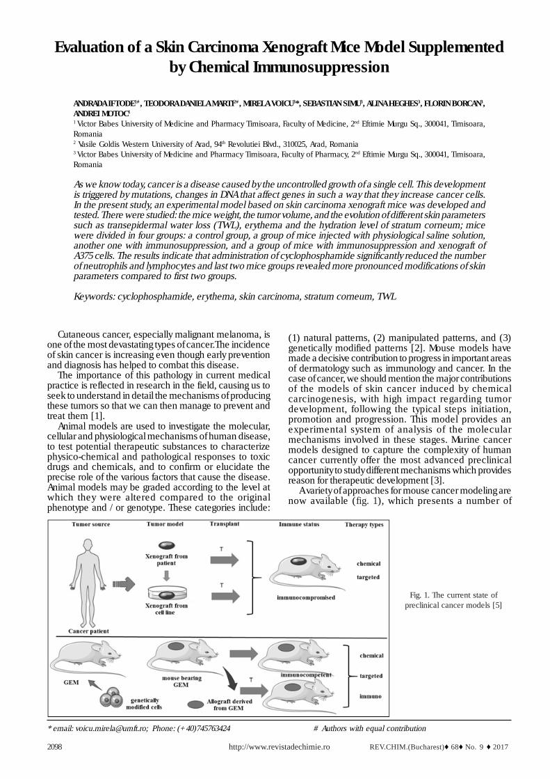
A variety of approaches for mouse cancer modeling arenow available (fig. 1), which presents a number of
Fig. 1. The current state ofpreclinical cancer models [5]
REV.CHIM.(Bucharest)♦ 68♦ No. 9 ♦ 2017 http://www.revistadechimie.ro 2099
advantages and disadvantages. Thus, the limitations ofstandard patterns by xenograft derived from the cell lineare discussed, models of genetically and biologicallyengineered mouse cancer are described,and also patternsderived from patient xenografts, reviewing the values andconstraints and highlighting recent developments [4].
The skin, the largest organ of the human body, performsa variety of functions. The measurement of skin pH isusedin clinical and preclinical research, to evaluate dangerouschanges in pH after outdoor exposure and to assess thecondition of the disease with acute or chronic changes. Abalanced pH level protects the skin against pollution,prevents infections, irritations, slows the aging process andkeeps the skin shiny and healthy.
The loss of transepidermal water (TWL or TEWL) is aterm associated with dermatology. It is defined asmeasuring the amount of water that passes from the insideof the body (human, animal or plant) through the epidermis(skin) to the outside of the body. The loss of transepidermalwater occurs through diffusion and evaporation processes,this process is continuous and cannot be controlled by thehuman body. TWL in mammals is known as “insensitivewater loss” because it is a process over which organismshave little physiological control. TWL measurements maybe useful for identifying skin lesions caused by certainchemical substances, physical strokes or pathologicalconditions such as eczema, because the TWL ratesincrease proportionally to the damage level and favor skindrying. Therefore, transepidermal water loss becomes asignificant factor in dehydration associated with severalmajor disease states [6].
In order to understand as accurately, concretely andrapidly as possible the mechanisms involved in thesediseases, laboratory animals, especially mice are used.The human skin structure compared to the mouse wasanalyzed in detail in order to make the most accuratepassage from preclinical experiments to clinical.Separation of the epidermis of the dermis is made by amembrane that is not very broad and has a sinuousappearance. Derm is about 90% of the skin’s thickness,having a high content of both collagen and elastin, whichare secreted by fibroblasts, thus giving the skin its elasticproperties. The dermis is separated from the lower tissuesby a layer of adipocytes, whose fat accumulation has adamping effect (of pillow).
The structural characteristics of the skin in laboratoryanimals, such as mice, rats, guinea pigs, rabbits, differ fromthose of human skin, e.g.: these species have a skin with athinner epidermis, the dermo-epidermal junction isrelatively flat without the sinuous appearance, a structurallyvaguely organized dermisand a rudimentary dermal system[7].
Mouse pairs exhibit a collision of hair follicles, whilehuman skin has very large interfolicular areas, with a veryrare occurrence of pilose follicles. The human skin has athick epidermis (with many layers of cells) and a dermisthicker than the mouse skin, and is characterized bydownward projections of epidermal ridges reaching thedermis as well. This characteristic is more prominent inpsoriasis and correlates with elongation of the dermalpapillae, which is known as papillomatosis, beingsometimes confused with follicular hyperplasia that occursin mouse models with inflammatory skin diseases. Themost common types of immune system cells in the humanepidermis are Langerhans cells and CD8-positive T cells.In the mouse epidermis there is a prominent population ofepidermal dendritic T cell type Vy5, which are absent inthe human epidermis. Both the human and mouse derms
are populated with macrophages, mastocytes,conventional αβ T cells, and a small population of inbornlymphoid cells. In mouse skin there is an importantcontribution of γδ T cells responsible for immunesurveillance and production of interleukin-17 [8].
Experimental partObtaining of experimental model
Adult, female, Balb/c mice, aged 10-12 weeks werepurchased from Charles River (Sulzfeld, Germany). Theywere kept in standard conditions: light-dark cycles, foodand water ad libitum, almost constant temperature (25oC)and humidity (60%) and were divided into four groups (5mice/group) as follows: Group 1 - control group; Group 2 -injected with physiological saline solution (solvent forcyclophosphamide); Group 3 - with immunosuppression;Group 4 - with immunosuppression and xenograft of A375.
Cyclophosphamide used in this research to induceimmunosuppression is a powder that can be dissolved inphysiological saline solution. Thus, cyclophosphamide wasadministered in 3 doses IM (final concentration: 180 mg/kg mouse body) as follows: 5 days prior to tumor cellinoculation, 3 days before inoculation and one day beforeinoculation of tumor cells.
The IM cyclophosphamide solution and the dosageswere calculated according to the protocol, and then thesubcutaneous tumor cells were inoculated. The protocolinvolves the following steps: (a) mice were weighed beforeeach administration of cyclophosphamide solution; (b) thedoses and the volume of solution required for injection werecalculated; (c) the solution was injected on the daysprovided, at the same time; (d) subcutaneous A375 tumorcells (1x10⁷cells/mouse) were inoculated. The weight ofthe mice was also monitored after the last dose ofcyclophosphamide as well as throughout the study(immunosuppression is associated with weight decrease).
Measurement of skin parametersBiochemical and physiological parameters of the skin
were monitored: transepidermal water loss (TWL),erythema and skin hydration level were conducted using aMultiprobe Adapter System (MPA5) from Courage-Khazaka(Koln, Germany). The determinations of skin parameterswere performed in triplicate at the same moment of theday, by the same operator, in a narrow range of temperature(25°C) and air humidity (55%).
The research was studied and approved by the EthicsCommittee from Victor Babes University of Medicine andPharmacy Timisoara.
StatisticsThe measurements of this study were done in triplicate
for each determination and the results appear as mean ±standard error. One-way Anova and post-tests were usedto determine the statistical difference between Groups 2,3, 4 and the control Group 1, according other studies of ourresearch team [9-12]. p<0.05 was considered statisticallysignificant; *, ** and *** indicate p<0.05, p<0.01 and<0.001.
Results and discussionsCyclophosphamide was subcutaneously injected in
animals of Group 3 and 4 using a 180 mg/kg mouse bodyon days 5, 3 and 1 before the tumor cells were injected.Administration of cyclophosphamide significantly reducedthe number of neutrophils and lymphocytes in the animalsof these groups. Moreover, none of the mice showed signsof toxicity or premature death due to the medical treatment.

http://www.revistadechimie.ro REV.CHIM.(Bucharest)♦ 68♦ No. 9 ♦ 20172100
The evolution of weight over time during the experimentis shown in table 1.
Non-invasive measurements of skin parametersDuring the experiment, there were no major changes in
values of transsepidermal water loss for Groups 1 (control)and Group 2 (injected with physiological saline solution):TWL values indicate a very slight increase that is specificfor all compounds with no harmful effects. In Group 3 mice(immunosuppressed), the increased transepidemic waterloss was more pronounced than in the case of thoseinoculated with tumor cells (fig. 4).
In the case of erythema measurements, significantdifferences were recorded between the first two groupson the one hand and groups 3 and 4 on the other. Valueswere altered by 8 arbitrary units (Group 1) and 12 arbitraryunits (Group 2), while important changes, 51 arbitrary units(Group 3) and 65 arbitrary units (Group 4) were recordedafter 40-days experiment (fig. 5).
Fig. 2. The appearance and resorption of bubble post-inoculationA375 cells in mice subjected to immunosuppression
Approximately 1x10⁷ cells from each cell line weresubcutaneously injected in mice of Group 4. In all injectedanimals, a palpable and visible bubble was detected onthe third day after tumor injection with a 100% absorptionrate (fig. 2).
It can be seen from the graph (fig. 3) that the meantumor volume increased radically every week until the 20th
day. Subsequently, tumor volume was shown to increasesteadily until the 35th day. By the end of the experiment,tumor volume began to decline in some animals involvedin the study.
Fig. 3. Evolution of tumor volume during the experimentTable 1THE EVOLUTION OF WEIGHT DURING THE EXPERIMENT IN BALB C
MICE USED AND DIVIDED INTO THE 4 GROUPS
Fig. 5. Mean values of erythema during experiment in the 4 groups
Fig. 4. Mean values of transepidermal water loss duringexperiment in the 4 groups
REV.CHIM.(Bucharest)♦ 68♦ No. 9 ♦ 2017 http://www.revistadechimie.ro 2101
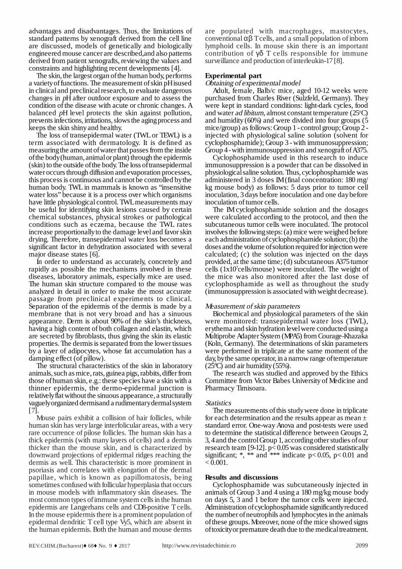
Fig. 6. Mean values of skin hydration during experiment in the 4groups
Another physiological parameter of the skin studied inthe present experiment was the water content of thestratum corneum, also known as skin hydration. In thiscase, a decrease in groups 3 and 4 was observed, morepronounced compared to groups 1 and 2 (fig. 6).
ConclusionsSkin cancer affects one in three people who are
diagnosed with cancer worldwide, and carcinoma - amalignant tumor of epithelial cells - is the most commontype of skin cancer and the easiest to treat if it is diagnosedquickly and right. This paper describes a research basedon a new experimental model of skin carcinoma xenograftat mice, supplemented by chemical immunosuppressionof cyclophosphamide. Adult female, Balb/c mice were thesubject of this study and they were divided in differentgroups: (1) control group; (2) injected with physiologicalsaline solution; (3) with immunosuppression; (4) withimmunosuppression and xenograft of A375 cells. It wasfound that cyclophosphamice not present any signs oftoxicity or premature death; the tumor volume increasesvery much in the fifth week of experiment and the valuesof skin parameters (TWL, erythema and skin hydration)
were significantly modified in the case of last two micegroups compared to the values obtained in the case of firsttwo mice groups.
References1. SOICA, C., DANCIU, C., SAVOIU-BALINT, G., BORCAN, F., AMBRUS,R., ZUPKO, I., BOJIN, F., CORICOVAC, D., CIURLEA, S., AVRAM, S.,DEHELEAN, C.A., OLARIU, T., MATUSZ, P. Int. J. Mol. Sci., 15, 2014, p.8235.2. WOLF, J.C. Alternative Animal Models. In: HASCHEK, W.M.,ROUSSEAUX, C.G., WALLIG, M.A. (Eds.) Haschek and Rousseaux’sHandbook of Toxicologic Pathology, 3rd Edition, Cambridge,Massachusetts: Academic Press. 2013.3. VAN DYKE, T., JACKS, T. Cell, 108, 2002, p. 135.4. MINDA, D., PAVEL, I.Z., BORCAN, F., CORICOVAC, D., PINZARU, I.,ANDRICA, F., MORGOVAN, C., NITA, L.D., SOICA, C., MUNTEAN, D.,TOMA, C.C. Rev. Chim. (Bucharest), 66, no. 3, 2015, p. 373.5. DAY, C.P., MERLINO, G., VAN DYKE, T. Cell, 163, 2015, p. 39.6. FLUHR, J.W., ELSNER, P., BERARDESCA, E., MAIBACH, H.I.Bioengineering of the skin: water and stratum corneum, 2nd Edition,Boca Raton, Florida: CRC Press. 20047. MATHUR, S., KAUR, P., SHARMA, M., KATYAL, A., SINGH, B., TIWARI,M., CHANDRA, R. Phytomed, 11, 2004, p. 452.8. SIMOES, M.C., SOUSA, J.J., PAIS A.A. Cancer Lett., 357, 2015, p. 8.9. OPREAN, C., ZAMBORI, C., BORCAN, F., SOICA, C., ZUPKO, I.,MINORICS, R., BOJIN, F., AMBRUS, R., MUNTEAN, D., DANCIU, C.,PINZARU, I.A., DEHELEAN, C., PAUNESCU, V., TANASIE, G. Pharm.Biol., 54, 2016, p. 2714.10. TRANDAFIRESCU, C., LEDETI, I., CORICOVAC, D.E., SOICA, C.M.,PINZARU, I., DEHELEAN, C.A., IACOB, R.E., BORCAN, F. Mat. Plast.,53, no. 2, 2016, p. 205.11. OPREAN, C., BORCAN, F., PAVEL, I., DEMA, A., DANCIU, C., SOICA,C., DEHELEAN, C., NICU, A., ARDELEAN, A., CRISTEA, M., IVAN, A.,TATU, C., BOJIN, F. In Vivo, 30, 2016, p. 633.12. MUNTEANU, M.F., ARDELEAN, A., BORCAN, F., TRIFUNSCHI, S.I.,GLIGOR, R., ARDELEAN, S.A., CORICOVAC, D., PINZARU, I., ANDRICA,F., BORCAN, L-C. Curr. Drug Delivery, 2017, [Epub ahead of print]
Manuscript received: 19.02.2017


![Failure of a patient-derived xenograft for brain tumor ... · nude or non-obese diabetic/severe combined immu-nodeficient (NOD/SCID) mice [, 15–18], a concept 7 generally referred](https://img.dokumen.tips/doc/110x75/5fda463fed2f21150206154b/failure-of-a-patient-derived-xenograft-for-brain-tumor-nude-or-non-obese-diabeticsevere.jpg)