Embed Size (px)
Citation preview

The Philips eL18-4 PureWave linear array transducer is our first high-performance transducer featuring ultra-broadband PureWave crystal technology with multi-row array configuration, allowing for fine-elevation focusing capability.
Ultrasound
Clinical case study
AuthorLuis F. Goncalves, MD
Phoenix Children’s Hospital Phoenix, AZ USA
Evaluating vascular flow in suspected placenta accreta
eL18-4 PureWave linear arraytransducer
CategoryFetal assessment
Overview
Accurate diagnosis of placenta accreta
is important in determining timing of
delivery as well as for surgical planning.1
For example, if a placenta percreta is
suspected prenatally, multiple specialists
may need to be involved to secure a
successful outcome. One of the clues for
placenta accreta is abnormal placental
vascularity seen as vascular lacunae,
appearing as disorganized venous
channels in the placental substance.
Patient history
A 45-year-old female was referred for
fetal MRI and ultrasound at 26 weeks
and three days for suspected placenta
accreta. Suspicious vascularity was
noted on the prior ultrasound performed
at an outside institution.
Figures 1, 2 and 3 2D images of placental anatomy.
©2018 Koninklijke Philips N.V. All rights are reserved.Philips reserves the right to make changes in specifications and/or to discontinue any product at any time without notice or obligation and will not be liable for any consequences resulting from the use of this publication.
philips.com
Printed in The Netherlands.4522 991 36781 * JUN 2018
Results from case studies are not predictive of results in other cases. Results in other cases may vary.
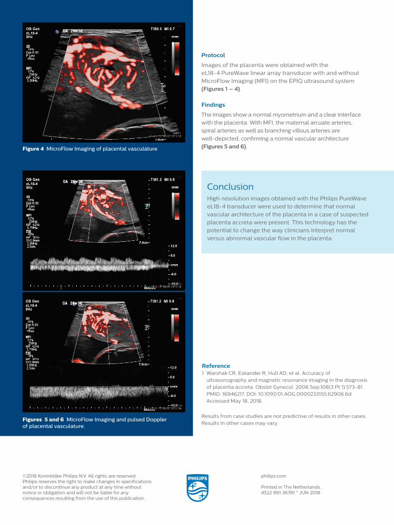
ConclusionHigh-resolution images obtained with the Philips PureWave
eL18-4 transducer were used to determine that normal
vascular architecture of the placenta in a case of suspected
placenta accreta were present. This technology has the
potential to change the way clinicians interpret normal
versus abnormal vascular flow in the placenta.
Figure 4 MicroFlow Imaging of placental vasculature
Reference1 Warshak CR, Eskander R, Hull AD, et al. Accuracy of ultrasonography and magnetic resonance imaging in the diagnosis of placenta accreta. Obstet Gynecol. 2006 Sep;108(3 Pt 1):573-81. PMID: 16946217. DOI: 10.1097/01.AOG.0000233155.62906.6d Accessed May 18, 2018.
Protocol
Images of the placenta were obtained with the
eL18-4 PureWave linear array transducer with and without
MicroFlow Imaging (MFI) on the EPIQ ultrasound system
(Figures 1 – 4).
Findings
The images show a normal myometrium and a clear interface
with the placenta. With MFI, the maternal arcuate arteries,
spiral arteries as well as branching villous arteries are
well-depicted, confirming a normal vascular architecture
(Figures 5 and 6).
Figures 5 and 6 MicroFlow Imaging and pulsed Doppler of placental vasculature.