Embed Size (px)
Citation preview
ORIGINAL ARTICLE
Magnetic resonance imaging in 300 cases ofplacenta accreta: surgical correlation of newfindingsJOSE M. PALACIOS JARAQUEMADA AND CLAUDIO H. BRUNO
From Southern Scientific Foundation, Lomas de Zamora, Buenos Aires, and School of Medicine, University ofBuenos Aires, Buenos Aires, Argentina
Acta Obstet Gynecol Scand 2005; 84: 716–724. # Acta Obstet Gynecol Scand 84 2005
Background. To establish the usefulness of placental magnetic resonance in patients witha diagnosis of placenta accreta through the correlation of diagnostic images and surgicalfindings.Methods. Three hundred patients with ultrasound signs of placenta accreta were studied.In 252 patients, magnetic resonance imaging (MRI) was performed in a closed 1.5T-resonator, and in 48 patients, open 0.23 T-set was used. T1 and T2 slices in the threeplanes were performed, and placental invasion was classified in depth levels and topo-graphic areas in relation to the posterior vesical wall. The final degree of invasion wasestablished during surgery according to clinical and anatomical criteria. The informationobtained with MRI was analyzed, thus establishing its relevance to the change in surgicaltechnique.Results. In 286 cases, MRI provided topographic information of placental invasion, andin 90 patients, it modified invasion levels. Undiagnosed parametrium extent was deter-mined in 11 cases, and 11 other cases were reclassified as placenta previa. Changes inconduct following MRI study included: recommendation to modify surgery date at week35, recommendation for prophylactic ureteral catheterization, recommendation for theuse of intraoperative blood salvage, possibility of approach through Pfannenstiel incision,probability of segmental myometrial approach, probability of aortic clamping, need toinvestigate subclinical disseminated intravascular coagulation, need of posterior pelvicdissection, and the possibility of uterine conservation.Conclusions. Magnetic resonance imaging turned out to be essential to define the topographyand area of placental invasion. New findings modified surgical tactic and technique, allowinga reduction in historical morbidity and a significant increase in conservative surgeries.
Key words: magnetic resonance imaging; placenta accreta; surgical correlation; placentalinvasion levels; uterine conservation
Submitted 3 August, 2004Accepted 10 January, 2005
Placenta accreta, increta and percreta are entities ofhigh maternal morbidity and mortality (1) andconstitute in themselves several degrees of placentalinvasion. Abnormal placental adherence is usuallyassociated with serious hemorrhage and myome-trial damage of different size, facts that conditionhysterectomy as the most frequent treatment.
Placenta previa coexisting with anterior oriterative cesarean is the main risk factor in the
Abbreviations:DIC: disseminated intravascular coagulation; FDP: fibrinogen-degradation products; MRI: magnetic resonance imaging;pMRI: placental magnetic resonance imaging; PAD: placentaladhesive disorders.
Surgical Correlation Centers: Swiss-Argentine Clinic, MaterDei Clinic, IADT Clinic, Otamendi Clinic, CEMIC Hospital,Durand Hospital, Metropolitan Hospital, and Buenos AiresProvince Hospital Network, Buenos Aires, Argentina.
# Acta Obstet Gynecol Scand 84 (2005)
Acta Obstet Gynecol Scand 2005: 84: 716–724 Copyright # Acta Obstet Gynecol Scand 2005
Printed in UK. All rights reservedActa Obstetricia et
Gynecologica Scandinavica
development of adherent placenta (2). This asso-ciation constitutes the most important clinicalrisk factor to suspect placental invasion. Sec-ondly, curettage, previous uterine surgeries, andinfections can be included.
Ultrasound is the first-rate method to diagnoseplacental invasion (3). However, it is usual tonotice discrepancies between ultrasound diagno-sis and surgical complexity, which make adequatetherapeutic planning difficult. Therefore, it isbelieved that an accurate presurgery diagnosiswould make it possible to minimize risks andplan a safer surgery.
Color Doppler and power Doppler were usedin order to reduce the diagnostic error of conven-tional ultrasound (4,5). These techniques opti-mized the detail of the uterine–placentalinterface and, consequently, its diagnosis. Never-theless, when it was necessary to characterize thelesion topography in relation to soft pelvic tis-sues, a placental magnetic resonance imaging(pMRI) was required (6).
Even though each type of placental adherencehas a certain surgical tactic and technique, from adiagnostic point of view, we could group placentaaccreta and its varieties under the label placentaladhesive disorders (PAD).
Studies that have correlated pMRI and PADare limited to small series, case presentations,retrospective analyses, or to variations in imageacquisition (7–9). However, due to its excellentdefinition, several authors considered it necessaryto make studies of statistical value to determinepMRI’s real value in PAD diagnosis. It wasthought that accurate diagnosis would undoubt-edly imply a better surgical opportunity (10).
This study aims at establishing pMRI’s useful-ness in the diagnosis and treatment of PAD andat determining the correlation between diagnosticimages and surgical results.
Materials and methods
Between May 1994 and May 2004, 300 pregnant womenwith ultrasound signs of placenta accreta were studiedbefore MRI was performed between the 25th and 28thgestation weeks. These patients were referred from hospitalsand private health care centers belonging to an estimatedpopulation area of 11 000 000 inhabitants, with an averageof 40 000 living newborns per year, and a cesarean ratebetween 30 and 60%.
Diagnostic ultrasounds were performed at private medicalinstitutions (79%) and at university centers (21%). All thestudies were obtained and informed by well-known opera-tors, with over 5 years experience. Ultrasound scanners withlinear and sectorial 3.5 and 5 MHz transducers and also 5and 7 MHz transvaginal transducers were employed. Ultra-sound characterization for placenta accreta and its varietieswas generally established according to Finberg and Williams(11) diagnostic criteria.
Ultrasound results were compared with those of pMRI inorder to establish the usefulness of the new information inconnection with the final surgical outcome.
In 252 patients, pMRI was performed in a Picker EdgeTM
1.5 closed resonator (Picker, Cleveland, Ohio, USA).Sequences T1: SE, TE 8, TR 140, FOV 45–50, TK 8, RES160� 256, PS 0.602, FLIP 120�, and SAR 1.81. Breath holdand sequences T2: FSE 32; TE 168; TR 3500; FOV 28;THCK 8; GAP 1.5; RES 192� 256; PS 1.328; FLIP 90;NSA 1; SAR 2.25. In the remaining 48 patients, a 0.23 Topen Picker Outlook ProviewTM resonator was used (Mar-coni, Vantaa, Finland) HOLD FE-92/6.0, FA 90�, SI. Thk/sep: 10/11 mm, FOV 200� 256, Phase enc: HOR, ETL 1. Allpatients ingested 750 ml of water 45 min before the study,and slices in the three spatial planes were used. All pMRIwere performed between the 30th and the 33rd gestationweeks. The examination time was nearly 30 min and almostalways well tolerated, and approximately only 12% of thepatients suffered from claustrophobia and requested theopen resonator.
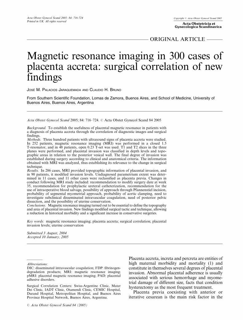
Sagittal images obtained through pMRI allowed the divi-sion of anterior placental invasion into two sectors (12). Bothwere delimited by a plane perpendicular to the upper bladderaxis, and the uterine sector bordering con the upper posteriorbladder wall is called S1, and the uterine sector adjacent to thelower posterior wall, S2 (Figs 1 and 2). The levels of placentalinvasion determined by pMRI were labeled: 0, for absence ofinvasion; A, for partial myometrial invasion; B, for total
Fig. 1. Sagittal cut of pelvis. The division plane can be seen insectors S1 (upper uterine segment) and S2 (lower uterinesegment). P, pubis; VA, vagina; B, bladder.
MRI in placenta accreta 717
# Acta Obstet Gynecol Scand 84 (2005)
myometrial invasion; and C, for invasions that involved thewhole of the myometrium and that included one or bothparametriums (seen in the pMRI’s coronal and axial slices).For the case of combined S1 and S2 invasions, groups wereformed according to the highest percentage of invasion.
The final degree of placental penetration and its specifictopography were established in the operating room accord-ing to clinical and anatomical criteria. With that purpose,placenta accreta was defined as that which adhered itselffirmly to the endometrium, and that which, when detached,showed non-self-controlled bleeding, placenta increta as thatwhich required curettage to remove invasive tissue deeplyimplanted in the myometrium, and placenta percreta as thatinvasion which involved all uterine layers, being able toexceed the uterus and invade neighboring organs.
Information that had not been provided by previous stud-ies was considered new, and data which modified surgicaltactic or technique were considered relevant information.
pMRI findings were compared with final surgical results.With that aim, specific surgery was performed following a pre-established protocol (13). There, laparotomy and myometrialapproach was considered according to the area and depth ofplacental invasion, being fit for uterine reconstructive surgerythose placental invasions which did not affect more than 50%of uterine axial circumference. The surgical protocol includedprecise parameters for vascular control which, according tothe case, would be performed in the pelvic vessels (parame-trium) or in the infrarenal abdominal aorta. Once fetal extrac-tion and pelvic fascial dissection were concluded,myometrium and vesical reconstruction was made accordingto specific technique or to hysterectomy when necessary.
Tests of statistical significance Chi-square were per-formed, between S1 and S2 levels established by pMRItopographic classification. Parameters of comparisonincluded the possibility of uterine conservation, hysterec-tomy, and the volume of transfused blood.
Results
Mother’s age ranged between 16 and 46 years.Patients reported 87 miscarriages, 61 of whichwhere solved without curettage. Myomectomybackground was recorded in 29 patients, 14cases belonging to corporal myomas and 8 tofundal myomas. In the seven remaining cases,their exact location could not be defined due tolack of reliable documentation.
Placental implantation corresponded to 206previous locations, 69 anterior ones, and 25 tolower anterior ones.
Analysis of the relation between the number ofcesarean and invasion levels can be examined inTable I. The most frequent invasion was the A typewith 72.88%, and B and C invasions were for oneto two cesareans in 5.42% and for three or morein 19%, respectively. C invasions (parametrium inva-sions) were evidenced only through pMRI. Ultra-sound type, its diagnosis, and also the new inform-ation obtained by pMRI can be seen in TableII.
pMRI modified the interpretation of invasiondegree in 90 patients (30%), and classified inva-sion topography in 286 patients (95.33%). In 14cases, invasion anatomy through studies withincomplete series could not be defined, causedby fetal movement or intolerance to study.
Surgical results were closely related to the kindand level of invasion evidenced through pMRI(Table III). Changes in conduct secondary topMRI study included: surgery rescheduling atweek 35 (68 cases), ureteral catheterization (11cases), indication of intraoperative blood salvage(43 cases), presence of specialists (43 cases), pos-sibility of Pfannenstiel incision (19 cases), seg-mental myometrial approach (34 cases), aorticvascular control (22 cases), detection of subclini-cal disseminated intravascular coagulation (21cases), posterior pelvic dissection (15 cases), andpossibility of uterine conservation (236 cases).
In 281 patients (93.66%), direct and personal cor-relation was made between images and surgicalfindings. In the 19 remaining cases, surgical correla-tion was performed by others.
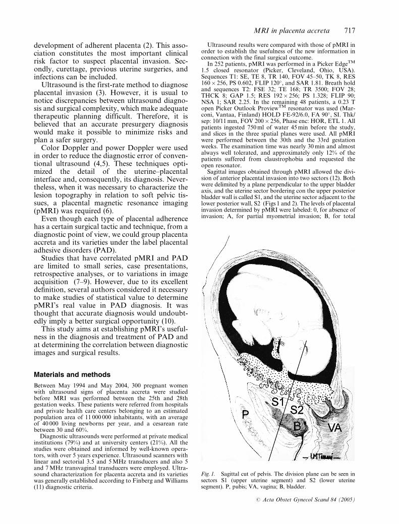
Fig. 2. Frontal view of pelvis. Both types of anterior invasionS1 (upper uterine segment) and S2 (lower uterine segment) can beobserved. VA, Vagina.
Table I. Previous cesarean and degree of invasion (regardless of pMRIclassification)
pMRI 0 A B C
One cesarean 5 22 3 1Two cesarean 6 38 9 3Three cesarean 0 73 16 2Four or more 0 82 27 13
Total 300 11 215 55 19
pMRI, placental magnetic resonance imaging. 0, no invasion; A, partialinvasion; B, total invasion; C, parametrial invasion.
718 J. M. Palacios Jaraquemada & C. H. Bruno
# Acta Obstet Gynecol Scand 84 (2005)
Total hysterectomy was indicated in 30 patients,out of which, 22 cases corresponded to massivemyometrial destruction (over 50% of uterine axialcircumference). In the other eight cases, hysterect-omy was advised because of coagulopathy.
pMRI accurately characterized the level andtopography of invasion in 97.66% of the cases.Four false positives for placenta percreta (1.33%)and three false negatives for placental invasion(1%) were recorded.
Because the uterine conservation was performedin most cases, the histology of placental invasionwas enable only in 30 cases, 20 of them were pla-centa percretas (B and C invasion), and the other 10were classified as placenta accretas (A invasion).
Chi-square test-established significant differences(P< 0.001) between S1 and S2 invasion levels(pMRI) related to the possibility of uterine conser-vation, hysterectomy, and transfused blood volume.
Discussion
It is usual to observe discrepancies among theultrasound report, histology, and surgical com-plexity of PAD (14,15). Repetition of thesedifferences would imply the usefulness of repla-
cing the morphological classification with a topo-graphic one. This change would allow theanalysis of diagnostic information with the mainaim of planning the surgery.
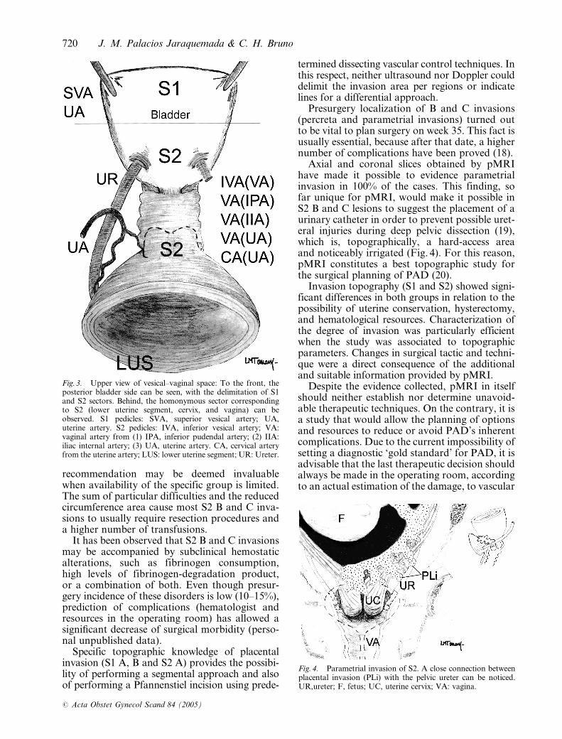
The division into S1 and S2 topographic areasis directly related to the degree of local irrigation(16). Sector S1 is irrigated by the uterine andupper vesical arteries, vessels which are generallyof easy access and which promote quick hemos-tasis. However, sector S2 has anastomotic irriga-tion and originated in multiple pedicles (Fig. 3).The visceral intersection formed among thevesical trigonum, the uterine cervix, and thevagina is irrigated by deeply located vessels (pelvicsubperitoneal), a fact which significantly increasesS2’s surgical complexity and the possibility ofhemorrhage.
S2 B and C invasions imply a higher probabil-ity of using specific procedures, such as aorticclamping (17) or posterior ureteral dissectionthat is mainly due to the difficulty to perform abloodless dissection or at least controlled within avascular magma located deeply inside the pelvis.Besides, and because of, its higher hemorrhagicprobability, it is convenient to use intraoperativeblood salvage in this kind of invasions. This
Table II. Previous diagnosis and pMRI new information
pMRI NF
AB US TV US DP US US diagnosis Degree S1/S2 location
Group 1 Placenta accreta: 26 Invasion 0: 7 S1: 20(n¼ 38) 38 0 0 Placenta increta: 12 Invasion B: 11
Invasion C: 3S2: 11
Group 2 Placenta accreta: 102 Invasion 0: 4 S1: 79(n¼ 134) 134 43 0 Placenta increta: 30
Placenta percreta: 2Invasion B: 17Invasion C: 3
S2: 47
Group 3 Placenta accreta: 88 Invasion B: 40 S1: 82(n¼ 128) 128 102 97 Placenta increta: 38
Placenta percreta: 2Invasion C: 5 S2: 45
Total 300 145 97 300 NF: 90 (30%) NF: 284 (94.66%)
AB US, abdominal ultrasound; D US: Doppler ultrasound; pMRI, placental magnetic resonance imaging; NF, new findings; TV US, transvaginal ultrasound. 0, noinvasion; A, partial invasion; B, total invasion; C, parametrial invasion. Group 1, AB US; group 2, AB USþ TV US; group 3, AB USþ TV USþDoppler.
Table III. Analysis by localization and outcome (pMRI classification)
0 A B C
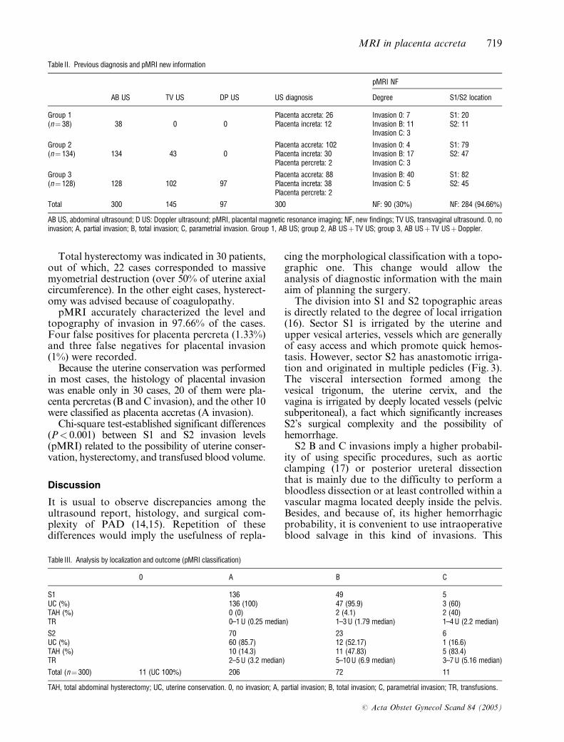
S1 136 49 5UC (%) 136 (100) 47 (95.9) 3 (60)TAH (%) 0 (0) 2 (4.1) 2 (40)TR 0–1U (0.25 median) 1–3U (1.79 median) 1–4U (2.2 median)
S2 70 23 6UC (%) 60 (85.7) 12 (52.17) 1 (16.6)TAH (%) 10 (14.3) 11 (47.83) 5 (83.4)TR 2–5U (3.2 median) 5–10U (6.9 median) 3–7U (5.16 median)
Total (n¼ 300) 11 (UC 100%) 206 72 11
TAH, total abdominal hysterectomy; UC, uterine conservation. 0, no invasion; A, partial invasion; B, total invasion; C, parametrial invasion; TR, transfusions.
MRI in placenta accreta 719
# Acta Obstet Gynecol Scand 84 (2005)
recommendation may be deemed invaluablewhen availability of the specific group is limited.The sum of particular difficulties and the reducedcircumference area cause most S2 B and C inva-sions to usually require resection procedures anda higher number of transfusions.
It has been observed that S2 B and C invasionsmay be accompanied by subclinical hemostaticalterations, such as fibrinogen consumption,high levels of fibrinogen-degradation product,or a combination of both. Even though presur-gery incidence of these disorders is low (10–15%),prediction of complications (hematologist andresources in the operating room) has allowed asignificant decrease of surgical morbidity (perso-nal unpublished data).
Specific topographic knowledge of placentalinvasion (S1 A, B and S2 A) provides the possibi-lity of performing a segmental approach and alsoof performing a Pfannenstiel incision using prede-
termined dissecting vascular control techniques. Inthis respect, neither ultrasound nor Doppler coulddelimit the invasion area per regions or indicatelines for a differential approach.
Presurgery localization of B and C invasions(percreta and parametrial invasions) turned outto be vital to plan surgery on week 35. This fact isusually essential, because after that date, a highernumber of complications have been proved (18).
Axial and coronal slices obtained by pMRIhave made it possible to evidence parametrialinvasion in 100% of the cases. This finding, sofar unique for pMRI, would make it possible inS2 B and C lesions to suggest the placement of aurinary catheter in order to prevent possible uret-eral injuries during deep pelvic dissection (19),which is, topographically, a hard-access areaand noticeably irrigated (Fig. 4). For this reason,pMRI constitutes a best topographic study forthe surgical planning of PAD (20).
Invasion topography (S1 and S2) showed signi-ficant differences in both groups in relation to thepossibility of uterine conservation, hysterectomy,and hematological resources. Characterization ofthe degree of invasion was particularly efficientwhen the study was associated to topographicparameters. Changes in surgical tactic and techni-que were a direct consequence of the additionaland suitable information provided by pMRI.
Despite the evidence collected, pMRI in itselfshould neither establish nor determine unavoid-able therapeutic techniques. On the contrary, it isa study that would allow the planning of optionsand resources to reduce or avoid PAD’s inherentcomplications. Due to the current impossibility ofsetting a diagnostic ‘gold standard’ for PAD, it isadvisable that the last therapeutic decision shouldalways be made in the operating room, accordingto an actual estimation of the damage, to vascular
Fig. 3. Upper view of vesical–vaginal space: To the front, theposterior bladder side can be seen, with the delimitation of S1and S2 sectors. Behind, the homonymous sector correspondingto S2 (lower uterine segment, cervix, and vagina) can beobserved. S1 pedicles: SVA, superior vesical artery; UA,uterine artery. S2 pedicles: IVA, inferior vesical artery; VA:vaginal artery from (1) IPA, inferior pudendal artery; (2) IIA:iliac internal artery; (3) UA, uterine artery. CA, cervical arteryfrom the uterine artery; LUS: lower uterine segment; UR: Ureter.
Fig. 4. Parametrial invasion of S2. A close connection betweenplacental invasion (PLi) with the pelvic ureter can be noticed.UR,ureter; F, fetus; UC, uterine cervix; VA: vagina.
720 J. M. Palacios Jaraquemada & C. H. Bruno
# Acta Obstet Gynecol Scand 84 (2005)
control probabilities, and to the ability to per-form adequate tissue reconstruction.
As 30% of the patients have been relocated to0, B or C types, it is advisable not to establishdefinitive actions before the topographic study(pMRI) has been completed and the final diag-nosis through direct inspection has been verified.
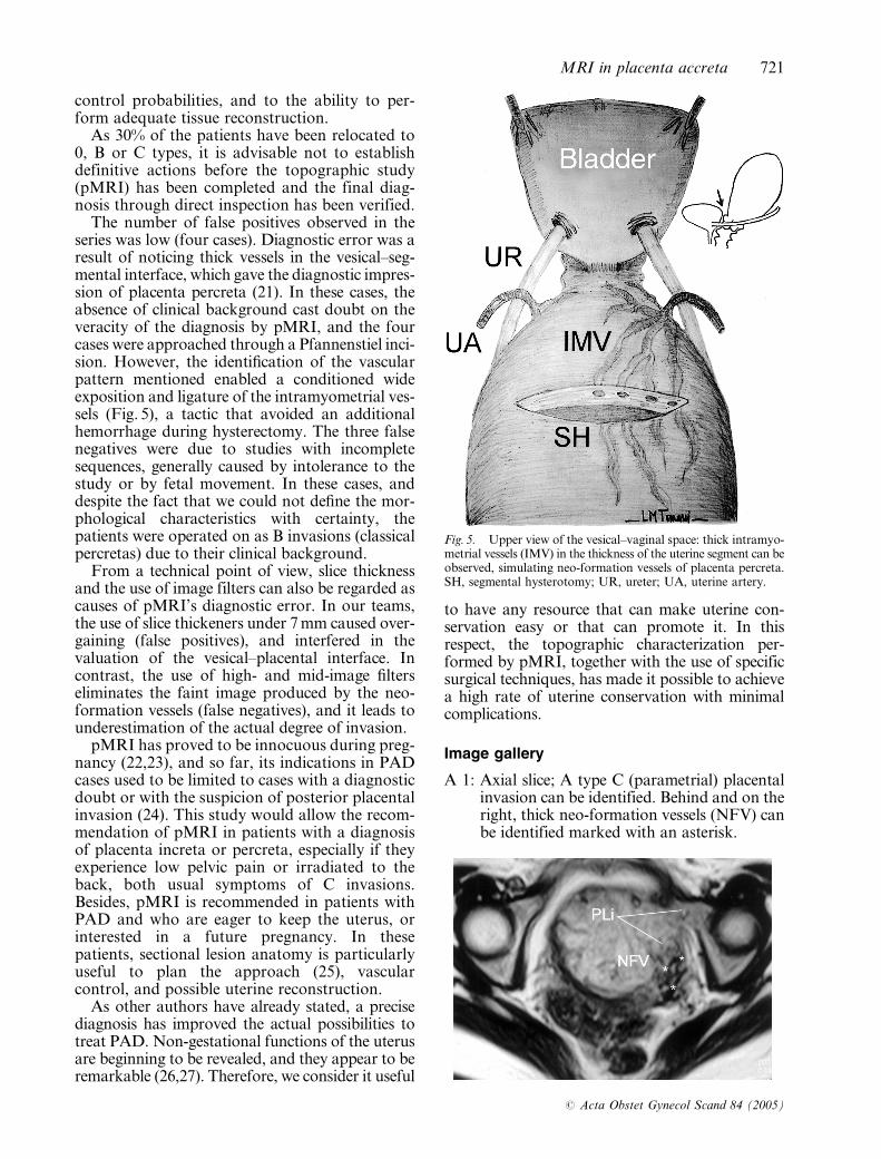
The number of false positives observed in theseries was low (four cases). Diagnostic error was aresult of noticing thick vessels in the vesical–seg-mental interface, which gave the diagnostic impres-sion of placenta percreta (21). In these cases, theabsence of clinical background cast doubt on theveracity of the diagnosis by pMRI, and the fourcases were approached through a Pfannenstiel inci-sion. However, the identification of the vascularpattern mentioned enabled a conditioned wideexposition and ligature of the intramyometrial ves-sels (Fig. 5), a tactic that avoided an additionalhemorrhage during hysterectomy. The three falsenegatives were due to studies with incompletesequences, generally caused by intolerance to thestudy or by fetal movement. In these cases, anddespite the fact that we could not define the mor-phological characteristics with certainty, thepatients were operated on as B invasions (classicalpercretas) due to their clinical background.
From a technical point of view, slice thicknessand the use of image filters can also be regarded ascauses of pMRI’s diagnostic error. In our teams,the use of slice thickeners under 7 mm caused over-gaining (false positives), and interfered in thevaluation of the vesical–placental interface. Incontrast, the use of high- and mid-image filterseliminates the faint image produced by the neo-formation vessels (false negatives), and it leads tounderestimation of the actual degree of invasion.
pMRI has proved to be innocuous during preg-nancy (22,23), and so far, its indications in PADcases used to be limited to cases with a diagnosticdoubt or with the suspicion of posterior placentalinvasion (24). This study would allow the recom-mendation of pMRI in patients with a diagnosisof placenta increta or percreta, especially if theyexperience low pelvic pain or irradiated to theback, both usual symptoms of C invasions.Besides, pMRI is recommended in patients withPAD and who are eager to keep the uterus, orinterested in a future pregnancy. In thesepatients, sectional lesion anatomy is particularlyuseful to plan the approach (25), vascularcontrol, and possible uterine reconstruction.
As other authors have already stated, a precisediagnosis has improved the actual possibilities totreat PAD. Non-gestational functions of the uterusare beginning to be revealed, and they appear to beremarkable (26,27). Therefore, we consider it useful
to have any resource that can make uterine con-servation easy or that can promote it. In thisrespect, the topographic characterization per-formed by pMRI, together with the use of specificsurgical techniques, has made it possible to achievea high rate of uterine conservation with minimalcomplications.
Image gallery
A 1: Axial slice; A type C (parametrial) placentalinvasion can be identified. Behind and on theright, thick neo-formation vessels (NFV) canbe identified marked with an asterisk.
Fig. 5. Upper view of the vesical–vaginal space: thick intramyo-metrial vessels (IMV) in the thickness of the uterine segment can beobserved, simulating neo-formation vessels of placenta percreta.SH, segmental hysterotomy; UR, ureter; UA, uterine artery.
MRI in placenta accreta 721
# Acta Obstet Gynecol Scand 84 (2005)
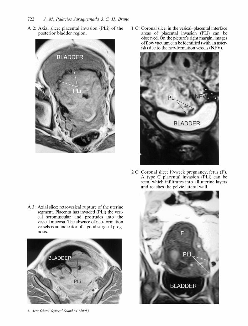
A 2: Axial slice; placental invasion (PLi) of theposterior bladder region.
A 3: Axial slice; retrovesical rupture of the uterinesegment. Placenta has invaded (PLi) the vesi-cal seromuscular and protrudes into thevesical mucosa. The absence of neo-formationvessels is an indicator of a good surgical prog-nosis.
1 C: Coronal slice; in the vesical–placental interfaceareas of placental invasion (PLi) can beobserved. On the picture’s right margin, imagesof flow vacuum can be identified (with an aster-isk) due to the neo-formation vessels (NFV).
2 C: Coronal slice; 19-week pregnancy, fetus (F).A type C placental invasion (PLi) can beseen, which infiltrates into all uterine layersand reaches the pelvic lateral wall.
722 J. M. Palacios Jaraquemada & C. H. Bruno
# Acta Obstet Gynecol Scand 84 (2005)
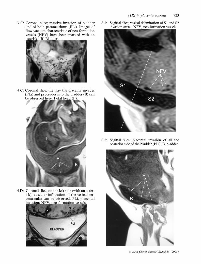
3 C: Coronal slice; massive invasion of bladderand of both parametriums (PLi). Images offlow vacuum characteristic of neo-formationvessels (NFV) have been marked with anasterisk. (B) Bladder.
4 C: Coronal slice; the way the placenta invades(PLi) and protrudes into the bladder (B) canbe observed here. Fetal head (F).
4 D: Coronal slice; on the left side (with an aster-isk), vascular infiltration of the vesical ser-omuscular can be observed. PLi, placentalinvasion; NFV, neo-formation vessels.
S 1: Sagittal slice; vesical delimitation of S1 and S2invasion areas. NFV, neo-formation vessels.
S 2: Sagittal slice; placental invasion of all theposterior side of the bladder (PLi), B, bladder.
MRI in placenta accreta 723
# Acta Obstet Gynecol Scand 84 (2005)
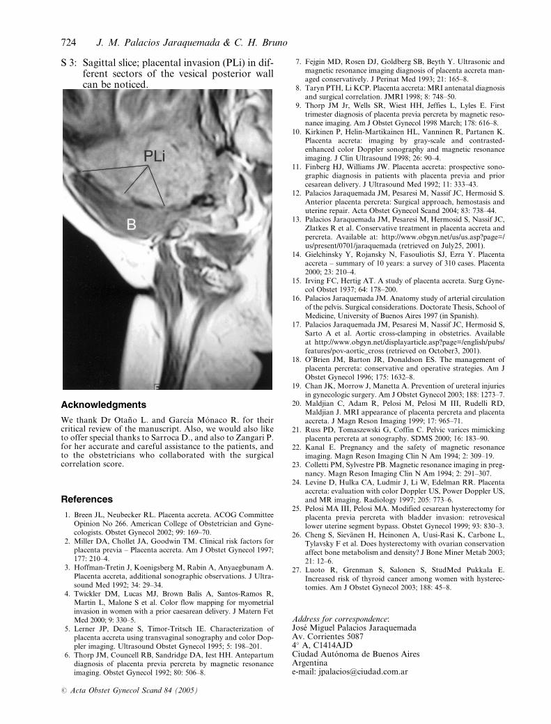
S 3: Sagittal slice; placental invasion (PLi) in dif-ferent sectors of the vesical posterior wallcan be noticed.
Acknowledgments
We thank Dr Otano L. and Garcıa Monaco R. for theircritical review of the manuscript. Also, we would also liketo offer special thanks to Sarroca D., and also to Zangari P.for her accurate and careful assistance to the patients, andto the obstetricians who collaborated with the surgicalcorrelation score.
References
1. Breen JL, Neubecker RL. Placenta accreta. ACOG CommitteeOpinion No 266. American College of Obstetrician and Gyne-cologists. Obstet Gynecol 2002; 99: 169–70.
2. Miller DA, Chollet JA, Goodwin TM. Clinical risk factors forplacenta previa – Placenta accreta. Am J Obstet Gynecol 1997;177: 210–4.
3. Hoffman-Tretin J, Koenigsberg M, Rabin A, Anyaegbunam A.Placenta accreta, additional sonographic observations. J Ultra-sound Med 1992; 34: 29–34.
4. Twickler DM, Lucas MJ, Brown Balis A, Santos-Ramos R,Martin L, Malone S et al. Color flow mapping for myometrialinvasion in women with a prior caesarean delivery. J Matern FetMed 2000; 9: 330–5.
5. Lerner JP, Deane S, Timor-Tritsch IE. Characterization ofplacenta accreta using transvaginal sonography and color Dop-pler imaging. Ultrasound Obstet Gynecol 1995; 5: 198–201.
6. Thorp JM, Councell RB, Sandridge DA, Iest HH. Antepartumdiagnosis of placenta previa percreta by magnetic resonanceimaging. Obstet Gynecol 1992; 80: 506–8.
7. Fejgin MD, Rosen DJ, Goldberg SB, Beyth Y. Ultrasonic andmagnetic resonance imaging diagnosis of placenta accreta man-aged conservatively. J Perinat Med 1993; 21: 165–8.
8. Taryn PTH, Li KCP. Placenta accreta: MRI antenatal diagnosisand surgical correlation. JMRI 1998; 8: 748–50.
9. Thorp JM Jr, Wells SR, Wiest HH, Jeffies L, Lyles E. Firsttrimester diagnosis of placenta previa percreta by magnetic reso-nance imaging. Am J Obstet Gynecol 1998 March; 178: 616–8.
10. Kirkinen P, Helin-Martikainen HL, Vanninen R, Partanen K.Placenta accreta: imaging by gray-scale and contrasted-enhanced color Doppler sonography and magnetic resonanceimaging. J Clin Ultrasound 1998; 26: 90–4.
11. Finberg HJ, Williams JW. Placenta accreta: prospective sono-graphic diagnosis in patients with placenta previa and priorcesarean delivery. J Ultrasound Med 1992; 11: 333–43.
12. Palacios Jaraquemada JM, Pesaresi M, Nassif JC, Hermosid S.Anterior placenta percreta: Surgical approach, hemostasis anduterine repair. Acta Obstet Gynecol Scand 2004; 83: 738–44.
13. Palacios Jaraquemada JM, Pesaresi M, Hermosid S, Nassif JC,Zlatkes R et al. Conservative treatment in placenta accreta andpercreta. Available at: http://www.obgyn.net/us/us.asp?page=/us/present/0701/jaraquemada (retrieved on July25, 2001).
14. Gielchinsky Y, Rojansky N, Fasouliotis SJ, Ezra Y. Placentaaccreta – summary of 10 years: a survey of 310 cases. Placenta2000; 23: 210–4.
15. Irving FC, Hertig AT. A study of placenta accreta. Surg Gyne-col Obstet 1937; 64: 178–200.
16. Palacios Jaraquemada JM. Anatomy study of arterial circulationof the pelvis. Surgical considerations. Doctorate Thesis, School ofMedicine, University of Buenos Aires 1997 (in Spanish).
17. Palacios Jaraquemada JM, Pesaresi M, Nassif JC, Hermosid S,Sarto A et al. Aortic cross-clamping in obstetrics. Availableat http://www.obgyn.net/displayarticle.asp?page=/english/pubs/features/pov-aortic_cross (retrieved on October3, 2001).
18. O’Brien JM, Barton JR, Donaldson ES. The management ofplacenta percreta: conservative and operative strategies. Am JObstet Gynecol 1996; 175: 1632–8.
19. Chan JK, Morrow J, Manetta A. Prevention of ureteral injuriesin gynecologic surgery. Am J Obstet Gynecol 2003; 188: 1273–7.
20. Maldjian C, Adam R, Pelosi M, Pelosi M III, Rudelli RD,Maldjian J. MRI appearance of placenta percreta and placentaaccreta. J Magn Reson Imaging 1999; 17: 965–71.
21. Russ PD, Tomaszewski G, Coffin C. Pelvic varices mimickingplacenta percreta at sonography. SDMS 2000; 16: 183–90.
22. Kanal E. Pregnancy and the safety of magnetic resonanceimaging. Magn Reson Imaging Clin N Am 1994; 2: 309–19.
23. Colletti PM, Sylvestre PB. Magnetic resonance imaging in preg-nancy. Magn Reson Imaging Clin N Am 1994; 2: 291–307.
24. Levine D, Hulka CA, Ludmir J, Li W, Edelman RR. Placentaaccreta: evaluation with color Doppler US, Power Doppler US,and MR imaging. Radiology 1997; 205: 773–6.
25. Pelosi MA III, Pelosi MA. Modified cesarean hysterectomy forplacenta previa percreta with bladder invasion: retrovesicallower uterine segment bypass. Obstet Gynecol 1999; 93: 830–3.
26. Cheng S, Sievanen H, Heinonen A, Uusi-Rasi K, Carbone L,Tylavsky F et al. Does hysterectomy with ovarian conservationaffect bone metabolism and density? J Bone Miner Metab 2003;21: 12–6.
27. Luoto R, Grenman S, Salonen S, StudMed Pukkala E.Increased risk of thyroid cancer among women with hysterec-tomies. Am J Obstet Gynecol 2003; 188: 45–8.
Address for correspondence:Jose Miguel Palacios JaraquemadaAv. Corrientes 50874� A, C1414AJDCiudad Autonoma de Buenos AiresArgentinae-mail: [email protected]
724 J. M. Palacios Jaraquemada & C. H. Bruno
# Acta Obstet Gynecol Scand 84 (2005)