Embed Size (px)
Citation preview

Evaluating Computational Pathology at the US FDA and
Related ResearchBrandon D. Gallas
US FDA, Center for Devices and Radiological HealthOffice of Science and Engineering Laboratories
Division of Imaging, Diagnostics, and Software Reliability

Scientific Evaluation of Computational Pathology and
Related ResearchBrandon D. Gallas
US FDA, Center for Devices and Radiological HealthOffice of Science and Engineering Laboratories
Division of Imaging, Diagnostics, and Software Reliability

OutlineEvaluating Computer Aids in Radiology at the FDA• What about computational pathology?
My Research in Pathology• eeDAP: Evaluation Environment for Digital and Analog Pathology
• eeDAP Studies– Compare scanners to microscope– Pathologist microscope viewing behavior– Measure registration accuracy
• CDRH Medical Device Development Tool program (MDDT)– eeDAP– Annotating Images to validate algorithms
European Congress of Pathology, Bilbao, Spain, 9/9/2018www.fda.gov 3

Medical Device Classification• Risk‐Based Paradigm
– Medical devices are classified and regulated according to their degree of risk to the public
• Intended Use / Indications for Use (IFU)
European Congress of Pathology, Bilbao, Spain, 9/9/2018www.fda.gov 4
Class I Class II Class III
Low Risk High Risk

Some Submission Types for Medical Devices
• 510(k) Premarket Notification– Path to market for the majority of medical devices– Requires determination that a new device is substantially equivalent to a legally marketed device
(predicate device)– Guidance:https://www.fda.gov/medicaldevices/deviceregulationandguidance/howtomarketyourdevice/premarketsubmiss
ions/premarketnotification510k/ucm134572.htm
• Premarket Approval (PMA)– Class III devices– Demonstrate reasonable assurance of safety and effectiveness
• Very device specific• Standalone submission• No comparison to a predicate
– Guidance:https://www.fda.gov/MedicalDevices/DeviceRegulationandGuidance/HowtoMarketYourDevice/PremarketSubmissions/PremarketApprovalPMA/ucm050289.htm
European Congress of Pathology, Bilbao, Spain, 9/9/2018www.fda.gov 5

Some Submission Types for Medical Devices
• De Novo– Novel devices that have not previously been classified are by default Class III (and hence, PMA
devices)– De novo is a petition for down‐classification (Class III to typically Class II)– De novo petition proposes “Special Controls” that would be needed to assure the safety and
effectiveness of the device– A granted de novo establishes a new device type, a new regulation, and necessary general (and
special) controls– Once the de novo is granted, the device is eligible to serve as a predicate
• All subsequent class II followers can use it as a predicate in their 510(k) submissions
• Guidance:https://www.fda.gov/downloads/MedicalDevices/DeviceRegulationandGuidance/GuidanceDocuments/ucm080197.pdf
European Congress of Pathology, Bilbao, Spain, 9/9/2018www.fda.gov 6
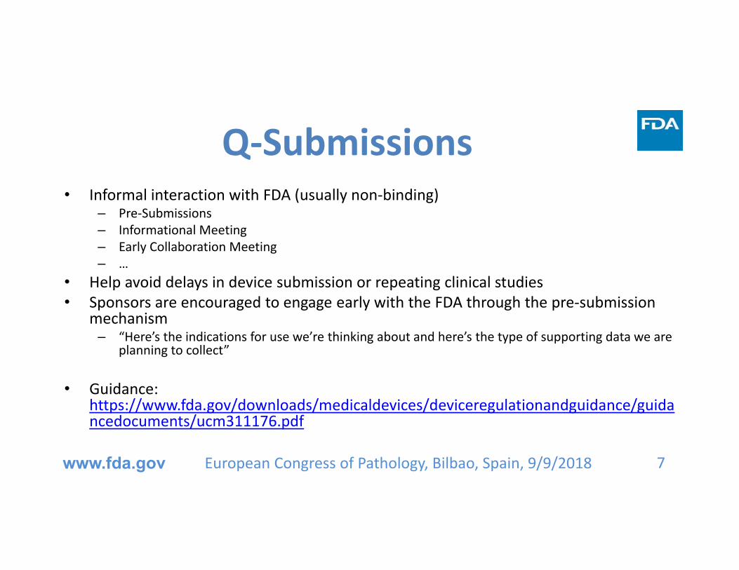
Q‐Submissions• Informal interaction with FDA (usually non‐binding)
– Pre‐Submissions– Informational Meeting– Early Collaboration Meeting– …
• Help avoid delays in device submission or repeating clinical studies• Sponsors are encouraged to engage early with the FDA through the pre‐submission
mechanism– “Here’s the indications for use we’re thinking about and here’s the type of supporting data we are
planning to collect”
• Guidance: https://www.fda.gov/downloads/medicaldevices/deviceregulationandguidance/guidancedocuments/ucm311176.pdf
European Congress of Pathology, Bilbao, Spain, 9/9/2018www.fda.gov 7
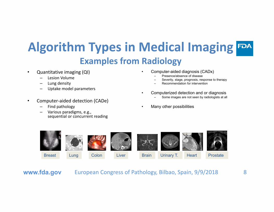
Algorithm Types in Medical ImagingExamples from Radiology
• Quantitative imaging (QI)– Lesion Volume– Lung density– Uptake model parameters
• Computer‐aided detection (CADe)– Find pathology– Various paradigms, e.g.,
sequential or concurrent reading
• Computer-aided diagnosis (CADx)– Presence/absence of disease– Severity, stage, prognosis, response to therapy– Recommendation for intervention
• Computerized detection and or diagnosis– Some images are not seen by radiologists at all
• Many other possibilities
European Congress of Pathology, Bilbao, Spain, 9/9/2018www.fda.gov 8
Breast BrainLung Colon Liver Urinary T. Heart Prostate

Core Content of 510(k) Submissions for computer aids in (Radiology)
• Find a predicate
• Description– Indications for use– Patient and clinician population– Clinical workflow– Imaging system and protocols
• Technological Characteristics – Algorithm design and function– Processing steps– Features– Models and classifiers– Training paradigm
• Imaging modality– Manufacturer and Model– Imaging parameters and techniques
• Databases: Training and Testing– Must be Independent
• Reference standard
• Assessment– Depends on algorithm type– Stand Alone– Clinical Performance: reader in‐the‐loop
European Congress of Pathology, Bilbao, Spain, 9/9/2018www.fda.gov 9

Standalone performance• Performance of algorithm by itself, independent of any interaction with user– Intrinsic functionality of device
European Congress of Pathology, Bilbao, Spain, 9/9/2018www.fda.gov 10
Apply AI/ML Tool
AcquireTest Dataset
Apply Scoring
StatisticalPerformance
Analysis
Establish Ground Truth
Hidde
n du
ring presen
tatio
nHidden during presentation

Clinical: Reader performance• Assessment of clinicians’ performance utilizing the device
– Many possible study designs• Prospective/retrospective• Multi‐reader multi‐case designs
European Congress of Pathology, Bilbao, Spain, 9/9/2018www.fda.gov 11
Establish Ground Truth
AcquireTest Dataset
StatisticalPerformance
Analysis
Apply Scoring
Clinical readwithout aid
Apply AI/ML Tool
Clinical readwith aid
Apply Scoring
Hidde
n du
ring presen
tatio
nHidden during presentation

Radiology CADe Guidances• Computer‐Assisted Detection Devices Applied to Radiology Images and Radiology
Device Data – Premarket Notification [510(k)] Submissions– http://www.fda.gov/RegulatoryInformation/Guidances/ucm187249.htm
• Clinical Performance Assessment: Considerations for Computer‐Assisted Detection Devices Applied to Radiology Images and Radiology Device Data ‐ Premarket Approval (PMA) and Premarket Notification [510(k)] Submissions
– http://www.fda.gov/RegulatoryInformation/Guidances/ucm187277.htm
• Software as a Medical Device (SAMD): Clinical Evaluation– https://www.fda.gov/medicaldevices/digitalhealth/softwareasamedicaldevice/default.htm
• Roadmap for other algorithm types
European Congress of Pathology, Bilbao, Spain, 9/9/2018www.fda.gov 12

Predicates in RadiologySpecial controls generally follow CADe guidance
• CADx: QuantX– DEN170022 (7/2017)– POK: computer‐assisted diagnostic software
for lesions suspicious for cancer• CADe + CADx: OsteoDetect
– DEN180005 (5/2018)– QBS: radiological computer assisted
detection/diagnosis software for fracture• Triage: ContaCT
– DEN170073 (2/2018)– QAS: radiological computer‐assisted triage
and notification software• Automatic Detection: IDx‐DR
– DEN180001 (4/2018)– PIB: diabetic retinopathy detection device
European Congress of Pathology, Bilbao, Spain, 9/9/2018www.fda.gov 13
https://www.quantinsights.com/
https://www.viz.ai/viz‐lvo/https://www.eyediagnosis.net/idx‐dr
https://www.slashgear.com/osteodetect‐ai‐tool‐finds‐wrist‐fractures‐gets‐fda‐approval‐28532138/

Interoperability vs. SpecializationLessons from Radiology
• First submission often tied to specific system
• Expand indications over time– New imaging system– Algorithm updates/improvements
• Expand indications via– New 510k– PMA Supplement
• Device and performance familiarity mayallow for less burdensome methods
Less burdensome methods• Studies with fewer readers or cases
• Reuse cases for evaluating test performance
• Re‐acquire digital images with alternate systems
• Stand‐alone performance only
• No statistical hypothesis test
• Technical arguments
European Congress of Pathology, Bilbao, Spain, 9/9/2018www.fda.gov 14
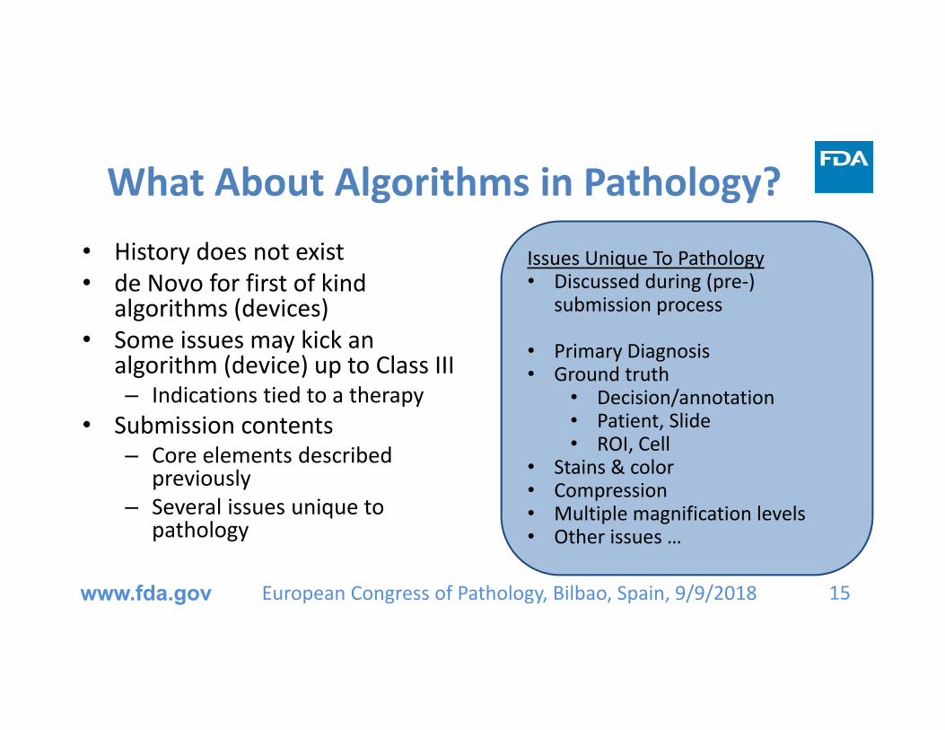
What About Algorithms in Pathology?• History does not exist• de Novo for first of kind
algorithms (devices)• Some issues may kick an
algorithm (device) up to Class III– Indications tied to a therapy
• Submission contents– Core elements described
previously– Several issues unique to
pathology
European Congress of Pathology, Bilbao, Spain, 9/9/2018www.fda.gov 15
Issues Unique To Pathology• Discussed during (pre‐)
submission process
• Primary Diagnosis• Ground truth
• Decision/annotation• Patient, Slide• ROI, Cell
• Stains & color• Compression• Multiple magnification levels• Other issues …
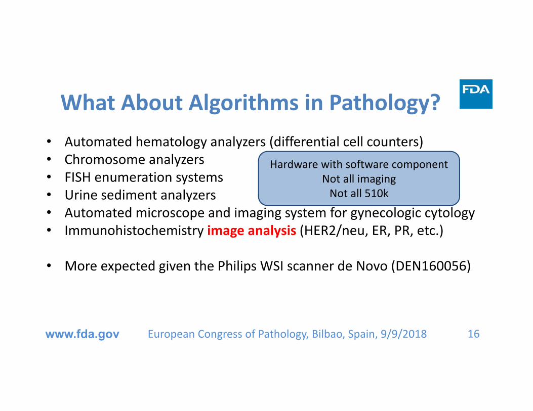
What About Algorithms in Pathology?• Automated hematology analyzers (differential cell counters)• Chromosome analyzers • FISH enumeration systems• Urine sediment analyzers• Automated microscope and imaging system for gynecologic cytology• Immunohistochemistry image analysis (HER2/neu, ER, PR, etc.)
• More expected given the Philips WSI scanner de Novo (DEN160056)
European Congress of Pathology, Bilbao, Spain, 9/9/2018www.fda.gov 16
Hardware with software componentNot all imagingNot all 510k

GenASIs HiPath IHC Family (K140957)
• 510k database: Quick search “IHC”– https://www.accessdata.fda.gov/scripts/cdrh/cfdocs/cfPMN/pmn.cfm
• Indications for use:– The GenASIs HiPath IHC Family provides image capture, management,
analysis, and viewing of specific immunohistochemically stained slides. It is intended for in vitro diagnostic use as an aid to the pathologist in the display, detection, counting, review and classification of tissues and cells of clinical interest based on particular morphology, color, intensity, size, pattern and shape:
– HER2, PR, ER, Ki67
European Congress of Pathology, Bilbao, Spain, 9/9/2018www.fda.gov 17

GenASIs HiPath IHC Family (K140957)
• Four predicates for the four different antibodies– K111543: Virtuoso System for IHC HER2 (4B5)– K111869: Virtuoso System for IHC PR (1E2)– K130515: Virtuoso System for IHC ER (SP1)– K111755: Virtuoso System for IHC Ki67 (30‐9)
• Image and Region Of Interest (ROI) selected by the pathologist • Device Components: Microscope, CCD color camera, PC, keyboard, Mouse, Color
Monitor, X‐Y stage and rack for loading 1 glass slide.
• Differences with predicates largely based on image acquisition– CCD on microscope versus slide scanner
European Congress of Pathology, Bilbao, Spain, 9/9/2018www.fda.gov 18
Other Virtuoso 510k’s expand indications to different• Stainer platform• Scanner

PMA for Cytology with Computer Aid
19
• Gynecologic Cytology Imaging Systems– Cytyc/Hologic ThinPrep Imaging
System (P020002)– Becton Dickinson/TriPath FocalPoint
Guided Screening System– Papanicolaou Stain– Detection algorithm, neural network– Images not saved– Cytologist reviews locations with
microscope
www.fda.gov European Congress of Pathology, Bilbao, Spain, 9/9/2018
Hidde
n du
ring presen
tatio
nHidden during presentation

Computer Aids in Radiology• R2 ImageChecker (P970058)
– The ImageChecker M1000 is a computer system intended to identify and mark regions of interest on routine screening mammograms to bring them to the attention of the radiologist after initial reading has been completed. Thus, the system assists the radiologist in minimizing observational oversights by identifying areas on the original mammogram that may warrant a second review.
European Congress of Pathology, Bilbao, Spain, 9/9/2018www.fda.gov 20
Hidde
n du
ring presen
tatio
nHidden during presentation

200315. New Manufacturing facility16. Choice of new operating points (high and low sensitivity), operates on analog and GE FFDM images, operates on GE FFDM images “formatted for presentation”, reduces false‐negatives of oversized malignant calcification clusters17. Alternative film digitizer18. Indications expanded to Fischer Senoscan FFDM200319. Indications expanded to Hologic Selenia FFDM200520. Indications expanded to include Siemens Novation FFDM21. More operating points200622. Change label to include specificity (previously it was sensitivity and false marks per image)200724. New manufacturing facility2001225. Algorithm updates and indications expanded to GE Senograph Essential201426. Indications expanded to C‐view images Hologic Selenia Dimensions (Tomosynthesis) system201627. New manufacturing facility
1998Approval of Original submission1. Hardware changes and minor bugs and enhancements19992. Performance change3. Post approval study protocol4. New marker (correlated masses)5. Alternative film digitizer20006. Performance change7. Label change with respect to efficacy8. New marker (subtle vs. obvious masses)20019. New marker (subtle vs. obvious calcifications)10. Indications expanded from screening to diagnosis11. Indications expanded to digital images (GE Senographe 2000)200212. Label change with respect to efficacy13. Transparent marker (see image under marker)14. Label change
R2 ImageChecker Submission History
European Congress of Pathology, Bilbao, Spain, 9/9/2018www.fda.gov 21
Hidde
n du
ring presen
tatio
nHidden during presentation

My Research and Projects• eeDAP: Evaluation Environment for Digital and Analog Pathology
• eeDAP Studies– Compare scanners to microscope– Pathologist microscope viewing behavior– Measure registration accuracy
• MDDT: Medical Device Development Tool– CDRH program– eeDAP– Annotating Images to validate algorithms
European Congress of Pathology, Bilbao, Spain, 9/9/2018www.fda.gov 22

eeDAP:Evaluation Environment for Digital and Analog Pathology
European Congress of Pathology, Bilbao, Spain, 9/9/2018www.fda.gov 23
Monitor, Computer, motorized stage with joystick,microscope with mounted camera, reticle in eyepiece
https://github.com/DIDSR/eeDAP
• Register glass slide and WSI
• Allow pathologists to evaluate same fields of view on microscope and WSI
Camera imageof glass slide
WSI Patch

eeDAP:Removes search from technology evaluation• eeDAP can eliminate
location variability for faster and more precise results.
European Congress of Pathology, Bilbao, Spain, 9/9/2018www.fda.gov 24
Clinical practicePathologists choose
Fields of View to evaluate
Pathologist 1Pathologist 2Pathologist 3Pathologist 4
Technology EvaluationAll pathologists evaluatesame Fields of View H&E 20x
H&E 40x

Compare scanners to microscopeInstall, Demo, Trainat Memorial Sloan Kettering Study Design
• 4 slides from Mark Simpson at NCI– HE: canine oral melanoma
• 10 ROIs per slide from tumor– ROI = 800 x 800 pixels @ 0.25um/pixel
= 200um x 200um= 17% of the entire FOV (0.24 mm2)
• Task: Mark and count mitotic figures (MF)
• eeDAP integrates ImageScope– Show ROIs – Mark cells
www.fda.gov 25
eeDAP on loanto MSK
European Congress of Pathology, Bilbao, Spain, 9/9/2018

Compare scanners to microscope
• High‐throughput reader study
• Same microscope frame … 14 heads!
• Stage mounts fine
• Camera mounts fine
European Congress of Pathology, Bilbao, Spain, 9/9/2018www.fda.gov 26

Compare scanners to microscope• Four scanners and microscope• Five study pathologists
– 157 candidate MFs• Three truthing pathologists• True MFs
– Start with candidates unanimously identified on microscope
– Add candidates determined to be true MFs
• Truthing panel• Group setting• Digital microscope (VisionTek)
European Congress of Pathology, Bilbao, Spain, 9/9/2018www.fda.gov 27
Accuracy = Average of Sensitivity & Specificity
Uncertainty accounts for reader and case variability
Bonferroni correction for multiple hypotheses:Compare Each Scanner to Microscope
P=0.002P=0.012
P=0.068
P=0.001

iMRMC:Statistical Analysis Tool for Reader Studies
• MRMC analysis– Multiple readers– Multiple cases– Uncertainty accounts for reader and case
variability
• Statistical analysis tool– Percent Correct– Area Under the ROC curve
• GitHub java application GUI– https://github.com/DIDSR/iMRMC
• CRAN R package– https://cran.r‐
project.org/web/packages/iMRMC/index.html
European Congress of Pathology, Bilbao, Spain, 9/9/2018www.fda.gov 28
DataInput
DataAnalysis
Study Sizing

Current and previous study
European Congress of Pathology, Bilbao, Spain, 9/9/2018www.fda.gov 29
Readers Per Candidate, total = 92
readersPerCandidate1
Den
sity
0 1 2 3 4
0.0
0.1
0.2
0.3
0.4
0
45
1214
21
23%
49%
15%13%
Distribution of agreement results per candidateTotal number of candidates = 157
Hidde
n du
ring presen
tatio
nHidden during presentation

MSKCC results• All 5 observers detected 157 candidate mitotic cells,
using all WSIs and microscopy. All counts by all observers using all observation methods are showed in Table 1. Using microscopy, 29 potential candidate mitotic cells were detected by all five observers, 8 candidates by four observers, 17 candidates by three observers, 13 candidates by two observers, 28 candidates by only one observer. The remaining 62 candidates remained undetected by microscopy; they were detected only using WSI.
European Congress of Pathology, Bilbao, Spain, 9/9/2018www.fda.gov 30
Hidde
n du
ring presen
tatio
nHidden during presentation

Readers per CandidateReaders Per Candidate, total = 92
readersPerCandidate1
Den
sity
0 1 2 3 4
0.0
0.1
0.2
0.3
0.4
0
45
1214
21
• Do you think this is a lot of reader variability?
• 45/92 = 49% marked by only one
• 21/92 = 23% unanimously marked
• Build these candidates into next study: Classification task
• Need some low‐probability candidates from ROIs with zero or one candidates ‐> yield 34
European Congress of Pathology, Bilbao, Spain, 9/9/2018www.fda.gov 31
Hidde
n du
ring presen
tatio
nHidden during presentation

Readers per Candidate:Multi‐head study
• Similar characteristics as before
• 79/158 = 49% marked by only one
• 21/158 = 23% unanimously marked– 13 agree with previous, 8 new ones
• How well does AI correlate with this scoring?
European Congress of Pathology, Bilbao, Spain, 9/9/2018www.fda.gov 32
Readers Per Candidate, total = 158
readersPerCandidate2
Den
sity
0 2 4 6 8 10
0.0
0.1
0.2
0.3
0.4
0.5
0
79
127 6 5
94
9 6
21
Hidde
n du
ring presen
tatio
nHidden during presentation

Pathologist Microscope Viewing Behavior:Collaboration with Cold Spring Harbor Laboratory and
Northwell Health New eeDAP workflow• Pathologist driven navigation of
slides on the microscope– Collect main diagnosis, grade, type,
etc.– Provide confidence ratings
• Continuously record– Stage position (+ mouse clicks)– Eyepiece camera video– Audio
• Registration after the fact
www.fda.gov 33
• Register the study slides– Glass and WSI
• Visit locations/objects– Pre‐determined list of “tasks”
• ROIs … candidate MFs– Evaluate each location/object
• Perform the “task”
Original eeDAP workflow
European Congress of Pathology, Bilbao, Spain, 9/9/2018

Pathologist Microscope Viewing Behavior:Collaboration with Cold Spring Harbor Laboratory and
Northwell Health
www.fda.gov 34
23:00
1:00:00 1:33:00
Registration after the fact
European Congress of Pathology, Bilbao, Spain, 9/9/2018
Video: Collecting3 registration anchors
Static images:3 registration anchors

Pathologist Microscope Viewing Behavior:Collaboration with Cold Spring Harbor Laboratory and
Northwell Health
www.fda.gov 35
Camera WSI
Video helps identify where to look/register
Registration after the fact
European Congress of Pathology, Bilbao, Spain, 9/9/2018
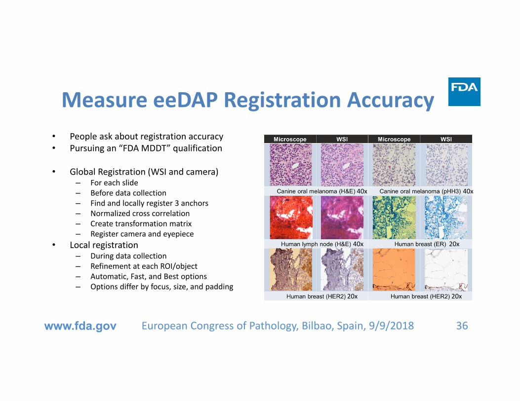
Measure eeDAP Registration Accuracy• People ask about registration accuracy• Pursuing an “FDA MDDT” qualification
• Global Registration (WSI and camera)– For each slide– Before data collection– Find and locally register 3 anchors– Normalized cross correlation– Create transformation matrix– Register camera and eyepiece
• Local registration– During data collection– Refinement at each ROI/object– Automatic, Fast, and Best options– Options differ by focus, size, and padding
European Congress of Pathology, Bilbao, Spain, 9/9/2018www.fda.gov 36
20x
20x20x
40x
40x 40x

Measure eeDAP Registration Accuracy
European Congress of Pathology, Bilbao, Spain, 9/9/2018www.fda.gov 37
Reticle: 10 mm with 100 divisions
40X: 250 µm with 2.5 µm divisions
20X: 500 µm with 5.0 µm divisions
WSI patch with virtual reticle shows target location.
Observer identifies target in microscope FOV and measures distance from center with ruler reticle.

Measure eeDAP Registration Accuracy
FDA study• 120 measurements
– 6 slides– 10 measurements per slide– 2 participants (replicate study)
• Global registration– Mean error = 37.62 µm (~3 cells)– Standard Deviation = 28 µm
• Local registration after focusing– Better than 95% of measurements <
5 µm
CSHL study• Designed and executed at CSHL• Very similar results
www.fda.gov 38European Congress of Pathology, Bilbao, Spain, 9/9/2018

Measure eeDAP Registration Accuracy
FDA study• 120 measurements
– 6 slides– 10 measurements per slide– 2 participants (replicate study)
• Global Registration Results
• Global registration– Mean error = 37.62 µm (~3 cells)
• Local registration after focusing– Better than 95% of measurements < 5 µm
CSHL study• 400 measurements
⁻ 20 slides (10 Rat H&E + 10 human H&E), scanned magnification 20X
⁻ 10 measurements per slide⁻ 2 participants (replicate study)
• Global registration⁻ Mean error = 31.35 um
• Local registration after focusing⁻ Better than 95% of measurements <5um
www.fda.gov 39European Congress of Pathology, Bilbao, Spain, 9/9/2018
Hidde
n du
ring presen
tatio
nHidden during presentation

MDDT: Medical Device Development Tool(CDRH program)
• Definition: Method, material, or measurement used to assess the effectiveness, safety, or performance of a medical device
• Qualified for a context of use• Facilitates submission and its review
(Point to the qualification package)• Encourages
– Innovation– Collaboration– Chance for community to impact regulatory
process
• Clinical outcome assessments– Surrogate outcomes– Patient reported outcomes
• Biomarker tests– Measure biological process
(gold standard)– Measure response to intervention
• Nonclinical assessment models– Computational models (simulations)– Probes and phantoms for bench tests– eeDAP!– Image databases with truth annotations
European Congress of Pathology, Bilbao, Spain, 9/9/2018www.fda.gov 40
Guidance on the web! https://www.fda.gov/medicaldevices/scienceandresearch/medicaldevicedevelopmenttoolsmddt/

MDDT:Annotating Images to validate algorithms
• Elevator Pitch:– Create dataset of images with truth annotations– To be available to algorithm developers for FDA submission (Performance
Evaluation)
• Follow example of ACR– American College of Radiology– https://www.acrdsi.org/Use‐Case‐Development
• TOUCH‐AI: Technology‐Oriented Use Cases for Healthcare AI• CERTIFY‐AI: ACR Digital Science Institute validation service
European Congress of Pathology, Bilbao, Spain, 9/9/2018www.fda.gov 41

MDDT:Annotating Images to validate algorithms
ACR TOUCH‐AI: Concepts and Tools• Open framework for defining use cases• Community‐Contributed use cases• Use cases reviewed by ACR committees• Use cases reviewed by FDA (MDDT)
• CARDS: Computer‐Assisted Reporting and Decision Support Tools for Radiologists– XML‐based Proceduralized Definitions– Logic relating Common Data Elements (CDE’s) to patient management
• Reference implementation– No‐frills user interface defined by CARDS– Structured evaluation (inputs): check boxes, menus, numeric fields– Standardized report (outputs)– Framework for value‐added vendors: PACS, VRS, AI
European Congress of Pathology, Bilbao, Spain, 9/9/2018www.fda.gov 42
• Can be unique to institution• Can be reviewed by ACR committees• Can be reviewed by FDA (MDDT) • “Scores”
(Labels, Counts, Segmentations,Measurements, Units)• Image‐based• Other Dx• Cut offs
Add No‐Frills Reference Viewer

MDDT:Annotating Images to validate algorithms
ACR TOUCH‐AI: Use case core contents• Clinical implementation (FDA: device description)
– Value proposition, narrative(s), workflow description
• Considerations for dataset development (FDA: indications for use)– (FDA: intended imaging procedures and protocols)– (FDA: intended patient population)
• Technical specifications (CARDS, XML): – Inputs, outputs
• Future development (CARDS, XML)– Inputs, outputs, extensions, comparison over time
European Congress of Pathology, Bilbao, Spain, 9/9/2018www.fda.gov 43

MDDT:Annotating Images to validate algorithms
ACR CERTIFY‐AI: Work In Progress
• STARD: Standards for Reporting of Diagnostic Accuracy Studies– Study design: prospective/retrospective, …– Reader and case sampling/description– Reference standard: “Scores” specified in use case– Performance metric: stand‐alone vs. reader in‐the‐loop– Analysis method: MRMC? Missing/indeterminate data? Sizing?– Study limitations
European Congress of Pathology, Bilbao, Spain, 9/9/2018www.fda.gov 44

MDDT:Annotating Images to validate algorithmsPossible Use Cases (Tasks)
• Automated detection of breast cancer metastases in lymph node WSIs
• Classify tumor infiltrating lymphocytes and score ROIs by density of TILS
• Classify tumor bed cells, ROI cellularity
• Counting mitotic figures and scoring proliferation in H&E
My Goals (not requirements)• Collect truth on the microscope
– Reference standard– Continuous 3D object. Not digitized.– Pathologist familiarity– Not tied to specific scanner
• Collect truth from multiple pathologists– Acknowledge pathologist variability– Reduce pathologist variability– Account for pathologist variability
– Number of readers depends on reader variability
www.fda.gov 45European Congress of Pathology, Bilbao, Spain, 9/9/2018

MDDT:Annotating Images to validate algorithmsPossible Use Cases (Tasks)
• Automated detection of breast cancer metastases in lymph node WSIs
• Classify tumor infiltrating lymphocytes and score ROIs by density of TILS
• Classify tumor bed cells, ROI cellularity
• Counting mitotic figures and scoring proliferation in H&E
Leveraging Challenges• (High Throughput)
• Can’t do this by myself– Nurturing partnerships– Need partners to share the load
• Willing to do heavy lifting– Drafting/reviewing FDA proposal and
submission– Reader study design– Reader study execution– Reader study analysis
www.fda.gov 46European Congress of Pathology, Bilbao, Spain, 9/9/2018

MDDT:Annotating Images to validate algorithmsPossible Use Cases: Tasks
• Automated detection of breast cancer metastases in lymph node WSIs
• Classify tumor infiltrating lymphocytes and score ROIs by density of TILS
• Classify tumor bed cells
• Counting mitotic figures and scoring proliferation in H&E
Leveraging Challenges• CAMELYON 16 & 17• Point of Contact: Jeroen van der Laak
– Challenge Organizer– Radboud University Medical Center– Nijmegen, The Netherlands
• Starting material transfer agreement (MTA)
– Camelyon16 glass slides– Algorithms available
• FDA algorithm• MDDT Issue: Camelyon16 images and
truth released
www.fda.gov 47European Congress of Pathology, Bilbao, Spain, 9/9/2018

MDDT:Annotating Images to validate algorithmsPossible Use Cases: Tasks
• Automated detection of breast cancer metastases in lymph node WSIs
• Classify tumor infiltrating lymphocytes and score ROIs by density of TILS
• Classify tumor bed cells
• Counting mitotic figures and scoring proliferation in H&E
Leveraging Challenges• Future challenge planned• Point of Contact: Roberto Salgado
– Chair: International Immuno‐oncology Working Group
• Large, motivated, working group with many pathologists and image sets from drug trials
• Massive Analysis and QC (MAQC) Society
– Project: “Reproducible machine learning for pathology image analysis”
www.fda.gov 48European Congress of Pathology, Bilbao, Spain, 9/9/2018

MDDT:Annotating Images to validate algorithmsPossible Use Cases: Tasks
• Automated detection of breast cancer metastases in lymph node WSIs
• Classify tumor infiltrating lymphocytes and score ROIs by density of TILS
• Classify tumor bed cells
• Counting mitotic figures and scoring proliferation in H&E
Leveraging Challenges• SPIE Medical Imaging conference
– February 2019– The international society for optics and
photonics– CAD and Digital Pathology tracks
• Point of Contact: FDA colleagues!– Sunnybrook Research Institute, University
of Toronto, University of Chicago, University of Michigan, NIH/NCI, Harvard University, Stony Brook University, Universitiy of Buffalo, Western University, Fraunhofer (Medical Imaging Computing) MEVIS, Nagoya University
www.fda.gov 49European Congress of Pathology, Bilbao, Spain, 9/9/2018

SPIE Medical ImagingChallenge
• Anne Martel University of Toronto ([email protected])• Shazia Akbar, University of Toronto ([email protected])• Nick Petrick, U.S. FDA ([email protected])• Marios Gavrielides, U.S. FDA ([email protected])• Berkman Sahiner, U.S. FDA ([email protected])• Kenny Cha, U.S. FDA ([email protected])• Sam Armato, University of Chicago (s‐[email protected])• Karen Drukker, University of Chicago ([email protected])• Lubomir Hadjiiski, University of Michigan ([email protected])• Keyvan Farahani, NIH/NCI ([email protected])• Jayashree Kalpathy‐Cramer, Harvard University
([email protected])• Diane Cline, SPIE ([email protected])• Joel Saltz, Stony Brook University ([email protected])• John Tomaszewski, Kaleida Health ([email protected])• Aaron Ward, Western University ([email protected])• Horst Hahn, Fraunhofer MEVIS ([email protected])• Kensaku Mori, Nagoya University ([email protected]‐u.ac.jp)
• Sunnybrook Research Institute, University of Toronto• University of Chicago• University of Michigan• NIH/NCI• Harvard University• Stony Brook University• Universitiy of Buffalo• Western University• Fraunhofer (Medical Imaging Computing) MEVIS• Nagoya University
European Congress of Pathology, Bilbao, Spain, 9/9/2018www.fda.gov 50
Hidde
n du
ring presen
tatio
nHidden during presentation

MDDT:Annotating Images to validate algorithmsPossible Use Cases: Tasks
• Automated detection of breast cancer metastases in lymph node WSIs
• Classify tumor infiltrating lymphocytes and score ROIs by density of TILS
• Classify tumor bed cells
• Counting mitotic figures and scoring proliferation in H&E
Leveraging Challenges• Tumor Proliferation Assessment
Challenge 2016 | TUPAC16• Point of contact: Mitko Veta
– Challenge Organizer– Eindhoven University of Technology (TU/e)
• No glass slides available but preparing for next challenge: breast cancer prognosis
www.fda.gov 51European Congress of Pathology, Bilbao, Spain, 9/9/2018

MDDT:Annotating Images to validate algorithms
• Next up …
• American Society for Clinical Pathology Annual Meeting (October 3)– Call for proposal to conduct perception studies (June 21)– NCI funding did not come through (September 7) … making calls … help?
– Demonstrate and get experience running eeDAP in conference environment (high‐throughput)
– Offer CME for study participants– Sourced lung tumor tissue for TILS counting/scoring– Data collection: Pre‐defined ROIs or Pathologist Guided?
European Congress of Pathology, Bilbao, Spain, 9/9/2018www.fda.gov 52

Summary• FDA has been evaluating computer aids in
radiology for two decades– Core content– There is guidance, examples, and predicates– Start with limited indication … grow
indications
• FDA history with computational pathology device
– De Novo request of Whole Slide Imaging (WSI) system for primary diagnosis was granted (PIPS, Philips, April 2017)
– No devices on the market for that scanner/technology today
– Request feedback on your submission plans
• eeDAP is at my research core … it is a tool– Microscope is still dominant/reference
modality• Large datasets are needed for training
– Smaller high‐quality data sets are needed for testing
• Reader variability– Account for it in performance evaluation– Statistics and Truthing
• MDDT: – Demonstrate and get experience with data
collection– Plan for defining use cases– Nurturing partnerships– Looking for partners to share the load
European Congress of Pathology, Bilbao, Spain, 9/9/2018www.fda.gov 53

Resources• eeDAP: evaluation environment for digital and analog pathology
– https://github.com/DIDSR/eeDAP
• iMRMC statistical analysis tool– GitHub: https://github.com/DIDSR/iMRMC– CRAN R package: https://cran.r‐project.org/web/packages/iMRMC/index.html
• WSI Working Group– https://nciphub.org/groups/wsi_working_group
• MDDT: Medical Device Development Tools– https://www.fda.gov/medicaldevices/scienceandresearch/medicaldevicedevelopmenttoolsmddt/
• CADe– http://www.fda.gov/RegulatoryInformation/Guidances/ucm187249.htm– http://www.fda.gov/RegulatoryInformation/Guidances/ucm187277.htm
• Software as a Medical Device (SAMD): Clinical Evaluation– https://www.fda.gov/medicaldevices/digitalhealth/softwareasamedicaldevice/default.htm
• Requests for Feedback on Medical Device Submissions– https://www.fda.gov/downloads/medicaldevices/deviceregulationandguidance/guidancedocuments/ucm311176.pdf
• De Novo Classification Process – https://www.fda.gov/downloads/MedicalDevices/DeviceRegulationandGuidance/GuidanceDocuments/ucm080197.pdf
• How to Prepare a Traditional 510(k)– https://www.fda.gov/medicaldevices/deviceregulationandguidance/howtomarketyourdevice/premarketsubmissions/premarketnotification510k/ucm134572.htm
European Congress of Pathology, Bilbao, Spain, 9/9/2018www.fda.gov 55