Embed Size (px)
Citation preview

Estructura del DNA
El DNA es una molécula de gran tamaño formada por dos hebras de polinucleótidos enrolladas entre sí para formar una doble hélice de 2.0 nm de diámetro. Cada cadena contiene los desoxirribonucleósidos de purina y de pirimidina
A diagrammatic representation of the double helix. The backbone consists of deoxyribose sugars (S) joined by phosphates (P) in phosphodiester bridges. The arrows at the top and bottom of the chains point in the 5′ to 3′ direction. The ribbons represent the sugar phosphate backbones

Components of the nucleic acids. (a) The nitrogen bases of DNA and RNA. Note the numbering system of the rings. In attaching itself to the 1′ carbon of the sugar phosphate, a pyrimidine base bonds through N-1 and a purine base bonds at N-9. (b) Part of a DNA chain. The numbers on the sugar of the nucleotide contain a prime (′) after them because the rings of the nitrogen bases are also numbered. In DNA, a hydrogen is present on the 2′-carbon of the pentose sugar. In RNA, an OH group occupies this position. The nucleotides are linked by a phosphodiester bond.

Genetic information flow and the components of the nucleic acids. (a) An overview of the types of informational macromolecules. (b) Part of a DNA chain. The numbers on the sugar of the nucleotide contain a prime (′) to differentiate them from the numbering onthe rings of the nitrogen bases. In DNA, a hydrogen is present on the 2′-carbon of the pentose sugar. In RNA, an OH group occupies this position. The nucleotides are linked by a phosphodiester bond. (c) The nitrogen bases of DNA and RNA and the specific pairing between cytosine (C) and guanine (G) and between thymine (T) and adenine (A) via hydrogen bonds. Uracil (U) instead of thymine is present in RNA. Note the numbering system of the rings in that a pyrimidine base bonds through N-1 to the sugar– phosphate backbone and that a purine base bonds through N-9. Atoms that are found in the major groove of the double helix and that interact with proteins are highlighted in pink.

Nucleósido y nucleótido

DNA structure. Complementary and antiparallel nature of DNA. Note that one chain ends in a 5′-phosphate group, whereas the other ends in a 3′-hydroxyl. The purple bases represent the pyrimidines cytosine (C) and thymine (T), and theyellow bases represent the purines adenine (A) and guanine (G).
DNA structure
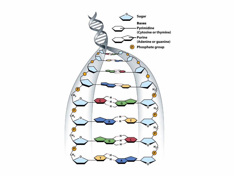

Nitrogenous bases• Pyrimidine bases: Cytosine (C), Thymine (T), Uracil
(U, in RNA)
• Purine bases: Adenine (A), Guanine (G)

A-T hydrogen bonding

G-C hydrogen bonding

DNA denaturation

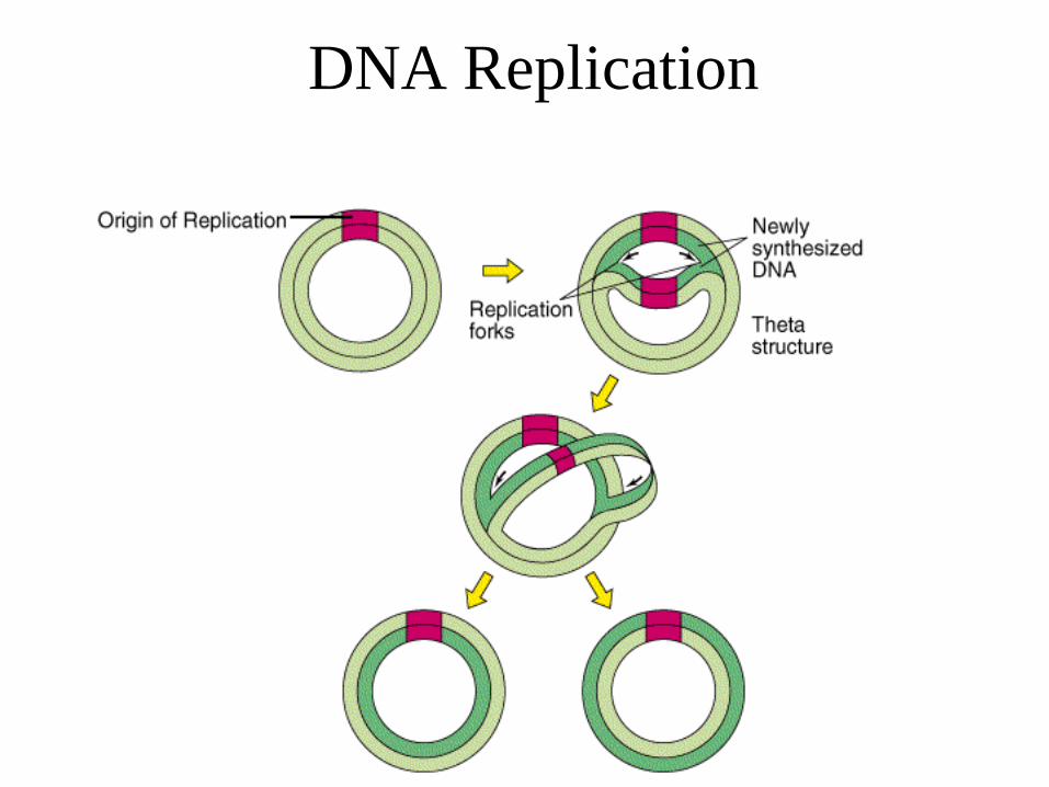
Replicación DNA
La replicación del DNA es semiconservadora, pues sólo se sintetiza la mitad del DNA, mientras que la otra mitad proviene de la molécula original.
Semiconservative DNA Replication. The replication fork of DNA showing the synthesis of two progeny strands. Newly synthesized strands are purple. Each copy contains one new and one old strand.

Semiconservative replication

DNA Replication: addition of a nucleotide
Extension of a DNA chain by adding a deoxyribonucleotide triphosphate at the 3′end. Growth proceeds from the 5′-phosphate to the 3′-hydroxyl end. DNA polymerase catalyzes the reaction. The four precursors are deoxythymidine triphosphate (dTTP), deoxyadenosine triphosphate (dATP), deoxyguanosine triphosphate (dGTP), and deoxycytidine triphosphate (dCTP). Upon nucleotide insertion, the two terminal phosphates of the triphosphate are split off as pyrophosphate (PPi). Thus, two energy-rich phosphate bonds are consumed when adding each nucleotide.

Intervienen las siguientes enzimas:
• DNA helicasa, separa las dos cadenas de DNA desenrollando la doble hélice
• DNA polimerasa, adiciona los nucleótidos complementarios. Actúa en el sentido 5’ → 3’
• DNA ligasa, une los fragmentos discontinuos de DNA: fragmentos de Okazaki
• DNA topoisomerasa II, Actúa en la replicación del DNA para reducir la tensión molecular causada por el superenrollamiento. Alivia la tensión mientras en DNA de la doble hebra está siendo desenrollada por la helicasa.
Se necesita, además:
• DNA patrón o molde
• Cebador o primer
• Sustratos de la reacción, sillares de construcción:

BASE
NITROGENADA NUCLEÓSIDO NUCLEÓTIDOS
(Base + pentosa) Nucleótido
monofosfato
Nucleótido
difosfato
Nucleótido
trifosfato
RNA
Adenina Adenosina AMP ADP ATP
Guanina Guanosina GMP GDP GTP
Citosina Citidina CMP CDP CTP
Uracilo Uridina UMP UDP UTP
DNA
Adenina Desoxiadenosina dAMP dADP dATP
Guanina Desoxiguanosina dGMP dGDP dGTP
Citosina Desoxicitidina dCMP dCDP dCTP
Timina Desoxitimidina dTMP dTDP dTTP

Events at the DNA replication fork

The replisome
The replisome consists of two copies of DNA polymerase III and DNA gyrase, plus helicase and primase (together forming the primosome), and many copies of single-strand DNA-binding protein. The Tau subunits hold the two DNA polymerase assemblies and helicase together. Just upstream of the rest of the replisome, DNA gyrase removes supercoils in the DNA to be replicated. Note that the two polymerases are replicating the two individual strands of DNA in opposite directions. Consequently, the lagging-strand template loops around so that the whole replisome moves in the same direction along the chromosome.
DNA Replication

Fidelidad de la replicación del DNA
• Además de la inserción de nucleótidos en la cadena
de replicación la DNA pol III ejerce también una
actividad 3’ → 5’ exonucleasa → corrección de
lectura
• La corrección de lectura de la exonucleasa se efectúa
en procariotas, eucariotas y en los sistemas de
replicación de DNA viral

• Además de la capacidad exonucleasa, los
procariotas y eucariotas contienen proteínas
endonucleasas capaces de suprimir nucleótidos
insertados erróneamente mucho después que la
DNA polimerasa ha pasado el punto de error.
• La combinación de endonucleasas y exonucleasas asegura la replicación casi libre de errores de las secuencias extremadamente largas de DNA que constituyen el DNA genómico

Proofreading by DNA
polymerase III

Tautomería: Las bases nitrogenadas se encuentran habitualmente en su forma cetónica y con menos frecuencia aparecen en su forma tautomérica enólica. Las formas tautoméricas o enólicas de las bases nitrogenadas (A*, T*, G* y C*) muestran relaciones de apareamiento distintas: A*-C, T*-G, G*-T y C*-A. Los errores en el apareamiento incorrecto de las bases nitrogenadas pueden ser detectados por la función correctora de pruebas de la DNA polimerasa III