Embed Size (px)
Citation preview
Research ArticleEssentiality of CTNNB1 in Malignant Transformation of HumanEmbryonic Stem Cells under Long-Term Suboptimal Conditions
Jie Liu,1 Sicong Zeng,1,2 Yang Wang,1 Juan Yu,3 Qi Ouyang,1,3 Liang Hu,1,3 Di Zhou,1,3
Ge Lin,1,3 and Yi Sun 1,2,3
1Institute of Reproductive & Stem Cell Engineering, School of Basic Medical Science, Central South University,Changsha 410001, China2Key Laboratory of Stem Cells and Reproductive Engineering, Ministry of Health, Changsha 410001, China3National Engineering and Research Center of Human Stem Cells, Changsha 410001, China
Correspondence should be addressed to Yi Sun; [email protected]
Received 11 March 2020; Revised 14 August 2020; Accepted 28 August 2020; Published 24 September 2020
Academic Editor: Darius Widera
Copyright © 2020 Jie Liu et al. This is an open access article distributed under the Creative Commons Attribution License, whichpermits unrestricted use, distribution, and reproduction in any medium, provided the original work is properly cited.
Human embryonic stem cells (hESCs) gradually accumulate abnormal karyotypes during long-term suboptimal culture, whichhinder their application in regenerative medicine. Previous studies demonstrated that the activation of CTNNB1 might beimplicated in this process. Hence, the hESC line with stably silenced CTNNB1 was established to further explore the role ofCTNNB1 in the malignant transformation of hESCs. It was shown to play a vital role in the maintenance of the physiologicalproperties of stem cells, such as proliferation, migration, differentiation, and telomere regulation. Furthermore, the malignanttransformation of hESCs was induced by continuous exposure to 0.001μg/ml mitomycin C (MMC). The results showed thatCTNNB1 and its target genes, including proto-oncogenes CCND1 and C-MYC, were aberrantly upregulated in hESCs afterMMC treatment. Moreover, the high expression of CTNNB1 accelerated cell transition from G0/G1 phase to the S phase andstimulated the growth of cells containing breakage-fusion-bridge (BFB) cycles. Conversely, CTNNB1 silencing inhibited theseeffects and triggered a survival crisis. The current data indicated that CTNNB1 is intimately associated with the physiologicalproperties of stem cells; however, the aberrant expression of CTNNB1 is involved in the malignant transformation of hESCs,which might advance the process by facilitating telomere-related unstable cell proliferation. Thus, the aberrant CTNNB1 levelmight serve as a potential biomarker for detecting the malignant transformation of hESCs.
1. Introduction
Human embryonic stem cells (hESCs) are derived from theinner cell mass of blastocysts with the potential of unlimitedself-renewal and pluripotent differentiation that makes it acandidate source of cells for regenerative medicine [1].Numerous studies have demonstrated that the accumulatedchromosomal aberrations in long-term suboptimal culturedhESCs are similar to those found in tumorigenesis and mightinterfere with the clinical application [2–4]. Consistently, ourprevious studies found that the human embryonic stem cellline, chHES-3, gained increasing karyotypic abnormalitiesand progressed toward malignancy under long-term subopti-
mal culture in vitro [5, 6]. We also found that trace levels ofmitomycin C (MMC), a DNA-damaging agent widely usedfor the preparation of feeder cells to support hESC growth,remained in the culture system which might be a majorcause of these abnormalities [7]. Furthermore, we demon-strated that CTNNB1 was aberrantly upregulated in karyo-typically aberrant hESCs under suboptimal cultureconditions. However, under optimized culture conditions,hESCs with different passages maintained normal karyotype,and the expression of CTNNB1 did not display significantchanges in karyotypically normal hESCs, thereby suggestinga link between CTNNB1 and the malignant transformationof hESCs [6].
HindawiStem Cells InternationalVolume 2020, Article ID 5823676, 14 pageshttps://doi.org/10.1155/2020/5823676
In humans, the Wnt/β-catenin signaling pathway has apivotal function in early embryogenesis [8]. As an essentialdownstream mediator of this signaling, β-catenin (the pro-tein encoded by the CTNNB1 gene) is involved in the regula-tion and coordination of cell renewal, cell fate specification,and cell differentiation [9]. Deletion of CTNNB1 results in aperi-implantation lethal phenotype in knockout mice, sug-gesting the vital role of CTNNB1 during embryogenesis.The functional studies of CTNNB1 in ESCs primarily focusedon the regulatory characteristics of pluripotency and self-renewal [10]. However, the aberrant activation or mutationin CTNNB1 is associated with several diseases as well as can-cers, such as colon cancer, pancreatic cancer, lung cancer,ovarian cancer, hepatoblastoma, and thymoma [11, 12]. Inrecent years, the key functions of CTNNB1 in tumorigenesishave been gradually revealed; it may facilitate the carcino-genic events by promoting cell proliferation and inhibitingcell apoptosis [13]. Our previous studies suggested thatCTNNB1 was also aberrantly upregulated in the malignantprogression of hESCs, but the role of CTNNB1 in this processremains unclear.
It is widely accepted that telomere is not only correlatedto self-renewal ability and pluripotency of ESCs but also tothe advanced invasive stage and poor prognosis of tumors[14–16]. Telomeres are composed of tandem repeats of the(TTAGGG)n DNA sequence and associated protein com-plexes that exert a protective effect on the chromosome ends.In normal somatic cells, the telomeres are shortened in eachround of cellular division [17]. After telomere degradationreaches a critical level, uncapped telomeres induce replicativesenescence or apoptosis to maintain genomic integrity [18].Intriguingly, telomere maintenance is a key feature of humanmalignant cells and is required for the infinite proliferationand maintenance of other cancer hallmarks [19]. Our previ-ous studies indicated that both abnormal shortening andelongation are associated with the tumorigenesis of hESCs,and the telomere dysfunction is responsible for complexchromosomal aberrations [20]. Accumulating evidence sug-gested that telomeres are crucial for cellular homeostasisand that telomere dysfunction can initiate genome instabilityand potentially trigger events that culminate in cancer [21].As successive cell divisions occur, telomere dysfunctionaccumulates chromosomal instability and encourages thefusion of chromosome ends [22]. This break-fusion-bridge(BFB) event results in substantial chromosomal rearrange-ments, especially translocations and aneuploidy [23]. Theseprocesses promote malignant cellular transformation viastochastic inactivation of tumor suppressor genes and theactivation of oncogenes [24].
Although these studies indicated that CTNNB1 and telo-mere are involved in the maintenance of stem cell character-istics and genomic stability, their correlation with themalignant transformation of hESCs remains to be elucidated[25, 26]. In this study, we established a CTNNB1-deficienthuman embryonic stem cell line by short-hairpin RNA(shRNA) lentivirus to investigate the role of CTNNB1 inmaintaining the stem cell physiological properties and malig-nant transformation of hESCs. The current data revealed thatCTNNB1 deficiency not only suppresses the capacity of pro-
liferation, migration, and differentiation of hESCs but alsoshortens the telomere length by reducing the telomeraseactivity. Further investigation indicated that the overexpres-sion of CTNNB1 and its target genes, including proto-oncogenes CCND1 and C-MYC, was accompanied by severeBFB events, which drives the complex chromosomal rear-rangements and consequent malignant transformation ofstem cells. Moreover, telomere regulation is intimatelyrelated to this process. These results provided new insightsinto the complex biology of the malignant transformationof hESCs and suggested that CTNNB1 might be a potentialbiomarker for this process.
2. Materials and Methods
2.1. Cell Culture. The chHES-3 cells with normal karyotype(Normal), karyotypically aberrant chHES-3 cells with simpleduplication karyotype (SIMP) and complex karyotype(COMP), and all the other hESC lines were established andcultured in our laboratory as previously reported [27].Briefly, cells were grown on ICR MEF (Harlan Laboratories,USA) feeders inactivated by 10μg/ml mitomycin C (Sigma-Aldrich, USA) and cultured in serum-free DFSR medium,containing knockout DMEM/F12 (Gibco-BRL, USA) supple-mented with 15% serum replacement (Gibco-BRL, USA),0.1mM β-mercaptoethanol (Sigma-Aldrich, USA), 1% nones-sential amino acids (Gibco-BRL, USA), 2mM L-glutamine(Gibco-BRL, USA), and 4ng/ml human recombinant basicfibroblast growth factor (Gibco-BRL, USA). The cells werepassaged by mechanical dissection every 6 days. The humanembryonal carcinoma cell (hECCs) line NTERA-2 cl.D1(EC) was cultured on matrigel-coated plates under conditionsdescribed previously for this cell line [28, 29]. The derivationexperiment was approved and guided by the ethical committeeof CITIC-Xiangya Reproductive & Genetic Hospital (ethicalpermission number 2001-01).
2.2. ITRAQ Proteomics Analysis. ITRAQ proteomics analysisamong Normal, SIMP, and COMP chHES-3 cells, as well asNTERA-2(EC) cells, was carried out as previously reported[29]. Briefly, cells were washed three times with ice-coldPBS collected by centrifugation at 1000 rpm and were sus-pended in 200μl of lysis buffer (7M urea, 1mg/ml DNase I,1mM Na3VO4, and 1mM PMSF) at 4°C. The cell lysatewas subjected to intermittent sonication using a Vibra Cell™high-intensity ultrasonic processor (Jencon, UK). Theprotein concentration of cleared lysates was determined by2-D Quantification kit (Amersham Biosciences, Sweden)according to the manufacturer’s instructions.
Approximately 100μg of proteins were reduced with5mM tris-carboxyethyl phosphine hydrochloride (TCEP)for 60min at 37°C, alkylated with 10mM methylethanethio-sulfonate (MMTS) for 20min at room temperature (RT), andthen diluted 10 times with deionized water prior to the diges-tion with 2μl of 0.25μg/μl sequencing grade trypsin (Pro-mega, USA) overnight at 37°C. Peptides generated werelabeled with iTRAQ reagents according to the manufacturer’sprotocol (Applied Biosystems/MDS SCIEX, USA). The sam-ples were labeled with the respective tags as follows: chHES-3
2 Stem Cells International
cell subsets including Normal, SIMP, and COMP chHES-3cells were labeled with reporter tags 114, 115, and 116, respec-tively. And hECCs were labeled with reporter tags 117.
ITRAQ-labeled tryptic peptide samples were fractionatedby Isoelectric Focusing (IEF) on immobilized pH gradient(pH 3-10, 18 cm long, Amersham Biosciences, Sweden).These fractions were lyophilized in a vacuum concentratorand subjected to C-18 cleanup using a C18 Discovery DSC-18 SPE column (100mg capacity, Supelco, USA). Thecleaned fractions were then lyophilized again and stored at-20°C prior to mass spectrometric analysis. Ten microlitersof sample was injected into the nano-LC−ESI−MS/MS sys-tem for each analysis. Mass spectrometry was performedusing a QStar Elite Hybrid ESI Quadrupole time-of-flighttandem mass spectrometer (ESI-Q-TOF-MS/MS, AppliedBiosystems/MDS-Sciex, Canada) coupled to an online capil-lary liquid chromatography system (Dionex, The Nether-lands). The mass spectrometer was set to perform dataacquisition in the positive ion mode, with a selected massrange of 300–1800m/z. The time of summation of MS/MSevents was set to be 2 s. This refers to the amount of timeallowed for the machine to accumulate MS/MS events beforeswitching back to MS scan. The two most abundant chargedpeptides above a 20-count threshold were selected forMS/MS and dynamically excluded for 30 s with ±50mDamass tolerance. Protein identification and quantification foriTRAQ samples were carried out using the ProteinPilot soft-ware (version 2.0; Applied Biosystems/MDS-Sciex, USA).The database search was performed by setting cysteine mod-ification by MMTS as a fixed modification. Other parametersinclude mass tolerance of up to 0.2Da, maximum of onemissed cleavage of trypsin, oxidation of methionine, N-terminal iTRAQ labeling, and iTRAQ labeled-lysine. Relativequantification of proteins in the case of iTRAQ is performedon the MS/MS scans and is the ratio of the areas under thepeaks at 114, 115, 116, and 117Da, which were the massesof the tags that correspond to the iTRAQ reagents used tolabel the samples. The statistical calculation for iTRAQ-based detection and relative quantification was performedusing the Paragon Algorithm 19 embedded within the Pro-teinPilot software. Following data analysis by the ProteinPi-lot software, the protein summary results were exportedinto an Excel sheet and manually inspected and processed.Briefly, for protein identification and quantitative analysis,95% confidence was used. Protein identification must bebased on at least two unique peptides, and the p values forthe relative quantification by iTRAQ must be <0.05. Proteinhits that do not satisfy these criteria are removed.
2.3. Stable Transfection. CTNNB1 shRNAs plasmid, pLKO.1-puro-shCTNNB1 (pLKO.1 puro shRNA beta-catenin, Plas-mid #18803) were from Addgene, with empty vector(pLKO.1 puro, Plasmid, #8453) as control. Viral particleswere packaged in virus packaging cell line 293FT and col-lected according to the manufacturer’s instructions, thenadded to mTeSR™1 medium (StemCell Technologies, Can-ada) with 8 ng/ml polybrene and incubated overnight; thesecond infection was performed on the next day. After infec-tion, puromycin resistance hESC clones were selected in the
medium containing 1μg/ml puromycin (Sigma-Aldrich,USA) for two weeks. The positive clones were picked andexpanded to establish cell lines, and stable transfection celllines were determined by the Western blot analysis.
2.4. Western Blot Analysis. The cells were harvested fromdishes, washed twice with cold PBS, and lysed in RIPA lysisbuffer (Beyotime, China) for 60 minutes on ice, followed bycentrifuging at 11,000× g for 15min at 4°C to remove celldebris. Then, the supernatant was collected and the proteinconcentrations were determined by BCA protein assay(Sigma-Aldrich, USA). After the addition of 2× loadingbuffer, eighty micrograms of lysates were boiled at 95°C for5min and were separated on 10% or 12% SDS-PAGE gels.Proteins were subsequently electrotransferred to PVDFmembranes (Millipore, Germany). After blocking with 5%nonfat dry milk in TBS-T containing 0.1% Tween-20 for2 h at room temperature, the membranes were probed withanti-CTNNB1 (88 kDa) or anti-β-Actin (43 kDa) diluted1 : 1000-1 : 2000 overnight at 4°C, followed by incubation ina 1 : 2000 dilution of secondary antibodies conjugated tohorseradish peroxidase for 1 h at room temperature. Proteinbands were detected using the ECL detection system,followed by exposure on Hyperfilm (Amersham Biosciences,Sweden). All the western immunoblots were performed atleast three times. In each experiment, membranes were alsoprobed with anti-β-Actin antibody to correct for differencesin protein loading. Image analysis system (Image J for win-dows) has been applied to analyze the strap of Western blot.
2.5. Immunofluorescence Staining and Alkaline PhosphataseActivity Assay. Antibodies used for immunofluorescencestaining and Western blot analysis are summarized in Sup-plementary Table 1. CTNNB1, ESCs-specific surfacemarkers, and three germ layers markers were tested byimmunostaining. The ESCs-specific surface markers wereconsisted of OCT-4, TRA-1-60, TRA-1-81, SSEA-4, SSEA-3,SSEA-1, and NANOG. Three germ layer markers consistedof β-tubulin (ectoderm), SMA (mesoderm), and AFP(endoderm). Cells were fixed in 4% paraformaldehyde for15min, permeabilized with 0.2% Triton X-100 for 10min,and blocked in 4% goat serum in PBS for 1h. Cells wereincubated with primary antibody overnight at 4°C. Then, thecells were stained using Alexa Fluor (Invitrogen, USA)secondary antibody for 1h. Nuclei were counterstained with4′,6-diamidino-2-phenylindole (DAPI, KPL, USA). Alkalinephosphatase activity was detected according to the protocolof the Fast-Red Substrate (Zymed Laboratories, USA).
2.6. Reverse Transcription-PCR (RT-PCR). Total RNA wasextracted using TRIzol reagents (Gibco-BRL, USA) accord-ing to the manufacturer’s instructions. Two micrograms ofRNA per sample was reverse-transcribed into first-strandcDNA by using the Transcriptor First Strand cDNA Synthe-sis Kit (Roche Diagnostics, Germany). For RT-PCR assay, thethermal cycling conditions were as follows: initial denatur-ation at 95°C for 2min, followed by 35 cycles of amplification(95°C for 30 s, 54–64°C for 30 s, and 72°C for 30 s) and a finalextension at 72°C for 5min. The PCR products were
3Stem Cells International
separated on a 1.5% agarose gel by electrophoresis and visu-alized on a UV transilluminator. Primer pairs are shown inSupplementary Table 2. Image analysis system (Image J forwindows) has been applied to analyze the strap of RT-PCR.
2.7. Real-Time Quantitative PCR (RT-qPCR). RT-qPCRamplifications were performed on the Roche LightCycler sys-tem (Roche Diagnostics, Germany) with SYBR Green I dye.The cDNA was submitted to real-time PCR using the follow-ing primer pairs in Supplementary Table 2. The amplificationconditions included an initial denaturation step at 95°C for5min followed by 40 cycles of 95°C for 30 s, 56°C for 30 s,and 72°C for 30 s. After each run, the cycle threshold (CT)values were provided from real-time PCR instrumentationby the LightCycler software. The analysis of the relativegene expression was performed by using the 2-ΔΔCT methoddescribed by Livak and Schmittgen [30]. Evaluation of 2-ΔΔCT indicates the fold change in gene expression relative tothe internal standard gene GAPDH and takes into accountthe standard deviation. Individual CT values were based onthree separate measurements. The specificity of the PCRamplification was directly verified by the melt curveanalysis of the final products in the iCycler.
2.8. Karyotype Analysis. The hESCs were cultured inmTeSR™1 medium (StemCell Technologies, Canada) for 3-5 days and then treated with a 0.06μg/ml KaryoMAX® Col-cemid™ solution (Gibco-BRL, USA) for 2.5 h. After washingwith PBS three times, the cells were incubated in Accutase(Millipore, USA) at 37°C for 2-3min and harvested usingstandard procedures, followed by standard G-banding forkaryotyping. At least 50 metaphase spreads were examinedfor each sample using an Olympus fluorescence microscopeBX51 (Olympus, Japan) with LUCIA KARYOTYPE software(Lucia, Czech Republic).
2.9. Embryoid Bodies Formation. Spontaneously in vitro dif-ferentiation was conducted through embryoid body (EB) for-mation. hESC colonies were mechanically dissociated intosmall clumps and detached to grow as aggregates in suspen-sion for 7 days to form embryoid bodies in DFSR mediumwithout bFGF. The embryoid bodies were then transferredto a gelatin-coated six-well plate for adherent culture for 3-8 days in the same medium.
2.10. Analysis of Cell Growth in Vitro. For cell proliferationand cytotoxicity assay, the aliquots of cell suspension con-taining 500 cells in 100μl of medium were transferred intothe individual well of 96-well tissue culture plates and weregrown for 7 days. Every 24 h, 10μl of Cell Counting Kit-8(BBI Life Sciences Co., China) was added to wells, and themedium was removed after 4 h of incubation. The absor-bance of each well was read with a Bio-Tek InstrumentsEL310 Microplate Autoreader (BioTek Instruments, USA)at 450nm. The percentage of cell growth was calculated bythe comparison of the A450 readings versus the first day ofabsorbance. Each experiment was performed at least threetimes in triplicate.
For the analysis of the cell proliferation, 1 × 10⁷ ES cellswere cultured for 12 h in 20ml EdU (5-ethynyl-20-deoxyur-
idine) medium and then harvested and stained by theClick-iTTM EdU Alexa Fluor 488 Cell Proliferation AssayKit (Invitrogen, USA) in accordance with the manufacturer’sprotocol. Fluorescence data were collected using a FACSCalibur (Becton Dickinson, USA).
2.11. Cell Cycle Analysis by Flow Cytometry. For cell cycleanalysis, 1 × 10⁶ cells were harvested, washed twice with coldPBS buffer, and fixed with 70% cold ethanol. After incubationat 4°C overnight, cells were washed with PBS, resuspended inFACS buffer containing RNase A (0.2μg/ml) and propidiumiodide (20μg/ml, Sigma-Aldrich, USA), and incubated at37°C for 30min. The stained cells were analyzed on a FACS-can flow cytometer (Becton Dickinson, USA) with excitationat 488nm and the emission recorded 675nm long pass (FL4,mitoxantrone) filters, and the data were analyzed by theModFIT/LT software.
2.12. Wound Healing Assay. Cell migration was detected bywound healing assay, which is a measure of the speed of thecollective motion of the cells. Plate 5 × 10⁵ cells in a plastic-bottomed 6-well dish. Mitomycin C (10μg/ml) was incu-bated for 2 h to arrest mitosis when the cells were fused to90%. To simulate wounding, scratch a confluent monolayerwith a pipette tip for a gap size of 0.5mm, rinse thewound with DPBS to remove debris, and replace it witha serum-free medium for continuous culture. Record thewound healing images at 0 h, 18 h until the gap is approx-imately half-closed. Image analysis system (Image J forwindows) has been applied to analyze the scratch area ateach time point and calculate the cell mobility.
2.13. Telomerase Activity. Quantitative measurement of telo-merase activity was performed using the TRAPeze Telome-rase Detection Kit (Millipore, USA) according to themanufacturer’s instruction. In brief, after the addition of100-200μl CHAPS buffer, the cells were lysed on ice for30min and then centrifuged at 12,000 rpm for 20min at4°C. The supernatant was collected, and the protein concen-trations were determined by standard procedures (BCAprotein assay, Sigma-Aldrich, USA). In each reaction, avolume of 0.33μg protein equivalent was added to a48μl reaction solution consisting of TRAP buffer, dNTPMix, TS primer, RP primer mix, and 2U Hotstart Taqpolymerase (Qiagen, USA). After incubated at RT for30min and stop at 80°C for 10min, telomerase activitywas determined by real-time quantification of the PCRproducts using a Roche Lightcycler II.
2.14. Telomere Length Quantitative FISH. For Q-FISH analy-ses on metaphase, cells were grown for 3–4 days after passageand stalled at metaphase by incubation with 0.1mg/l colce-mid for 1.5 h. After that, cells were collected, treated in0.56% KCl buffer for 10min, and fixed 3 times in fixationbuffer (methanol to acetic acid, 3 : 1). Metaphase spreadswere prepared by dropping fixed cells onto cleared glassslides. The Q-FISH procedure was performed as previouslydescribed [31]. Cell line LY-S and LY-R were employed togenerate the calibration curve in every experiment. Fluores-cence images were captured on fixed exposure timing and
4 Stem Cells International
insensitivity, and telomere length was determined bymeasuring the telomere fluorescence intensity, which wasevaluated by using the Tel-TELO program.
2.15. Break-Fusion-Bridge Detection. For anaphase bridgescoring, cells were cultured on coverslips to approximately80% confluence and fixed with 4% paraformaldehyde (PFA,Sigma-Aldrich, USA) at room temperature (RT), rinsed twicein PBS, allowed to dry, and counterstained with DAPI. Thenumber of anaphase bridges per total anaphase figures wasthen evaluated.
2.16. Statistical Analysis. All observations were confirmed byat least three independent experiments. Analysis of variance(ANOVA) followed by a Fisher’s protected least significantdifference (LSD) test was used to analyze all experiments,and the results were expressed as mean ± standard deviation(SD). These analyses were done using the Statistical Packagefor Social Science software (SPSS for Windows, version 10.0).A value (p < 0:05) was considered statistically significant.
3. Results
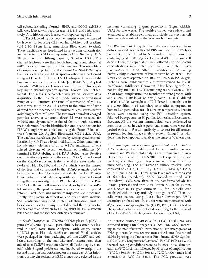
3.1. Quantitative Proteomics Showed Upregulated Expressionof CTNNB1 Proteins in a Malignant Transformed Stem CellLine.Our previous studies reported that the chHES-3 cell lineunderwent karyotypic changes from simple to complex andexperienced dysregulation of self-renewal pathway and dys-function of related oncogenes during long-term suboptimalculture, leading to malignant transformation (Yang et al.[5]). Hence, chHES-3 is a specific cell model for the investiga-tion of the mechanism underlying stem cell malignant trans-formation. Significantly, the intracellular Wnt signalingpathway was preferentially activated in malignant trans-formed chHES-3 cells, especially the expression of CTNNB1was aberrantly upregulated, which might be associatedwith this process [6]. To further investigate the differentialexpression of CTNNB1 during the chHES-3 malignanttransformation, Normal (chHES-3 cells retained a normalkaryotype), SIMP (chHES-3 with a simple duplication kar-yotype), and COMP (chHES-3 with the accumulation ofmore chromosomal abnormalities) chHES-3, as well ashuman embryonal carcinoma cell (hECCs) line NTERA-2cl.D1 (EC) were analyzed by quantitative proteomics. Fourunique reporter ions (m/z = 114‐117) were used to labelthe Normal, SIMP, COMP chHES-3 cells, and EC cells,respectively. Consistent with a previous study [6], theMS/MS spectra and iTRAQ ratios of the peptides fromβ-catenin showed a high expression of CTNNB1 in karyo-typically aberrant chHES-3 (Figure 1).
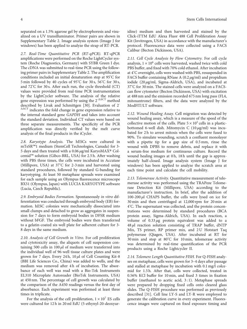
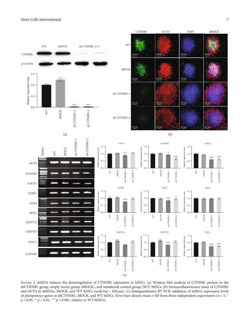
3.2. Reduced Differentiation Capacity in CTNNB1-DepletedStem Cells. CTNNB1-deficient hESC colonies were estab-lished by shRNA infection, and the CTNNB1 expressionand stem cell characteristics were explored after interference(Figures 2 and 3 and Supplemental Figure 2). The silencingefficiency of CTNNB1 was measured by Western blot andimmunofluorescence assays. The results showed thatcompared to the wild-type (WT) and empty vector group(MOCK), the expression of CTNNB1 was remarkably
decreased in the shCTNNB1 group, while no significantdifference was detected between the MOCK and WTgroups (Figures 2(a) and 2(b)). The G-band analysis afterlong-term culture showed that hESCs after CTNNB1interference retained a normal karyotype (SupplementalFigure 1(b)). Immunofluorescence and RT-PCR resultsindicated that the downregulation of the CTNNB1 leveldecreased the expression of intrinsic pluripotency factors,including OCT4, NANOG, TDGF1, TERF1, KLF4, REX1,CRIPTO, and THY-1 (Figures 2(b) and 2(c)).
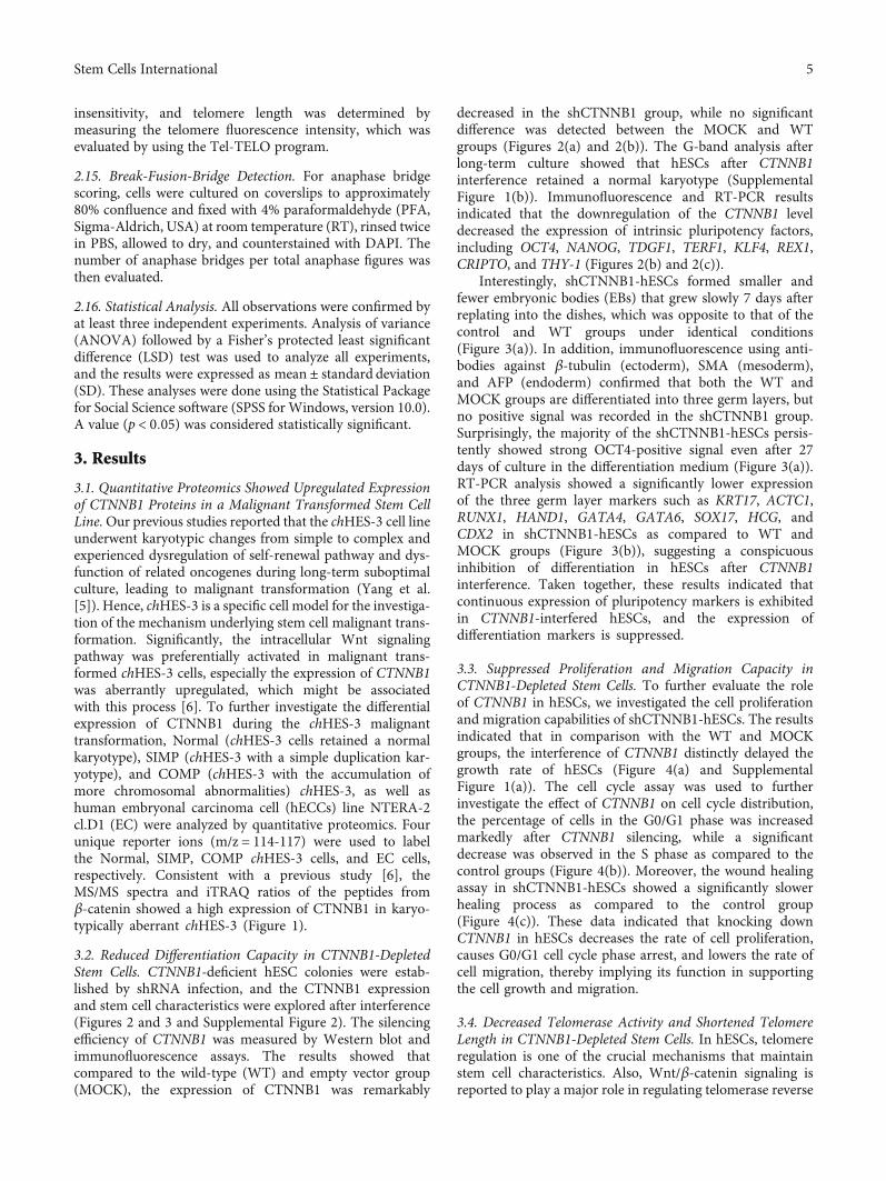
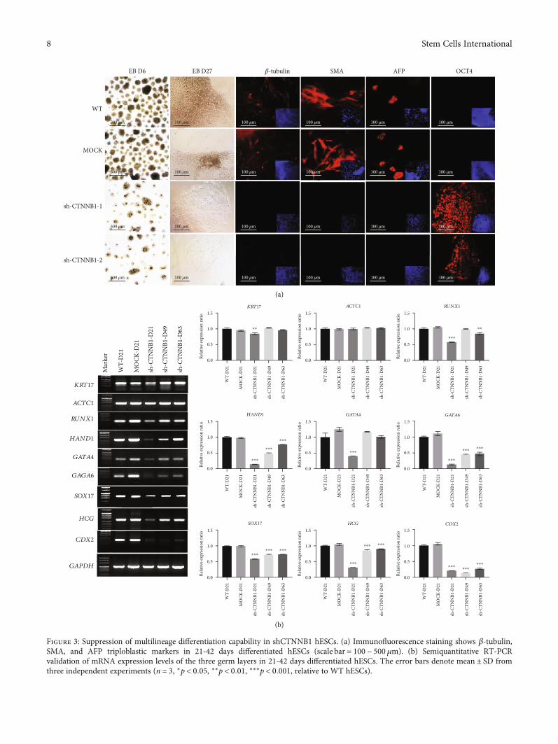
Interestingly, shCTNNB1-hESCs formed smaller andfewer embryonic bodies (EBs) that grew slowly 7 days afterreplating into the dishes, which was opposite to that of thecontrol and WT groups under identical conditions(Figure 3(a)). In addition, immunofluorescence using anti-bodies against β-tubulin (ectoderm), SMA (mesoderm),and AFP (endoderm) confirmed that both the WT andMOCK groups are differentiated into three germ layers, butno positive signal was recorded in the shCTNNB1 group.Surprisingly, the majority of the shCTNNB1-hESCs persis-tently showed strong OCT4-positive signal even after 27days of culture in the differentiation medium (Figure 3(a)).RT-PCR analysis showed a significantly lower expressionof the three germ layer markers such as KRT17, ACTC1,RUNX1, HAND1, GATA4, GATA6, SOX17, HCG, andCDX2 in shCTNNB1-hESCs as compared to WT andMOCK groups (Figure 3(b)), suggesting a conspicuousinhibition of differentiation in hESCs after CTNNB1interference. Taken together, these results indicated thatcontinuous expression of pluripotency markers is exhibitedin CTNNB1-interfered hESCs, and the expression ofdifferentiation markers is suppressed.
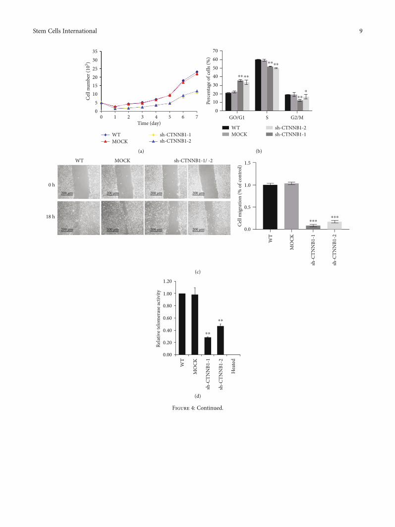
3.3. Suppressed Proliferation and Migration Capacity inCTNNB1-Depleted Stem Cells. To further evaluate the roleof CTNNB1 in hESCs, we investigated the cell proliferationand migration capabilities of shCTNNB1-hESCs. The resultsindicated that in comparison with the WT and MOCKgroups, the interference of CTNNB1 distinctly delayed thegrowth rate of hESCs (Figure 4(a) and SupplementalFigure 1(a)). The cell cycle assay was used to furtherinvestigate the effect of CTNNB1 on cell cycle distribution,the percentage of cells in the G0/G1 phase was increasedmarkedly after CTNNB1 silencing, while a significantdecrease was observed in the S phase as compared to thecontrol groups (Figure 4(b)). Moreover, the wound healingassay in shCTNNB1-hESCs showed a significantly slowerhealing process as compared to the control group(Figure 4(c)). These data indicated that knocking downCTNNB1 in hESCs decreases the rate of cell proliferation,causes G0/G1 cell cycle phase arrest, and lowers the rate ofcell migration, thereby implying its function in supportingthe cell growth and migration.
3.4. Decreased Telomerase Activity and Shortened TelomereLength in CTNNB1-Depleted Stem Cells. In hESCs, telomereregulation is one of the crucial mechanisms that maintainstem cell characteristics. Also, Wnt/β-catenin signaling isreported to play a major role in regulating telomerase reverse
5Stem Cells International
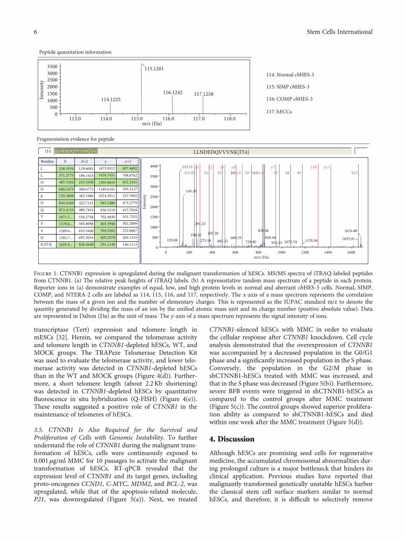
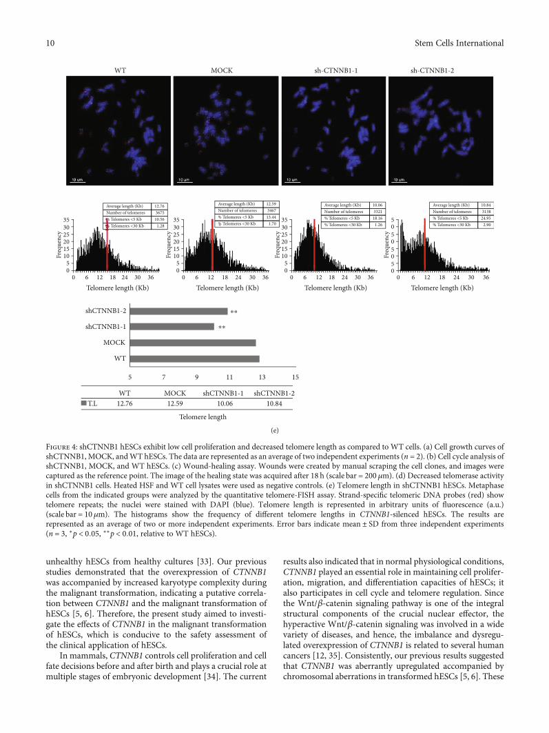
transcriptase (Tert) expression and telomere length inmESCs [32]. Herein, we compared the telomerase activityand telomere length in CTNNB1-depleted hESCs, WT, andMOCK groups. The TRAPeze Telomerase Detection Kitwas used to evaluate the telomerase activity, and lower telo-merase activity was detected in CTNNB1-depleted hESCsthan in the WT and MOCK groups (Figure 4(d)). Further-more, a short telomere length (about 2.2Kb shortening)was detected in CTNNB1-depleted hESCs by quantitativefluorescence in situ hybridization (Q-FISH) (Figure 4(e)).These results suggested a positive role of CTNNB1 in themaintenance of telomeres of hESCs.
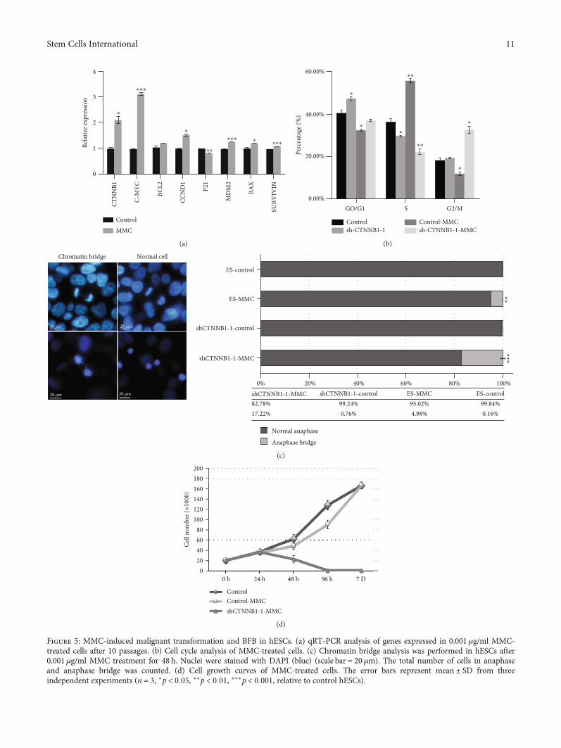
3.5. CTNNB1 Is Also Required for the Survival andProliferation of Cells with Genomic Instability. To furtherunderstand the role of CTNNB1 during the malignant trans-formation of hESCs, cells were continuously exposed to0.001μg/ml MMC for 10 passages to activate the malignanttransformation of hESCs. RT-qPCR revealed that theexpression level of CTNNB1 and its target genes, includingproto-oncogenes CCND1, C-MYC, MDM2, and BCL-2, wasupregulated, while that of the apoptosis-related molecule,P21, was downregulated (Figure 5(a)). Next, we treated
CTNNB1-silenced hESCs with MMC in order to evaluatethe cellular response after CTNNB1 knockdown. Cell cycleanalysis demonstrated that the overexpression of CTNNB1was accompanied by a decreased population in the G0/G1phase and a significantly increased population in the S phase.Conversely, the population in the G2/M phase inshCTNNB1-hESCs treated with MMC was increased, andthat in the S phase was decreased (Figure 5(b)). Furthermore,severe BFB events were triggered in shCTNNB1-hESCs ascompared to the control groups after MMC treatment(Figure 5(c)). The control groups showed superior prolifera-tion ability as compared to shCTNNB1-hESCs and diedwithin one week after the MMC treatment (Figure 5(d)).
4. Discussion
Although hESCs are promising seed cells for regenerativemedicine, the accumulated chromosomal abnormalities dur-ing prolonged culture is a major bottleneck that hinders itsclinical application. Previous studies have reported thatmalignantly transformed genetically unstable hESCs harborthe classical stem cell surface markers similar to normalhESCs, and therefore, it is difficult to selectively remove
Peptide quantitation information
114: Normal chHES-3
115: SIMP chHES-3
116: COMP chHES-3
117: hECCs
350030002500200015001000
5000
113.0
0
129.08
291.23
145.10
115.11b3+B1
y1 y2 y3 y4 y7 y10 y11b2 b3 b4y10+2 y10+2 b7 b8 b9 b12b5 b12
258.20 405.26
273.18 485.32600.35
729.40
838.46
828.46954.55 1071.54 1170.56 1655.91
1676.88
200 400 600 800m/z (Da)
1000 1200 1400 1600
Fragmentation evidence for peptide
IT4
Residue
L 258.1934 129.6003 1673.9312 837.4692
708.8762652.3341595.3127
537.7992
473.2779
415.7644
351.7352
302.2009
252.6667
203.1325
146.1111
1416.7451
1303.6610
1189.61811074.5911
945.5486
830.5218
702.4630603.3946504.3262
405.2578
291.2149
186.1424243.1638
300.6773365.1986
422.7121
486.7414
536.2756585.8098
635.3440692.3654
828.4640
371.2775485.3204
600.3473729.3899
844.4169
972.4755
1071.5...1170.6...
1269.6...
1383.7...
1655.9...
L
N
D
ED
QV
V
V
NK[IT4]
b b+2 y y+2
LLNDEDQVVVNK[IT4]
114.0
114.1225
115.1201
116.1242 117.1258
115.0m/z (Da)
116.0 117.0 118.0
3500
4000
3000
2500
2000Inte
nsity
1500
1000
500
0
Inte
nsity
LLNDEDQVVVNK[IT4]
Figure 1: CTNNB1 expression is upregulated during the malignant transformation of hESCs. MS/MS spectra of iTRAQ-labeled peptidesfrom CTNNB1. (a) The relative peak heights of iTRAQ labels. (b) A representative tandem mass spectrum of a peptide in each protein.Reporter ions in (a) demonstrate examples of equal, low, and high protein levels in normal and aberrant chHES-3 cells. Normal, SIMP,COMP, and NTERA-2 cells are labeled as 114, 115, 116, and 117, respectively. The x-axis of a mass spectrum represents the correlationbetween the mass of a given ion and the number of elementary charges. This is represented as the IUPAC standard m/z to denote thequantity generated by dividing the mass of an ion by the unified atomic mass unit and its charge number (positive absolute value). Dataare represented in Dalton (Da) as the unit of mass. The y-axis of a mass spectrum represents the signal intensity of ions.
6 Stem Cells International
WT
CTNNB1
𝛽-ACTIN
MOCK sh-CTNNB1-1/-2
1.5
1.0
0.5
0.0
WT
Relat
ive e
xpre
ssio
n ra
tio
MO
CK
sh-C
TNN
B1-1
sh-C
TNN
B1-2
⁎⁎⁎ ⁎⁎⁎
(a)
100 𝜇m 100 𝜇m 100 𝜇m 100 𝜇m
100 𝜇m 100 𝜇m 100 𝜇m 100 𝜇m
100 𝜇m 100 𝜇m 100 𝜇m 100 𝜇m
100 𝜇m 100 𝜇m 100 𝜇m
sh-CTNNB1-2
sh-CTNNB1-1
MOCK
WT
CTNNB1 OCT4 DAPI MERGE
(b)
OCT4 NANOG TDGF1
TERF1 KLF4 REX1
LEFTYA CRIPTO THY-1
1.5
1.0
0.5
0.0
1.5
1.0
0.5
0.0
1.5
1.0
0.5
0.0
WT
Relat
ive e
xpre
ssio
n ra
tio
Relat
ive e
xpre
ssio
n ra
tio
1.5
1.0
0.5
0.0Relat
ive e
xpre
ssio
n ra
tio
Relat
ive e
xpre
ssio
n ra
tio
1.5
1.0
0.5
0.0
1.5
1.0
0.5
0.0Relat
ive e
xpre
ssio
n ra
tio
Relat
ive e
xpre
ssio
n ra
tio
1.5
1.0
0.5
0.0
1.5
1.0
0.5
0.0
1.5
1.0
0.5
0.0Relat
ive e
xpre
ssio
n ra
tio
Relat
ive e
xpre
ssio
n ra
tio
Relat
ive e
xpre
ssio
n ra
tio
MO
CK
sh-C
TNN
B1-1
sh-C
TNN
B1-2
WT
MO
CK
sh-C
TNN
B1-1
sh-C
TNN
B1-2
WT
MO
CK
sh-C
TNN
B1-1
sh-C
TNN
B1-2
WT
MO
CK
sh-C
TNN
B1-1
sh-C
TNN
B1-2
WT
MO
CK
sh-C
TNN
B1-1
sh-C
TNN
B1-2
WT
MO
CK
sh-C
TNN
B1-1
sh-C
TNN
B1-2
WT
MO
CK
sh-C
TNN
B1-1
sh-C
TNN
B1-2
WT
MO
CK
sh-C
TNN
B1-1
sh-C
TNN
B1-2
WT
MO
CK
sh-C
TNN
B1-1
sh-C
TNN
B1-2
Mar
ker
WT
MO
CK
sh-C
TNN
B1-1
sh-C
TNN
B1-2
OCT4
NANOG
TDGF1
TERF1
KLF4
REX1
LEFTYA
CRIPTO
THY-1
GAPDH
⁎⁎ ⁎
⁎⁎⁎ ⁎⁎⁎ ⁎⁎⁎
⁎⁎⁎⁎⁎⁎
⁎⁎⁎⁎⁎⁎ ⁎⁎⁎
⁎
(c)
Figure 2: shRNA induces the downregulation of CTNNB1 expression in hESCs. (a) Western blot analysis of CTNNB1 protein in theshCTNNB1 group, empty vector group (MOCK), and uninfected control group (WT) hESCs. (b) Immunofluorescence assay of CTNNB1and OCT4 in shRNAs, MOCK, and WT hESCs (scale bar = 100μm). (c) Semiquantitative RT-PCR validation of mRNA expression levelsof pluripotency genes in shCTNNB1, MOCK, and WT hESCs. Error bars denote mean ± SD from three independent experiments (n = 3, ∗p < 0:05, ∗∗p < 0:01, ∗∗∗p < 0:001, relative to WT hESCs).
7Stem Cells International
100 𝜇m 100 𝜇m 100 𝜇m 100 𝜇m
100 𝜇m 100 𝜇m 100 𝜇m 100 𝜇m
100 𝜇m 100 𝜇m 100 𝜇m 100 𝜇m
100 𝜇m
500 𝜇m
500 𝜇m
500 𝜇m
500 𝜇m 100 𝜇m
100 𝜇m
100 𝜇m
100 𝜇m
100 𝜇m 100 𝜇m 100 𝜇m
sh-CTNNB1-2
sh-CTNNB1-1
MOCK
WT
EB D6 EB D27 𝛽-tubulin SMA AFP OCT4
(a)
Mar
ker
WT-
D21
MO
CK-D
21
sh-C
TNN
B1-D
21
sh-C
TNN
B1-D
49
sh-C
TNN
B1-D
63
WT-
D21
MO
CK-D
21
sh-C
TNN
B1-D
21
sh-C
TNN
B1-D
49
sh-C
TNN
B1-D
63
WT-
D21
MO
CK-D
21
sh-C
TNN
B1-D
21
sh-C
TNN
B1-D
49
sh-C
TNN
B1-D
63
WT-
D21
MO
CK-D
21
sh-C
TNN
B1-D
21
sh-C
TNN
B1-D
49
sh-C
TNN
B1-D
63
WT-
D21
MO
CK-D
21
sh-C
TNN
B1-D
21
sh-C
TNN
B1-D
49
sh-C
TNN
B1-D
63
WT-
D21
MO
CK-D
21
sh-C
TNN
B1-D
21
sh-C
TNN
B1-D
49
sh-C
TNN
B1-D
63
KRT17
KRT17
ACTC1
ACTC1
RUNX1
RUNX1
HAND1
HAND1
GATA4
GATA4
GAGA6
GATA6
SOX17
SOX17HCG
HCG
CDX2
CDX2
GAPDH
1.5
1.0
0.5
0.0Relat
ive e
xpre
ssio
n ra
tio
1.5
1.0
0.5
0.0Relat
ive e
xpre
ssio
n ra
tio
1.5
1.0
0.5
0.0Relat
ive e
xpre
ssio
n ra
tio
WT-
D21
MO
CK-D
21
sh-C
TNN
B1-D
21
sh-C
TNN
B1-D
49
sh-C
TNN
B1-D
63
WT-
D21
MO
CK-D
21
sh-C
TNN
B1-D
21
sh-C
TNN
B1-D
49
sh-C
TNN
B1-D
63
1.5
1.0
0.5
0.0Relat
ive e
xpre
ssio
n ra
tio
1.5
1.0
0.5
0.0Relat
ive e
xpre
ssio
n ra
tio
WT-
D21
MO
CK-D
21
sh-C
TNN
B1-D
21
sh-C
TNN
B1-D
49
sh-C
TNN
B1-D
63
WT-
D21
MO
CK-D
21
sh-C
TNN
B1-D
21
sh-C
TNN
B1-D
49
sh-C
TNN
B1-D
63
1.5
1.0
0.5
0.0Relat
ive e
xpre
ssio
n ra
tio
1.5
1.0
0.5
0.0Relat
ive e
xpre
ssio
n ra
tio
1.5
1.0
0.5
0.0Relat
ive e
xpre
ssio
n ra
tio
1.5
1.0
0.5
0.0Relat
ive e
xpre
ssio
n ra
tio
⁎⁎⁎⁎⁎⁎
⁎⁎⁎⁎⁎⁎
⁎⁎⁎
⁎⁎⁎
⁎⁎⁎
⁎⁎⁎
⁎⁎⁎
⁎⁎⁎⁎⁎⁎⁎⁎⁎
⁎⁎⁎
⁎⁎⁎⁎⁎⁎
⁎⁎⁎
⁎⁎⁎
⁎⁎⁎⁎
(b)
Figure 3: Suppression of multilineage differentiation capability in shCTNNB1 hESCs. (a) Immunofluorescence staining shows β-tubulin,SMA, and AFP triploblastic markers in 21-42 days differentiated hESCs (scale bar = 100 − 500 μm). (b) Semiquantitative RT-PCRvalidation of mRNA expression levels of the three germ layers in 21-42 days differentiated hESCs. The error bars denote mean ± SD fromthree independent experiments (n = 3, ∗p < 0:05, ∗∗p < 0:01, ∗∗∗p < 0:001, relative to WT hESCs).
8 Stem Cells International
353025201510
50
0 1 2 3Time (day)
Cell
num
ber (
105 )
4 6 75
WTMOCK
sh-CTNNB1-1sh-CTNNB1-2
(a)
6070
5040
20
30
100
GO/G1 S G2/M
Perc
enta
ge o
f cel
ls (%
)
⁎⁎⁎⁎
⁎⁎⁎⁎
⁎⁎⁎
WTMOCK
sh-CTNNB1-2sh-CTNNB1-1
(b)
1.5
1.0
0.5
0.0
WT
WT
0 h
18 h
MOCK sh-CTNNB1-1/ -2
Cell
mig
ratio
n (%
of c
ontr
ol)
MO
CK
sh-C
TNN
B1-1
sh-C
TNN
B1-2
⁎⁎⁎⁎⁎⁎
200 𝜇m
200 𝜇m
200 𝜇m
200 𝜇m
200 𝜇m
200 𝜇m
200 𝜇m
200 𝜇m
(c)
1.20
1.00
0.80
0.60
0.40
0.20
0.00
Relat
ive t
elom
eras
e act
ivity
WT
MO
CK
sh-C
TNN
B1-1
sh-C
TNN
B1-2
Hea
ted
⁎⁎
⁎⁎
(d)
Figure 4: Continued.
9Stem Cells International
unhealthy hESCs from healthy cultures [33]. Our previousstudies demonstrated that the overexpression of CTNNB1was accompanied by increased karyotype complexity duringthe malignant transformation, indicating a putative correla-tion between CTNNB1 and the malignant transformation ofhESCs [5, 6]. Therefore, the present study aimed to investi-gate the effects of CTNNB1 in the malignant transformationof hESCs, which is conducive to the safety assessment ofthe clinical application of hESCs.
In mammals, CTNNB1 controls cell proliferation and cellfate decisions before and after birth and plays a crucial role atmultiple stages of embryonic development [34]. The current
results also indicated that in normal physiological conditions,CTNNB1 played an essential role in maintaining cell prolifer-ation, migration, and differentiation capacities of hESCs; italso participates in cell cycle and telomere regulation. Sincethe Wnt/β-catenin signaling pathway is one of the integralstructural components of the crucial nuclear effector, thehyperactive Wnt/β-catenin signaling was involved in a widevariety of diseases, and hence, the imbalance and dysregu-lated overexpression of CTNNB1 is related to several humancancers [12, 35]. Consistently, our previous results suggestedthat CTNNB1 was aberrantly upregulated accompanied bychromosomal aberrations in transformed hESCs [5, 6]. These
35
Freq
uenc
y
3025201510
50
0 6
T.L 12.76
5 7 9 11 13 15
WT
WT
WT
MOCK
MOCK
MOCK
shCTNNB1-1
shCTNNB1-1
sh-CTNNB1-1
shCTNNB1-2
shCTNNB1-2
sh-CTNNB1-2
12.59 10.06 10.84
Telomere length
12 18 24
Telomere length (Kb)30 36
35
Freq
uenc
y
3025201510
50
0 6 12 18 24
Telomere length (Kb)30 36
35
Freq
uenc
y
3025201510
50
0 6 12 18 24
Telomere length (Kb)30 36
50505050
Freq
uenc
y
0 6 12 18 24
Telomere length (Kb)30 36
⁎⁎
⁎⁎
Average length (Kb)Number of telomeres% Telomeres <5 Kb% Telomeres <30 Kb
12.763675
10.561.28
Average length (Kb)Number of telomeres% Telomeres <5 Kb% Telomeres <30 Kb
12.593467
13.441.70
Average length (Kb)Number of telomeres% Telomeres <5 Kb% Telomeres <30 Kb
10.063321
18.161.26
Average length (Kb)Number of telomeres% Telomeres <5 Kb% Telomeres <30 Kb
10.843138
24.952.90
(e)
Figure 4: shCTNNB1 hESCs exhibit low cell proliferation and decreased telomere length as compared to WT cells. (a) Cell growth curves ofshCTNNB1, MOCK, andWT hESCs. The data are represented as an average of two independent experiments (n = 2). (b) Cell cycle analysis ofshCTNNB1, MOCK, and WT hESCs. (c) Wound-healing assay. Wounds were created by manual scraping the cell clones, and images werecaptured as the reference point. The image of the healing state was acquired after 18 h (scale bar = 200μm). (d) Decreased telomerase activityin shCTNNB1 cells. Heated HSF and WT cell lysates were used as negative controls. (e) Telomere length in shCTNNB1 hESCs. Metaphasecells from the indicated groups were analyzed by the quantitative telomere-FISH assay. Strand-specific telomeric DNA probes (red) showtelomere repeats; the nuclei were stained with DAPI (blue). Telomere length is represented in arbitrary units of fluorescence (a.u.)(scale bar = 10μm). The histograms show the frequency of different telomere lengths in CTNNB1-silenced hESCs. The results arerepresented as an average of two or more independent experiments. Error bars indicate mean ± SD from three independent experiments(n = 3, ∗p < 0:05, ∗∗p < 0:01, relative to WT hESCs).
10 Stem Cells International
4
3
2
1
0
Rela
tive e
xpre
ssio
n
CTN
NB1
C-M
YC
BCL2
CCN
D1
P21
MD
M2
BAX
SURV
IVIN
⁎⁎⁎
⁎
⁎
⁎⁎
⁎⁎⁎ ⁎ ⁎⁎⁎
ControlMMC
(a)
60.00%
40.00%
20.00%
0.00%GO/G1 S G2/M
Perc
enta
ge (%
)
⁎
⁎⁎
⁎⁎
⁎⁎
⁎
⁎
Controlsh-CTNNB1-1
Control-MMCsh-CTNNB1-1-MMC
(b)
ES-control
ES-MMC
shCTNNB1-1-control
shCTNNB1-1-MMC
0%shCTNNB1-1-MMC shCTNNB1-1-control82.78%17.22%
99.24%0.76% 4.98% 0.16%
99.84%ES-control
95.02%ES-MMC
20% 40% 60% 80% 100%
Normal anaphase
Anaphase bridge
Chromatin bridge Normal cell
20 𝜇m20 𝜇m
20 𝜇m 20 𝜇m
⁎⁎
⁎⁎⁎
(c)
200180160140120100
80
Cell
num
ber (
×100
0)
604020
00 h 24 h 48 h 96 h 7 D0 h 24 h 48 h 96 h 7 D
ControlControl-MMCshCTNNB1-1-MMC
(d)
Figure 5: MMC-induced malignant transformation and BFB in hESCs. (a) qRT-PCR analysis of genes expressed in 0.001 μg/ml MMC-treated cells after 10 passages. (b) Cell cycle analysis of MMC-treated cells. (c) Chromatin bridge analysis was performed in hESCs after0.001μg/ml MMC treatment for 48 h. Nuclei were stained with DAPI (blue) (scale bar = 20μm). The total number of cells in anaphaseand anaphase bridge was counted. (d) Cell growth curves of MMC-treated cells. The error bars represent mean ± SD from threeindependent experiments (n = 3, ∗p < 0:05, ∗∗p < 0:01, ∗∗∗p < 0:001, relative to control hESCs).
11Stem Cells International
findings indicated that CTNNB1 plays an essential role in themaintenance of the physiological functions of hESCs;however, the dysregulation of CTNNB1 might be related tothe malignant transformation of hESCs.
Previously, we reported that the trace level of MMC,which is an adverse factor for malignant transformation ofhESCs, causes karyotype abnormalities and carcinogenesisin long-term cultured hESCs [7]. Thus, we optimized our cul-ture conditions by using γ-inactivated feeder cells, control-ling the density of feeder cells, and passaging by manuallymechanical cutting. As a result, hESCs retained a stable nor-mal karyotype even after more than two years of cultivationunder optimized conditions [6, 7]. In this study, we inducedthe malignant transformation of hESCs by continuous expo-sure to 0.001μg/ml MMC; subsequently, the cellularresponse was assessed. The data demonstrated that after theMMC treatment, the expression of CTNNB1 and its targetgenes, especially proto-oncogenes CCND1, C-MYC, MDM2,and BAX, was upregulated, while that of the apoptosis-related molecule, P21, was downregulated. Moreover, theoverexpression of CTNNB1 accelerated the cell transitionfrom G0/G1 to S phase. Conversely, CTNNB1 depletioninduced the G2/M cell cycle checkpoint arrest after MMCtreatment. CCND1 is a crucial downstream target gene ofthe Wnt/β-catenin signaling pathway, which coordinatesthe cell cycle progression [36, 37]. CCND1 dysregulationleads to uncontrolled cell cycle progression, and its overex-pression is correlated to genomic instability, rapid cellgrowth, cell bypass of critical cellular checkpoints, and neo-plastic growth [37, 38]. In addition, pathologically activeMYC and MDM2 genes drive an abnormally rapid cell cycleto dysregulate cell proliferation [39]. BAX is a proapoptoticgene and might affect cell proliferation by directly modulat-ing the cell cycle inhibitor P21 [40, 41]. Thus, our resultsdemonstrated that elevated expression of CTNNB1 targetsCCND1 upregulation and P21 downregulation, which in turnaccelerates the transition of chromosomally unstable cellsfrom G0/G1 to S phase. Conversely, CTNNB1 depleted inhESCs induced G2/M phase arrest and survival crisis afterMMC treatment.
Telomere is a conserved DNA-protein complex thatmaintains the stability of the chromosomes. Telomeres loseabout 200 nucleotides per cell division.When a telomere deg-radation reaches a threshold level, uncapped telomeres acti-vate cell senescence or apoptosis by enforcing the cell cyclearrest [18, 42]. Telomere dysfunction promotes tumorigene-sis by inducing chromosomal instability in tumor-initiatingcells, bypassing the cell cycle checkpoint, and increasing theproliferative competition of transformed cells [43, 44]. Asreported, hTERT promoter mutations and CTNNB1 muta-tions are significantly associated, and telomere and CTNNB1
have a cooperative effect in tumorigenesis [25]. In this study,the MMC treatment elevated the number of telomere BFBcycles in CTNNB1-depleted cells and significantly reducedthe proliferation and survival of CTNNB1-depleted cellswithin 7 days. This might be due to the mismatched end-to-end chromosome fusions and recombination effectuatedby uncapped chromosomes leading to mitotic arrest andaffecting the cellular fate in precrisis [45, 46]. Conversely,cells with MMC-induced overexpression of CTNNB1 over-comes the survival crisis and contributed to uncontrolledproliferation. Moreover, CTNNB1 maintains the telomerefunction by regulating the telomerase activity. Takentogether, the current results indicated that CTNNB1 silencingmay exacerbate telomere deprotection, thereby increasingthe sensitivity of cells to mutagens and promoting cell deathduring cell cycle arrest. Typically, the overexpression ofCTNNB1 conferred a proliferative advantage, culture adapt-ability, and resistance to apoptosis on chromosomally unsta-ble cells by mediating telomere maintenance, and thereforedriving the malignant transformation of hESCs.
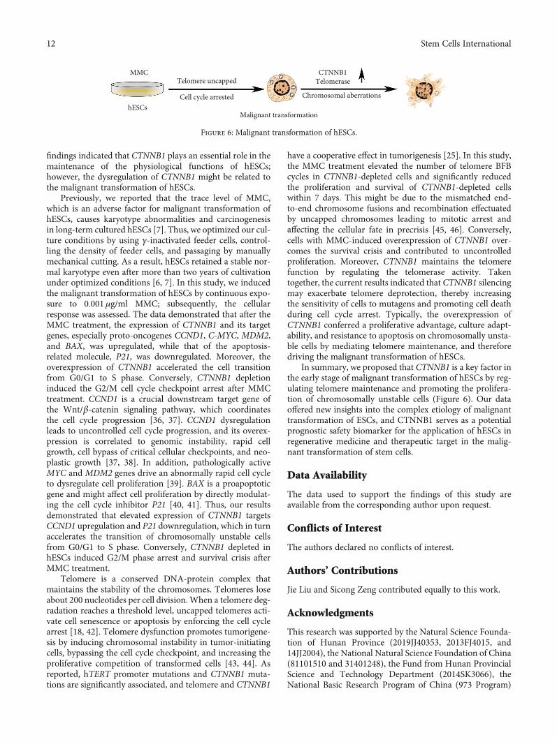
In summary, we proposed that CTNNB1 is a key factor inthe early stage of malignant transformation of hESCs by reg-ulating telomere maintenance and promoting the prolifera-tion of chromosomally unstable cells (Figure 6). Our dataoffered new insights into the complex etiology of malignanttransformation of ESCs, and CTNNB1 serves as a potentialprognostic safety biomarker for the application of hESCs inregenerative medicine and therapeutic target in the malig-nant transformation of stem cells.
Data Availability
The data used to support the findings of this study areavailable from the corresponding author upon request.
Conflicts of Interest
The authors declared no conflicts of interest.
Authors’ Contributions
Jie Liu and Sicong Zeng contributed equally to this work.
Acknowledgments
This research was supported by the Natural Science Founda-tion of Hunan Province (2019JJ40353, 2013FJ4015, and14JJ2004), the National Natural Science Foundation of China(81101510 and 31401248), the Fund from Hunan ProvincialScience and Technology Department (2014SK3066), theNational Basic Research Program of China (973 Program)
Telomere uncappedMMC
hESCsCell cycle arrested
Malignant transformation
Chromosomal aberrations
CTNNB1Telomerase
Figure 6: Malignant transformation of hESCs.
12 Stem Cells International
(2012CB944901), National Science and Technology MajorProject of the Ministry of Science and Technology of HunanProvince, China (2017SK1032), Postgraduate ScientificResearch Innovation Project of Hunan Province(CX20190236), and the Fundamental Research Funds forCentral Universities of the Central South University(2018zzts834 and 2017zzts030).
Supplementary Materials
Supplementary Figure 1: (a) Cell proliferation analysis byEdU labeling was performed in stably transfected shCTNNB1groups, stably transfected empty vector group (MOCK),and uninfected control group (WT), respectively (mean ±SD, n =3; ∗p < 0:05 vs. WT). (b) The G-band analysis aftera long-term culture showed that shCTNNB1 hESCs retain anormal karyotype. Supplementary Figure 2: Expression ofpluripotency markers in shCTNNB1 cells. Cellular morphol-ogy of hESCs (scale bar =100 μm). Alkaline phosphatase(AKP) activity (scale bar =200 μm) and the expression ofTRA-1-60, TRA-1-81, SSEA-3, SSEA-4, SSEA-1, andNANOG were detected by immunofluorescence staining inshCTNNB1 hESCs. Nuclei were stained with DAPI (blue;insets) (scale bar =100 μm). Supplementary Table 1. Primaryantibodies used for immunofluorescence staining and West-ern blot analysis. Supplementary Table 2. Primers for RT-PCR and RT-qPCR. (Supplementary Materials)
References
[1] J. A. Thomson, J. Itskovitz-Eldor, S. S. Shapiro et al., “Embry-onic stem cell lines derived from human blastocysts,” Science,vol. 282, no. 5391, pp. 1145–1147, 1998.
[2] P. Rebuzzini, M. Zuccotti, C. A. Redi, and S. Garagna, “Chro-mosomal abnormalities in embryonic and somatic stem cells,”Cytogenetic and Genome Research, vol. 147, no. 1, pp. 1–9,2016.
[3] P. Rebuzzini, M. Zuccotti, C. A. Redi, and S. Garagna, “Achil-les’ heel of pluripotent stem cells: genetic, genomic and epige-netic variations during prolonged culture,” Cellular andMolecular Life Sciences, vol. 73, no. 13, pp. 2453–2466, 2016.
[4] C. Kermi, A. Aze, and D. Maiorano, “Preserving GenomeIntegrity During the Early Embryonic DNA ReplicationCycles,” Genes, vol. 10, no. 5, p. 398, 2019.
[5] S. Yang, G. Lin, Y. Q. Tan et al., “Tumor progression ofculture-adapted human embryonic stem cells during long-term culture,” Genes, Chromosomes & Cancer, vol. 47, no. 8,pp. 665–679, 2008.
[6] Y. Sun, Y. Yang, S. Zeng, Y. Tan, G. Lu, and G. Lin, “Identifi-cation of proteins related to epigenetic regulation in the malig-nant transformation of aberrant karyotypic human embryonicstem cells by quantitative proteomics,” PLoS One, vol. 9, no. 1,article e85823, 2014.
[7] D. Zhou, G. Lin, S. C. Zeng et al., “Trace levels of mitomycin Cdisrupt genomic integrity and lead to DNA damage responsedefect in long-term-cultured human embryonic stem cells,”Archives of Toxicology, vol. 89, no. 1, pp. 33–45, 2015.
[8] Z. Liu, J. Guo, Y. Wang et al., “CFTR- β-catenin interactionregulates mouse embryonic stem cell differentiation and
embryonic development,” Cell Death and Differentiation,vol. 24, no. 1, pp. 98–110, 2017.
[9] Z. Steinhart and S. Angers, “Wnt signaling in development andtissue homeostasis,” Development, vol. 145, no. 11, articledev146589, 2018.
[10] M. Majidinia, J. Aghazadeh, R. Jahanban-Esfahlani, andB. Yousefi, “The roles of Wnt/β-catenin pathway in tissuedevelopment and regenerative medicine,” Journal of CellularPhysiology, vol. 233, no. 8, pp. 5598–5612, 2018.
[11] S. Shang, F. Hua, and Z. W. Hu, “The regulation of β-cateninactivity and function in cancer: therapeutic opportunities,”Oncotarget, vol. 8, no. 20, pp. 33972–33989, 2017.
[12] B. L. Pan, L. Wu, L. Pan et al., “Up-regulation of microRNA-340 promotes osteosarcoma cell apoptosis while suppressingproliferation, migration, and invasion by inactivating theCTNNB1-mediated Notch signaling pathway,” BioscienceReports, vol. 38, no. 4, 2018.
[13] Y. H. Chang, T. Y. Chu, and D. C. Ding, “WNT/β-Catenin sig-naling pathway regulates non-tumorigenesis of human embry-onic stem cells co-cultured with human umbilical cordmesenchymal stem cells,” Scientific Reports, vol. 7, no. 1, article41913, 2017.
[14] A. Bernal and L. Tusell, “Telomeres: Implications for CancerDevelopment,” International Journal of Molecular Sciences,vol. 19, no. 1, p. 294, 2018.
[15] F. Li, Y. Ge, D. Liu, and Z. Songyang, “The role of telomere-binding modulators in pluripotent stem cells,” Protein & Cell,vol. 11, no. 1, pp. 60–70, 2020.
[16] X. Chen, W. J. Tang, J. B. Shi, M. M. Liu, and X. H. Liu, “Ther-apeutic strategies for targeting telomerase in cancer,” Medici-nal Research Reviews, vol. 40, no. 2, pp. 532–585, 2020.
[17] J. W. Shay, “Role of telomeres and telomerase in aging andcancer,” Cancer Discovery, vol. 6, no. 6, pp. 584–593, 2016.
[18] M. De Vitis, F. Berardinelli, and A. Sgura, “Telomere lengthmaintenance in cancer: at the crossroad between telomeraseand alternative lengthening of telomeres (ALT),” Interna-tional Journal of Molecular Sciences, vol. 19, no. 2, p. 606,2018.
[19] X. Yuan, M. Dai, and D. Xu, “Telomere-related markers forcancer,” Current Topics in Medicinal Chemistry, vol. 20,no. 6, pp. 410–432, 2020.
[20] S. Zeng, L. Liu, Y. Sun et al., “Telomerase-mediated telomereelongation from human blastocysts to embryonic stem cells,”Journal of Cell Science, vol. 127, no. 4, pp. 752–762, 2014.
[21] R. Bhargava, M. Fischer, and R. J. O’Sullivan, “Genome rear-rangements associated with aberrant telomere maintenance,”Current Opinion in Genetics & Development, vol. 60, pp. 31–40, 2020.
[22] K. J. Turner, V. Vasu, and D. K. Griffin, “Telomere biology andhuman phenotype,” Cells, vol. 8, no. 1, p. 73, 2019.
[23] M. K. Graham and A. Meeker, “Telomeres and telomerase inprostate cancer development and therapy,” Nature ReviewsUrology, vol. 14, no. 10, pp. 607–619, 2017.
[24] C. Laberthonniere, F. Magdinier, and J. D. Robin, “Bring it toan end: does telomeres size matter?,” Cells, vol. 8, no. 1,p. 30, 2019.
[25] J. Zucman-Rossi, A. Villanueva, J. C. Nault, and J. M. Llovet,“Genetic landscape and biomarkers of hepatocellular carci-noma,” Gastroenterology, vol. 149, no. 5, pp. 1226–1239.e4,2015.
13Stem Cells International
[26] F. Newell, Y. Kong, J. S. Wilmott et al., “Whole-genome land-scape of mucosal melanoma reveals diverse drivers and thera-peutic targets,” Nat Commun, vol. 10, no. 1, p. 3163, 2019.
[27] G. Lin, Y. Xie, Q. OuYang et al., “HLA-matching potential ofan established human embryonic stem cell bank in China,”Cell Stem Cell, vol. 5, no. 5, pp. 461–465, 2009.
[28] P. W. Andrews, I. Damjanov, D. Simon et al., “Pluripotentembryonal carcinoma clones derived from the human terato-carcinoma cell line Tera-2. Differentiation in vivo andin vitro,” Laboratory Investigation, vol. 50, no. 2, pp. 147–162, 1984.
[29] S. Yang, G. Lin, L. Deng, and G. X. Lu, “Tumourigenic charac-teristics of embryonal carcinoma cells as a model for studyingtumour progression of human embryonic stem cells,” Cell Pro-liferation, vol. 45, no. 4, pp. 299–310, 2012.
[30] K. J. Livak and T. D. Schmittgen, “Analysis of relative geneexpression data using real-time quantitative PCR and the2(-Delta Delta C(T)) method,” Methods, vol. 25, no. 4,pp. 402–408, 2001.
[31] I. Ourliac-Garnier and A. Londono-Vallejo, “Telomere lengthanalysis by quantitative fluorescent in situ hybridization (Q-FISH),”Methods inMolecular Biology, vol. 1587, pp. 29–39, 2017.
[32] Z. Xu, A. M. Robitaille, J. D. Berndt et al., “Wnt/β-catenin sig-naling promotes self-renewal and inhibits the primed statetransition in naïve human embryonic stem cells,” Proceedingsof the National Academy of Sciences of the United States ofAmerica, vol. 113, no. 42, pp. E6382–E6390, 2016.
[33] M. P. Henry, J. R. Hawkins, J. Boyle, and J. M. Bridger, “Thegenomic health of human pluripotent stem cells: genomicinstability and the consequences on nuclear organization,”Frontiers in Genetics, vol. 9, p. 623, 2019.
[34] A. A. A. van de Moosdijk, Y. B. C. van de Grift, S. M. A. deMan, A. L. Zeeman, and R. van Amerongen, “A novel Axin2knock-in mouse model for visualization and lineage tracingof WNT/CTNNB1 responsive cells,” Genesis, 2020.
[35] E. H. van Schie and R. van Amerongen, “AberrantWNT/CTNNB1 signaling as a therapeutic target in humanbreast cancer: weighing the evidence,” Frontiers in Cell andDevelopment Biology, vol. 8, p. 25, 2020.
[36] A. Bisso, M. Filipuzzi, G. P. Gamarra Figueroa et al., “Cooper-ation between MYC and β‐Catenin in liver tumorigenesisrequires Yap/Taz,” Hepatology, 2020.
[37] Q. Wang, G. He, M. Hou et al., “Cell Cycle Regulation byAlternative Polyadenylation of CCND1,” Scientific Reports,vol. 8, no. 1, p. 6824, 2018.
[38] M. Katoh, “Multi-layered prevention and treatment of chronicinflammation, organ fibrosis and cancer associated withcanonical WNT/β-catenin signaling activation (review),”International Journal of Molecular Medicine, vol. 42, no. 2,pp. 713–725, 2018.
[39] B. A. Scholz, N. Sumida, C. D. M. de Lima et al., “WNT signal-ing and AHCTF1 promote oncogenic MYC expressionthrough super-enhancer-mediated gene gating,” NatureGenetics, vol. 51, no. 12, pp. 1723–1731, 2019.
[40] S. Brayer, A. Joannes, M. Jaillet et al., “The pro-apoptotic BAXprotein influences cell growth and differentiation from thenucleus in healthy interphasic cells,” Cell Cycle, vol. 16,no. 21, pp. 2108–2118, 2017.
[41] A. Karimian, Y. Ahmadi, and B. Yousefi, “Multiple functionsof p21 in cell cycle, apoptosis and transcriptional regulationafter DNA damage,” DNA Repair, vol. 42, pp. 63–71, 2016.
[42] M. T. Hayashi, A. J. Cesare, T. Rivera, and J. Karlseder, “Celldeath during crisis is mediated by mitotic telomere deprotec-tion,” Nature, vol. 522, no. 7557, pp. 492–496, 2015.
[43] C. Gunes, A. I. Avila, and K. L. Rudolph, “Telomeres in can-cer,” Differentiation, vol. 99, pp. 41–50, 2018.
[44] O. N. Perera, A. P. Sobinoff, E. T. Teber et al., “Telomerasepromotes formation of a telomere protective complex incancer cells,” Science Advances, vol. 5, no. 10, article eaav4409,2019.
[45] J. Maciejowski, Y. Li, N. Bosco, P. J. Campbell, and T. deLange, “Chromothripsis and kataegis induced by telomerecrisis,” Cell, vol. 163, no. 7, pp. 1641–1654, 2015.
[46] P. Feijoo, D. Dominguez, L. Tusell, and A. Genesca, “Telo-mere-dependent genomic integrity: evolution of the fusion-bridge-breakage cycle concept,” Current PharmaceuticalDesign, vol. 20, no. 41, pp. 6375–6385, 2014.
14 Stem Cells International