Embed Size (px)
Citation preview

ERRORS IN THE DETECTION AND ERRORS IN THE DETECTION AND IDENTIFICATION OF HEMOGLOBIN IDENTIFICATION OF HEMOGLOBIN
VARIENTSVARIENTS
Peter J. Howanitz MDPeter J. Howanitz MDProfessor and Vice ChairProfessor and Vice ChairDepartment of PathologyDepartment of Pathology
SUNY Downstate, Brooklyn NY, USASUNY Downstate, Brooklyn NY, USA(([email protected]))

GOALS AND OBJECTIVESGOALS AND OBJECTIVES
• Describe Measurements Of HemoglobinsDescribe Measurements Of Hemoglobins• Introduce Role of HPLCIntroduce Role of HPLC• Case StudiesCase Studies• New Finding--Only A1C Detects VariantNew Finding--Only A1C Detects Variant• Questions And AnswersQuestions And Answers

REASONS FOR HEMOGLOBIN REASONS FOR HEMOGLOBIN ID ID AND QUANTIFICATION AND QUANTIFICATION
• Newborn ScreeningNewborn Screening
• Prenatal ScreeningPrenatal Screening
• Follow-up Newborn ScreeningFollow-up Newborn Screening
• Diagnosis Cause of MicrocytosisDiagnosis Cause of Microcytosis
• Anemia, Polycythemia, Chronic Anemia, Polycythemia, Chronic HemolysisHemolysis
• Hemoglobinopathy Blood ReplacementHemoglobinopathy Blood Replacement
• Unexplained A1c ResultsUnexplained A1c Results


WHY USE HPLC?WHY USE HPLC?
• AdvantagesAdvantages– Throughput 11 Specimens/hour, 24 Hr Cal.Throughput 11 Specimens/hour, 24 Hr Cal.– Analytic Sensitivity @ Low ConcentrationsAnalytic Sensitivity @ Low Concentrations– Improved PrecisionImproved Precision– Better SeparationBetter Separation– Less Referrals For IDLess Referrals For ID
• DisadvantagesDisadvantages– More ComplexMore Complex→ Higher Skill Level→ Higher Skill Level– Co-elution Of HemoglobinsCo-elution Of Hemoglobins

Hemoglobin Hemoglobin Electrophoresis Electrophoresis PatternsPatterns

STRUCTURE HEMOGLOBINS STRUCTURE HEMOGLOBINS
HemoglobiHemoglobinn
Globin ChainGlobin Chain Adult LevelAdult Level
AA αα22ββ22 A A >95%>95%
AA22 αα22δδ22 2-3%2-3%
FF αα22γγ22 F< 2.0%F< 2.0%

COMMON HEMOGLOBIN COMMON HEMOGLOBIN POINT POINT MUTATIONS MUTATIONS• Alpha Chain Variants Alpha Chain Variants
– G Philadelphia (G Philadelphia (αα68 Asn→Lys)68 Asn→Lys)• Beta Chain VariantsBeta Chain Variants
– S (S (ββ6 Glu→Val)6 Glu→Val)– C (C (ββ6 Glu→Lys)6 Glu→Lys)– E (E (ββ26 Glu→Lys)26 Glu→Lys)– D Los Angeles (D Los Angeles (ββ22 Glu→Gln)22 Glu→Gln)
• Delta Chain VariantsDelta Chain Variants---- A2’ (A2’ (δδ16 Gly→Arg)16 Gly→Arg)

INTERPRETATION OF HPLC INTERPRETATION OF HPLC RESULTSRESULTS
• Hemoglobin Retention TimeHemoglobin Retention Time• Variant Hemoglobin Percentage*Variant Hemoglobin Percentage*• AA22 Percentage* Percentage* • Number of Variants*Number of Variants*• CBC Indices*CBC Indices*• Transfusion HistoryTransfusion History• AgeAge• Clinical Course*Clinical Course*
• * Changed By Thalassemia* Changed By Thalassemia

BIO-RAD VARIANT BIO-RAD VARIANT WINDOWSWINDOWSPEAK NAMEPEAK NAME RETENTIORETENTIO
N TIME N TIME (MIN)(MIN)
PEAK NAMEPEAK NAME RETENTIORETENTION TIME N TIME (MIN)(MIN)
F WindowF Window 0.98-0.98-1.201.20
AA22 WindowWindow
3.30-3.903.30-3.90
P2 P2 WindowWindow
1.24-1.24-1.401.40
D WindowD Window 3.90-4.303.90-4.30
P3 P3 WindowWindow
1.40-1.40-1.901.90
S WindowS Window 4.30-4.904.30-4.90
AA00 WindowWindow
1.90-1.90-3.103.10
C WindowC Window 4.90-5.304.90-5.30

INTREPRATION OF INTREPRATION OF RESULTSRESULTS# Abnormal # Abnormal Peaks (%)Peaks (%)
A%A% A2%A2% VARIANTVARIANT EXAMPLEXAMPLEE
1 (25-40)1 (25-40) 50-6050-60 3.5-4.53.5-4.5 ββ--ChainChain AS, ACAS, AC
2 (25, 1.0)2 (25, 1.0) 70-8070-80 1.5-1.5-2.2*2.2*
αα--Chain Chain AG-PhilAG-Phil
2 (50,45)2 (50,45) 00 3.5-4.53.5-4.5 2 2 ββ--Chain Chain
SCSC
3 3
(12, (12, 20,14)20,14)
40-5040-50 2.0* 2.0*
1 1 αα--,1 ,1 ββ--, , 11αβαβ- - ChainChain
ASG- ASG- PhillyPhilly


INTREPRATION OF RESULTSINTREPRATION OF RESULTS
• Hemoglobin FHemoglobin F– >2-80% Babies>2-80% Babies– 90-100% Homozygous 90-100% Homozygous HHereditary ereditary
PPersistence ersistence FFetal etal HHemoglobin,emoglobin,ββ00, , δβδβ00-Thal-Thal– 15-40% Heterozygous HPFH15-40% Heterozygous HPFH– 10-25% SS, Hydroxyurea Treated10-25% SS, Hydroxyurea Treated– 3-10% Homozygous Hemoglobinopathies, 3-10% Homozygous Hemoglobinopathies,
Anemias, Leukemias, Malignancies, Anemias, Leukemias, Malignancies, – < 5% < 5% ββ-Thal, Lepore -Thal, Lepore


INTREPRATION OF INTREPRATION OF RESULTSRESULTS
• Hemoglobin AHemoglobin A– Increased P2-? Diabetes (Increased P2-? Diabetes (↑↑AA11C>7%)C>7%)
– Increased P3-(>P2) Old SpecimenIncreased P3-(>P2) Old Specimen– Inverse of Other HemoglobinsInverse of Other Hemoglobins– Focus on Abnormal HemoglobinsFocus on Abnormal Hemoglobins


HEMOGLOBIN AHEMOGLOBIN A22’’
• Elutes in S WindowElutes in S Window
• ΔΔ16 16 Gly→ArgGly→Arg
• Characteristic Low ACharacteristic Low A2 2 Percentage (1.0-2.5%)Percentage (1.0-2.5%)
• Most Common In Blacks (2%)Most Common In Blacks (2%)
• CBC NormalCBC Normal
• Little Consequence, Except Little Consequence, Except ββ-Thal (add A-Thal (add A22))

INTREPRATION OF INTREPRATION OF RESULTSRESULTS• Hemoglobin AHemoglobin A22
– Increased Increased •4.0-7.0% 4.0-7.0% ΒΒ-Thalassemia, S-Thalassemia, Sββ++ Thal Thal
•3.5-4.5% Hb AS, AC, SC, SS, CC3.5-4.5% Hb AS, AC, SC, SS, CC
•6.5-14.0% Hb Lepore6.5-14.0% Hb Lepore
•25-30% Hb E25-30% Hb E
– DecreasedDecreased•1.3-1.7% Iron Deficiency, Sideroblastic, Aplastic 1.3-1.7% Iron Deficiency, Sideroblastic, Aplastic
AnemiasAnemias
•1.5-2.3% 1.5-2.3% δδ Chain Variant (A2’), Chain Variant (A2’), αα Chain Variant Chain Variant


HEMOGLOBIN EHEMOGLOBIN E
• Found in SE Asia, Found in SE Asia, ββ26Glu→Lys26Glu→Lys
• Most Common Hemoglobinopathy WorldwideMost Common Hemoglobinopathy Worldwide• Complicated by Iron Def, Thalassemia, AComplicated by Iron Def, Thalassemia, A22
ElutionElution• Trait (Hb AE)Trait (Hb AE)
– Asymtomatic, No CBC AbnormalitiesAsymtomatic, No CBC Abnormalities• Disease (Hb EE)Disease (Hb EE)
– Mild Anemia, Target Cells, Mild Anemia, Target Cells, ↓↓RBC SurvivalRBC Survival– ↓↓Osmotic FragilityOsmotic Fragility– +Beta Thal = Severe, As Homozygous +Beta Thal = Severe, As Homozygous ββ-Thal-Thal– +Alpha Thal=↓Hb E+Alpha Thal=↓Hb E


HEMOGLOBIN D HEMOGLOBIN D
D Window On Bio-Rad Variant D Window On Bio-Rad Variant ΒΒ121Glu→Gln121Glu→Gln
Found In India (D-Punjab/D-Los Angeles)Found In India (D-Punjab/D-Los Angeles)Most Common D In U.S. Blacks (< 0.02%)Most Common D In U.S. Blacks (< 0.02%)Trait Asymtomatic, No Anemia, Normal CBCTrait Asymtomatic, No Anemia, Normal CBCDisease Asymtomatic, No Anemia/ HemolysisDisease Asymtomatic, No Anemia/ HemolysisD Los-AngelesS = Symptoms of Sickle Cell D Los-AngelesS = Symptoms of Sickle Cell
DiseaseDisease


HEMOGLOBIN G HEMOGLOBIN G PHILADELPHIAPHILADELPHIA
• Elutes In D-WindowElutes In D-Window• αα6868Asn→Lys Asn→Lys of Hb A and A of Hb A and A22
• Heterozygote-CBC NormalHeterozygote-CBC Normal– Most Common Most Common αα Chain Variant In Blacks, Chain Variant In Blacks,
Italians (25%), ChineseItalians (25%), Chinese– Associated With Associated With αα-Thal (30%, 45%G)-Thal (30%, 45%G)
• Association With S or C Common Association With S or C Common (Double Heterozygote) (Double Heterozygote)


HEMOGLOBIN SHEMOGLOBIN S
S Trait (Hemoglobin AS) S Trait (Hemoglobin AS) ββ66Glu→ValGlu→Val
Common In Blacks; Other Populations Common In Blacks; Other Populations Asymptomatic, Blood Sickles in VitroAsymptomatic, Blood Sickles in VitroProtective Against MalariaProtective Against Malaria
S Disease (Hemoglobin SS)S Disease (Hemoglobin SS)Severe Symptoms, Sickling in Vivo Severe Symptoms, Sickling in Vivo
Hydroxy Urea TreatmentHydroxy Urea Treatment→Induces F→Induces FCrisesCrises→Bone Pain, Hemolysis, Stroke, etc→Bone Pain, Hemolysis, Stroke, etcSimilar Symptoms Other Double Heterozygotes (SC)Similar Symptoms Other Double Heterozygotes (SC)


HEMOGLOBIN CHEMOGLOBIN C
• Prevalent in West Africa, 3% U.S BlacksPrevalent in West Africa, 3% U.S Blacks• Trait (Hb AC) Trait (Hb AC) ββ66Glu→LysGlu→Lys
– No Symptoms or Anemia, No Symptoms or Anemia, – Hypochromia, Up to 40% Target CellsHypochromia, Up to 40% Target Cells
• Disease (Hb CC)Disease (Hb CC)– Mild Hemolytic Anemia, SpenomeglyMild Hemolytic Anemia, Spenomegly– Rod Shaped Crystals in RBCsRod Shaped Crystals in RBCs– Normochromic, Normocytic Anemia, Normochromic, Normocytic Anemia, – 40-90% Target Cells40-90% Target Cells

MORE RARE VARIANTS?

BIORAD TURBO A1C-BIORAD TURBO A1C-CHROMATOGRAMCHROMATOGRAM

BIO-RAD A1C-AS BIO-RAD A1C-AS CHROMATOGRAMCHROMATOGRAM
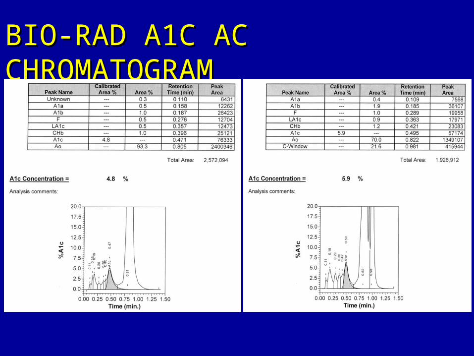
BIO-RAD A1C AC BIO-RAD A1C AC CHROMATOGRAMCHROMATOGRAM

BIO-RAD UNKNOWN BIO-RAD UNKNOWN VARIANT A1C VARIANT A1C CHROMATOGRAM TYPE 1CHROMATOGRAM TYPE 1

BIO-RAD UNKNOWN BIO-RAD UNKNOWN VARIANT A1C VARIANT A1C CHROMATOGRAM TYPE 2CHROMATOGRAM TYPE 2

HEMOGLOBIN A1C HEMOGLOBIN A1C CHROMATOGRAPHSCHROMATOGRAPHS
CONTROL PATIENT 1 PATIENT 2
A1C HPLC results of a control specimen and the patients’ specimens. Note the variant eluting at 0.872 & 0.853 minutes in chromatograms of patient 1 and patient 2 depicted by an arrow.

CONTROL PATIENT 1 PATIENT 2Hemoglobin HPLC results of a control specimen and the patients’ specimens. A hemoglobin variant is not identified in either chromatogram.
HEMOGLOBIN IDENTIFICATION CHROMATOGRAMS

HEMOGLOBIN IDENTIFICATION HEMOGLOBIN IDENTIFICATION CAPILLARY CAPILLARY ELECTROPHORETOGRAMSELECTROPHORETOGRAMS
CONTROL PATIENT 1 PATIENT 2
Capillary electrophoresis of a control specimen and the patients’ specimens. A hemoglobin variant is not identified in either electrophoretogram

HEMOGLOBIN HEMOGLOBIN ELECTROPHORESIS ELECTROPHORESIS
ACID GELALKALINE GELHemoglobin electrophoresis on alkaline and acid gel. The patient’s specimen migrates as S on alkaline gel, and a split A band on acid gel, identified as an arrow. Electrophoresis of the specimen from the second patient was identical to the first (not shown).Controls for C, S, F and A are the top two specimens in either gel.

GENETIC ANALYSIS OF GENETIC ANALYSIS OF VARIANTVARIANT
• DNA Sequence Analysis DNA Sequence Analysis – Alpha-2 Substitution Alpha-2 Substitution – Codon 95 CCG To CTG, Pro To LeuCodon 95 CCG To CTG, Pro To Leu
• Hemoglobin G-GeorgiaHemoglobin G-Georgia– Compatible With Other Lab Findings Compatible With Other Lab Findings

HEMOGLOBIN G-GEORGIAHEMOGLOBIN G-GEORGIA
• Five Cases In LiteratureFive Cases In Literature
• Found In Blacks & PortugueseFound In Blacks & Portuguese
• Increased 0Increased 022 Affinity, Decreased Affinity, Decreased Heme-Heme InteractionHeme-Heme Interaction
• No CBC AbnormalitiesNo CBC Abnormalities
• Double Heterozygote With S & CDouble Heterozygote With S & C

CONCLUSIONSCONCLUSIONS
• HPLC Valuable Laboratory TechniqueHPLC Valuable Laboratory Technique
• Discussed Common VariantsDiscussed Common Variants
• Interpreted Chromatograms–Case Interpreted Chromatograms–Case StudiesStudies
• New-Hemoglobin G-Georgia Not NotedNew-Hemoglobin G-Georgia Not Noted
• Important To ID A1c VariantsImportant To ID A1c Variants
• Questions?Questions?