Embed Size (px)
Citation preview
Grand Rounds November 20, 2014
SUNY Downstate Medical Center
Department of Ophthalmology
~Boleslav Kotlyar, MD~

Subjective
HPI: 28 yo Hispanic F presents for initial
eval, c/o gradually worsening vision and
trouble reading. Never worn glasses.
Denied photophobia
PMH/meds: none
PSH: none
FH: denies blindness
SH: No smoking/EtOH/drugs. Full diet.
Stay-at-home mom
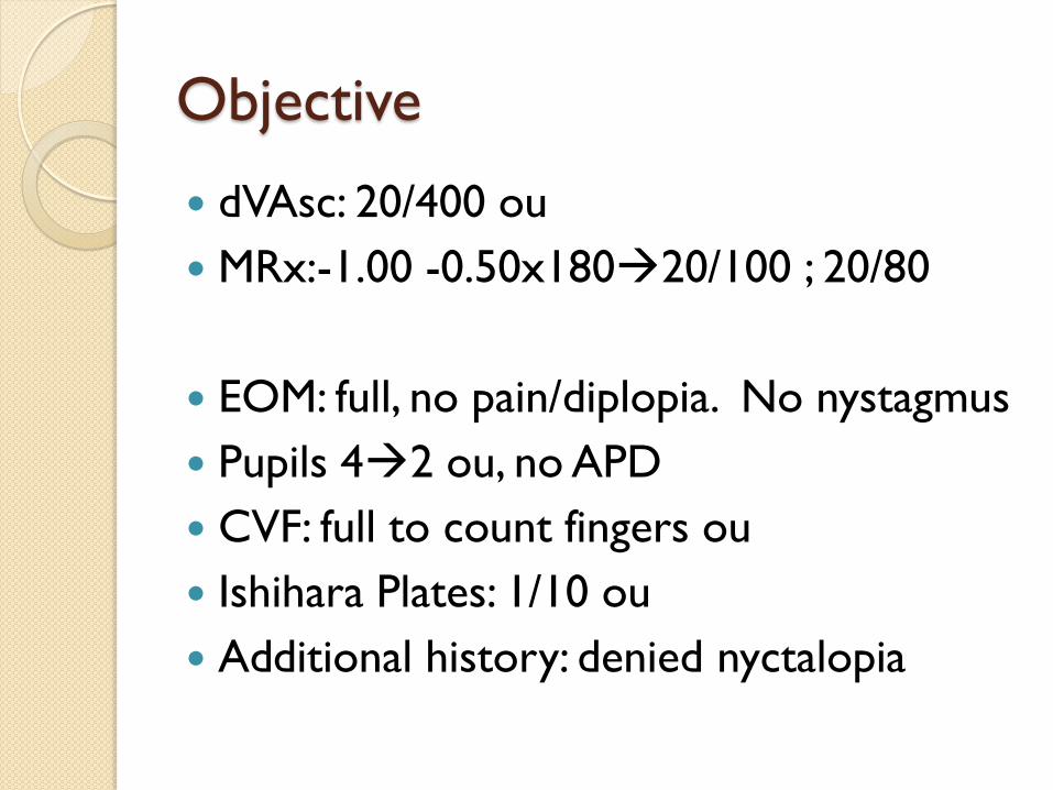
Objective
dVAsc: 20/400 ou
MRx:-1.00 -0.50x18020/100 ; 20/80
EOM: full, no pain/diplopia. No nystagmus
Pupils 42 ou, no APD
CVF: full to count fingers ou
Ishihara Plates: 1/10 ou
Additional history: denied nyctalopia
Slit Lamp Exam
LLA: wnl ou
C/S: W+Q ou
K: clear ou
AC: D+Q ou
I/P: round, reactive 4->2 ou, no APD
Lens: clear ou
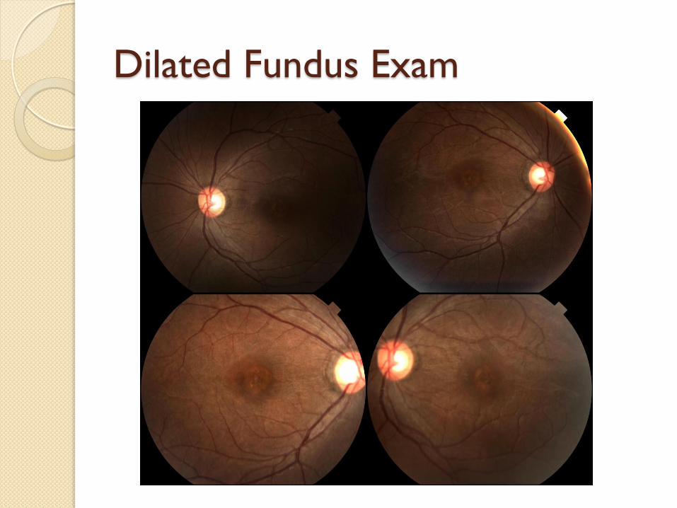
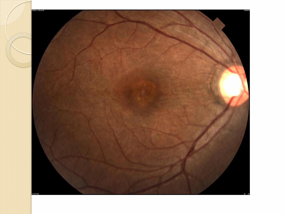
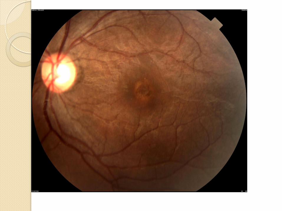
Dilated Fundus Exam

Autofluorescence
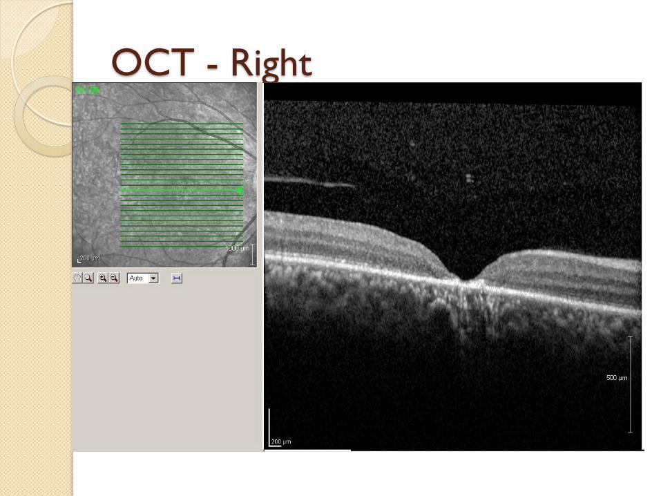
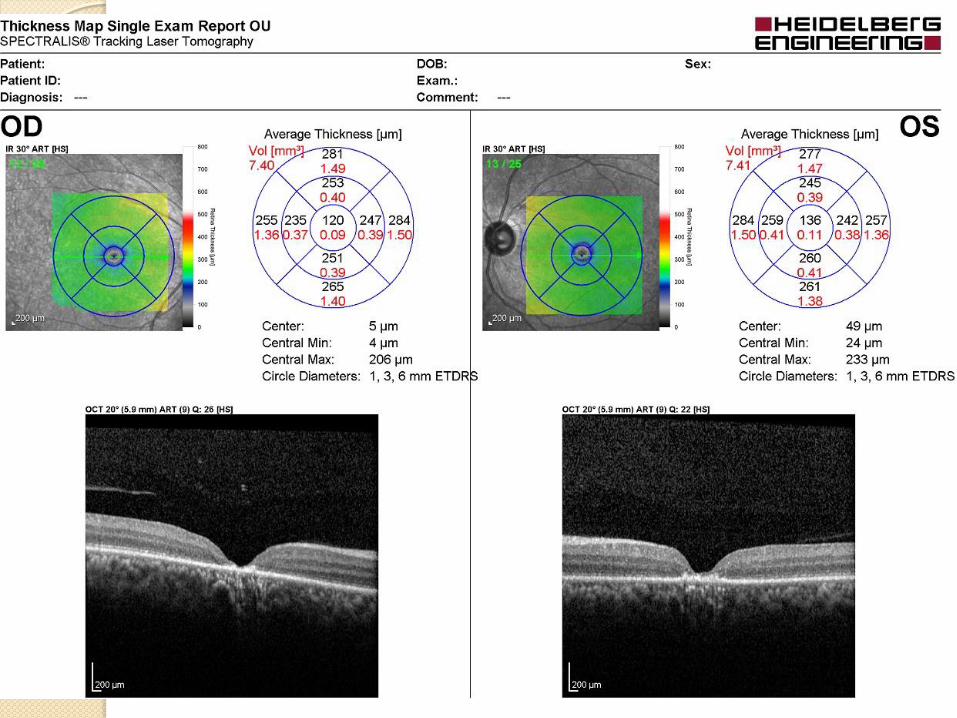
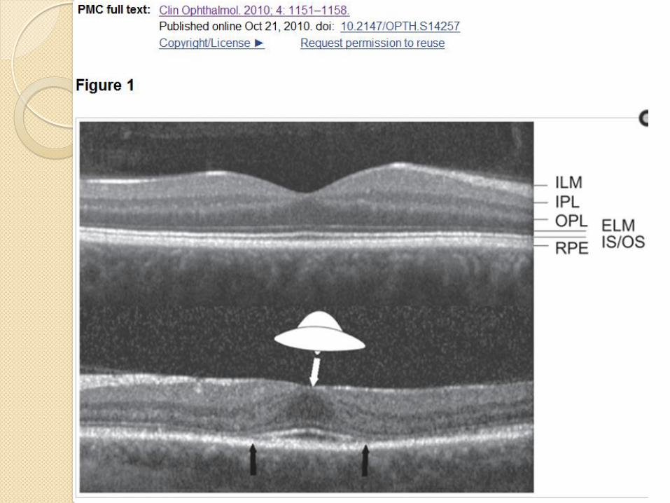
OCT - Right

OCT- Right
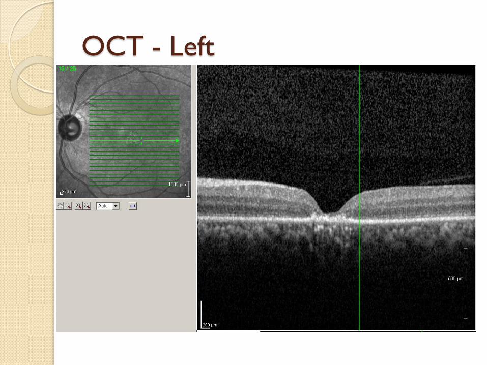
OCT - Left

DDx Stargardt Disease
Cone Dystrophies
Medication
Age-Related Macular Degeneration
Idiopathic Chronic Macular Holes
Benign Concentric Annular Dystrophy
Central Areolar Choroidal Dystrophy
Speilmeyer-Vogt-Batten-Mayou (Juvenile neuronal ceroid lipofucinosis)
North Carolina Dystrophy
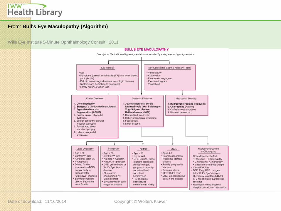
Date of download: 11/16/2014 Copyright © Wolters Kluwer
From: Bull's Eye Maculopathy (Algorithm)
Wills Eye Institute 5-Minute Ophthalmology Consult, 2011
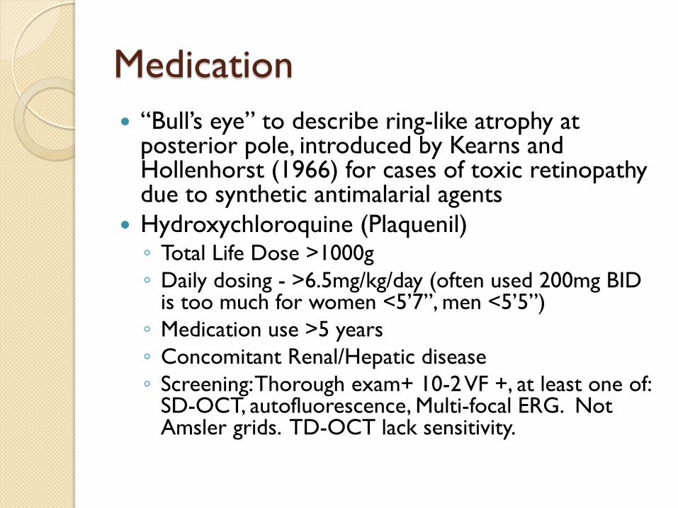
Medication
“Bull’s eye” to describe ring-like atrophy at posterior pole, introduced by Kearns and Hollenhorst (1966) for cases of toxic retinopathy due to synthetic antimalarial agents
Hydroxychloroquine (Plaquenil) ◦ Total Life Dose >1000g
◦ Daily dosing - >6.5mg/kg/day (often used 200mg BID is too much for women <5’7”, men <5’5”)
◦ Medication use >5 years
◦ Concomitant Renal/Hepatic disease
◦ Screening: Thorough exam+ 10-2 VF +, at least one of: SD-OCT, autofluorescence, Multi-focal ERG. Not Amsler grids. TD-OCT lack sensitivity.
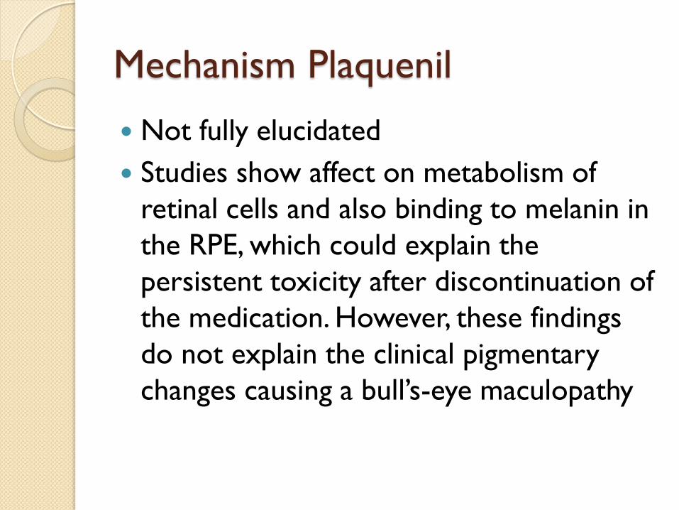
Mechanism Plaquenil
Not fully elucidated
Studies show affect on metabolism of
retinal cells and also binding to melanin in
the RPE, which could explain the
persistent toxicity after discontinuation of
the medication. However, these findings
do not explain the clinical pigmentary
changes causing a bull’s-eye maculopathy

Eye Net Magazine June 2011
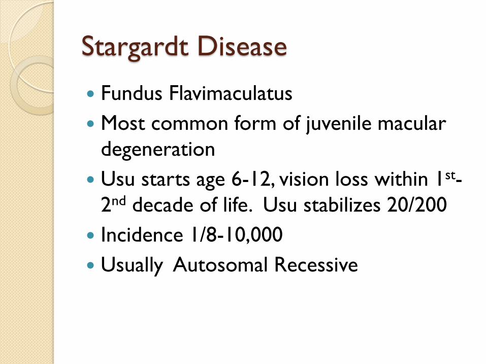
Stargardt Disease
Fundus Flavimaculatus
Most common form of juvenile macular
degeneration
Usu starts age 6-12, vision loss within 1st-
2nd decade of life. Usu stabilizes 20/200
Incidence 1/8-10,000
Usually Autosomal Recessive
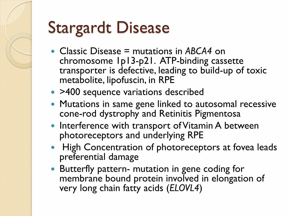
Stargardt Disease
Classic Disease = mutations in ABCA4 on chromosome 1p13-p21. ATP-binding cassette transporter is defective, leading to build-up of toxic metabolite, lipofuscin, in RPE
>400 sequence variations described
Mutations in same gene linked to autosomal recessive cone-rod dystrophy and Retinitis Pigmentosa
Interference with transport of Vitamin A between photoreceptors and underlying RPE
High Concentration of photoreceptors at fovea leads preferential damage
Butterfly pattern- mutation in gene coding for membrane bound protein involved in elongation of very long chain fatty acids (ELOVL4)
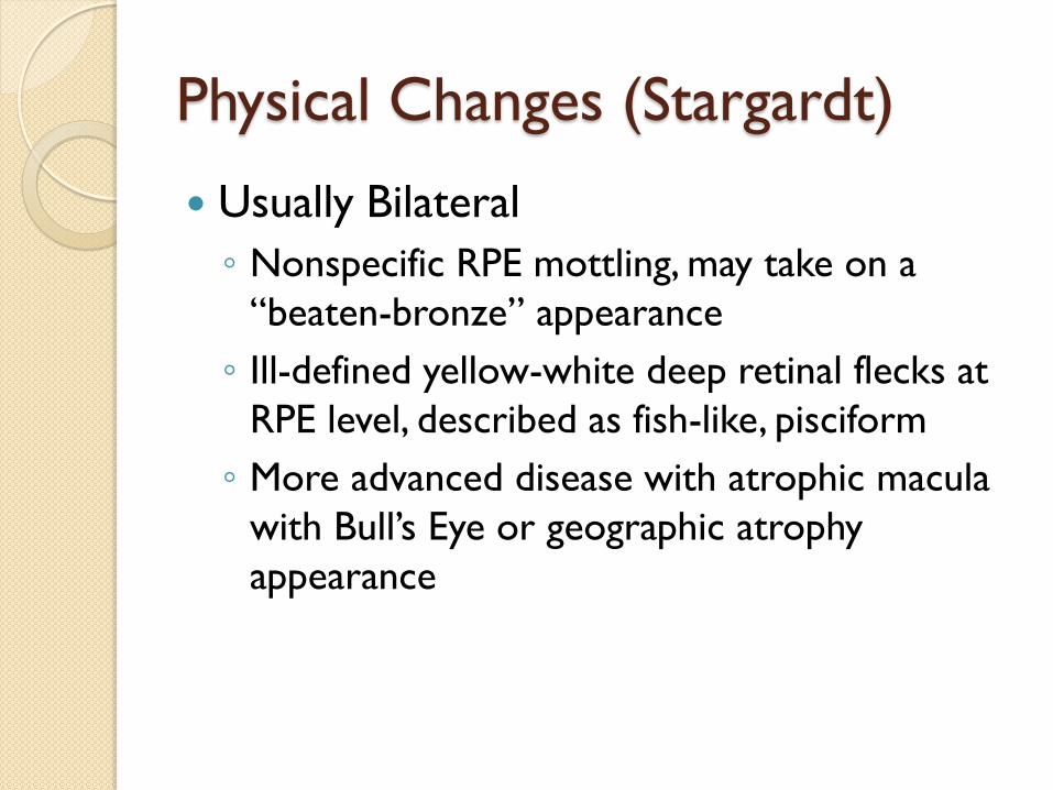
Physical Changes (Stargardt)
Usually Bilateral
◦ Nonspecific RPE mottling, may take on a
“beaten-bronze” appearance
◦ Ill-defined yellow-white deep retinal flecks at
RPE level, described as fish-like, pisciform
◦ More advanced disease with atrophic macula
with Bull’s Eye or geographic atrophy
appearance

Date of download: 11/19/2014 Copyright © Wolters Kluwer
From: Stargardt Disease
Pisciform lesions and macular bull's-eye atrophy in patient with Sargardt's disease.
Legend:
Wills Eye Institute 5-Minute Ophthalmology Consult, 2011
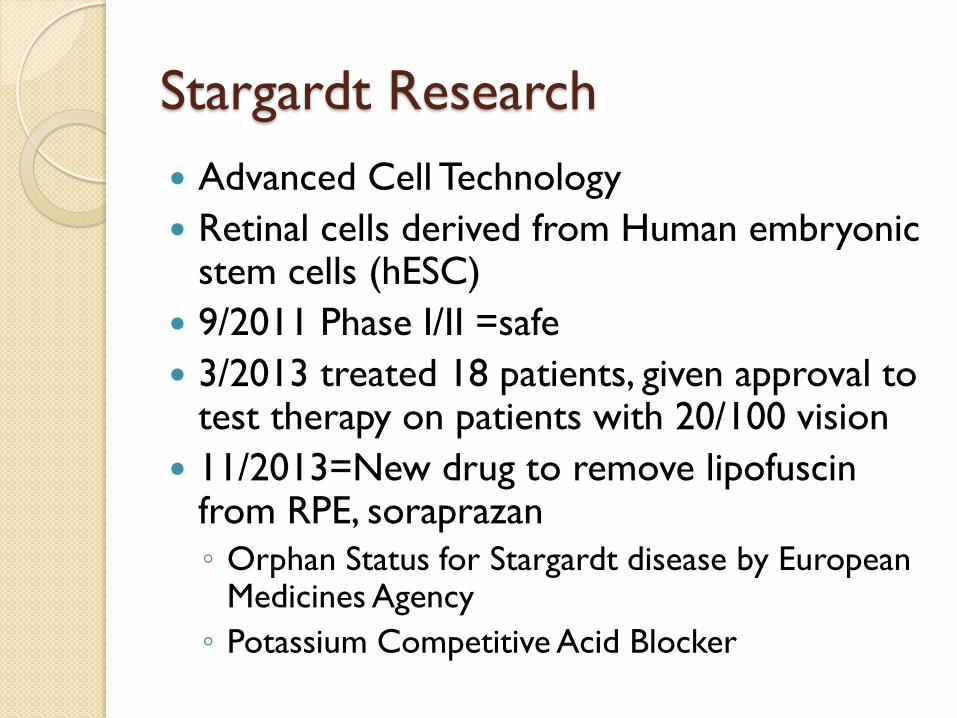
Stargardt Research
Advanced Cell Technology
Retinal cells derived from Human embryonic stem cells (hESC)
9/2011 Phase I/II =safe
3/2013 treated 18 patients, given approval to test therapy on patients with 20/100 vision
11/2013=New drug to remove lipofuscin from RPE, soraprazan
◦ Orphan Status for Stargardt disease by European Medicines Agency
◦ Potassium Competitive Acid Blocker
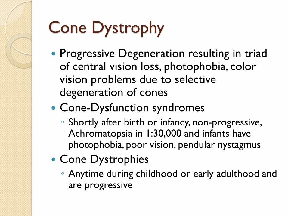
Cone Dystrophy
Progressive Degeneration resulting in triad of central vision loss, photophobia, color vision problems due to selective degeneration of cones
Cone-Dysfunction syndromes
◦ Shortly after birth or infancy, non-progressive, Achromatopsia in 1:30,000 and infants have photophobia, poor vision, pendular nystagmus
Cone Dystrophies
◦ Anytime during childhood or early adulthood and are progressive
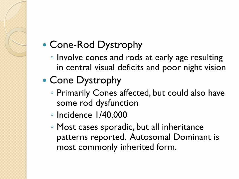
Cone-Rod Dystrophy
◦ Involve cones and rods at early age resulting in central visual deficits and poor night vision
Cone Dystrophy
◦ Primarily Cones affected, but could also have some rod dysfunction
◦ Incidence 1/40,000
◦ Most cases sporadic, but all inheritance patterns reported. Autosomal Dominant is most commonly inherited form.
Cone Dystrophy
Symptoms typically before 20 yo
Color vision problems occur early in
disease, unlike many other macular
dystrophies
Earlier onset – more severe disease
Nyctalopia makes rod disease more likely
Associated Systemic Conditions
Neurofibromatosis I
Spinocerebellar Ataxia type 7
Amelogenesis
Pierre-Marie Ataxia
Trichomegaly
Bardet-Biedl Syndrome
Alstrom Syndrome

Physical changes (Cone Dystrophy)
Initially normal as dysfunction occurs
before ophthalmologic changes
Then, variable from macular granularity to
well-demarcated, circular, depigmented
area of macular atrophy
Optic Discs may have temporal pallor
VA from 20/20 to CF
Color Plates often with varying degrees
of abnormality
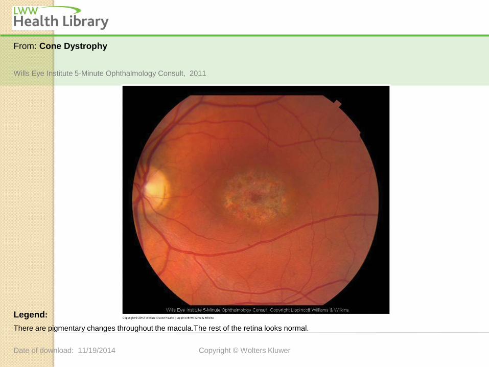
Date of download: 11/19/2014 Copyright © Wolters Kluwer
From: Cone Dystrophy
There are pigmentary changes throughout the macula.The rest of the retina looks normal.
Legend:
Wills Eye Institute 5-Minute Ophthalmology Consult, 2011

Diagnosis (Cone Dystrophy)
ERG (full-field and multi-focal)
◦ Characteristic markedly abnormal light
response (photopic- cone), with normal to
slightly abnormal dark response (scotopic-
rod)
◦ Selective decrease in photopic B-wave along
with decreased amplitude on 30-Hz flicker
may exist
OCT
◦ May show transverse photoreceptor loss with
disruption/focal loss of IS/OS junction

More Testing (Cone Dystrophy)
Fundus Autofluorescence
◦ May show foveolar hyper-autofluorescence
(nonspecific)
Fluorescein Angiography
◦ May show early hyperfluorescence
Visual Fields
◦ Often with full peripheral fields, but bilateral
central scotomas
No single test/finding sufficient for
diagnosis
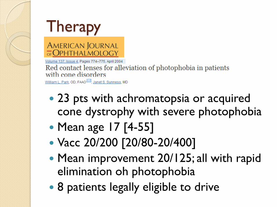
Therapy
23 pts with achromatopsia or acquired cone dystrophy with severe photophobia
Mean age 17 [4-55]
Vacc 20/200 [20/80-20/400]
Mean improvement 20/125; all with rapid elimination oh photophobia
8 patients legally eligible to drive
st
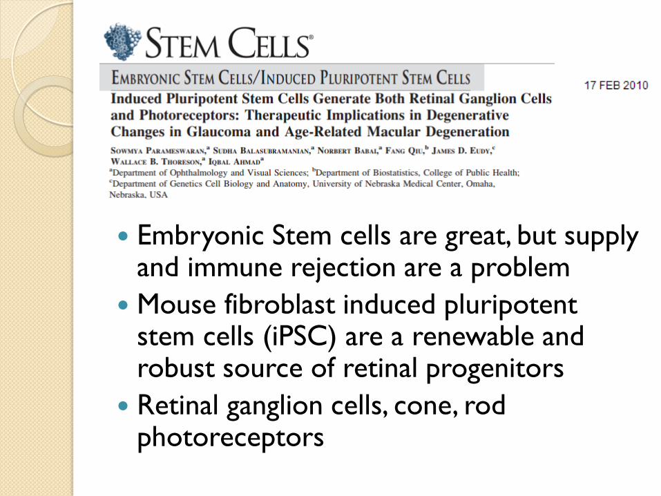
Embryonic Stem cells are great, but supply and immune rejection are a problem
Mouse fibroblast induced pluripotent stem cells (iPSC) are a renewable and robust source of retinal progenitors
Retinal ganglion cells, cone, rod photoreceptors
Our Patient
Patient is pending Fluorescein
Angiography
Referred to SUNY Optometry for ERG
Referred to Light House
Children were examined with our
pediatric ophthalmology staff (so far they
look great!)

Take Home Points
Look for causes for vision being worse
than you would expect
If you see a bull’s eye, obtain family
history, age of onset, night-time
symptoms, photophobia, color deficits,
medication use
Fundus photography, autofluorescence,
OCT, VF, ERG may help
Reflective Practice
This case represented application of
careful history taking, ophthalmic
examination and creation of a complete
differential diagnosis to evaluate and
treat complex retinal disorders
Core Competencies
Patient Care: The case involved thorough patient care, ability to explain findings, and need for treatment to the patient. Once diagnosed, the patient is receiving proper management and care.
Medical Knowledge This presentation allowed us to review the presentations, proper evaluation/work up, and differential of bull’s eye maculopathies
Practice-Based Learning and Improvement: This presentation included a current literature search of bull’s eye maculopathies
Interpersonal and Communication Skills: The patient was treated with respect and every effort was made to communicate with the patient and treat in accordance with her wishes.
Professionalism: The patient was treated in the proper manner.
Systems-Based Practice: The patient was discussed with colleagues and treated appropriately

Thank you!
Our patient
KCHC Eye Clinic Staff
Dr Rony Gelman
Dr Christopher Fecarotta

Works Cited Hansen, M, Schuman, S. Hydroxychloroquine-Induced Retinal Toxicity. EyeNet June 2011
Maguire, JI, Murchison, AP and Jaeger, EA . Wills Eye Institute 5-Minute Ophthalmology Consult. Philadelphia:
Wolters Kluwer Health/Lippincott Williams & Wilkins, 2011
Meunier, I et al. Spectral-Domain Optical Coherence Tomography in Hereditary Retinal Dystrophies. Selected
Topics in Optical Coherence Tomography, InTech. February 2012
Parameswaran, S et al. Induced pluripotent stem cells generate both retinal ganglion cells and photoreceptors:
therapeutic implications in degenerative changes in glaucoma and age-related macular degeneration. Stem Cells.
2010: 28: 695-703
Park, W, Sunness, J. Red Contact Lenses for alleviation of photobphobia in patients with cone disorders.
American Journal of Ophthalmology. 137:4: 774-775. April 2004
Pinicker’s A, Cruysberg JRM, AanDeKerk, AL. Main types of bull’s eye maculopathy Functional classification.
Documenta ophthalmologica 58, 257-267 (1984)