Embed Size (px)
Citation preview

EquinEHEaltHupdatE
For Horse Owners and VeterinariansVol. 12, Issue No. 1 – 2011
purduE univErsity scHool of vEtErinary MEdicinE
Contents...HealthPPID . . . . . . . . . . . . . . .pg . 6
Joint Injections . . . . . . .pg . 4
Hoof Abscesses . . . . . . .pg . 3
Community PracticeBlanketing Your Horse . . . . . . . . . . . . . .pg . 5
Upcoming Foaling Season . . . . . . pg . 7
News & NotesShelbyville Clinic . . . . .pg . 2
New Faculty . . . . . . . . .pg . 2 - Dr . Sandra Taylor - Dr . Stacey Tinkler
New Residents . . . . . . . .pg . 3 - Dr . Balazs Toth - Dr . Sarah Waxman
In DepthThe Economic Impact and Health Study of the Indiana Equine Industry . . . . . .pg . 1
The Economic Impact and Health Study of the Indiana Equine Industry
(continued on pg. 4)
The equine industry is complex . Animals, facilities, resources, skills, markets and health issues vary greatly and contribute to the complexity of the industry . In order to better understand these enterprises and their interactions, a study was conducted and analyzed by Susan E . Conners, Ph .D ., and Jonathan M . Furdek, Ph .D . of Purdue University Calumet; and Laurent Couetil, DVM, Ph .D . and Mark A . Russell, Ph .D . of Purdue University . Utilizing a trifold of academic resources stemming from Purdue Calumet’s Equine Business Manage-ment, Purdue University’s College of Agriculture and School of Veterinary Medicine, this comprehensive report provides insights into the multifaceted equine industry and focuses on the Indiana equine industry’s economic impact, health issues and the entrepreneurial trends within the industry . Purdue University is the only university in the country to have veterinary, animal science, and equine business programs all serving the equine industry . The complete report can be found on Purdue’s Equine Sports Medicine Center website: http://www .vet .purdue .edu/esmc/
This broad study is based on three online surveys that were conducted simultaneously in spring 2011 . They included a Horse Show Survey, an Indiana Race Track Survey, and an Indiana Equine Business and Owners Survey . The Indiana Equine Business and Own-ers Survey was broken down into several segments: an Equine Business Survey, an Equine Owners Survey, an Equine Entrepreneur Survey, and an Equine Health Survey . Estimation of economic impacts was determined utilizing state-specific IMPLAN multipliers to estimate the overall economic impact of the industry on the state GDP and on employment . The IMPLAN modeling system has been in use since 1979 and is currently used by more than 500 private consulting firms, university research centers, and government agencies .
The equine industry produces a $2 .6 billion economic impact in Indiana . The pro-jected direct and indirect tax revenue consequences from the economic activity generated by the equine industry are significant, despite an unstable economic environment . State and local tax revenues attributed to the equine industry exceed $160 million, while federal tax revenues attributed to the equine industry are approximately $107 million . The resulting tax impact exceeds $270 million .
This multimillion-dollar industry appears to be changing from an industry focused on work and recreation to an industry that more prominently engages in recreation and competition . Segments of the industry – namely racing, breeding, boarding and training – have significant export potential, providing facilities and services to those outside the state, and generating business activity and revenues in the state . A typical equine business opera-tion generates an average of $108,000 in revenue . The major expense for an equine business owner was in wages and salaries, followed by all taxes .
Purdue University is an equal access/equal opportunity/affirmative action university.If you have trouble accessing this document because of a disability, please contact PVM Web Communications at [email protected].

2
News & NotesRegional Equine Diagnostic and Surgical Center Update
Surgical suite ................ $ 500,000Large exam room ............ $ 100,000Standard exam room ...... $ 50,000Laboratory ...................... $ 25,000Surgery ........................... $ 25,000Induction/recovery area ... $ 25,000Surgical prep ................... $ 25,000Pharmacy ........................ $ 25,000
Imaging suite ................. $ 850,000MRI ................................. $ 250,000Video endoscope ............ $ 150,000Digital X-ray .................... $ 75,000Nuclear imaging .............. $ 50,000Ultrasound ...................... $ 30,000C-arm ............................. $ 20,000Advanced shockwave therapy ............................ $ 15,000
Conference center ........ $ 500,000Reception ....................... $ 250,000Lobby ............................. $ 50,000Consultation room .......... $ 30,000Veterinarian’s office ......... $ 25,000Business office ................ $ 15,000
Equine Diagnostic and Surgical Center $2,700,000Stall suite ...................... $ 100,000Holding stall .................... $ 10,000Mare & foal stall ............. $ 7,000Standard stall .................. $ 5,000Isolation stall ................... $ 5,000Lameness corridor ........... $ 15,000On-call suite .................... $ 15,000
Outdoor OpportunitiesFencing ........................... $ 200,000Landscaping .................... $ 50,000Roofed round pen ........... $ 40,000Signage .......................... $ 25,000Large paddock ................ $ 20,000Outdoor lameness track .. $ 15,000Overnight parking ........... $ 15,000Storage buildings ............ $ 15,000Small paddock ................ $ 10,000
Endowed PositionsSurgeon .......................... $ 1,500,000DVM ............................... $ 1,000,000Veterinary Technologist ... $ 500,000
Indoor Riding Arena .... $ 1,000,000
Equine Center Naming OpportunitiesContact Carol Willoughby, PVM director of advancement,
at (800) 830-0104 for additional details.
Displayed is the preliminary architectural plan for the Purdue University Regional Equine Diagnostic and Surgical Center at Shelbyville. These plans have been shared with the Dean’s Advisory Board and the internal planning committee and acted upon. We are pleased and excited at this time to share with you preliminary renderings and the site development plan.
On April 20, the School closed on the purchase of a 71- acre site where the facility will be located. Only 20 acres are needed for this facility’s purposes. The remainder of the Regional Equine Diagnostic and Surgical Center Update land will be retained for possible future development. The site will be owned by the Purdue Research Foundation and loaned to the University.
Thanks to the generosity of alumni and donors we have raised $3 million to date. The School has committed to raising the $12 million needed to support this project through private fundraising. These gifts have enabled the School to move steadily toward the completion of this new and exciting project that will deliver specialty referral services to a growing industry in Indiana.
Dr. Sandy Taylor is a native of West Richland, Washington, where she grew up on a small horse farm . She received her BS degree (1998) and DVM degree (2001) from Washington State University, and then completed a one-year in-ternship in equine medicine and surgery at San Luis Rey Equine Hospital in Bonsall, California . To obtain private practice ambulatory experience, she spent the following year as an associate ambulatory veterinarian at Tacoma Equine Hospital in Tacoma, Washington .
In order to pursue a career in large animal internal medicine, she then completed a residency in large animal internal medi-cine at the University of California, Davis, and received board certification in the American College of Veterinary Internal Medicine in 2006 . Throughout these endeavors, Dr . Taylor developed a passion for teaching and a desire to remain in academia . To this end, she entered graduate school at Wash-ington State University and obtained a PhD in 2010 . Her PhD research focused on equine immunology and virology, with specific attention to neutralizing antibody-mediated control of lentivirus infection . Her current research interests include neonatal septicemia, infectious disease, and treatment and prevention of equine sarcoid .
Dr. Stacy Tinkler hails from New York, where she grew up riding in the local hunter/jumper circuit as a child . She attended Cornell University and after ob-taining her bachelor’s degree in Animal Science, she spent several years living in Colorado taking care of horses and then traveling and working as an ESL (English as a second language) teacher in Spain . She pursued and obtained her DVM degree from the Univer-sity of Minnesota in 2005 . After graduation, she completed an
internship year in equine medicine and surgery at Chaparral Animal Hospital in Phoenix, Arizona . She then went on to help start up the equine ambulatory service operating out of the University of Minnesota for 2 years and then transitioned into a Large Animal Internal Medicine residency there and became a diplomate of the American College of Veterinary Internal Medicine in 2011 . Dr . Tinkler spent this past year as a post-doc at Purdue University researching acid-base disorders of race horses . Her professional interests include infectious disease, and metabolic disease .
Dr. Sandra D. Taylor New Large Animal Medicine Faculty
Dr. Stacy Tinkler New Equine Community
Practice Faculty
New Faculty:

Sole Abscesses in the HorseKristi Adam, DVM Student (Class of 2012)
Sole abscesses are the most common cause of acute lameness in the horse . They develop via foreign body penetration of the sole or direct migration of bacteria or fungus through separation in the sole-wall junction (white line) . Common foreign bodies entering the sole include rocks, nails, wire, or sharp pieces of wood . Other risk factors for sole abscesses include weak areas in the sole, puncture wounds, trauma, sole cracks, and housing in wet conditions .
Affected horses present with a sudden onset of acute lameness and can be non-weightbearing . Other signs of foot abscessation include bounding digital pulses at the level of the fetlock, heat in the affected foot, and swelling of the coronary band or pastern . The area of sole pain can be pinpointed with the use of hoof testers and a point of entry maybe visible following paring of the sole with a hoof knife . In some cases the point of entry is not readily visible and hoof testers may be inconclusive . In these cases further diagnostics should be performed which can include nerve blocks and radiographs of the foot .
The primary goal of treatment is abscess drainage . This is accomplished by desensitization of the foot with a nerve block and cleaning of the foot . In some instances this may require removal of the shoe . This can be performed by your veterinarian or farrier . The sole of the foot must be trimmed and pared to provide a clean surface . Ideally the abscess location can be identified with hoof testers . Once the foot is clean the foot is carefully examined for a dark spot which frequently leads to the abscess . The abscess is opened with a hoof knife to provide drainage . It is important for the drainage hole to be wide enough so that the abscess does not “seal over” until it is fully resolved . Once the abscess has been drained the foot should be protected with a bandage or a shoe can be replaced by your farrier and covered with a metal or plastic plate (treatment plate) . It is very important to keep the foot clean and dry while the abscess is resolving .
When a sole abscess is suspected but not found following cleaning of the hoof it is recommended to soak the foot in water and Epsom salt or use a poultice to soften the foot . This will encourage drainage of the abscess either from the sole or via the coronary band . Abscess drainage from the coronary band is commonly known as “gravel” . In some cases it may take 7-10 days before an abscess opens and drains when it cannot be identified on the sole of the hoof .
Other considerations for treatment of sole abscesses include a Tetanus toxoid booster to prevent the development of tetanus and the administration of antimicrobial and anti-inflammatory medication . Antibiotics are not generally given unless the extent of abscessation is severe or involves the coffin bone as drainage of the abscess is the most important treatment . Pain and inflammation associated with the abscess is managed with by administering phenylbutazone (Bute), 1 to 2 grams once or twice daily .
Complications of sole abscesses include chronic drainage, infection of the coffin bone, and rarely fracture of the coffin bone . If the lameness does not rapidly improve following drainage of the abscess your veterinarian should be contacted . Radiographs of the foot may be required if the horse does not respond to treatment .
Most horses will show significant improvement within the first 12-24 hours of treatment . The most noticeable sign of improvement is relief of lameness and willingness to walk on the affected limb . The site of abscess drainage will usually dry up within 7-10 days and the patient can return to work . Although, horses should not return to exercise until completely sound . Care should be taken to prevent future abscesses from occurring . This can be achieved by daily foot care such as cleaning out debris around the frog and sole and providing a clean, dry environment .
3
New Residents:
Dr. Balazs TothResident, Large Animal Medicine, 2010-2013
Education / Training:DVM, University of Svent Istvan, Budapest, Hungary 2005
Internship, Hagyard Equine Medical Institute, Lexington, KY, 2007-08
MS, University of California-Davis, Davis, CA, 2008 - 2010
Research / Scholarly InterestsInfectious diseases Neonatology Critical care medicine
Dr. Sarah WaxmanResident, Large Animal Surgery 2011-2014
Education / TrainingDVM, Kansas State University College of Veterinary Medicine; Manhattan, KS 2009
Internship, Wilhite and Frees Equine Hospital; Peculiar, MO, 2009-2010
Research / Scholarly InterestsResearch: Joint injection techniques Interests: GI disorders, wound healing
4
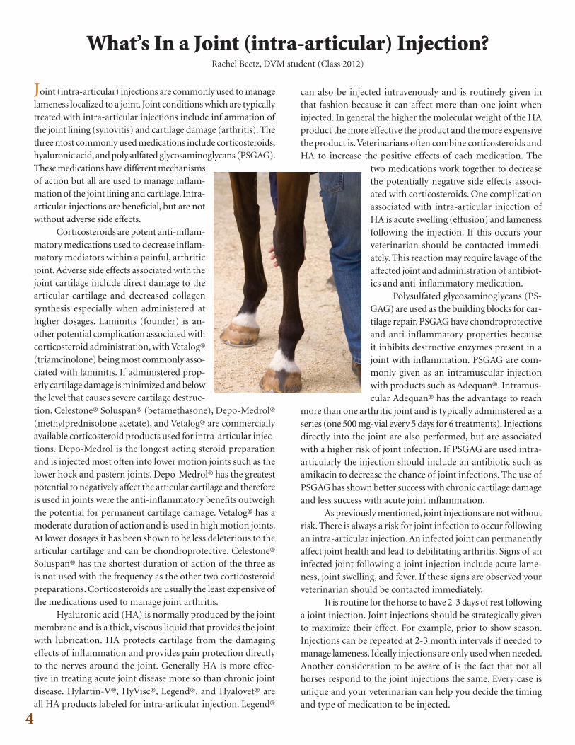
What’s In a Joint (intra-articular) Injection?Rachel Beetz, DVM student (Class 2012)
Joint (intra-articular) injections are commonly used to managelameness localized to a joint . Joint conditions which are typically treated with intra-articular injections include inflammation of the joint lining (synovitis) and cartilage damage (arthritis) . The three most commonly used medications include corticosteroids, hyaluronic acid, and polysulfated glycosaminoglycans (PSGAG) . These medications have different mechanisms of action but all are used to manage inflam-mation of the joint lining and cartilage . Intra-articular injections are beneficial, but are not without adverse side effects .
Corticosteroids are potent anti-inflam-matory medications used to decrease inflam-matory mediators within a painful, arthritic joint . Adverse side effects associated with the joint cartilage include direct damage to the articular cartilage and decreased collagen synthesis especially when administered at higher dosages . Laminitis (founder) is an-other potential complication associated with corticosteroid administration, with Vetalog® (triamcinolone) being most commonly asso-ciated with laminitis . If administered prop-erly cartilage damage is minimized and below the level that causes severe cartilage destruc-tion . Celestone® Soluspan® (betamethasone), Depo-Medrol® (methylprednisolone acetate), and Vetalog® are commercially available corticosteroid products used for intra-articular injec-tions . Depo-Medrol is the longest acting steroid preparation and is injected most often into lower motion joints such as the lower hock and pastern joints . Depo-Medrol® has the greatest potential to negatively affect the articular cartilage and therefore is used in joints were the anti-inflammatory benefits outweigh the potential for permanent cartilage damage . Vetalog® has a moderate duration of action and is used in high motion joints . At lower dosages it has been shown to be less deleterious to the articular cartilage and can be chondroprotective . Celestone® Soluspan® has the shortest duration of action of the three as is not used with the frequency as the other two corticosteroid preparations . Corticosteroids are usually the least expensive of the medications used to manage joint arthritis .
Hyaluronic acid (HA) is normally produced by the joint membrane and is a thick, viscous liquid that provides the joint with lubrication . HA protects cartilage from the damaging effects of inflammation and provides pain protection directly to the nerves around the joint . Generally HA is more effec-tive in treating acute joint disease more so than chronic joint disease . Hylartin-V®, HyVisc®, Legend®, and Hyalovet® are all HA products labeled for intra-articular injection . Legend®
can also be injected intravenously and is routinely given in that fashion because it can affect more than one joint when injected . In general the higher the molecular weight of the HA product the more effective the product and the more expensive the product is . Veterinarians often combine corticosteroids and HA to increase the positive effects of each medication . The
two medications work together to decrease the potentially negative side effects associ-ated with corticosteroids . One complication associated with intra-articular injection of HA is acute swelling (effusion) and lameness following the injection . If this occurs your veterinarian should be contacted immedi-ately . This reaction may require lavage of the affected joint and administration of antibiot-ics and anti-inflammatory medication .
Polysulfated glycosaminoglycans (PS-GAG) are used as the building blocks for car-tilage repair . PSGAG have chondroprotective and anti-inflammatory properties because it inhibits destructive enzymes present in a joint with inflammation . PSGAG are com-monly given as an intramuscular injection with products such as Adequan® . Intramus-cular Adequan® has the advantage to reach
more than one arthritic joint and is typically administered as a series (one 500 mg-vial every 5 days for 6 treatments) . Injections directly into the joint are also performed, but are associated with a higher risk of joint infection . If PSGAG are used intra-articularly the injection should include an antibiotic such as amikacin to decrease the chance of joint infections . The use of PSGAG has shown better success with chronic cartilage damage and less success with acute joint inflammation .
As previously mentioned, joint injections are not without risk . There is always a risk for joint infection to occur following an intra-articular injection . An infected joint can permanently affect joint health and lead to debilitating arthritis . Signs of an infected joint following a joint injection include acute lame-ness, joint swelling, and fever . If these signs are observed your veterinarian should be contacted immediately .
It is routine for the horse to have 2-3 days of rest following a joint injection . Joint injections should be strategically given to maximize their effect . For example, prior to show season . Injections can be repeated at 2-3 month intervals if needed to manage lameness . Ideally injections are only used when needed . Another consideration to be aware of is the fact that not all horses respond to the joint injections the same . Every case is unique and your veterinarian can help you decide the timing and type of medication to be injected .
5
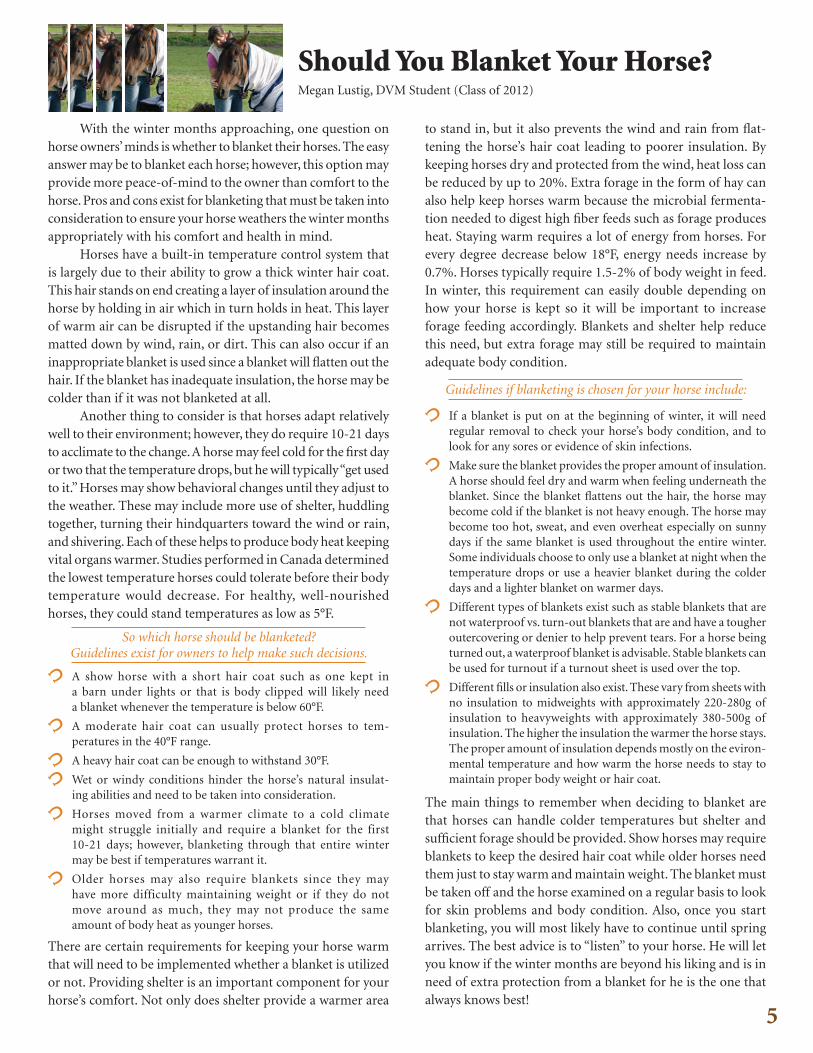
Should You Blanket Your Horse?Megan Lustig, DVM Student (Class of 2012)
With the winter months approaching, one question on horse owners’ minds is whether to blanket their horses . The easy answer may be to blanket each horse; however, this option may provide more peace-of-mind to the owner than comfort to the horse . Pros and cons exist for blanketing that must be taken into consideration to ensure your horse weathers the winter months appropriately with his comfort and health in mind .
Horses have a built-in temperature control system that is largely due to their ability to grow a thick winter hair coat . This hair stands on end creating a layer of insulation around the horse by holding in air which in turn holds in heat . This layer of warm air can be disrupted if the upstanding hair becomes matted down by wind, rain, or dirt . This can also occur if an inappropriate blanket is used since a blanket will flatten out the hair . If the blanket has inadequate insulation, the horse may be colder than if it was not blanketed at all .
Another thing to consider is that horses adapt relatively well to their environment; however, they do require 10-21 days to acclimate to the change . A horse may feel cold for the first day or two that the temperature drops, but he will typically “get used to it .” Horses may show behavioral changes until they adjust to the weather . These may include more use of shelter, huddling together, turning their hindquarters toward the wind or rain, and shivering . Each of these helps to produce body heat keeping vital organs warmer . Studies performed in Canada determined the lowest temperature horses could tolerate before their body temperature would decrease . For healthy, well-nourished horses, they could stand temperatures as low as 5°F .
So which horse should be blanketed? Guidelines exist for owners to help make such decisions.
A show horse with a short hair coat such as one kept in a barn under lights or that is body clipped will likely need a blanket whenever the temperature is below 60°F .
A moderate hair coat can usually protect horses to tem- peratures in the 40°F range .
A heavy hair coat can be enough to withstand 30°F .
Wet or windy conditions hinder the horse’s natural insulat- ing abilities and need to be taken into consideration .
Horses moved from a warmer climate to a cold climate might struggle initially and require a blanket for the first 10-21 days; however, blanketing through that entire winter may be best if temperatures warrant it .
Older horses may also require blankets since they may have more difficulty maintaining weight or if they do not move around as much, they may not produce the same amount of body heat as younger horses .
There are certain requirements for keeping your horse warm that will need to be implemented whether a blanket is utilized or not . Providing shelter is an important component for your horse’s comfort . Not only does shelter provide a warmer area
to stand in, but it also prevents the wind and rain from flat-tening the horse’s hair coat leading to poorer insulation . By keeping horses dry and protected from the wind, heat loss can be reduced by up to 20% . Extra forage in the form of hay can also help keep horses warm because the microbial fermenta-tion needed to digest high fiber feeds such as forage produces heat . Staying warm requires a lot of energy from horses . For every degree decrease below 18°F, energy needs increase by 0 .7% . Horses typically require 1 .5-2% of body weight in feed . In winter, this requirement can easily double depending on how your horse is kept so it will be important to increase forage feeding accordingly . Blankets and shelter help reduce this need, but extra forage may still be required to maintain adequate body condition .
Guidelines if blanketing is chosen for your horse include:
If a blanket is put on at the beginning of winter, it will need regular removal to check your horse’s body condition, and to look for any sores or evidence of skin infections .
Make sure the blanket provides the proper amount of insulation . A horse should feel dry and warm when feeling underneath the blanket . Since the blanket flattens out the hair, the horse may become cold if the blanket is not heavy enough . The horse may become too hot, sweat, and even overheat especially on sunny days if the same blanket is used throughout the entire winter . Some individuals choose to only use a blanket at night when the temperature drops or use a heavier blanket during the colder days and a lighter blanket on warmer days .
Different types of blankets exist such as stable blankets that are not waterproof vs . turn-out blankets that are and have a tougher outercovering or denier to help prevent tears . For a horse being turned out, a waterproof blanket is advisable . Stable blankets can be used for turnout if a turnout sheet is used over the top .
Different fills or insulation also exist . These vary from sheets with no insulation to midweights with approximately 220-280g of insulation to heavyweights with approximately 380-500g of insulation . The higher the insulation the warmer the horse stays . The proper amount of insulation depends mostly on the eviron-
mental temperature and how warm the horse needs to stay to maintain proper body weight or hair coat .
The main things to remember when deciding to blanket are that horses can handle colder temperatures but shelter and sufficient forage should be provided . Show horses may require blankets to keep the desired hair coat while older horses need them just to stay warm and maintain weight . The blanket must be taken off and the horse examined on a regular basis to look for skin problems and body condition . Also, once you start blanketing, you will most likely have to continue until spring arrives . The best advice is to “listen” to your horse . He will let you know if the winter months are beyond his liking and is in need of extra protection from a blanket for he is the one that always knows best!

Equine Pituitary Pars Intermedia DysfunctionLindsay Schwartzmeyer, DVM student (class 2012)
“The Hairy Horse”
for no obvious reason or has chronic sole abscesses should be tested for PPID . Statistics have shown that 52% of PPID horses and 33% of all geriatric horses will develop laminitis . It is important to understand that at times, laminitis is THE ONLY sign that a horse has PPID . Often they are positive when diagnostic testing is performed .
As you can see it is a good idea to treat and manage a PPID horse as early and best as possible to decrease the progression of the disease and the development of painful side effects . One of the first steps would be to accurately diagnosis PPID in the suspected horse, but that is easier said than done . Not all tests are accurate 100% of the time . In PPID horses the hormones that can be measured fluctuate due to each individual horse and with the photoperiod and season . The most common di-agnostic tests would include CBC/chemistry, dexamethasone suppression testing, and endogenous ACTH measurement, or a TRH stimulation test or domperidone stimulation test . In a study performed by Jill Beech it was shown that following serum ACTH or alpha-MSH concentrations was the best way to monitor the disease progression and response to treatment . Hirsutism, or the development of a long hair coat, is a good way to diagnose the condition with advanced PPID .
The best treatment is the drug Pergolide . When response to just pergolide is not sufficient, adding the drug Cyprohep-tadine may help . In addition to treating the pituitary gland, symptomatic treatment including good hoof care and correc-tive shoeing is needed . It is also important to maintain good dental care, aggressive anthelmintic treatment, and adequate nutritional support . Clipping of the coat during the warmer months will make the horse more comfortable and prevent excess sweating . This treatment may not work for every horse but will manage the disease very well in most . There is no cure for PPID, but pergolide plus excellent husbandry may extend your horse’s life . There is still much that is not a lot known about PPID . Research is being completed to help better understand the disease pathophysiology and it is possible that more dependable diagnostic tests and treatments will be discovered soon .
Early spring arrives and you notice your older horse is not shedding out or shedding out very slowly . There are long hairs remaining on the legs and under the chin, or his entire haircoat looks longer and more curly than before . Also you may recall that this horse grew a different winter coat than normal; longer, thicker, and curly . You may have thought that this was a normal change that occurs as the horse gets older . This is not the case, however, the long curly coat that doesn’t shed normally is a sign of Equine Pituitary Pars Intermedia Dysfunction (Equine Cushing’s Disease) .
What is Equine Pituitary Pars Intermedia Dysfunction (PPID) or Equine Cushings and why can it be a problem for your horse? The pituitary gland is the “master organ” in the brain that controls many of the hormones needed for normal function . PPID involves overgrowth of the pituitary gland cells which causes an increased production of the hormones alpha-MSH, beta-endorphin, and ACTH . The cause of the hyper-plastic pars intermedia of the pituitary gland is still largely unknown . What is known is that there is a loss of input by nerve cells from the brain that secrete the neurotransmitter dopamine . Without this input, the cells in the pituitary make too much of the hormones they normally secrete in very small amounts . The overproduction of those hormones cause the clinical signs seen in PPID .
Clinical signs associated with PPID include loss of muscle mass, abnormal fat disposition, with an increase in fat over the top line, immunosuppression, increased thirst and urination, increased respiratory rate . In advanced cases, type II diabetes insipidus, swelling over the eyes, sinusitis, chronic low grade
infections and hyperhidrosis (excessive sweating) can occur . According to a study by Mark T . Donaldson, one of
the most important clinical signs of PPID is laminitis . Any older horse that founders
References:Evaluation of suspected Pituitary Pars Intermedia Dysfunction in horses with laminitis, by Mark T. Donaldson, Alec J.R. Jorgensen, and Jill Beech; JAVMA Vol 224, No. 7, April 1, 2004Evaluation of plasma ACTH, alpha MSH and insulin concentrations during various photoperiods in clinically normal horses and ponies and those with Pituitary Pars Intermedia Dysfunction, by Jill Beech, Raymond C Boston, Dianne McFarlane, and Sue Lindborg; JAVMA, Vol 235, No. 6, September 15, 2009Clinical Forum: “Diagnosing Pituitary Pars Intermedia Dysfunction”, by Dianne MCFarlane; Equine Compendium, Vol 2, No. 4, July/August 2007
Equine Industry Study (continued from cover)
The health study details conditions causing death, diseases or loss of use for Indiana equines . These include 11 different condi-tion categories that may cause death or disease starting from birth and progressing through age categories . The greatest cause of death was old age, followed by colic and leg fracture . The greatest cause of loss of use was reported as hoof problems, followed by lameness and injuries . Furthermore, respondents estimated that the total cost to remedy health-related afflictions, along with the cost of lost use, was $803,960 . This represents a median cost of $500 for each of the 317 cases reported, with a range of $20 to $7,000 .
Revenues and expenses aside, the study shows that many equine operations do not perceive that they are businesses . Al-though most respondents have a high opinion of being an entre-preneur, they feel that they lack the resources required to run a business . Almost 60 percent of respondents who were in business reported not to have completed a business plan, while almost a third of respondents who already had their own business listed it as their primary source of income .
This indicates a need to further educate those in or those who desire to become a part of the equine industry, so that they can recognize their economic role and operate their enterprises more like businesses . The opportunity exists to assist these business owners or would-be entrepreneurs to generate wealth, jobs and tax revenue for the equine industry and the state .
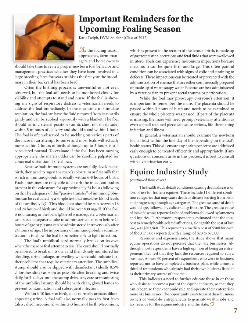
Important Reminders for the Upcoming Foaling SeasonKatie Delph, DVM Student (Class of 2012)
As the foaling seasonapproaches, farm man-agers and horse owners
should take time to review proper newborn foal behavior and management practices whether they have been involved in a large breeding farm for years or this is the first year the brood-mare in their backyard has been bred .
Often the birthing process is uneventful or not even observed, but the foal still needs to be monitored closely for viability and attempts to stand and nurse . If the foal is show-ing any signs of respiratory distress, a veterinarian needs to address the foal immediately . In the meantime to stimulate respiration, the foal can have the fluid removed from its nostrils gently and can be rubbed vigorously with a blanket . The foal should sit in a sternal position (on its chest not on its side) within 5 minutes of delivery and should stand within 1 hour . The foal is often observed to be suckling on various parts of the mare in an attempt to nurse and most foals will actually nurse within 2 hours of birth, although up to 3 hours is still considered normal . To evaluate if the foal has been nursing appropriately, the mare’s udder can be carefully palpated for abnormal distention if she allows .
Because foals’ immune systems are not fully developed at birth, they need to ingest the mare’s colostrum or first milk that is rich in immunoglobulins, ideally within 6-8 hours of birth . Foals’ intestines are only able to absorb the intact antibodies present in the colostrum for approximately 24 hours following birth . The adequacy of this “passive transfer” of immunoglobu-lins can be evaluated by a simple test that measures blood levels of the antibody IgG . This blood test should be run between 16 and 24 hours of birth and should be over 800 mg/dl . If the foal is not nursing or the foal’s IgG level is inadequate, a veterinarian can pass a nasogastric tube to administer colostrum before 24 hours of age or plasma can be administered intravenously after 24 hours of age . The importance of immunoglobulin adminis-tration is to allow the foal to be better able to fight infection .
The foal’s umbilical cord normally breaks on its own when the mare or foal attempt to rise . The cord should normally be allowed to break on its own and then closely monitored for bleeding, urine leakage, or swelling which could indicate fur-ther problems that require veterinary attention . The umbilical stump should also be dipped with disinfectant (ideally 0 .5% chlorohexidine) as soon as possible after breaking and twice daily for 3-4 days until the stump dries . Any care or monitoring of the umbilical stump should be with clean, gloved hands to prevent contamination and subsequent infection .
Within 6-10 hours of birth, a foal normally urinates dilute-appearing urine . A foal will also normally pass its first feces (also called meconium) within 2-3 hours of birth . Meconium,
which is present in the rectum of the fetus at birth, is made up of gastrointestinal secretions and fetal fluids that were swallowed in utero . Foals can experience meconium impactions because meconium can be quite firm and large . This often painful condition can be associated with signs of colic and straining to defecate . These impactions can be treated or prevented with the administration of enemas that are either commercially prepared or made up of warm soapy water . Enemas are best administered by a veterinarian to prevent rectal trauma or perforation .
While the foal may preoccupy everyone’s attention, it is important to remember the mare . The placenta should be passed within 3 hours of birth and needs to be examined to ensure the whole placenta was passed . If part of the placenta is missing, the mare will need prompt veterinary attention as even a small retained piece can cause serious, life-threatening infection and illness
In general, a veterinarian should examine the newborn foal at least within the first day of life depending on the foal’s health status . This will ensure any health concerns are addressed early enough to be treated efficiently and appropriately . If any questions or concerns arise in this process, it is best to consult with a veterinarian early .
7

Nonprofit Org.U.S. Postage
PAIDPurdue University
Purdue UniversitySchool of Veterinary Medicine Equine Sports Medicine Center 1248 Lynn Hall West Lafayette, Indiana 47907-1248
ADDRESS SERVICE REQUESTED
Phone: 765-494-8548 Fax: 765-496-2641www .vet .purdue .edu/esmc/
With generous support of Purdue University’s Veterinary Teaching Hospital and the School of Veterinary Medicine Dean’s Office .
Please address all correspondence related to this newsletter to the address above .
Editorial Board: Drs . Couétil L ., Hawkins J . and Tinkler S .
Design & layout by: Elaine Scott Design
EA/EOU
Purdue’s Equine Sports Medicine Center is dedicated to the educa-tion and support of Indiana horsemen and veterinarians through the study of the equine athlete . The Center offers comprehensive evalua-tions designed to diagnose and treat the causes of poor performance, to provide performance and fitness assessments, and to improve the rehabilitation of athletic horses . Other integral goals of the Center are to pioneer leading-edge research in the area of equine sports medicine, to provide the highest level of training to future equine veterinarians, and to offer quality continuing education to Indiana veterinarians and horsemen . For more information visit our website:
is published by:EquinE HEaltH updatE
Continuum© by Larry Anderson
The Equine Sports Medicine Center
www.vet.purdue.edu/esmc/
Purdue University is an equal access/equal opportunity/affirmative action university.If you have trouble accessing this document because of a disability, please contact PVM Web Communications at [email protected].