Embed Size (px)
Citation preview

Diagnostic and Interventional Imaging (2015) 96, 757—774
CONTINUING EDUCATION PROGRAM: FOCUS. . .
Epistaxis: The role of arterial embolization
A. Reyrea,∗, J. Michelb, L. Santinib, P. Dessib,V. Vidala, J.-M. Bartoli a, G. Moulina, A. Varoquauxa
a Service de radiologie adultes, CHU Timone, AP—HM, 264, rue Saint-Pierre,13385 Marseille cedex 5, Franceb Service de chirurgie ORL, CHU Timone, AP—HM, 264, rue Saint-Pierre,13385 Marseille cedex 5, France
KEYWORDSEpistaxis;CT angiography;Arteriography;Internal maxillaryand sphenopalatineartery;Embolization
Abstract Epistaxis is defined as flow of blood from the nasal fossae and is a common andbenign disorder in the great majority of cases which does not require medical care. It mayhowever become a genuine medical or surgical emergency because of the amount, repeatedepisodes or patient’s medical vulnerability (such as coronary artery disease patients). Epistaxismay be either primary or a symptom of an underlying disease. Four levels of problems need to beanswered faced with epistaxis: recognizing it, and in particular not missing ‘‘epistaxis’’ due toswallowed blood or venous hemorrhage, which falls outside of the scope of interventional radi-ology; establishing the amount and its repercussions, particularly as a decompensating factorin another disease; investigating its cause and in particular never missing a tumor (male adoles-cents); obtaining hemostasis. Epistaxis varies not only in type and cause but must be consideredin its clinical context. Arterial embolization is a treatment of choice for severe refractory epis-taxis and some hemorrhages. When carried out by trained operators, it is an effective methodwith few risks of complications and is increasingly being used in reference centers (Brinjikjiet al.). It remains, however, a method which is less widely used than surgery, particularly in theUnited States where in a series of 69,410 patients treated over the last 10 years for refractoryepistaxis, 92.6% underwent surgical ligation, 6.4% embolization and 1% combined treatments(Brinjikji et al.). Epistaxis is occasionally catastrophic and requires extremely urgent manage-ment. In each case, close collaboration with the surgeon, the presence of an intensive careanesthetist and at least sedation are all factors which improve management and thereforethe results of embolization. All patients and/or their friends/close family should have given‘‘reliable, clear and appropriate’’ information.
© 2015 Éditions francaises de radiologie. Published by Elsevier Masson SAS. All rights reserved.
∗ Corresponding author.E-mail address: [email protected] (A. Reyre).
http://dx.doi.org/10.1016/j.diii.2015.06.0062211-5684/© 2015 Éditions francaises de radiologie. Published by Elsevie
r Masson SAS. All rights reserved.
7
G
Epeeoef
pid
[rhatusOfi
ltal
eu
P
Aave
N
Tbtscp
aaaoblood supply (Fig. 3) and this is therefore the artery most
F
58
eneral points
pistaxis has a prevalence of approximately 60% in the adultopulation and is the commonest acute disorder seen inar-nose and throat clinical practice. The natural history ofpistaxis is that it recovers untreated in the great majorityf cases. According to Small et al. [1], only 6% of cases ofpistaxis require medical treatment although some may beatal without treatment.
The causes can be divided into two subgroups: an idio-athic (or essential) group and a ‘‘symptomatic’’ groupn which the epistaxis is the symptom of an underlyingisease.
More than 70% of cases are ‘‘idiopathic’’ or ‘‘essential’’2]. The majority of cases of essential epistaxis areelated to risk factors [3], which are hypertension,ypercholesterolemia, smoking, the NSAID (mostly aspirin)nd coagulation disorders. The organic causes of symp-omatic epistaxis (Fig. 1) are trauma, surgery partic-larly trans-sphenoidal [4], ENT tumors, malformationsuch as hereditary hemorrhagic telangiectasiae (Rendu-sler disease) [5] or occasionally carotid-cavernous sinusstulae [6].
Regardless of the cause, epistaxis can be serious andife threatening. When it is first seen, it is important
o assess the amount and repetition of blood lossesnd carry out a general laboratory assessment (coagu-ation, complete blood count and grouping). In severeo
u
igure 1. Causes of refractory epistaxis by incidence, from Christense
A. Reyre et al.
pistaxis, patients should be managed in a specialist ENTnit.
ractical radiological anatomy
full knowledge of the branches of the external carotidrtery is an essential. These should be identified by con-entional angiography after selective catheterization of thexternal carotid artery (Figs. 2—10).
ormal radiological anatomy
he sphenopalatine artery is the artery usually responsi-le for refractory epistaxis. This can be occluded surgicallyhrough an endonasal approach or it can be embolized. Pos-ible hazardous anastomoses with branches of the externalarotid artery can result in visual or central deficits per- orost-embolization.
The vascular blush is an anastomotic plexus made up ofrterial branches (second order) from the external carotidrtery, described by Kiesselbach. This is located in thentero-inferior part of the nasal septum and is the site ofver 90% of epistaxis. The sphenopalatine artery is its main
ften involved in idiopathic epistaxis.Other branches of the maxillary artery should also be
nderstood.
n et al. in 2005 [10].

Epistaxis: The role of arterial embolization 759
Figure 2. Dividing branches of the external carotid artery after selective catheterization. Selective arteriography of the external carotidartery (a), and flat panel CT angiography (b). 1: lingual artery; 2: facial artery; 3: occipital artery; 4: superficial temporal artery; 5: internalmaxillary artery; 6: ascending pharyngeal artery.
Figure 3. Sphenopalatine artery. Selective arteriography of the external carotid artery (a) and flat plane CT (FPCT) 3D oblique coronal(b) and axial (c) reconstructions. The sphenopalatine artery (a: white arrows) is a branch of the maxillary artery. It passes through the
asal
T
Amdd
i•
sphenopalatine foramen (b, c: white arrows) before entering the nand posterior septal branches (b: arrowheads).
Anastomoses between internal and externalcarotid artery territories
A variable number of anastomoses exist between branches ofthe internal and external carotid arteries (aIC-aEC) carryinga risk of neurological accidents with off-target emboliza-tion. These ‘‘hazardous’’ anastomoses should always belooked for (Fig. 11). Only the ethmoidal arteries routinelycontribute to the vascular supply of the nasal fossae. Theanterior and posterior ethmoidal arteries arise from the oph-
thalmic artery (which itself is the first collateral branch ofthe internal carotid artery) and supply the upper part of thesuperior nasal concha and the most superior part of the nasalfossae.cavity, where it divides into lateral posterior, posterior turbinate
he place of diagnostic imaging
routine supra-aortic vessel CT angiogram (SAOV) is recom-ended before any treatment procedure. If the epistaxis iseemed to be severe, treatment should be given withoutelay.
This must include an image in the arterial phase and anmage in the tissue phase. These have two uses:
etiologic: they can be used to investigate for an under-lying disease as the cause of the epistaxis (symptomatic
epistaxis):◦ tumor: investigation for a mass in the nasal fossae orcavum or vascular lesions (wall irregularities, pseudoa-neurysm),

760 A. Reyre et al.
Figure 4. Middle meningeal artery. Selective arteriography of the external carotid artery (a) and flat panel CT (FPCT) 3D oblique coronal(b) and axial (c) reconstructions. The middle meningeal artery (a, b: white arrow) is generally the second superior branch of the maxillaryartery after the superficial temporal artery (a, b: arrowhead). After its origin, it travels vertically in the infratemporal fossa enteringthe cranium (c: arrows) through the spinous foramen and supplies the dura mater. It is not responsible for epistaxis and does not requireembolization in this situation. Its anatomical position generally allows it to be preserved.
Figure 5. Infra-orbital artery. Selective arteriography of the external carotid (a) and flat panel 3D (FPCT) oblique coronal (b) and axial(c) reconstruction. The infra-orbital artery arises from the distal portion of the internal maxillary artery and enters the infra-orbital canalwhere it travels jointly with the infra-orbital nerve (a branch of V2). Before emerging through the infra-orbital foramen, it divides intoan orbital branch supplying the inferior rectus muscle and the lachrymal sac and an anterior branch supplying the superior dental tissuesand maxillary sinus mucosa. Some of its branches may anastomose superiorly with the facial artery (through the angular artery) or theophthalmic artery (through dorsal nasal branches of the ophthalmic artery). It is not therefore directly responsible for posterior epistaxisbut because of its anatomical site, it is rarely preserved.

Epistaxis: The role of arterial embolization 761
Figure 6. Descending palatine artery. Selective arteriography of the external carotid (a) and flat plane 3D (FPCT) oblique coronal (b)and axial (c) reconstruction. Descending palatine artery arises from the distal portion of the internal maxillary artery (a: arrow), crossesthe maxillary nerve (V2) next to the pterygopalatine ganglion in the pterygopalatine groove (b: arrowhead) and gives rise to a descendingvertical portion (b: arrow) in the greater palatine nerve canal (c: arrow) where it travels together with the nerve carrying the same name.It supplies the propharyngeal mucosa in the soft palate and upper gums. It is therefore not directly responsible for posterior epistaxis butbecause of its anatomical site, it is rarely preserved.
Figure 7. Inferior alveolar artery. Selective arteriography of the external carotid (a) and flat panel 3D (FPCT) oblique coronal (b) andaxial (c) reconstruction. The inferior alveolar artery arises from the initial portion of the internal maxillary artery (a: arrowhead) on itsinferior surface. It then runs downwards and anteriorly to join the inferior alveolar canal which it enters at the Spix spine (b, c: arrow). Itruns together with the inferior alveolar nerve to the mental foramen where it gives rise to a median incisor branch, a mental branch whichanastomoses with branches of the facial artery and a branch supplying the mylohyoid muscle. It is not responsible for epistaxis and doesnot require embolization in this situation. Because of its anatomical site, it can usually be preserved.

762 A. Reyre et al.
Figure 8. Ascending pharyngeal artery. Selective arteriography of the external carotid (a) and flat panel 3D (FPCT) oblique coronal (b) andaxial MIP (c) reconstruction. The ascending pharyngeal artery arises from the initial portion of the external carotid artery immediately abovethe bifurcation of the common carotid artery (a: arrow). It is the smallest branch of the external carotid artery and runs in an ascendingvertical plane in the carotid space. It separates into three vessels. The first is an anterior palatine vessel. The second is an anterior pharyngealvessel which contributes to the supply to the nasopharyngeal mucosal space and pharyngeal muscles (pharyngeal constrictors and styloidseptum muscles: b, c: arrow). Finally, the ascending pharyngeal artery gives rise to a posterior neuromeningeal vessel (a: arrowhead), whichenters the base of the cranium at the hypoglossal jugular foramen and which contributes to supplying the mixed nerves (vasa nervorum)(IX, X, XI) and the hypoglossal nerve (XII). The neuromeningeal trunk (posterior) of the ascending pharyngeal artery is not therefore directlyresponsible for posterior epistaxis and because of its anatomical site, it can usually be preserved. Embolization of this vessel carries risksof neuronal damage.
Figure 9. Ascending palatine artery. Selective arteriography of the external carotid (a) and flat panel 3D (FPCT) sagittal MIP (b) and axialMIP (c) reconstruction. The ascending palatine artery (a: arrows) varies in origin and usually arises from the initial portion of the externalcarotid artery (b: arrow) close to the facial artery [23] (b: arrowhead). Amongst other things, it supplies the palatine tonsils, the roof of thepalate and the Eustachian tube (c: arrows). Because of its many anastomoses, it may make a minor contribution to the posterior vascularsupply of the floor of the nasal fossae. It is believed to be difficult to catheterize and embolization of this vessel is therefore only rarely anangiographic target to treat refractory epistaxis.

Epistaxis: The role of arterial embolization 763
Figure 10. Facial artery. Hyperselective arteriography of the facial artery (a) and selective arteriography of the external carotid arteryin coronal MIP (b) and axial MIP (c) 3D flat plane capture CT plan (FPCT) reconstructions. A branch of the external carotid artery, the facialartery, supplies the superficial planes of the face. Through its terminal branches, it supplies the anterior nasal region through the superiorlabial artery (a: arrow) which gives rise to the infraseptal artery at the internal canthus (b: grey arrow) where it is not unusual for it to have
lizedips ason of
•
anastomoses with the ophthalmic artery system. It is usually embohazardous anastomoses and possible unsightly discoloration of the las a result of persistent epistaxis immediately following embolizati
◦ vascular malformation: investigation for arteriovenousshunts,
◦ rendu-Osler disease: investigation for telangiectasiae,◦ post-traumatic or postoperative: investigation for an
active leak, pseudoaneurysms or wall irregularities;
Figure 11. Example of aIC-aEC anastomoses in a 62-year-old female pathe external carotid artery (a) shows the presence of ‘‘blushes’’ in the
with the internal carotid system. Note the absence of opacification of
embolization. Selective angiography of the internal carotid artery (b) andshows a large anastomotic network between the internal and external cnasal fossae and nasopharynx. The ethmoidal (b: arrows) arteries are op(a: foramen lacerum artery, foramen rotundum artery and pterygoid forab: arrowheads) are both opacified by the two arterial systems showing adifficult. Note the aneurysms at the ending of the carotid artery (c: arro
second line and embolization should take account of potentially a result of colored microbeads Embolization is usually carried out
the sphenopalatine arteries.
preoperative:◦ with a view to endovascular treatment: anatomy of the
SAOV, atheroma (risk of CVA),◦ with a view to surgery: identification of the sphenopala-
tine foramen and ethmoidal arteries.
tient suffering from Rendu Osler disease. Selective angiography ofnasal fossa mucosa (a: arrow) without any anastomosis being seenthe internal maxillary artery because of prior proximal microcoil
3D flat panel image (c: flat panel CT) in sagittal MIP reconstructionarotid artery systems contributing posterior vascularization to theacified together with multiple arteries at the base of the craniummen artery; c: arrows). Branches of the sphenopalatine artery (a,
IC-aEC anastomoses and making possible embolization particularlywheads).

7
A
Bwt
M
Btinwn
S
Ts••
•
•
•
•
ba
T
T
ut
I
Ti
••
•
•
•
It
Ia
hecor
irctT6
SStodits emergence (sphenopalatine foramen) into the posteriorpart of the nasal fossa and is effective in 75% to 85% of cases(or even more in very experienced hands). Its limitations are
64
CT angiography is usually normal in essential epistaxis.
ssessment of severity
efore any treatment is given, it is essential to definehether the epistaxis is mild or severe. Diagrammatically,
wo clinical pictures can be distinguished.
ild epistaxis
lood flow is not particularly heavy and comes in drops fromhe nostril. It almost always begins unilaterally. ORL exam-nation is straightforward after the patent has wiped theirose and anterior rhinoscopy shows the site of the bleeding,hich is generally anterior in Kiesselbach’s plexus. This haso consequences on the patient’s general condition.
evere epistaxis
he severity of epistaxis depends on different factors, whichhould be assessed immediately:
the presence of hemorrhagic shock;a fall in hemoglobin: epistaxis is deemed to be severe ifthe blood hemoglobin is under 8 g;the amount: this is not so much assessed by whether theepistaxis is bilateral or anteroposterior but using objec-tive concepts such as heart rate (pulse), blood pressure,sweating and pallor. Volume is always difficult to esti-mate and is often over-estimated by the patient or thepatient’s friends/family, but can be underestimated if itis swallowed;the duration or repetition of epistaxis should be con-firmed;association with a disease liable to decompensate becauseof blood loss such as coronary artery disease or carotidartery stenosis;the presence of coagulation abnormalities (such as anti-coagulation therapy) occasionally makes control of thebleeding more difficult.
The nasal cavities are occasionally difficult to examineecause of the amount of the bleed which is often bilateralnd anteroposterior.
reatment of essential epistaxis
his depends on how benign or severe the epistaxis is.If the epistaxis is symptomatic, treatment is that of the
nderlying cause, which will be discussed in the next sec-ion.
f the epistaxis is benign
he treatments follow a ‘‘crescendo’’ sequence, increasingn step in the following order:
Local treatment: ‘‘minor measures’’:manual bidigital compression for very anterior bleeding;anterior meshing by applying hemostatic meshes(Merocel®);
Fp
A. Reyre et al.
endoscopic electrocoagulation with bipolar diathermyforceps under local anesthesia if an anterior lesion is vis-ible;if this fails:◦ anteroposterior tamponade: this is usually a treatment
for posterior rather than anterior epistaxis. In the lattersituation, anterior and posterior tamponade should beapplied using a dual balloon tube (Fig. 12);
if this fails:◦ here again, posterior epistaxis is usually the cause,◦ anteroposterior tamponade may be ineffective in a
number of cases (25 to 52% depending on the series),this is defined as failure of two posterior tamponadesover 48 h,
◦ in this situation, the epistaxis is said to be refractoryand is deemed to be severe. This therefore requiressurgery or endovascular treatment.
f the epistaxis is deemed to be severe fromhe outset (or refractory)
n practice, severe epistaxis should be treated initially bynteroposterior tamponade.
If it is life threatening (hemorrhagic shock, fall inemoglobin to under 8 g), if bleeding persists (refractorypistaxis) or on a background of a very serious underlyingause, it should be treated surgically (electrocoagulationf the sphenopalatine artery) or through an endovascularadiological approach (arterial embolization).
If anterior tamponade fails, the dual balloon tube isntroduced as far as the rhinopharynx (Fig. 12). The poste-ior balloon (arrow) is inflated moderately and blocked in thehoana. The anterior balloon (arrowhead) is then inflated inhe vestibule of the nostril to isolate the whole nasal fossa.he balloons should be filled with water and deflated every
hours to avoid mucosal necrosis.
urgeryurgery can be useful for uncontrolled bleeding. Differentypes of surgery are possible: endonasal electrocoagulationf the sphenopalatine artery (Fig. 13) involves using bipolariathermy forceps to coagulate the sphenopalatine artery at
igure 12. Dual balloon catheter for anterior and posterior tam-onade.

Epistaxis: The role of arterial embolization 765
Figure 13. Endonasal electrocoagulation (endoscopic) of the Fr
staiipt
sbt
stotrb
aacoa
OTccpearpeta
sphenopalatine artery for refractory epistaxis: the sphenopalatineartery is coagulated by bipolar diathermy forceps (arrow).
mucosal damage making it difficult to view the artery andheavy bleeding due to blood coagulopathies.
The ethmoidal arteries are usually ligated if emboliza-tion fails if post-embolization views show revascularizationof the nasal fossa by the anterior or posterior ethmoidalartery.
Ligation of the anterior ethmoidal artery is usually per-formed by a surgical approach through the surgical approachinternal canthus rather than endonasally. The posterior eth-moidal artery is ligated in recurrence of bleeding despiteligating the anterior ethmoidal artery.
Ligation of the external carotid artery through cervi-cotomy is a technique which has now been abandonedexcept in very rare cases as it obstructs access to futureembolization. Ligating the internal maxillary artery througha vestibular approach is a demanding technique which ispoorly suited to emergency use and is rarely carried out.
Endovascular treatmentAnterior essential epistaxes are not treated by embolizationunless symptomatic treatment and/or surgery have failed.Posterior epistaxis is a good indication for embolization [8](Fig. 14) if the epistaxis is severe from the outset (hemor-rhagic shock or large fall in hemoglobin with a hemoglobinof under 8 g) or if two posterior tamponades have failed overmore than 48 hours (refractory epistaxis).
Embolization technique:• femoral approach with a valve introducer (6F if possible);• selective catheterization of the common internal and
external carotid artery (if possible using a guide catheter)ipsilateral to the bleeding (and possibly contralateral ifthe bleeding side is not identified on ENT examination);
• microcatheterization (0.021 inch) of the arteries to beembolized. Flexible ‘‘navigating’’ microcatheters shouldbe used in preference to avoid arterial spasm.
The arterial architecture and bleeding areas should beidentified on anteroposterior and lateral views. Anasto-moses with the cerebral or ophthalmic arterial territoriesmust be identified, together with those between the
ae
a
igure 14. Topography of the arteries usually embolized forefractory epistaxis, from Christensen et al. [10].
phenopalatine and anterior ethmoidal arteries via theurbinate and infra-orbital arteries. Catheterization ofrteries to the neck (subclavian artery and its branches)s generally not required in idiopathic epistaxis althoughs essential in other cases of ENT hemorrhage, particularlyostoperatively in patients who have undergone laryngec-omy and are bleeding from their tracheostomy cannula.
Hemorrhage from the anterior ethmoidal artery requiresurgical treatment and is not treated endovascularlyecause of the dangers associated with microcatheterizinghe ophthalmic artery.
Embolization of the ipsilateral sphenopalatine artery isufficient in most cases and may be combined with emboliza-ion of the ipsilateral facial artery (Fig. 15), which is veryften anastomosed with the sphenopalatine artery throughhe infra-orbital artery (which significantly reduces theecurrence of hemorrhage) (Fig. 5). In bilateral epistaxis,oth sphenopalatine and facial arteries may be embolized.
In some cases, other branches of the external carotidrtery may restore flow to the ending of the sphenopalatinertery and the internal maxillary artery through counter-urrent anastomoses. These only occasionally develop aftercclusion of the main trunk and can then be catheterizednd occluded.
cclusion materialshe choice of occlusion material depends mostly on theause of the epistaxis. Microparticles are preferable in mostases of essential epistaxis when non-resorbable micro-articles with a diameter of over 500 microns in size arextremely effective. These should however be avoided ifnastomoses are present between the sphenopalatine ter-itory and the territory of the anterior ethmoidal artery,articularly if these two territories are contributing to thepistaxis. In order to be used, they require free-flow injec-ion and radioscopy monitoring to avoid any risk of refluxnd to identify anastomoses which are unmasked second-
rily. These microparticles can cause complications duringmbolization of the facial artery (skin necrosis and pain).Insertion of microcoils may be a good alternative: thesere either ‘‘flushable’’ or controlled release. They should
766 A. Reyre et al.
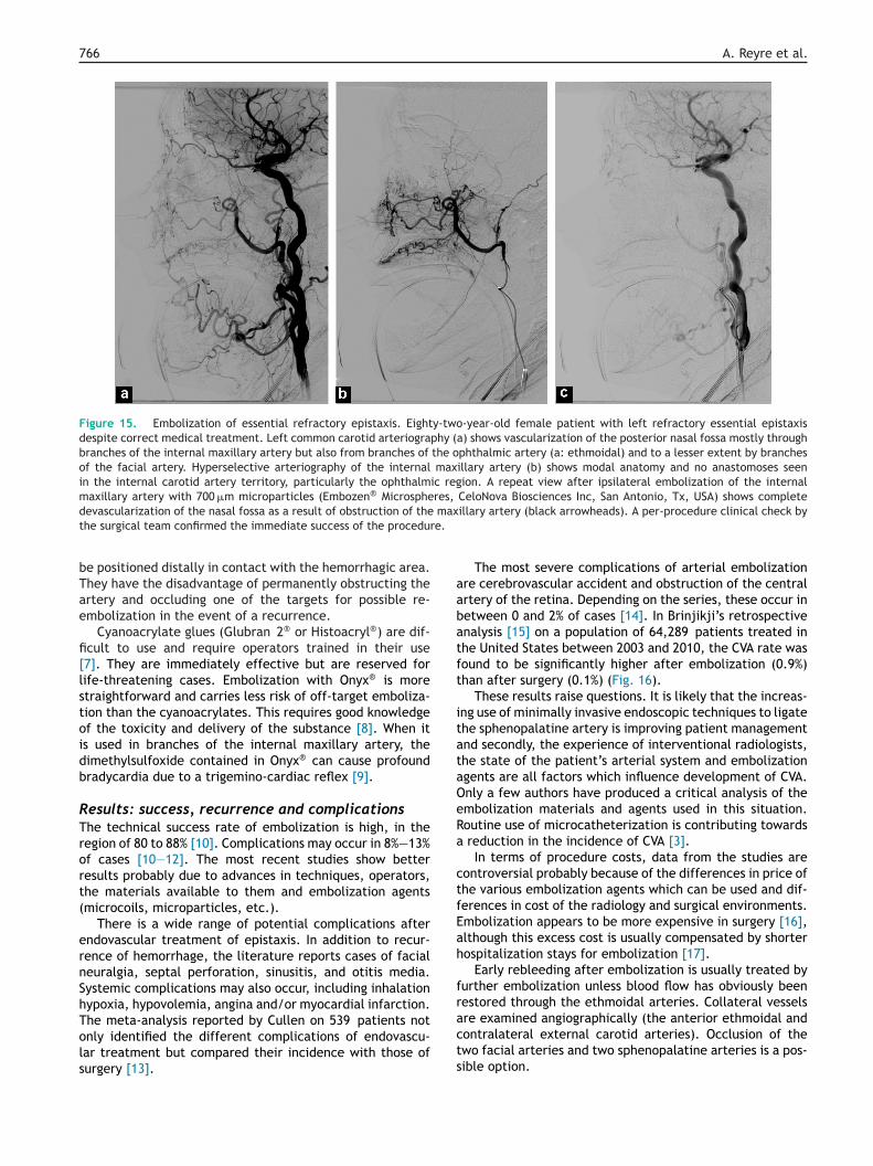
Figure 15. Embolization of essential refractory epistaxis. Eighty-two-year-old female patient with left refractory essential epistaxisdespite correct medical treatment. Left common carotid arteriography (a) shows vascularization of the posterior nasal fossa mostly throughbranches of the internal maxillary artery but also from branches of the ophthalmic artery (a: ethmoidal) and to a lesser extent by branchesof the facial artery. Hyperselective arteriography of the internal maxillary artery (b) shows modal anatomy and no anastomoses seenin the internal carotid artery territory, particularly the ophthalmic region. A repeat view after ipsilateral embolization of the internalmaxillary artery with 700 �m microparticles (Embozen® Microspheres, CeloNova Biosciences Inc, San Antonio, Tx, USA) shows completed maxt re.
bTae
fi[lstoidb
RTrort(
ernShTols
aabatft
itataOeRa
ctfEah
fr
evascularization of the nasal fossa as a result of obstruction of thehe surgical team confirmed the immediate success of the procedu
e positioned distally in contact with the hemorrhagic area.hey have the disadvantage of permanently obstructing thertery and occluding one of the targets for possible re-mbolization in the event of a recurrence.
Cyanoacrylate glues (Glubran 2® or Histoacryl®) are dif-cult to use and require operators trained in their use7]. They are immediately effective but are reserved forife-threatening cases. Embolization with Onyx® is moretraightforward and carries less risk of off-target emboliza-ion than the cyanoacrylates. This requires good knowledgef the toxicity and delivery of the substance [8]. When its used in branches of the internal maxillary artery, theimethylsulfoxide contained in Onyx® can cause profoundradycardia due to a trigemino-cardiac reflex [9].
esults: success, recurrence and complicationshe technical success rate of embolization is high, in theegion of 80 to 88% [10]. Complications may occur in 8%—13%f cases [10—12]. The most recent studies show betteresults probably due to advances in techniques, operators,he materials available to them and embolization agentsmicrocoils, microparticles, etc.).
There is a wide range of potential complications afterndovascular treatment of epistaxis. In addition to recur-ence of hemorrhage, the literature reports cases of facialeuralgia, septal perforation, sinusitis, and otitis media.ystemic complications may also occur, including inhalationypoxia, hypovolemia, angina and/or myocardial infarction.
he meta-analysis reported by Cullen on 539 patients notnly identified the different complications of endovascu-ar treatment but compared their incidence with those ofurgery [13].acts
illary artery (black arrowheads). A per-procedure clinical check by
The most severe complications of arterial embolizationre cerebrovascular accident and obstruction of the centralrtery of the retina. Depending on the series, these occur inetween 0 and 2% of cases [14]. In Brinjikji’s retrospectivenalysis [15] on a population of 64,289 patients treated inhe United States between 2003 and 2010, the CVA rate wasound to be significantly higher after embolization (0.9%)han after surgery (0.1%) (Fig. 16).
These results raise questions. It is likely that the increas-ng use of minimally invasive endoscopic techniques to ligatehe sphenopalatine artery is improving patient managementnd secondly, the experience of interventional radiologists,he state of the patient’s arterial system and embolizationgents are all factors which influence development of CVA.nly a few authors have produced a critical analysis of thembolization materials and agents used in this situation.outine use of microcatheterization is contributing towards
reduction in the incidence of CVA [3].In terms of procedure costs, data from the studies are
ontroversial probably because of the differences in price ofhe various embolization agents which can be used and dif-erences in cost of the radiology and surgical environments.mbolization appears to be more expensive in surgery [16],lthough this excess cost is usually compensated by shorterospitalization stays for embolization [17].
Early rebleeding after embolization is usually treated byurther embolization unless blood flow has obviously beenestored through the ethmoidal arteries. Collateral vessels
re examined angiographically (the anterior ethmoidal andontralateral external carotid arteries). Occlusion of thewo facial arteries and two sphenopalatine arteries is a pos-ible option.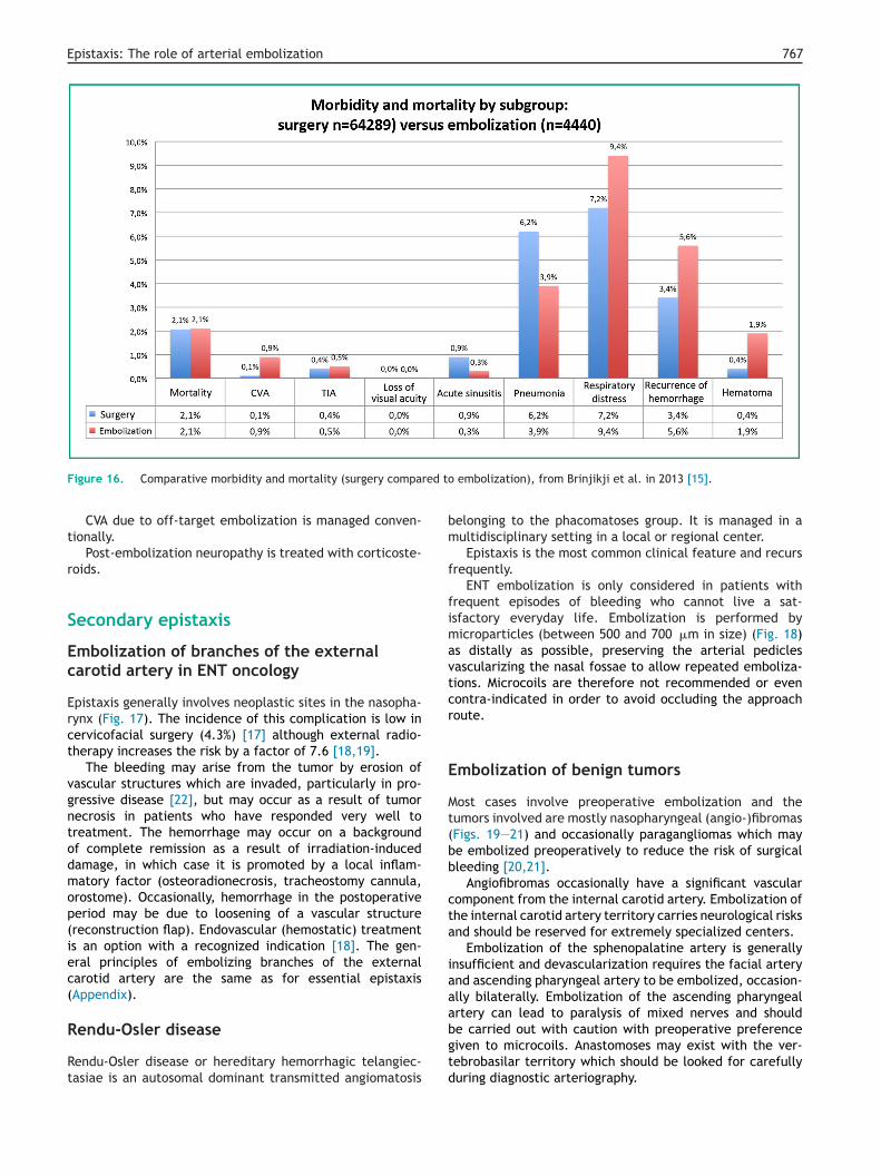
Epistaxis: The role of arterial embolization 767
ed to
bm
f
fimavtcr
E
Mt(bb
cta
iaaartery can lead to paralysis of mixed nerves and should
Figure 16. Comparative morbidity and mortality (surgery compar
CVA due to off-target embolization is managed conven-tionally.
Post-embolization neuropathy is treated with corticoste-roids.
Secondary epistaxis
Embolization of branches of the externalcarotid artery in ENT oncology
Epistaxis generally involves neoplastic sites in the nasopha-rynx (Fig. 17). The incidence of this complication is low incervicofacial surgery (4.3%) [17] although external radio-therapy increases the risk by a factor of 7.6 [18,19].
The bleeding may arise from the tumor by erosion ofvascular structures which are invaded, particularly in pro-gressive disease [22], but may occur as a result of tumornecrosis in patients who have responded very well totreatment. The hemorrhage may occur on a backgroundof complete remission as a result of irradiation-induceddamage, in which case it is promoted by a local inflam-matory factor (osteoradionecrosis, tracheostomy cannula,orostome). Occasionally, hemorrhage in the postoperativeperiod may be due to loosening of a vascular structure(reconstruction flap). Endovascular (hemostatic) treatmentis an option with a recognized indication [18]. The gen-eral principles of embolizing branches of the externalcarotid artery are the same as for essential epistaxis(Appendix).
Rendu-Osler disease
Rendu-Osler disease or hereditary hemorrhagic telangiec-tasiae is an autosomal dominant transmitted angiomatosis
bgtd
embolization), from Brinjikji et al. in 2013 [15].
elonging to the phacomatoses group. It is managed in aultidisciplinary setting in a local or regional center.Epistaxis is the most common clinical feature and recurs
requently.ENT embolization is only considered in patients with
requent episodes of bleeding who cannot live a sat-sfactory everyday life. Embolization is performed byicroparticles (between 500 and 700 �m in size) (Fig. 18)
s distally as possible, preserving the arterial pediclesascularizing the nasal fossae to allow repeated emboliza-ions. Microcoils are therefore not recommended or evenontra-indicated in order to avoid occluding the approachoute.
mbolization of benign tumors
ost cases involve preoperative embolization and theumors involved are mostly nasopharyngeal (angio-)fibromasFigs. 19—21) and occasionally paragangliomas which maye embolized preoperatively to reduce the risk of surgicalleeding [20,21].
Angiofibromas occasionally have a significant vascularomponent from the internal carotid artery. Embolization ofhe internal carotid artery territory carries neurological risksnd should be reserved for extremely specialized centers.
Embolization of the sphenopalatine artery is generallynsufficient and devascularization requires the facial arterynd ascending pharyngeal artery to be embolized, occasion-lly bilaterally. Embolization of the ascending pharyngeal
e carried out with caution with preoperative preferenceiven to microcoils. Anastomoses may exist with the ver-ebrobasilar territory which should be looked for carefullyuring diagnostic arteriography.

768 A. Reyre et al.
Figure 17. Epistaxis from a round, median nasopharyngeal tissue lesion vascularized on CT (a: arrowheads) seen as an isointense appear-ance on T2-weighted imaging (b: arrowheads), with reduced diffusion (ADC = 0.5 × 10⁻³ mm2/sec, c: arrowheads) and vivid enhancementafter IV of gadolinium chelate on T1-weighted image with fat saturation (d: arrowheads). Hyperselective catheterization of the anteriortrunk of the ascending pharyngeal artery (e: arrows) shows a tumor blush (e: arrowheads). Devascularization was achieved by insertingcontrolled release microcoils (f: arrow) showing devascularization of the tumor process in a later view (f: arrowheads). After embolization,biopsy showed a plasmacytoma allowing this to be managed specifically.
Figure 18. Embolization in Rendu-Osler disease. External carotid artery arteriography before embolization (a) showing distal hypervas-cularization of the nasal fossa by branches of the sphenopalatine artery (a: black arrows) and by branches of the facial artery (a: whitearrows). A repeat view after embolization (b) carried out using 700 �m microparticles shows satisfactory occlusion of the sphenopalatineaT
nd facial arteries. Note a symptomatic refeeding of the nasal fossa vascelangiectasiae are also present in the lingual artery (b: black arrow).ularization through the anterior ethmoidal artery (b: white arrow).

Epistaxis: The role of arterial embolization 769
Figure 19. Juvenile angiofibroma. T2-weighted axial (a), T1-weighted axial (b), T1-weighted after IV of gadolinium chelate and fatsaturation (c), ADC mapping (d), T2-weighted coronal (e) and T1-weighted 3D with gadolinium enhancement and fat saturation (f). A mass ispresent in the right nasal fossa (a—d: arrows) exhibiting a few ‘‘flow voids’’ on T2-weighted imaging (a: arrowheads) and vivid enhancementafter IV of gadolinium chelate (b, c), with no reduction in ADC values (1.4 × 10⁻³ mm2/sec, e: arrows) entering the sphenopalatine foramen(e, f: arrows).
Figure 20. Juvenile angiofibroma. 3D dynamic gadolinium-chelate enhanced MR angiography every 4 seconds in a sagittal plane (a—f)ptake
showing the hypervascular nature of the lesion (a—f: arrowheads), uarteries (b: arrows).
in which is almost synchronous with the basilar trunk and cerebral
770 A. Reyre et al.
Figure 21. Preoperative embolization of a nasopharyngeal fibroma. Nineteen-year-old male patient with a Radkowski classification type1B nasopharyngeal fibroma. Diagnostic angiography of the external carotid artery (reflux into the internal, a) shows a tumor ‘‘blush’’ (a:arrowheads) from the sphenopalatine artery (a: arrows). Devascularization was achieved by hyperselective catheterization and embolizationo mer,
e izatio
C
Emtpoc
C
Avtam
Ddci
Q
12A
B
f the arterial trunk with Onyx® 34 (ethylene vinyl alcohol copolypisodes of bradycardia. A repeat view shows complete devascular
onclusion
mbolization now currently offers huge benefit in theanagement of epistaxis. Its results are improved by mul-
idisciplinary collaboration (surgeons and intensive carehysicians), by strictly selecting its indications, knowledgef the vascular anatomy and mastering the process of micro-atheterization and the embolization agents.
Take-home messages• there are two types of epistaxis:
◦ essential,◦ symptomatic, secondary to underlying disease;
• arterial embolization is a treatment of choice forsevere refractory and symptomatic epistaxis;
• full knowledge of the anatomy of the branches of theexternal carotid artery is an essential requirementfor embolization. Its main indications are:◦ essential epistaxis which is serious from the outset
and life threatening or refractory after 48 hours ofcorrect medical treatment,
◦ severe epistaxis in ORL oncology either throughinvasion or postoperatively,
◦ preoperative devascularization of hypervasculartumors (paragangliomas and nasopharyngealfibromas),
◦ frequent episodes of epistaxis in Rendu-Oslerdisease;
• the treatment of benign essential epistaxis is initiallylocal and then ‘‘crescendo’’ escalation;
• hemorrhages from the anterior ethmoidal arteryrequire surgical treatment;
• the occlusion materials vary depending on the typeof epistaxis:◦ microparticles (from 500 to 700 microns) for
essential epistaxis or Rendu-Osler disease,◦ microcoils or glues, particularly in secondary
epistaxis or with hazardous anastomoses,◦ microcoils are not recommended in Rendu-Osler
disease;C
EV3 Micro Therapeutics Inc., Irvine, CA) (b: arrowheads), with non of the mass (c: arrowheads).
• microparticles can only be used when no hazardousanastomoses with the internal carotid artery orophthalmic artery territory are present and requirefree flow injection under radioscopy guidance;
• positioning microcoils may be a good alternative.These have the disadvantage of permanentlyobstructing the artery (re-embolization is impossiblein the event of recurrence);
• amongst the complications of arterial embolization,cerebrovascular accident and central retinal arteryobstruction are the most severe;
• the complication rate in a trained operator’s handsis no greater than that of surgery.
linical case
70-year-old man presents with recurrent low to moderateolume epistaxis. He has a past history of T2N0M0 conven-ional cell renal carcinoma, and a coronary stent in his leftnterior descending artery. Clinical examination reveals aass in the right nasal fossa.An MRI is performed including: T2-weighted images (A),
iffusion-weighted MR imaging (B), T1-weighted images (C),ynamic MR angiography (D), a time-intensity (infusion)urve (E) and T1-weighted gadolinium-chelate enhancedmage (F) (Fig. 22).
uestions
. Describe the abnormalities present on MR images.
. What are the correct answers?
. The epistaxis is secondary to a right nasal fossa tissuelesion.
. The hypervascular appearances with lavage (E) andthe high apparent diffusion coefficient of the lesion
−3 2
(1.8 × 10 mm /s; B) are both in favor of a hematoge-nous metastasis from a renal cancer.. The patient’s age and lack of invasion of the pterygopala-tine groove do not support an angiofibroma.
Epistaxis: The role of arterial embolization 771
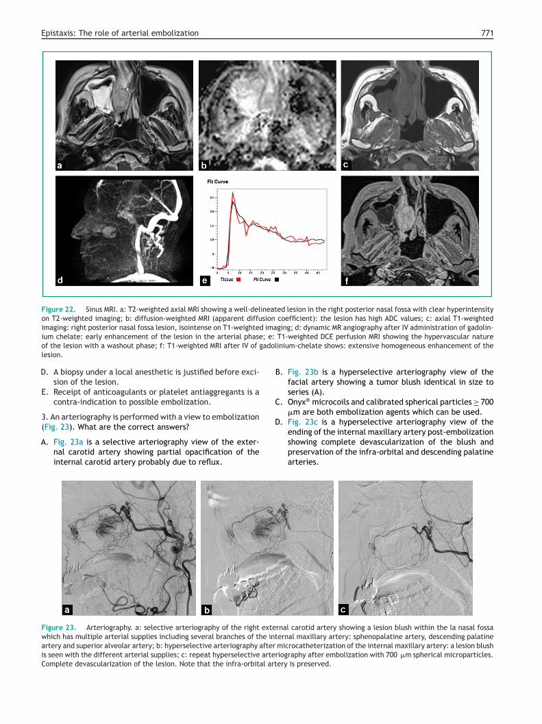
Figure 22. Sinus MRI. a: T2-weighted axial MRI showing a well-delineated lesion in the right posterior nasal fossa with clear hyperintensityon T2-weighted imaging; b: diffusion-weighted MRI (apparent diffusion coefficient): the lesion has high ADC values; c: axial T1-weightedimaging: right posterior nasal fossa lesion, isointense on T1-weighted imaging; d: dynamic MR angiography after IV administration of gadolin-
; e:
adol
D
A
B
C
D
ium chelate: early enhancement of the lesion in the arterial phaseof the lesion with a washout phase; f: T1-weighted MRI after IV of glesion.
. A biopsy under a local anesthetic is justified before exci-sion of the lesion.
E. Receipt of anticoagulants or platelet antiaggregants is acontra-indication to possible embolization.
3. An arteriography is performed with a view to embolization(Fig. 23). What are the correct answers?
. Fig. 23a is a selective arteriography view of the exter-nal carotid artery showing partial opacification of theinternal carotid artery probably due to reflux.
Figure 23. Arteriography. a: selective arteriography of the right extewhich has multiple arterial supplies including several branches of the intartery and superior alveolar artery; b: hyperselective arteriography after
is seen with the different arterial supplies; c: repeat hyperselective arterComplete devascularization of the lesion. Note that the infra-orbital art
T1-weighted DCE perfusion MRI showing the hypervascular natureinium-chelate shows: extensive homogeneous enhancement of the
. Fig. 23b is a hyperselective arteriography view of thefacial artery showing a tumor blush identical in size toseries (A).
. Onyx® microcoils and calibrated spherical particles ≥ 700�m are both embolization agents which can be used.
. Fig. 23c is a hyperselective arteriography view of the
ending of the internal maxillary artery post-embolizationshowing complete devascularization of the blush andpreservation of the infra-orbital and descending palatinearteries.rnal carotid artery showing a lesion blush within the la nasal fossaernal maxillary artery: sphenopalatine artery, descending palatinemicrocatheterization of the internal maxillary artery: a lesion blushiography after embolization with 700 �m spherical microparticles.ery is preserved.

7
E
l
c
r
c
o
a
s
eab
um7tbw
A
TtSCJG(t
D
Tc
At
E
MS
I
•
•
A
ILb
CAvW
HiEsp
I
Pr
s
eh
O
6ig(
ewoacb
I
••
••
72
. At this stage in the procedure (C), the lesion may carrya risk of revascularization from the contralateral orintracranial arterial system.
Answers1. A: MRI T2-weighted axial section: well-circumscribed
esion in the right posterior fossa with hypersignal.B: MRI diffusion-weighted sequence (apparent diffusion
oefficient): this lesion shows high ADC values.C: MRI T1-weighted axial section: isointense lesion of the
ight posterior fossa.D: dynamic MR angiography after IV of gadolinium-
helate: early lesion enhancement in the arterial phase.E: MRI perfusion sequence T1 DCE: hypervascular nature
f the lesion with washing stage.F: T1-weighted MRI after IV of gadolinium-chelate: large
nd homogeneous enhancement of the lesion.2. Correct answers: A, C. Comments:B: the high ADC suggests a benign lesion.D: a biopsy is contra-indicated in hypervascular nasal
inus lesions.E: embolization is particularly justified prior to surgical
xcision if the patient cannot avoid taking an anticoagulantnd/or platelet antiaggregant in order to reduce the risks ofleeding.
3. Correct answers: A, C, D, E. Comments:B: internal maxillary artery (ending). This patient
nderwent distal embolization with calibrated sphericalicroparticles ≥ 700 �m in size (Embozen® Mircrosphere
00 �m CeloNova©, C) followed by occlusion of the trunk ofhe internal maxillary artery with microcoils. No significantleeding occurred on endonasal excision. The final diagnosisas a benign angiomatous nasal fossa polyp.
cknowledgements
hanks to Alexis Jacquier (University Professor and Hospi-al Consultant), Hervé Brunel (Hospital Consultant), Jéromeoussan (Hospital Consultant), Aurélie Dehaene (Hospitalonsultant), Antonin Flavian (Senior Clinical Registrar),ean-Marie Caporossi (Senior Clinical Registrar), Chloéaudon (Senior Clinical Registrar), and to Jean Izaaryene
Senior Clinical Registrar) for their clinical and imaging con-ributions.
isclosure of interest
he authors declare that they have no conflicts of interestoncerning this article.
ppendix A. Appendix Practical guide to
he endovascular treatment of epistaxisnvironment
ultidisciplinary group in a radiology suite: ENTurgeon—Anesthetist—Interventional Radiologist.
‘
Ff
A. Reyre et al.
ndication
Posterior epistaxis, refractory after 48 hours of correctmedical treatmentSerious epistaxis, life threatening from the outset.
ssessment of patient status
n all casesaboratory assessment: PR APTT INR, CBC, platelets andlood group.
ontrolled hemorrhagessessment of cause and feasibility assessment: Supra-aorticessel CT angiography from the aortic arch to the circle ofillis and facial bone CT.
emodynamic stability not controlled byntensive careither endovascular treatment with embolization or surgeryhould be discussed with the ENT surgeons and intensive carehysicians.
n the operating suite
atient placed lying down with a head rest, sedated,adioscopy in position, infusion, informed and ‘‘reassured’’.
One operator, one operator’s assistant and an operatinguite technician.
Anesthetist/Intensive care physician for station and gen-ral anesthesia if required, monitoring and correctingemodynamic indices.
n the angiography table
F valve introducer. Consider 35 cm long introducer depend-ng on morphology of the iliofemoral arterial system. 6Fuide catheter (Envoy®, 100 cm) on 0.035′’ hydrophilic guideTerumo®) and antireflux valve.
Microcatheter (at least 135 cm long) suitable for thembolization agents (compatible with Onyx® if necessary)ith a straight or preshaped (45◦ or 90◦) tip and two radio-paque markers if controlled release coils are used. 45◦
nd 90◦ curved hydrophilic microguides. The introducers,atheters and microcatheters should be rinsed and infusedy bags and blood pressure.
n the angiography suite trolley
Microcoils suitable for the microcatheter.Calibrated microparticles (Luer-lock syringe prefilled to500—700 �m).Cyanoacrylates, (Histoacryle® or Glubran2®).Ethylene vinyl alcohol copolymer (Onyx®), rarely used inemergencies.
‘Conventional’’ procedure
emoral artery approach, valve introducer rinsed and per-used, guide catheter rinsed and perfused.

[
[
[
[
[
[
[
[
[
[
[
[
[
Epistaxis: The role of arterial embolization
• Injection into the common carotid artery after selectivecatheterization of the bleeding side with long series tostudy jugular venous return.
• Selective injection of the internal carotid artery withanteroposterior and lateral views (to study the ophthalmicartery and anterior ethmoidal artery).
• Selective injection of the external carotid artery, postero-anterior and lateral views including the nasal fossae in thefield.
• Investigation for the target and identifying hazardousanastomoses: foramen lacerum artery, clival branches,pterygoid artery, foramen ovalis artery, foramen rotun-dum artery and in the ophthalmic artery.
• Use of a microguide and microcatheter couple from thestart leaving a guide catheter at the origin of the externalcarotid artery. Hyperselective microcatheterization of thetarget (failing this the sphenopalatine artery).
• Use of arterial tracing. Do not catheterize the branches ofthe external carotid artery with the 0.035 guide to avoidspasm.
If hazardous anastomoses are present, place the micro-catheter beyond the anastomosis but always consider therisk of reflux during embolization. If necessary, carry outproximal embolization (of the trunk) of the hazardous anas-tomosis using microcoils before the microparticles.
In the event of arterial spasm, to enable free flowembolization, use an in situ intra-arterial perfusion ofnitrates (1 mg of Risordan®) with the agreement of theanesthetist-intensive care physician.
Embolization always free flow as close as possible to thetarget and a radioscopy control.
Angiography images are taken routinely at the end ofthe procedure to investigate for collateral circulation andrevascularization of the target. If the sphenopalatine arteryis revascularized from the facial artery, the facial arteryshould be embolized with very distal microspheres or micro-coils.
The final check investigates for revascularization of thenasal fossa particularly by the ethmoidal arteries.
Leave the femoral introducer in situ until hemodynamicand neurological control has been confirmed clinically by theORL team in the angiography suite.
References
[1] Small M, Murray JA, Maran AG. A study of patients with epis-taxis requiring admission to hospital Health Bull (Raleigh)1982;40(1):20—9.
[2] Leppänen M, Seppänen S, Laranne J, Kuoppala K. Microcatheterembolization of intractable idiopathic epistaxis. CardiovascIntervent Radiol 1999;22(6):499—503.
[3] Elden L, Montanera W, Terbrugge K, Willinsky R, LasjauniasP, Charles D. Angiographic embolization for the treatment ofepistaxis: a review of 108 cases. Otolaryngol Head Neck Surg1994;111(1):44—50.
[4] Cockroft KM, Carew JF, Trost D, Fraser RR, Chandler WF.Delayed epistaxis resulting from external carotid artery injuryrequiring embolization: a rare complication of transsphenoidalsurgery: case report. Neurosurgery 2000;47(1):236—9.
[
773
[5] Begbie ME, Wallace GMF, Shovlin CL. Hereditary haemorrhagictelangiectasia (Osler-Weber-Rendu syndrome): a view from the21st century. Postgrad Med J 2003;79(927):18—24.
[6] Gierthmuehlen M, Schumacher M, Zentner J, Hader C. Brain-stem compression caused by bilateral traumatic carotid cav-ernous fistulas: case report. Neurosurgery 2010;67(4):1160—4.
[7] Pollak JS, White RI. The use of cyanoacrylate adhe-sives in peripheral embolization. J Vasc Interv Radiol2001;12(8):907—13.
[8] Elhammady MS, Wolfe SQ, Farhat H, Moftakhar R, Aziz-SultanMA. Onyx embolization of carotid-cavernous fistulas. J Neuro-surg 2010:589—94.
[9] Puri AS, Thiex R, Zarzour H, Rahbar R, Orbach DB.Trigeminocardiac reflex in a child during pre-Onyx DMSO injec-tion for juvenile nasopharyngeal angiofibroma embolization.A case report. Interv Neuroradiol [Internet] 2011;17(1):13—6http://www.pubmedcentral.nih.gov/articlerender.fcgi?artid=3278028&tool=pmcentrez&rendertype=Abstract
10] Christensen NP, Smith DS, Barnwell SL, Wax MK. Arte-rial embolization in the management of posterior epistaxis.Otolaryngol Head Neck Surg 2005;133(5):748—53.
11] Moreau S, De Rugy MG, Babin E, Courtheoux P, Valdazo. Supras-elective embolization in intractable epistaxis: review of 45cases. Laryngoscope 1998;108(6):887—8.
12] Tseng EY, Narducci C, Willing SJ, Sillers MJ. Angiographicembolization for epistaxis: a review of 114 cases. Laryngoscope1998;108(4 Pt 1):615—9.
13] Cullen MM, Tami T. Comparison of internal maxillary arteryligation versus embolization for refractory posterior epistaxis.Otolaryngol Head Neck Surg 1998;118(5):636—42.
14] Mames RN, Snady-McCoy L, Guy J. Central retinal and pos-terior ciliary artery occlusion after particle embolization ofthe external carotid artery system. Ophthalmology 1991:527—31.
15] Brinjikji W, Kallmes DF, Cloft HJ. Trends in epistaxis emboliza-tion in the United States: a study of the nationwideinpatient sample 2003—2010. J Vasc Interv Radiol 2013;24(7):969—73.
16] Klotz DA, Winkle MR, Richmon J, Hengerer AS. Surgicalmanagement of posterior epistaxis: a changing paradigm.Laryngoscope 2002;112(9):1577—82.
17] Strong EB, Bell DA, Johnson LP, Jacobs JM. Intractable epis-taxis: transantral ligation vs. embolization: efficacy review andcost analysis. Otolaryngol Head Neck Surg 1995;113(6):674—8.
18] Bates MC, Shamsham FM. Endovascular management ofimpending carotid rupture in a patient with advanced headand neck cancer. J Endovasc Ther 2003;10(1):54—7.
19] Chen Y-F, Lo Y-C, Lin W-C, Lin C-H, Chiang H-J, Chen J-F,et al. Transarterial embolization for control of bleeding inpatients with head and neck cancer. Otolaryngol Head NeckSurg 2010;142(1):90—4.
20] Pyun HW, Lee DH, Yoo HM, Lee JH, Choi CG, Kim SJ, et al. Place-ment of covered stents for carotid blowout in patients withhead and neck cancer: follow-up results after rescue treat-ments. AJNR Am J Neuroradiol 2007;28(8):1594—8.
21] Chang F-C, Lirng J-F, Luo C-B, Wang S-J, Wu H-M, Guo W-Y,et al. Patients with head and neck cancers and associatedpost-irradiated carotid blowout syndrome: endovascular thera-peutic methods and outcomes. J Vasc Surg 2008;47(5):936—45.
22] Bhansali S, Wilner H, Jacobs JR. Arterial embolization forcontrol of bleeding in advanced head and neck carcinoma. J
Laryngol Otol 1986:1289—93.23] Lasjaunias P, Moret J. The ascending pharyngeal artery:normal and pathological radioanatomy. Neuroradiology1976;11(2):77—82.