Embed Size (px)
Citation preview

Epidermal Growth Factor Receptor (EGFR) SignalingRegulates Global Metabolic Pathways in EGFR-mutated LungAdenocarcinoma*□S
Received for publication, April 21, 2014, and in revised form, June 9, 2014 Published, JBC Papers in Press, June 13, 2014, DOI 10.1074/jbc.M114.575464
Hideki Makinoshima‡1, Masahiro Takita‡§, Shingo Matsumoto‡¶, Atsushi Yagishita‡, Satoshi Owada�,Hiroyasu Esumi�, and Katsuya Tsuchihara‡§
From the ‡Division of Translational Research, Exploratory Oncology Research & Clinical Trial Center, National Cancer Center,Kashiwa, Chiba 277-8577, Japan, §Department of Integrated Biosciences, Graduate School of Frontier Sciences, The University ofTokyo, Kashiwa, Chiba 277-8561, Japan, ¶Thoracic Oncology Division, National Cancer Center Hospital East, Kashiwa,Chiba 277-8577, Japan, and �Research Institute for Biomedical Sciences, Tokyo University of Science, Noda, Chiba 278-0022, Japan
Background: Genetic mutations in cancer-driver genes induce specific metabolic alterations in cancer cells.Results: EGF receptor signaling has an important role for glycolysis, pentose phosphate pathway, and pyrimidine biosynthesisin EGFR-mutated lung cancer.Conclusion: Our work reveals the relationship between the EGFR signaling axis and key metabolic changes.Significance: These data implicate a possible link between therapeutic response and regulation of metabolism in EGFR-mutated LAD.
Genetic mutations in tumor cells cause several unique meta-bolic phenotypes that are critical for cancer cell proliferation.Mutations in the tyrosine kinase epidermal growth factor recep-tor (EGFR) induce oncogenic addiction in lung adenocarcinoma(LAD). However, the linkage between oncogenic mutated EGFRand cancer cell metabolism has not yet been clearly elucidated.Here we show that EGFR signaling plays an important role inaerobic glycolysis in EGFR-mutated LAD cells. EGFR-tyrosinekinase inhibitors (TKIs) decreased lactate production, glucoseconsumption, and the glucose-induced extracellular acidifica-tion rate (ECAR), indicating that EGFR signaling maintainedaerobic glycolysis in LAD cells. Metabolomic analysis revealedthat metabolites in the glycolysis, pentose phosphate pathway(PPP), pyrimidine biosynthesis, and redox metabolism were sig-nificantly decreased after treatment of LAD cells with EGFR-TKI. On a molecular basis, the glucose transport carried out byglucose transporter 3 (GLUT3) was downregulated in TKI-sen-sitive LAD cells. Moreover, EGFR signaling activated carba-moyl-phosphate synthetase 2, aspartate transcarbamylase, anddihydroorotase (CAD), which catalyzes the first step in de novopyrimidine synthesis. We conclude that EGFR signaling regu-lates the global metabolic pathway in EGFR-mutated LAD cells.Our data provide evidence that may link therapeutic response tothe regulation of metabolism, which is an attractive target forthe development of more effective targeted therapies to treatpatients with EGFR-mutated LAD.
The discovery of oncogenic driver mutations allows us toidentify druggable targets and develop new therapies usingsmall molecule tyrosine kinase inhibitors (TKIs)2 aimed at therelevant patient populations (1–3). More than 50% of lung ade-nocarcinomas (LAD) from East Asian non-smokers harborEGFR mutations, and these tumors have been termed oncogeneaddicted to reflect their dependence on EGFR-mediated pro-survival signaling and their high susceptibility to apoptosisinduced by EGFR-TKIs (e.g. gefitinib and erlotinib) (4 –7). Thetyrosine kinase activity of EGFR is dysregulated by gene muta-tions that lead to aberrant EGFR signaling through pathwaysincluding the RAS/MAPK and PI3K/AKT pathways (8, 9). Themost frequently occurring mutations in the EGFR gene (in-frame deletion in exon 19 at codons 746 –750 or a single-basesubstitution L858R in exon 21) predict an improved clinicalresponse to first-line oral EGFR-TKIs compared with standardplatinum-based chemotherapy in patients with advanced non-small-cell lung carcinoma (NSCLC) (4, 8).
There is accumulating evidence that genetic mutations incancer-driver genes, tumor suppressors, and amplified onco-genes are linked to specific alterations in metabolic activity incancer cells, involving proteins such as isocitrate dehydroge-nase (IDH), fumarate hydratase (FH), MYC, K-RAS, and BRAF(10 –13). The Warburg effect, the phenomenon in which cancercells exhibit rapid glucose consumption with secretion of lac-tate despite abundant oxygen availability, has been recognizedsince the 1930s (14 –16). Indeed, glucose metabolism in cancercells is tightly regulated by many molecules at the transcrip-tional, translational, and post-translational levels (10, 17, 18).c-MYC is critically involved in the regulation of many growth-* This work was supported by the National Cancer Center Research and
Development Fund (25-A-6) and JSPS KAKENHI Grant Number 24300345(to K. T.).
□S This article contains supplemental Tables S1 and S2.Author’s Choice—Final version full access.
1 To whom correspondence should be addressed: Division of TranslationalResearch, Exploratory Oncology Research & Clinical Trial Center, NationalCancer Center, Kashiwa, Chiba 277-8577, Japan. Tel.: 81-4-7134-6855; Fax:81-4-7134-6865; E-mail: [email protected].
2 The abbreviations used are: TKI, tyrosine kinase inhibitor; EGFR, epidermalgrowth factor receptor; LAD, lung adenocarcinoma; IC50, half maximalinhibitory concentration; PPP, pentose phosphate pathway; ECAR, extra-cellular acidification rate; OCR, oxygen consumption rate; GLUT, glucosetransporter; CAD, carbamoyl-phosphate synthetase 2, aspartate transcar-bamylase, and dihydroorotase.
THE JOURNAL OF BIOLOGICAL CHEMISTRY VOL. 289, NO. 30, pp. 20813–20823, July 25, 2014Author’s Choice © 2014 by The American Society for Biochemistry and Molecular Biology, Inc. Published in the U.S.A.
JULY 25, 2014 • VOLUME 289 • NUMBER 30 JOURNAL OF BIOLOGICAL CHEMISTRY 20813
by guest on April 19, 2019
http://ww
w.jbc.org/
Dow
nloaded from
promoting signal transduction pathways and glucose metabo-lism genes, including GLUT1, hexokinase 2 (HK2), pyruvatekinase muscle (PKM2), and lactate dehydrogenase A (LDHA)(10, 19). Through the up-regulation of these genes, c-MYC con-tributes directly to the Warburg effect (19). The enzymaticactivities of glycolytic enzymes such as HK2, phosphofructoki-nase (PFK), PKM2, and LDHA are modulated by post-transla-tional modification (18). For example, PKM2 is phosphorylatedin its tyrosine residue (Y105) with low activity in human cancercells, resulting in increased lactate production, which is one-step downstream from PKM2 in glycolysis, even under aerobicconditions (14, 17). Furthermore, PKM2 promotes the War-burg effect through EGF-stimulated EGFR activation and theMAPK signaling pathway (20, 21). In brain cancer, the activat-ing EGFRvIII mutation induces enhanced glycolysis by promot-ing glycolytic gene expression through the Myc/Max pathway(22). However, the specific role of mutated EGFR for aerobicglycolysis in lung cancer has not yet been clearly described.
In this work, we demonstrate that EGFR signaling is requiredfor lactate production under aerobic growth conditions in LADcells. EGFR signaling maintains key metabolites in glycolysisand PPP by regulating glucose transport through GLUT3expression. In addition to glucose metabolism, we show thatEGFR signaling up-regulates de novo pyrimidine biosynthesis.Moreover, we describe the altered metabolic profiles in TKI-sensitive LAD cells in response to erlotinib. Our results implythat EGFR signaling plays a central role in modulating globalmetabolic pathways in EGFR-mutated LAD.
EXPERIMENTAL PROCEDURES
Materials—Cell lines were purchased from the Immuno-Bi-ological Laboratories (Fujioka, Japan) and American Type Cul-ture Collection (ATCC). RPMI 1640 (R8758 and R1383), phos-phate-buffered saline (PBS), 2-deoxy-D-glucose (2DG) werepurchased from Sigma-Aldrich. Fetal bovine serum (FBS) waspurchased from Biowest (Nuaille, France). Dimethyl sulfoxide(DMSO) and glucose were purchased from Wako Pure Chem-icals Industries (Osaka, Japan). Gefitinib and erlotinib werepurchased from Santa Cruz Biotechnology (Dallas, TX). CellCounting Kit-8 was purchased from Dojindo Laboratories(Kumamoto, Japan). Lactate assay kit II and glucose assay kit IIwere purchased from BioVision (Milpitas). FluxPak XF24 assaypack and XF glycolysis stress test kit were purchased from Sea-horse Bioscience (North Billerica). Countess Automated CellCounter including Trypan Blue and chamber slides was pur-chased from Invitrogen (Carlsbad, CA). Primary antibodiesspecific for EGFR, phospho-EGFR Tyr-1068, AKT, phospho-AKT Ser473, ERK1/2, phospho-ERK1/2 Thr202/Tyr204,GSK3�/�, phospho-GSK3�/� Ser21/9, c-MYC, PKM2, phos-pho-PKM2 Tyr105, GYS, phospho-GYS Ser641, LDHA, phos-pho-LDHA Tyr-15, HK2, S6K, phospho-S6K Thr421/Ser424,CAD, phospho-CAD (Ser-1859), and �-actin were purchasedfrom Cell Signaling Technologies (Danvers, MA) and GLS,GLUT1, GLUT3, PDHA1, and phospho-PDHA1 Ser-293 fromAbcam (Cambridge, UK), respectively. The peroxidase-linkedsecondary antibodies for WB, HRP-linked Sheep anti-mouseIgG and Donkey anti-rabbit IgG, were purchased from GEHealthcare Biosciences (Pittsburgh, PA). Fluorescein (FITC)-
conjugated goat anti-rabbit IgG for FACS was purchased fromBeckman Coulter (Fullerton, CA). Oligomycin was purchasedfrom Merck Millipore (Darmstadt, Germany). SYBR Premix ExTaq was purchased from TaKaRa Bio (Shiga, Japan). Ribonu-clease A (RNase A) was purchased from Roche Applied Science(Penzberg, Germany) and contaminated DNase was inactivatedat 80 °C for 30 min. 3-O-(3H-methyl)-D-glucose (3-OMeG) waspurchased from Perkin Elmer (Waltham, MA).
Cell Survival Assay and Proliferation Assay—EGFR mutantLAD cells were seeded in RPMI 1640 containing various con-centrations of EGFR inhibitors in 96-well cell culture plates.After 72 h of incubation, cell viability was analyzed using aWST-8 assay using the Cell Counting Kit-8 (Dojindo, Japan).To count the number of viable cells, Trypan Blue-negative cellswere counted using a Countess Automated Cell Counter(Invitrogen).
Lactate and Glucose Assay—Lactate and glucose in culturemedium were measured with the respective lactate assay kit IIand glucose assay kit II according to the manufacturer’s instruc-tions (BioVision, Mountain View, CA). Briefly, after centrifu-gation (3,500 rpm, 15 min, 4 °C), cell culture medium superna-tants were frozen at �20 °C. Samples were later thawed, dilutedin assay buffer, and mixed with lactate or glucose reaction mix-ture for 30 min. The optical density of the mixture in each wellwas read at 450 nm on a microplate reader (Molecular Devices).The lactate concentration was calculated from a standard curveand normalized to cell numbers and culture time. Glucose con-sumption was calculated from a standard curve, subtractingbackground from cell-free medium, and normalizing to cellnumbers and time.
Measurement of ECAR and OCR—ECAR and OCR weremeasured with a XF glycolysis stress test kit according to themanufacturer’s instructions (Seahorse Bioscience). In brief,4.5 � 104 cells were plated onto XF24 plates in RPMI 1640 (10%FBS, 2 mM glutamine) and incubated at 37 °C, 5% CO2 over-night. Cells were washed with assay medium (minus glucoseand unbuffered RPMI 1640 (SIGMA R1383)), replaced withassay medium, and then placed at 37 °C in a CO2-free incubatorfor 30 min. ECAR and OCR were monitored using a SeahorseBioscience XF24 Extracellular Flux Analyzer over time andeach cycle consisted of 3 min mixing, 3 min waiting and 3 minmeasuring. Glucose, oligomycin, and 2DG were diluted intoXF24 media and loaded into the accompanying cartridge toachieve final concentrations of 10 mM, 5 �M, and 100 mM,respectively. Injections of the drugs into the medium occurredat the time points specified.
Western Blotting—Cells were lysed in RIPA buffer (150 mM
NaCl, 1% Triton X-100, 0.5% sodium deoxycholate, 0.1% SDS,50 mM Tris, pH 8.0) on ice for 10 min, sonicated, and centri-fuged at 15,000 � g for 10 min. The protein content of super-natants was measured by BCA assay (Pierce). Identical amountsof protein samples were separated via 4 –20% SDS/PAGE,transferred to PVDF membranes, and incubated overnight withprimary antibodies (1:1000 dilution). The primary antibodiesused in this study are listed in the materials. ECL anti-rabbitIgG HRP-linked whole antibody (1:10,000; GE Healthcare) andECL anti-mouse IgG HRP-linked whole antibody (1:10,000; GEHealthcare) were used as secondary antibodies. Signals were
Regulation of Cancer Metabolism in EGFR-mutated Lung Cancer
20814 JOURNAL OF BIOLOGICAL CHEMISTRY VOLUME 289 • NUMBER 30 • JULY 25, 2014
by guest on April 19, 2019
http://ww
w.jbc.org/
Dow
nloaded from
detected using ECL Western blotting detection reagent (GEHealthcare) and x-ray films (GE Healthcare).
Quantitative RT-PCR—Cells were washed with PBS and totalRNA from the LAD cell lines was isolated with TRIzol Reagent(Invitrogen). Complementary DNA (cDNA) was synthesizedusing the SuperScript VILO cDNA synthesis kit (Invitrogen).Synthesized primers were purchased from TaKaRa Bio (Japan).Real-time RT-PCR was carried out with specific primers and a7500 detection system (Applied Biosystems). �-Actin was usedfor normalization as control and the relative quantitation valuecompared with the calibrator for that target is expressed as2�(Ct�Cc).
Metabolite Measurements—Metabolic extracts were pre-pared from 2–5 � 106 cells with methanol containing InternalStandard Solution (Human Metabolome Technologies; HMT,Inc., Tsuruoka, Japan) and analyzed using a capillary electro-phoresis (CE)-connected ESI-TOFMS and CE-MS/MS system(HMT, CARCINOSCOPE). 2–5 � 106 cells were used for theextraction of intracellular metabolites. Culture medium wasremoved from the dish, and cells were washed twice in 5% man-nitol solution (10 ml first and then 2 ml). Cells were then treatedwith 800 �l of methanol and 550 �l of Milli-Q water containinginternal standards (H3304 –1002, HMT, Inc., Tsuruoka, Japan).
The metabolite extract was transferred into a microfuge tubeand centrifuged at 2,300 � g and 4 °C for 5 min. Next, the upperaqueous layer was centrifugally filtered through a Millipore5-kDa cutoff filter at 9,100 � g and 4 °C for 120 min to removeproteins. The filtrate was centrifugally concentrated and re-suspended in 50 �l of Milli-Q water for CE-MS analysis. Cat-ionic compounds were analyzed in the positive mode of CE-TOFMS and anionic compounds were analyzed in the positiveand negative modes of CE-MS/MS according to the methodsdeveloped by Soga et al. (23–25). To obtain peak informationincluding m/z, migration time (MT), and peak area, detectedpeaks by CE-TOFMS and CE-MS/MS were extracted usingautomatic integration software (MasterHands, Keio University,Tsuruoka, Japan and MassHunter Quantitative AnalysisB.04.00, Agilent Technologies, Santa Clara, CA, respectively).The peaks were annotated with putative metabolites from theHMT metabolite database based on their MTs in CE and m/zvalues determined by TOFMS. The tolerance range for the peakannotation was configured at �0.5 min for MT and �10 ppmfor m/z. In addition, concentrations of metabolites were calcu-lated by normalizing the peak area of each metabolite withrespect to the area of the internal standard and by using stan-dard curves, which were obtained by three-point calibrations.
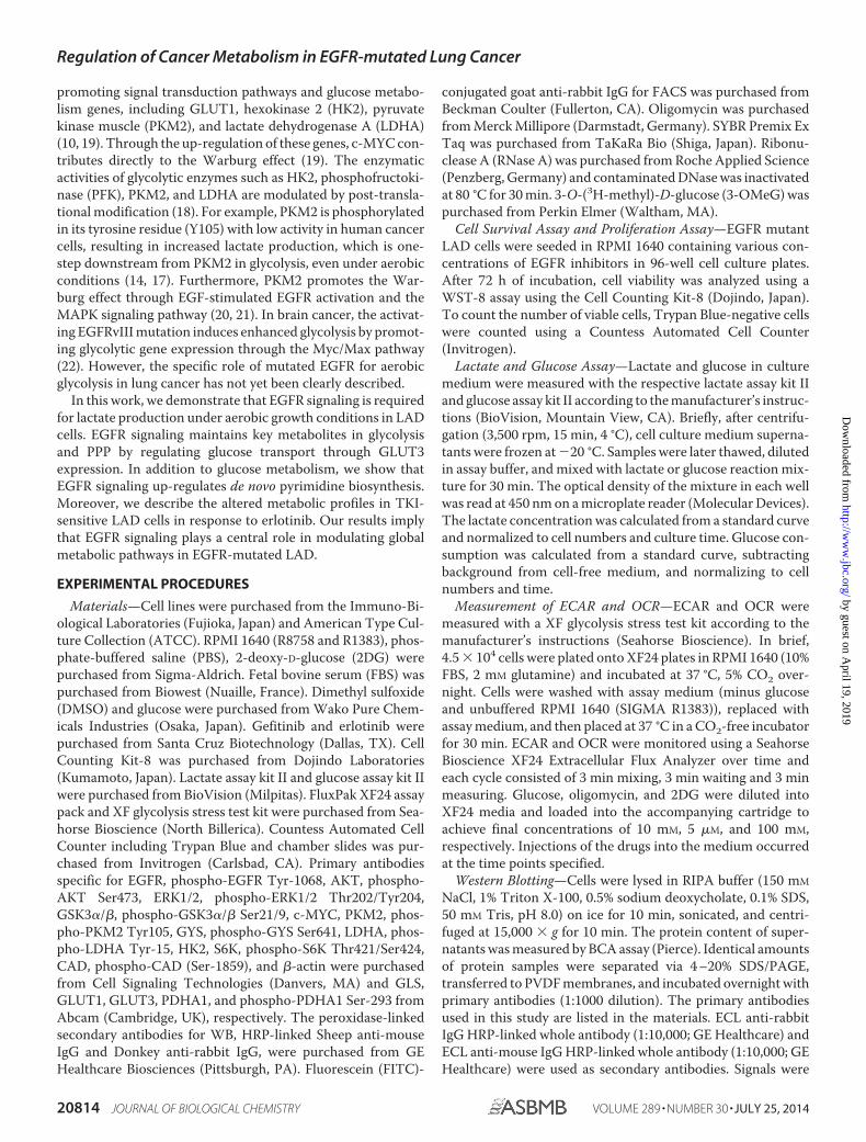
FIGURE 1. EGFR-TKI treatment represses lactate production in TKI-sensitive LAD cells. A, WST-8 assay with gefitinib. Cells were treated with the indicatedinhibitors for 72 h, and the viability was assessed by the WST-8 assay. Data are shown as the mean � S.D. (n � 6). Blue line: HCC827, red line: PC-9, green line:H1975. The in vitro half-maximal inhibitory concentration (IC50) for the growth of EGFR-mutated LAD cell lines was determined such that HCC827 to gefitinib:0.085 �M, PC-9 to gefitinib: 0.031 �M, and H1975 to gefitinib: �10 �M. B, WST-8 assay with erlotinib. Cells were treated with the indicated concentrations for72 h, and viability was assessed by the WST-8 assay. The data are shown as the mean � S.D. (n � 6). Blue line: HCC827, red line: PC-9, green line: H1975. The in vitrohalf-maximal inhibitory concentration (IC50) for the growth of EGFR-mutated LAD cell lines was determined such that HCC827 to 0.065 �M, PC-9 to 0.067 �M andH1975 to 8.8 �M. C, medium color in HCC827 and PC-9 was altered by addition of EGFR-TKIs (1 �M) for 24 h. The phenol red in culture media exhibits a gradualcolor transition from red to yellow over the pH range 8.0 to 6.6. D, cell growth responses at 6 h to 1 �M of gefitinib or erlotinib were measured using a TrypanBlue staining. The cell number of HCC827 (blue), PC-9 (red), and H1975 were shown. GEF; gefitinib, ERLO; erlotinib. The data are shown as the mean � S.D. (n �4). *, p � 0.05; **, p � 0.01 versus control by two-tailed Student’s t test. E, extracellular lactate production in HCC827 (blue), PC-9 (red) and H1975 (green) cell linesat 6 h post-TKI treatment. Error bars indicate S.D. (n � 6). *, p � 0.05; **, p � 0.01 versus control by two-tailed Student’s t test.
Regulation of Cancer Metabolism in EGFR-mutated Lung Cancer
JULY 25, 2014 • VOLUME 289 • NUMBER 30 JOURNAL OF BIOLOGICAL CHEMISTRY 20815
by guest on April 19, 2019
http://ww
w.jbc.org/
Dow
nloaded from

Expression of Glucose Transporter and Glucose TransportAssay—To detect expression of membrane-bound GLUTs,cells were fixed with 80% ethanol and incubated with anti-GLUT3 antibody (Abcam) and stained with the appropriateFITC-conjugated anti-rabbit IgG antibody (Jackson ImmunoResearch). Quantification of FITC-fluorescent intensity wasperformed using a FACSCanto II (BD Biosciences). Proceduresfor 3-OMeG uptake assay were previously described (26). LADcells were treated with indicated TKIs for 6 h before glucosetransport assay. Uptake was performed from 0.5 min to 10 minand radioactivity in the cells was quantified with Tri-Carb3110TR low activity liquid scintillation analyzer (PerkinElmer).
Statistical Analyses—Unless otherwise indicated, resultswere reported as the mean � S.D. Statistical analyses were doneby two-tailed Student’s t test. For metabolomic data analysis weused Welch t test and p values were indicated as *, �0.05; **,�0.01; and ***, �0.001.
RESULTS
Lactate Production Was Decreased in TKI-sensitive LADCells after EGFR-TKI Treatment—We initially characterizedthe EGFR-mutated lung adenocarcinoma cell lines used in this
study by measuring cell viability in the absence or presence ofEGFR-TKIs after 72 h. All three LAD cell lines have the EGFRmutation in either exon 19 or exon 21. Cell line HCC827 carriedthe delE746-A750 mutation, PC-9 exhibited delE746-A750 andNCI-H1975 (H1975) carried EGFR L858R�T790M (27, 28).The H1975 cells have the T790M mutation, which causesresistance to gefitinib and erlotinib (29). HCC827 and PC-9 celllines were highly sensitive to the EGFR-TKI gefitinib anderlotinib in the nanomolar range as compared with the TKI-resistant cell H1975 (Fig. 1, A and B). These data are consistentwith previous findings (27, 28, 30).
In dose response assays with EGFR inhibitors, we observeddifferences in the color of the culture medium in the presence ofTKIs against EGFR, especially in the growth cultures of TKI-sen-sitive cell lines (Fig. 1C). In culture media, phenol red exhibits agradual color transition from red to yellow at lower pH values as aresult of lactate production (31). Therefore, we explored the gly-colytic capacity of EGFR-mutated LAD cells. Since a 72-h incuba-tion with TKIs leads to a dramatic reduction in cell viability insensitive cell lines, we set up experimental conditions where TKItreatment was given at a relatively higher concentration (1 �M) and
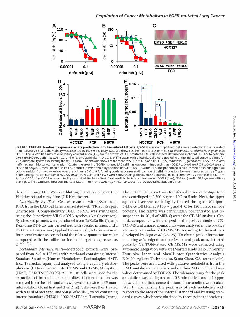
FIGURE 2. Glucose consumption and flux analysis monitoring glucose metabolism. A, glucose consumption rate in HCC827 (blue), PC-9 (red), and H1975(green) cells. Cells were cultured for 24 h in the absence or presence of EGFR-TKIs and glucose concentration in culture supernatant was quantified. Cell-freemedium was used as a background control. Error bars indicate S.D. (n � 6). *, p � 0.05; **, p � 0.01 versus control by two-tailed Student’s t test. B, measurementof ECAR over time. After 6-h treatment with TKIs, cells were applied to flux assay. ECAR was measured every 9 min. The addition of glucose, oligomycin, and2-deoxy-D-glucose (2DG) was carried out at the time point indicated by the arrows. Error bars indicate S.D. C, ECAR values of HCC827 (blue), PC-9 (red), andH1975(green) cells at 36 min of flux assay. Error bars indicate S.D. (n � 24 –30). **, p � 0.005; ***, p � 0.001 versus control by two-tailed Student’s t test. All cellswere treated with the indicated TKIs (1 �M) for 6 h before each assay. GEF, gefitinib; ERLO, erlotinib. D, OCR values of HCC827 (blue), PC-9 (red), and H1975 (green)cells at 36 min of flux assay. Error bars indicate S.D. (n � 24 –30). *, p � 0.001 versus control by two-tailed Student’s t test. All cells were treated with the indicatedTKIs (1 �M) for 6 h before each assay. GEF, gefitinib; ERLO, erlotinib.
Regulation of Cancer Metabolism in EGFR-mutated Lung Cancer
20816 JOURNAL OF BIOLOGICAL CHEMISTRY VOLUME 289 • NUMBER 30 • JULY 25, 2014
by guest on April 19, 2019
http://ww
w.jbc.org/
Dow
nloaded from
shorter time (6 h) to allow all cells to grow equally and therebystandardize the number of viable cells analyzed (Fig. 1D). Interest-ingly, we discovered that exposure of the cells to TKIs for up to 6 hsignificantly lowered the rate of lactate accumulation in themedium of TKI-sensitive LAD cell lines but not in resistant cells(Fig. 1E, *, p � 0.05; **, p � 0.01 t test).
Glycolytic Activities Were Down-regulated in TKI-sensitiveLAD Cells after Inhibition of EGFR Signaling—Next, we quan-tified the glucose consumption rate and found that inhibition ofEGFR signaling significantly lowered the rate of glucose con-sumption from the growth medium of TKI-sensitive HCC827and PC9 cells but not in the TKI-resistant H1975 cells (Fig. 2A*, p � 0.05; **, p � 0.01 t test).
To better define lactate production derived from glucose, wemeasured the glucose-induced extracellular acidification rate(ECAR), an indicator of lactate production, and the oxygenconsumption rate (OCR), an indicator of oxidative phosphory-lation (OXPHOS), using a flux analyzer. Basal levels of ECAR atthe beginning of measurements, which indicated non-glyco-
lytic acidification, were low in HCC827 cells (Fig. 2B). Equiva-lent ECAR was observed in HCC827 cells both pre- and post-treatment with an ATPase inhibitor oligomycin to inducemaximum cellular glycolytic capacity (Fig. 2B). At the final step,the addition of 2-deoxy-D-glucose (2DG), an inhibitor for gly-colysis, completely shut down extracellular acidification (Fig.2B). ECAR was statistically higher in DMSO controls comparedwith TKI-treated HCC827 and PC-9 cells (Fig. 2C *, p � 0.01; **,p � 0.005; ***, p � 0.001 t test). In contrast to ECAR, OCR waschanged in TKI-treated HCC827, but not in PC-9 and H1975cells (Fig. 2D).
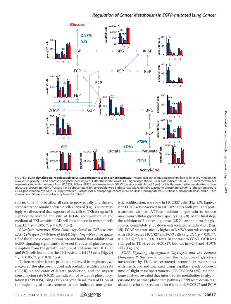
EGFR Signaling Up-regulates Glycolysis and the PentosePhosphate Pathway—To confirm the reduction of glycolysismetabolites by TKIs, we extracted intracellular metaboliteswith methanol and analyzed using capillary electrophoresistime-of-flight mass spectrometry (CE-TOFMS) (25). Metabo-lome analysis revealed that intermediate metabolites in glycol-ysis and the pentose phosphate pathway (PPP) were down-reg-ulated by erlotinib treatment for 6 h in both HCC827 and PC-9
FIGURE 3. EGFR signaling up-regulates glycolysis and the pentose phosphate pathway. Intracellular concentration (pmol/million cells) of key metabolitesinvolved in glycolysis and pentose phosphate pathway (PPP) after the inhibition of EGFR signaling is shown. Error bars indicate S.D. (n � 3). Total metaboliteswere extracted with methanol from HCC827, PC9 or H1975 cells treated with DMSO (blue) or erlotinib (red, 1 �M) for 6 h. Representative metabolites such asglucose 6-phosphate (G6P), fructose 1,6-bisphosphate (FBP), glyceraldehyde 3-phosphate (G3P), dihydroxyacetone phosphate (DHAP), 3-phosphoglycerate(3PG), phosphoenolpyruvate (PEP), pyruvate (PA), lactate (LA), 6-phosphogluconate (6PG), ribulose 5-phosphate (Ru5P), ribose 5-phosphate (R5P), and ATP areshown here. Others are listed in supplemental Table 1.
Regulation of Cancer Metabolism in EGFR-mutated Lung Cancer
JULY 25, 2014 • VOLUME 289 • NUMBER 30 JOURNAL OF BIOLOGICAL CHEMISTRY 20817
by guest on April 19, 2019
http://ww
w.jbc.org/
Dow
nloaded from
cells (Fig. 3 and supplemental Table S1). We observed that keyglycolysis and PPP metabolites such as fructose 1,6-bisphos-phate (FBP), dihydroxyacetone phosphate (DHAP), 3-phos-phoglycerate (3PG), phosphoenolpyruvate (PEP), lactate (LA),and 6-phosphogluconate (6PG) were decreased in TKI-sensi-tive HCC827 and PC9 cells after 6 h of erlotinib treatment, butnot in TKI-resistant H1975 cells (Fig. 3 and supplemental TableS1). Glucose 6-phosphate (G6P), glyceraldehyde 3-phosphate(G3P), pyruvate (PA), ribulose 5-phosphate (Ribu5P), andribose 5-phosphate (R5P) were significantly reduced in bothHCC827 and PC9 cells. The amount of adenosine triphosphate(ATP), which is the molecular unit of currency of intracellularenergy transfer, was not changed in any of the tested three celllines after erlotinib treatment. The reduction of glucose utiliza-tion after TKI treatment was observed in both glycolysis andpentose phosphate pathways, suggesting that EGFR signalingmight regulate a glucose transport or hexokinase activity inTKI-sensitive LAD cells.
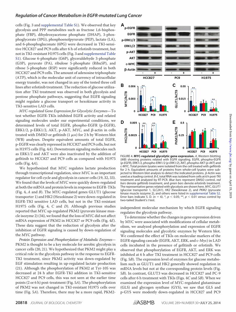
MYC-regulated Gene Expression for Glycolytic Enzymes—Totest whether EGFR-TKIs inhibited EGFR activity and relatedsignaling molecules under our experimental conditions, wedetermined levels of total EGFR, phospho-EGFR (p-EGFR),ERK1/2, p-ERK1/2, AKT, p-AKT, MYC, and �-actin in cellstreated with DMSO or gefitinib (1 �M) for 2 h by Western blot(WB) analyses. Despite equivalent amounts of total EGFR,p-EGFR was clearly repressed in HCC827 and PC9 cells, but notin H1975 cells (Fig. 4A). Downstream signaling molecules suchas ERK1/2 and AKT were also inactivated by the addition ofgefitinib to HCC827 and PC9 cells as compared with H1975cells (Fig. 4A).
We hypothesized that MYC regulates lactate productionthrough transcriptional regulation, since MYC is an importantregulator for cell cycle and glycolysis in cancer cells (19, 32, 33).We found that the levels of MYC were quickly down-regulatedat both the mRNA and protein levels in response to EGFR-TKIs(Fig. 4, A and B). The MYC-regulated genes GLUT1 (glucosetransporter 1) and HK2 (Hexokinase 2) were down-regulated inEGFR-TKI sensitive LAD cells, but not in the TKI-resistantH1975 cells (Fig. 4, C and D). Although previous studiesreported that MYC up-regulated PKM2 (pyruvate kinase mus-cle isozyme 2) (34), we found that the loss of MYC did not affectmRNA expression of PKM2 in HCC827 or PC9 cells (Fig. 4E).These data suggest that the reduction of glycolysis after theinhibition of EGFR signaling is caused by down-regulation ofthe MYC pathway.
Protein Expression and Phosphorylation of Metabolic Enzymes—PKM2 is thought to be a key molecule for aerobic glycolysis incancer cells (20, 21). We hypothesized that PKM2 might play acritical role in the glycolysis pathway in the response to EGFR-TKI treatment, since PKM2 activity was down-regulated byEGF stimulation resulting in up-regulated lactate production(21). Although the phosphorylation of PKM2 at Tyr-105 wasdecreased at 24 h after EGFR-TKI addition in TKI-sensitiveHCC827 and PC9 cells, this was not seen at the earlier timepoints (2 or 6 h) post-treatment (Fig. 5A). The phosphorylationof PKM2 was not changed in TKI-resistant H1975 cells overtime (Fig. 5A). Therefore, there may be a more rapid, PKM2-
independent molecular mechanism by which EGFR signalingregulates the glycolysis pathway.
To determine whether the changes in gene expression drivenby MYC were associated with modifications of cellular metab-olism, we analyzed phosphorylation and expression of EGFRsignaling molecules and glycolytic enzymes by Western blot.We confirmed the effect of TKIs on molecular markers of theEGFR signaling cascade (EGFR, AKT, ERK, and c-Myc) in LADcells incubated in the presence of gefitinib or erlotinib. Weobserved that phosphorylation of EGFR, AKT, and ERK wasinhibited at 6 h after TKI treatment in HCC827 and PC9 cells(Fig. 5B). The expression level of enzymes for glucose metabo-lism such as GLUT1 and HK2 generally showed regulation inmRNA levels but not at the corresponding protein levels (Fig.5B). In contrast, GLUT3 was decreased in HCC827 and PC-9cells after 6 h treatment with TKIs (Figs. 4C and 5B). When weexamined the expression level of MYC-regulated glutaminase(GLS) and glycogen synthase (GYS), we saw that GLS andp-GYS were modestly down-regulated in HCC827 and PC9
FIGURE 4. MYC-regulated glycolytic gene expression. A, Western blotting(WB) showing proteins related with EGFR signaling. EGFR, phospho-EGFR(p-EGFR), ERK1/2, phospho-ERK1/2 (p-ERK1/2), AKT, phospho-AKT (p-AKT) andc-MYC. Total protein lysates were isolated from the cell treated with gefitinibfor 2 h. Equivalent amounts of proteins from whole-cell lysates were sub-jected to Western blot analysis to detect the indicated proteins. �-Actin wasused as a loading control. B-E, total RNA was isolated from cells at 6 h post-TKItreatment and analyzed by RT-PCR. Blue bars represent DMSO control, redbars denote gefitinib treatment, and green bars denote erlotinib treatment.The representative genes related with glycolysis are shown here. MYC, GLUT1(glucose transporter 1, SLC2A1), HK2 (hexokinase 2), and PKM2 (pyruvatekinase muscle isozyme 2), and others were listed in supplemental Table S2.Error bars indicate S. D. (n � 6). *, p � 0.05; **, p � 0.01 versus control bytwo-tailed Student’s t test.
Regulation of Cancer Metabolism in EGFR-mutated Lung Cancer
20818 JOURNAL OF BIOLOGICAL CHEMISTRY VOLUME 289 • NUMBER 30 • JULY 25, 2014
by guest on April 19, 2019
http://ww
w.jbc.org/
Dow
nloaded from
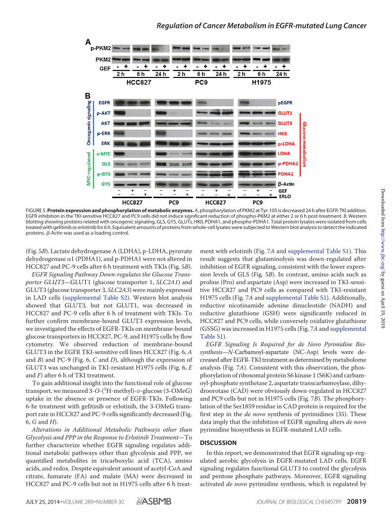
(Fig. 5B). Lactate dehydrogenase A (LDHA), p-LDHA, pyruvatedehydrogenase �1 (PDHA1), and p-PDHA1 were not altered inHCC827 and PC-9 cells after 6 h treatment with TKIs (Fig. 5B).
EGFR Signaling Pathway Down-regulates the Glucose Trans-porter GLUT3—GLUT1 (glucose transporter 1, SLC2A1) andGLUT3 (glucose transporter 3, SLC2A3) were mainly expressedin LAD cells (supplemental Table S2). Western blot analysisshowed that GLUT3, but not GLUT1, was decreased inHCC827 and PC-9 cells after 6 h of treatment with TKIs. Tofurther confirm membrane-bound GLUT3 expression levels,we investigated the effects of EGFR-TKIs on membrane-boundglucose transporters in HCC827, PC-9, and H1975 cells by flowcytometry. We observed reduction of membrane-boundGLUT3 in the EGFR TKI-sensitive cell lines HCC827 (Fig. 6, Aand B) and PC-9 (Fig. 6, C and D), although the expression ofGLUT3 was unchanged in TKI-resistant H1975 cells (Fig. 6, Eand F) after 6 h of TKI treatment.
To gain additional insight into the functional role of glucosetransport, we measured 3-O-(3H-methyl)-D-glucose (3-OMeG)uptake in the absence or presence of EGFR-TKIs. Following6-hr treatment with gefitinib or erlotinib, the 3-OMeG trans-port rate in HCC827 and PC-9 cells significantly decreased (Fig.6, G and H).
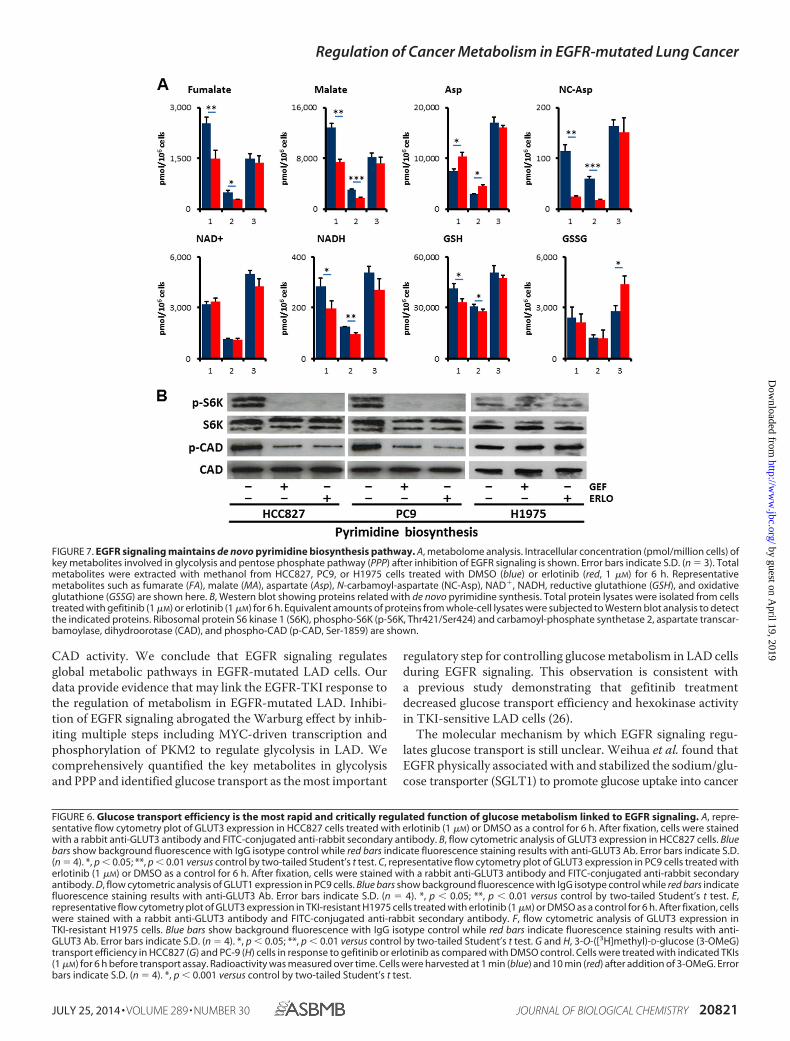
Alterations in Additional Metabolic Pathways other thanGlycolysis and PPP in the Response to Erlotinib Treatment—Tofurther characterize whether EGFR signaling regulates addi-tional metabolic pathways other than glycolysis and PPP, wequantified metabolites in tricarboxylic acid (TCA), aminoacids, and redox. Despite equivalent amount of acetyl-CoA andcitrate, fumarate (FA) and malate (MA) were decreased inHCC827 and PC-9 cells but not in H1975 cells after 6 h treat-
ment with erlotinib (Fig. 7A and supplemental Table S1). Thisresult suggests that glutaminolysis was down-regulated afterinhibition of EGFR signaling, consistent with the lower expres-sion levels of GLS (Fig. 5B). In contrast, amino acids such asproline (Pro) and aspartate (Asp) were increased in TKI-sensi-tive HCC827 and PC9 cells as compared with TKI-resistantH1975 cells (Fig. 7A and supplemental Table S1). Additionally,reductive nicotinamide adenine dinucleotide (NADH) andreductive glutathione (GSH) were significantly reduced inHCC827 and PC9 cells, while conversely oxidative glutathione(GSSG) was increased in H1975 cells (Fig. 7A and supplementalTable S1).
EGFR Signaling Is Required for de Novo Pyrimidine Bio-synthesis—N-Carbamoyl-aspartate (NC-Asp) levels were de-creased after EGFR-TKI treatment as determined by metabolomeanalysis (Fig. 7A). Consistent with this observation, the phos-phorylation of ribosomal protein S6 kinase 1 (S6K) and carbam-oyl-phosphate synthetase 2, aspartate transcarbamoylase, dihy-droorotase (CAD) were obviously down-regulated in HCC827and PC9 cells but not in H1975 cells (Fig. 7B). The phosphory-lation of the Ser1859 residue in CAD protein is required for thefirst step in the de novo synthesis of pyrimidines (35). Thesedata imply that the inhibition of EGFR signaling alters de novopyrimidine biosynthesis in EGFR-mutated LAD cells.
DISCUSSION
In this report, we demonstrated that EGFR signaling up-reg-ulated aerobic glycolysis in EGFR-mutated LAD cells. EGFRsignaling regulates functional GLUT3 to control the glycolysisand pentose phosphate pathways. Moreover, EGFR signalingactivated de novo pyrimidine synthesis, which is regulated by
FIGURE 5. Protein expression and phosphorylation of metabolic enzymes. A, phosphorylation of PKM2 at Tyr-105 is decreased 24 h after EGFR-TKI addition.EGFR inhibition in the TKI-sensitive HCC827 and PC9 cells did not induce significant reduction of phospho-PKM2 at either 2 or 6 h post-treatment. B, Westernblotting showing proteins related with oncogenic signaling, GLS, GYS, GLUTs, HKII, PDHA1, and phospho-PDHA1. Total protein lysates were isolated from cellstreated with gefitinib or erlotinib for 6 h. Equivalent amounts of proteins from whole-cell lysates were subjected to Western blot analysis to detect the indicatedproteins. �-Actin was used as a loading control.
Regulation of Cancer Metabolism in EGFR-mutated Lung Cancer
JULY 25, 2014 • VOLUME 289 • NUMBER 30 JOURNAL OF BIOLOGICAL CHEMISTRY 20819
by guest on April 19, 2019
http://ww
w.jbc.org/
Dow
nloaded from

Regulation of Cancer Metabolism in EGFR-mutated Lung Cancer
20820 JOURNAL OF BIOLOGICAL CHEMISTRY VOLUME 289 • NUMBER 30 • JULY 25, 2014
by guest on April 19, 2019
http://ww
w.jbc.org/
Dow
nloaded from
CAD activity. We conclude that EGFR signaling regulatesglobal metabolic pathways in EGFR-mutated LAD cells. Ourdata provide evidence that may link the EGFR-TKI response tothe regulation of metabolism in EGFR-mutated LAD. Inhibi-tion of EGFR signaling abrogated the Warburg effect by inhib-iting multiple steps including MYC-driven transcription andphosphorylation of PKM2 to regulate glycolysis in LAD. Wecomprehensively quantified the key metabolites in glycolysisand PPP and identified glucose transport as the most important
regulatory step for controlling glucose metabolism in LAD cellsduring EGFR signaling. This observation is consistent witha previous study demonstrating that gefitinib treatmentdecreased glucose transport efficiency and hexokinase activityin TKI-sensitive LAD cells (26).
The molecular mechanism by which EGFR signaling regu-lates glucose transport is still unclear. Weihua et al. found thatEGFR physically associated with and stabilized the sodium/glu-cose transporter (SGLT1) to promote glucose uptake into cancer
FIGURE 6. Glucose transport efficiency is the most rapid and critically regulated function of glucose metabolism linked to EGFR signaling. A, repre-sentative flow cytometry plot of GLUT3 expression in HCC827 cells treated with erlotinib (1 �M) or DMSO as a control for 6 h. After fixation, cells were stainedwith a rabbit anti-GLUT3 antibody and FITC-conjugated anti-rabbit secondary antibody. B, flow cytometric analysis of GLUT3 expression in HCC827 cells. Bluebars show background fluorescence with IgG isotype control while red bars indicate fluorescence staining results with anti-GLUT3 Ab. Error bars indicate S.D.(n � 4). *, p � 0.05; **, p � 0.01 versus control by two-tailed Student’s t test. C, representative flow cytometry plot of GLUT3 expression in PC9 cells treated witherlotinib (1 �M) or DMSO as a control for 6 h. After fixation, cells were stained with a rabbit anti-GLUT3 antibody and FITC-conjugated anti-rabbit secondaryantibody. D, flow cytometric analysis of GLUT1 expression in PC9 cells. Blue bars show background fluorescence with IgG isotype control while red bars indicatefluorescence staining results with anti-GLUT3 Ab. Error bars indicate S.D. (n � 4). *, p � 0.05; **, p � 0.01 versus control by two-tailed Student’s t test. E,representative flow cytometry plot of GLUT3 expression in TKI-resistant H1975 cells treated with erlotinib (1 �M) or DMSO as a control for 6 h. After fixation, cellswere stained with a rabbit anti-GLUT3 antibody and FITC-conjugated anti-rabbit secondary antibody. F, flow cytometric analysis of GLUT3 expression inTKI-resistant H1975 cells. Blue bars show background fluorescence with IgG isotype control while red bars indicate fluorescence staining results with anti-GLUT3 Ab. Error bars indicate S.D. (n � 4). *, p � 0.05; **, p � 0.01 versus control by two-tailed Student’s t test. G and H, 3-O-([3H]methyl)-D-glucose (3-OMeG)transport efficiency in HCC827 (G) and PC-9 (H) cells in response to gefitinib or erlotinib as compared with DMSO control. Cells were treated with indicated TKIs(1 �M) for 6 h before transport assay. Radioactivity was measured over time. Cells were harvested at 1 min (blue) and 10 min (red) after addition of 3-OMeG. Errorbars indicate S.D. (n � 4). *, p � 0.001 versus control by two-tailed Student’s t test.
FIGURE 7. EGFR signaling maintains de novo pyrimidine biosynthesis pathway. A, metabolome analysis. Intracellular concentration (pmol/million cells) ofkey metabolites involved in glycolysis and pentose phosphate pathway (PPP) after inhibition of EGFR signaling is shown. Error bars indicate S.D. (n � 3). Totalmetabolites were extracted with methanol from HCC827, PC9, or H1975 cells treated with DMSO (blue) or erlotinib (red, 1 �M) for 6 h. Representativemetabolites such as fumarate (FA), malate (MA), aspartate (Asp), N-carbamoyl-aspartate (NC-Asp), NAD�, NADH, reductive glutathione (GSH), and oxidativeglutathione (GSSG) are shown here. B, Western blot showing proteins related with de novo pyrimidine synthesis. Total protein lysates were isolated from cellstreated with gefitinib (1 �M) or erlotinib (1 �M) for 6 h. Equivalent amounts of proteins from whole-cell lysates were subjected to Western blot analysis to detectthe indicated proteins. Ribosomal protein S6 kinase 1 (S6K), phospho-S6K (p-S6K, Thr421/Ser424) and carbamoyl-phosphate synthetase 2, aspartate transcar-bamoylase, dihydroorotase (CAD), and phospho-CAD (p-CAD, Ser-1859) are shown.
Regulation of Cancer Metabolism in EGFR-mutated Lung Cancer
JULY 25, 2014 • VOLUME 289 • NUMBER 30 JOURNAL OF BIOLOGICAL CHEMISTRY 20821
by guest on April 19, 2019
http://ww
w.jbc.org/
Dow
nloaded from
cells (36). However, this function did not require EGFR kinaseactivity. In this report, we found that TKIs to EGFR, gefitinib, anderlotinib, repressed aerobic glycolysis and PPP in EGFR-mutatedLAD cells. Although SGLT1 directly interacts with EGFR, EGFRsignaling may regulate GLUT translocation in an indirect manner.Mutated EGFRs found in LAD have constitutive tyrosine kinaseactivity, resulting in activation of downstream RAS/MAPK andPI3K/AKT pathways (Figs. 4A and 5B). In adipocytes and skeletalmuscle, insulin and the PI3K/AKT pathway mediate GLUT4translocation (37, 38). To promote glucose uptake into muscle andfat cells, insulin stimulates the translocation of GLUT4 from intra-cellular membranes to the cell surface. Insulin signals go throughAS160 (Akt substrate of 160 kDa) and Tbc1Ds to modulate RabGTPase, and through Rho GTPase TC10a to act on other targets(37, 38). The EGFR-PI3K/AKT axis might control GLUT translo-cation to the plasma membrane in EGFR-mutated LAD cells. Toprove this, we would need to characterize in more detail themolecular mechanisms that control GLUT expression, activity,and translocation.
A recent study showed that AMPK-dependent degradationof thioredoxin-interacting protein (TXNIP) upon stress led toenhanced glucose uptake via GLUT1 (39). Another researchreport showed that tumor-associated mutant p53 (mutp53)stimulated the Warburg effect in cancer cells as a new mutp53gain of function (40). Mutp53 did not affect the expression ofGLUT1, but promoted aerobic glycolysis by inducing GLUT1translocation to the plasma membrane, which was mediated byactivated RHOA and its downstream effector ROCK. In thisstudy, the EGFR-TKI-sensitive LAD cell lines HCC827 and PC9possess mutp53, but not the H1975 cell line. A possible molec-ular mechanism is that either EGFR signaling may regulateGLUT translocation by directly activating the RHOA/ROCKpathway or the mutp53 pathway that in turn activates RHOA/ROCK function. Further experiments would be required todetermine whether EGFR signaling controls glucose transportthrough the TXNIP or mutp53 pathway.
New therapeutic strategies are currently needed to overcomethe EGFR T790M-mediated acquired resistance observed in theclinic (8). A recent Phase III study of afatinib monotherapy failed toshow overall survival benefit in patients with acquired resistance toreversible EGFR-TKIs(41). Kim et al. showed that targeting of gly-colysis was an effective therapeutic option to overcome the limitedefficacy of afatinib in LAD cells with EGFR T790M (42). Treat-ment with 2DG completely shut down lactate production inEGFR-mutated LAD cells (Fig. 2B), since 2DG is a glucose analogthat competes with glucose for cellular uptake. Therefore, combi-nation therapies of EGFR-TKIs and drugs that block the glycolysispathway such as GLUT-inhibitors would be expected to be muchmore effective for TKI-resistant cases.
Molecular targeting therapy using TKIs is currently one ofthe most successful forms of treatment in the clinic, andincludes imatinib targeting BCR-ABL in chronic myeloid leu-kemia (CML) and gefitinib/erlotinib in EGFR-mutated LAD(3). Despite high therapeutic responses to EGFR-TKI treat-ment, it is clear that not all patients experience benefit; thus,there is still a need to identify potential non-responders andmatch patients with the most effective therapies (4). Monitor-ing of tumor glucose utilization by [18F]fluorodeoxyglucose
(FDG)-positron emission tomography (PET) was implementedfor the early prediction of treatment response to EGFR-TKIs inNSCLC (26, 43). In this report, we demonstrate that TKIs toEGFR, gefitinib and erlotinib, repress aerobic glycolysis inEGFR-mutated LAD cells. Those correlations strongly suggestthat intermediate metabolites in the pentose phosphate path-way, glycolysis, and pyrimidine biosynthesis such as FBP,DHAP, LA, 6PG, and NC-Asp could serve as well-defined bio-markers to predict response to EGFR-TKI therapy.
The application of metabolomics in oncology has focused itsability to identify biomarkers for cancer diagnosis, prognosis,and therapeutic efficacy (44). In our previous study, we com-pared the metabolomics of normal and tumor tissues surgicallyresected pairwise from nine lung patients using CE-TOFMS toelucidate tumor-specific metabolism (45). Significantly highlactate concentrations and elevated activating phosphorylationlevels of phosphofructokinase and pyruvate kinase in lungtumors confirmed hyperactive glycolysis (45). Here we showthat EGFR signaling regulates many metabolites in EGFR-mu-tated LAD cells under in vitro culture conditions; however,whether EGFR-TKIs have the same effects in vivo is stillunknown. To build upon this work, further investigations willexplore these concepts in relevant animal models and in LADtissue biopsy samples using bronchoscope before and afterEGFR-TKI therapy. In vivo validation of these concepts willhave significant implications for future diagnostic and thera-peutic possibilities for patients.
Acknowledgments—We thank all of Dr. Tsuchihara’s laboratorymembers for help. We also thank Dr. Phillip Wong for carefully read-ing the manuscript and providing critical comments.
REFERENCES1. da Cunha Santos, G., Shepherd, F. A., and Tsao, M. S. (2011) EGFR mu-
tations and lung cancer. Annu. Rev. Pathol. 6, 49 – 692. Irmer, D., Funk, J. O., and Blaukat, A. (2007) EGFR kinase domain muta-
tions - functional impact and relevance for lung cancer therapy. Oncogene26, 5693–5701
3. Levitzki, A. (2013) Tyrosine kinase inhibitors: views of selectivity, sensi-tivity, and clinical performance. Annu. Rev. Pharmacol. Toxicol. 53,161–185
4. Linardou, H., Dahabreh, I. J., Bafaloukos, D., Kosmidis, P., and Murray, S.(2009) Somatic EGFR mutations and efficacy of tyrosine kinase inhibitorsin NSCLC. Nat. Rev. Clin. Oncol. 6, 352–366
5. Suzuki, A., Mimaki, S., Yamane, Y., Kawase, A., Matsushima, K., Suzuki, M.,Goto, K., Sugano, S., Esumi, H., Suzuki, Y., and Tsuchihara, K. (2013) Identi-fication and characterization of cancer mutations in Japanese lung adenocar-cinoma without sequencing of normal tissue counterparts. PLoS One 8,e73484
6. Imielinski, M., Berger, A. H., Hammerman, P. S., Hernandez, B., Pugh,T. J., Hodis, E., Cho, J., Suh, J., Capelletti, M., Sivachenko, A., Sougnez, C.,Auclair, D., Lawrence, M. S., Stojanov, P., Cibulskis, K., Choi, K., de Waal,L., Sharifnia, T., Brooks, A., Greulich, H., Banerji, S., Zander, T., Seidel, D.,Leenders, F., Ansén, S., Ludwig, C., Engel-Riedel, W., Stoelben, E., Wolf, J.,Goparju, C., Thompson, K., Winckler, W., Kwiatkowski, D., Johnson, B. E.,Jänne, P. A., Miller, V. A., Pao, W., Travis, W. D., Pass, H. I., Gabriel, S. B.,Lander, E. S., Thomas, R. K., Garraway, L. A., Getz, G., and Meyerson, M.(2012) Mapping the Hallmarks of Lung Adenocarcinoma with MassivelyParallel Sequencing. Cell 150, 1107–1120
7. Mellinghoff, I. (2007) Why do cancer cells become “addicted” to onco-genic epidermal growth factor receptor? Plos Med. 4, 1620 –1622
Regulation of Cancer Metabolism in EGFR-mutated Lung Cancer
20822 JOURNAL OF BIOLOGICAL CHEMISTRY VOLUME 289 • NUMBER 30 • JULY 25, 2014
by guest on April 19, 2019
http://ww
w.jbc.org/
Dow
nloaded from
8. Pao, W., and Chmielecki, J. (2010) Rational, biologically based treatmentof EGFR-mutant non-small-cell lung cancer. Nature Reviews Cancer 10,760 –774
9. Lemmon, M. A., and Schlessinger, J. (2010) Cell signaling by receptortyrosine kinases. Cell 141, 1117–1134
10. Cairns, R. A., Harris, I. S., and Mak, T. W. (2011) Regulation of cancer cellmetabolism. Nat. Rev. Cancer 11, 85–95
11. Levine, A. J., and Puzio-Kuter, A. M. (2010) The control of the metabolicswitch in cancers by oncogenes and tumor suppressor genes. Science 330,1340 –1344
12. Cheong, H., Lu, C., Lindsten, T., and Thompson, C. B. (2012) Therapeutictargets in cancer cell metabolism and autophagy. Nat. Biotechnol. 30,671– 678
13. Ying, H., Kimmelman, A. C., Lyssiotis, C. A., Hua, S., Chu, G. C., Fletcher-Sananikone, E., Locasale, J. W., Son, J., Zhang, H., Coloff, J. L., Yan, H.,Wang, W., Chen, S., Viale, A., Zheng, H., Paik, J. H., Lim, C., Guimaraes,A. R., Martin, E. S., Chang, J., Hezel, A. F., Perry, S. R., Hu, J., Gan, B., Xiao,Y., Asara, J. M., Weissleder, R., Wang, Y. A., Chin, L., Cantley, L. C., andDePinho, R. A. (2012) Oncogenic Kras maintains pancreatic tumorsthrough regulation of anabolic glucose metabolism. Cell 149, 656 – 670
14. Lunt, S. Y., and Vander Heiden, M. G. (2011) Aerobic glycolysis: meetingthe metabolic requirements of cell proliferation. Annu. Rev. Cell Dev. Biol.27, 441– 464
15. Soga, T. (2013) Cancer metabolism: key players in metabolic reprogram-ming. Cancer Sci. 104, 275–281
16. Vander Heiden, M. G., Cantley, L. C., and Thompson, C. B. (2009) Under-standing the Warburg effect: the metabolic requirements of cell prolifer-ation. Science 324, 1029 –1033
17. Hitosugi, T., Kang, S., Vander Heiden, M. G., Chung, T. W., Elf, S., Lythgoe,K., Dong, S., Lonial, S., Wang, X., Chen, G. Z., Xie, J., Gu, T. L., Polakiewicz,R. D., Roesel, J. L., Boggon, T. J., Khuri, F. R., Gilliland, D. G., Cantley, L. C.,Kaufman, J., and Chen, J. (2009) Tyrosine phosphorylation inhibits PKM2 topromote the Warburg effect and tumor growth. Sci. Signal 2, ra73
18. Hitosugi, T., and Chen, J. (2013) Post-translational modifications and theWarburg effect. Oncogene doi:10.1038/onc.2013.406
19. Miller, D. M., Thomas, S. D., Islam, A., Muench, D., and Sedoris, K. (2012)c-Myc and cancer metabolism. Clin. Cancer Res. 18, 5546 –5553
20. Yang, W., and Lu, Z. (2013) Nuclear PKM2 regulates the Warburg effect.Cell Cycle 12, 3154 –3158
21. Wong, N., Ojo, D., Yan, J., and Tang, D. (2014) PKM2 contributes tocancer metabolism. Cancer Lett. doi: 10.1016/j.canlet. 2014.01.031
22. Babic, I., Anderson, E. S., Tanaka, K., Guo, D., Masui, K., Li, B., Zhu, S., Gu, Y.,Villa, G. R., Akhavan, D., Nathanson, D., Gini, B., Mareninov, S., Li, R., Cama-cho, C. E., Kurdistani, S. K., Eskin, A., Nelson, S. F., Yong, W. H., Cavenee,W. K., Cloughesy, T. F., Christofk, H. R., Black, D. L., and Mischel, P. S. (2013)EGFR mutation-induced alternative splicing of Max contributes to growth ofglycolytic tumors in brain cancer. Cell Metab. 17, 1000–1008
23. Soga, T., and Heiger, D. N. (2000) Amino acid analysis by capillary elec-trophoresis electrospray ionization mass spectrometry. Anal. Chem. 72,1236 –1241
24. Soga, T., Ueno, Y., Naraoka, H., Ohashi, Y., Tomita, M., and Nishioka, T.(2002) Simultaneous determination of anionic intermediates for Bacillussubtilis metabolic pathways by capillary electrophoresis electrospray ion-ization mass spectrometry. Anal. Chem. 74, 2233–2239
25. Soga, T., Ohashi, Y., Ueno, Y., Naraoka, H., Tomita, M., and Nishioka, T.(2003) Quantitative metabolome analysis using capillary electrophoresismass spectrometry. J. Proteome Res. 2, 488 – 494
26. Su, H., Bodenstein, C., Dumont, R. A., Seimbille, Y., Dubinett, S., Phelps,M. E., Herschman, H., Czernin, J., and Weber, W. (2006) Monitoringtumor glucose utilization by positron emission tomography for the pre-diction of treatment response to epidermal growth factor receptor kinaseinhibitors. Clin. Cancer Res. 12, 5659 –5667
27. Okamoto, K., Okamoto, I., Okamoto, W., Tanaka, K., Takezawa, K., Ku-wata, K., Yamaguchi, H., Nishio, K., and Nakagawa, K. (2010) Role ofsurvivin in EGFR inhibitor-induced apoptosis in non-small cell lung can-cers positive for EGFR mutations. Cancer Res. 70, 10402–10410
28. Donev, I. S., Wang, W., Yamada, T., Li, Q., Takeuchi, S., Matsumoto, K.,Yamori, T., Nishioka, Y., Sone, S., and Yano, S. (2011) Transient PI3K inhibi-
tion induces apoptosis and overcomes HGF-mediated resistance to EGFR-TKIs in EGFR mutant lung cancer. Clin. Cancer Res. 17, 2260–2269
29. Zhou, W., Ercan, D., Chen, L., Yun, C. H., Li, D., Capelletti, M., Cortot,A. B., Chirieac, L., Iacob, R. E., Padera, R., Engen, J. R., Wong, K. K., Eck,M. J., Gray, N. S., and Jänne, P. A. (2009) Novel mutant-selective EGFRkinase inhibitors against EGFR T790M. Nature 462, 1070 –1074
30. Sudo, M., Chin, T. M., Mori, S., Doan, N. B., Said, J. W., Akashi, M., andKoeffler, H. P. (2013) Inhibiting proliferation of gefitinib-resistant, non-small cell lung cancer. Cancer Chemother. Pharmacol. 71, 1325–1334
31. Sonveaux, P., Végran, F., Schroeder, T., Wergin, M. C., Verrax, J., Rabbani,Z. N., De Saedeleer, C. J., Kennedy, K. M., Diepart, C., Jordan, B. F., Kelley,M. J., Gallez, B., Wahl, M. L., Feron, O., and Dewhirst, M. W. (2008)Targeting lactate-fueled respiration selectively kills hypoxic tumor cells inmice. J. Clin. Invest. 118, 3930 –3942
32. Morrish, F., Neretti, N., Sedivy, J. M., and Hockenbery, D. M. (2008) Theoncogene c-Myc coordinates regulation of metabolic networks to enablerapid cell cycle entry. Cell Cycle 7, 1054 –1066
33. Osthus, R. C., Shim, H., Kim, S., Li, Q., Reddy, R., Mukherjee, M., Xu, Y.,Wonsey, D., Lee, L. A., and Dang, C. V. (2000) Deregulation of glucosetransporter 1 and glycolytic gene expression by c-Myc. J. Biol. Chem. 275,21797–21800
34. David, C. J., Chen, M., Assanah, M., Canoll, P., and Manley, J. L. (2010)HnRNP proteins controlled by c-Myc deregulate pyruvate kinase mRNAsplicing in cancer. Nature 463, 364 –368
35. Ben-Sahra, I., Howell, J. J., Asara, J. M., and Manning, B. D. (2013) Stimu-lation of de novo pyrimidine synthesis by growth signaling through mTORand S6K1. Science 339, 1323–1328
36. Weihua, Z., Tsan, R., Huang, W. C., Wu, Q., Chiu, C. H., Fidler, I. J., andHung, M. C. (2008) Survival of cancer cells is maintained by EGFR inde-pendent of its kinase activity. Cancer Cell 13, 385–393
37. Watson, R. T., and Pessin, J. E. (2006) Bridging the GAP between insulinsignaling and GLUT4 translocation. Trends Biochem. Sci. 31, 215–222
38. Bogan, J. S. (2012) Regulation of glucose transporter translocation inhealth and diabetes. Annu. Rev. Biochem. 81, 507–532
39. Wu, N., Zheng, B., Shaywitz, A., Dagon, Y., Tower, C., Bellinger, G., Shen,C. H., Wen, J., Asara, J., McGraw, T. E., Kahn, B. B., and Cantley, L. C.(2013) AMPK-dependent degradation of TXNIP upon energy stress leadsto enhanced glucose uptake via GLUT1. Mol. Cell 49, 1167–1175
40. Zhang, C., Liu, J., Liang, Y., Wu, R., Zhao, Y., Hong, X., Lin, M., Yu, H., Liu,L., Levine, A. J., Hu, W., and Feng, Z. (2013) Tumour-associated mutantp53 drives the Warburg effect. Nat. Commun. 4, 2935
41. Miller, V. A., Hirsh, V., Cadranel, J., Chen, Y. M., Park, K., Kim, S. W.,Zhou, C., Su, W. C., Wang, M., Sun, Y., Heo, D. S., Crino, L., Tan, E. H.,Chao, T. Y., Shahidi, M., Cong, X. J., Lorence, R. M., and Yang, J. C. (2012)Afatinib versus placebo for patients with advanced, metastatic non-small-cell lung cancer after failure of erlotinib, gefitinib, or both, and one or twolines of chemotherapy (LUX-Lung 1): a phase 2b/3 randomised trial. Lan-cet Oncol. 13, 528 –538
42. Kim, S. M., Yun, M. R., Hong, Y. K., Solca, F., Kim, J. H., Kim, H. J., andCho, B. C. (2013) Glycolysis inhibition sensitizes non-small cell lung can-cer with T790M mutation to irreversible EGFR inhibitors via translationalsuppression of Mcl-1 by AMPK activation. Mol. Cancer Ther. 12,2145–2156
43. Takahashi, R., Hirata, H., Tachibana, I., Shimosegawa, E., Inoue, A., Na-gatomo, I., Takeda, Y., Kida, H., Goya, S., Kijima, T., Yoshida, M., Kum-agai, T., Kumanogoh, A., Okumura, M., Hatazawa, J., and Kawase, I. (2012)Early [18F]fluorodeoxyglucose positron emission tomography at two daysof gefitinib treatment predicts clinical outcome in patients with adenocar-cinoma of the lung. Clin. Cancer Res. 18, 220 –228
44. Spratlin, J. L., Serkova, N. J., and Eckhardt, S. G. (2009) Clinical applica-tions of metabolomics in oncology: a review. Clin. Cancer Res. 15,431– 440
45. Kami, K., Fujimori, T., Sato, H., Sato, M., Yamamoto, H., Ohashi, Y.,Sugiyama, N., Ishihama, Y., Onozuka, H., Ochiai, A., Esumi, H., Soga, T.,and Tomita, M. (2013) Metabolomic profiling of lung and prostate tumortissues by capillary electrophoresis time-of-flight mass spectrometry.Metabolomics 9, 444 – 453
Regulation of Cancer Metabolism in EGFR-mutated Lung Cancer
JULY 25, 2014 • VOLUME 289 • NUMBER 30 JOURNAL OF BIOLOGICAL CHEMISTRY 20823
by guest on April 19, 2019
http://ww
w.jbc.org/
Dow
nloaded from
Owada, Hiroyasu Esumi and Katsuya TsuchiharaHideki Makinoshima, Masahiro Takita, Shingo Matsumoto, Atsushi Yagishita, Satoshi
Pathways in EGFR-mutated Lung AdenocarcinomaEpidermal Growth Factor Receptor (EGFR) Signaling Regulates Global Metabolic
doi: 10.1074/jbc.M114.575464 originally published online June 13, 20142014, 289:20813-20823.J. Biol. Chem.
10.1074/jbc.M114.575464Access the most updated version of this article at doi:
Alerts:
When a correction for this article is posted•
When this article is cited•
to choose from all of JBC's e-mail alertsClick here
Supplemental material:
http://www.jbc.org/content/suppl/2014/06/13/M114.575464.DC1
http://www.jbc.org/content/289/30/20813.full.html#ref-list-1
This article cites 45 references, 12 of which can be accessed free at
by guest on April 19, 2019
http://ww
w.jbc.org/
Dow
nloaded from