Embed Size (px)
Citation preview

ORIGINAL PAPER
Enzymatic hydrolysis of camel milk caseinand its antioxidant properties
Devendra Kumar1 & Manish Kumar Chatli1 &
Raghvendar Singh2& Nitin Mehta1 & Pavan Kumar1
Received: 13 August 2015 /Revised: 30 November 2015 /Accepted: 3 December 2015 /Published online: 5 January 2016# INRA and Springer-Verlag France 2016
Abstract The aim of this work was to investigate the enzymatic hydrolysis of wholecasein from camel milk with proteolytic enzymes, viz. alcalase, α-chymotrypsin andpapain, and to assess the antioxidant activity of hydrolysates. Casein powder wasreconstituted (5% TS) and the selected enzymes were added at 1:100 (enzyme/sub-strate). Hydrolysis was carried out for 6 h at 55 °C for alcalase and papain and 37 °C forα-chymotrypsin, and samples were drawn at 2-h interval. The hydrolysates wereanalysed for pH, degree of hydrolysis (DH) and antioxidant activities, viz. 2, 2′-azinobis (3-ethylbenzthiazoline-6-sulphonic acid) (ABTS), 2, 2′-diphenyl-1-picrylhydrazyl (DPPH) and ferric reducing antioxidant power (FRAP) assay. Thedecrease in pH of the hydrolysates was observed with the progress of hydrolysis time,and as compared to alcalase, the rate of pH decrease was higher for α-chymotrypsinand papain. The DH increased significantly (P<0.05) up to 6 h on hydrolysis withalcalase and papain, whereas with chymotrypsinolysis, DH increased significantly(P<0.05) up to 4 h; thereafter, a non-significant increase was observed. The antioxidantactivity assessed by ABTS, DPPH and FRAP assay increased significantly (P<0.05)with the increase in hydrolysis time as well as the degree of hydrolysis. Thechymotrypsinolysis exhibited higher antioxidant activity as compared to alcalase andpapain. The results obtained in this study suggested that camel milk casein could beused as an ingredient in nutraceuticals or health-oriented foods.
Keywords Camel milk . Enzymatic hydrolysis . Casein hydrolysate . Degree ofhydrolysis . Antioxidant activity
Dairy Sci. & Technol. (2016) 96:391–404DOI 10.1007/s13594-015-0275-9
* Devendra [email protected]
1 Department of Livestock Products Technology, College of Veterinary Science, Guru Angad DevVeterinary and Animal Sciences University, Ludhiana 141 004 Punjab, India
2 National Research Centre on Camel, P.B. No. 07, Bikaner, Rajasthan, India

1 Introduction
The one-humped camels (Camelus dromedarius) are well-known producers ofmilk which differs from bovine milk in the composition and structure of itsprotein content and thus has different functional and medicinal properties.Caseins (CNs) are the major proteins in camel milk, and α-, β- and κ-CNconstitutes about 65, 21 and 3.47% respectively, of total caseins (Kappeleret al. 2003). Camel milk shows similarity to human milk as it contains a highamount of β-CN; this could reflect its higher digestibility and lower incidenceof allergy in infants, as β-CN is more sensitive to peptic hydrolysis than α-CN(El-Agamy et al. 2009). The estimated molecular masses of β-CN and α-CN incamel milk using SDS-PAGE technique were reported to be 28.6 kDa(Mohamed 1993) and 35 kDa (Farah 1996), respectively, which is higher thanthose in bovine milk casein. Camel milk α-lactalbumin was reported to have amolecular weight of 14.6 kDa having 123 residues, which is similar to that ofbovine, human and goat milk (Beg et al. 1985).
The peptides derived from milk proteins have been shown to exert various func-tionalities such as antioxidant activities, anti-cancer activities, reduction of bloodpressure (ACE), opioid activities, mineral binding, growth stimulation and antimicro-bial activities (Clare and Swaisgood 2000; Meisel 2005). Hence, caseins may playimportant biological functions after being hydrolyzed with different proteases. Some ofthe authors also reported that casein-derived bioactive peptides decrease the risk ofheart disease, diabetes and cancer (Rival et al. 2001; Aimutis 2004). The antioxidantproperties of the peptides are related to their composition, structure and hydrophobicity(Chen et al. 1998), position of amino acid residue (Chen et al. 1995) and molecularweight (Li et al. 2008).
The bioactivity of peptides obtained from camel milk casein has not beenextensively studied so far. Salami et al. (2008) reported higher extent ofhydrolysis of camel caseins than that of bovine caseins upon treatment withpancreatic enzymes. Antimicrobial, radical-scavenging and angiotensin l-converting enzyme inhibitory activities of camel milk have also been studiedby Jrad et al. (2014), after hydrolysis by pepsin and pancreatin. Despite that,recent studies suggested that camel milk could have significant therapeuticattributes such as anti-cancer and anti-diabetic properties (Agrawal et al.2003; Magjeed 2005), search for milk-based bioactive peptides has been fo-cused until now mainly on bovine and to smaller extent on ovine and caprinemilk proteins.
Therefore, this study was undertaken to produce casein hydrolysates fromcamel milk using proteolytic enzymes from different sources such as alcalase(microbial), α-chymotrypsin (animal) and papain (plant) and to investigate theantioxidant activity of the hydrolysates. All the three enzymes are endopepti-dase and have been proven as one of the best enzymes for preparation of foodprotein hydrolysates (Rossini et al. 2009; Centenaro et al. 2014; Salami et al.2011). The enzyme α-chymotrypsin was selected to explore the possible effectthrough gastrointestinal digestion whereas the other two enzymes, alcalase andpapain, were used to compare the activity in same hydrolysis conditions andpossibility to explore the large-scale production.
392 D. Kumar et al.

2 Materials and methods
2.1 Chemical and reagents
Enzyme alcalase (EC 3.4.21.62, activity ≥5 units.g−1 protein) was procured fromSigma-Aldrich Chemical Co., India, and α-chymotrypsin (EC 3.4.21.1, activity35 units.mg−1 protein) and papain (EC.3.4.22.2, activity ≥10 units.mg−1 protein)were procured from MP Biomedicals, India. Fine chemicals such as 2, 2′-azinobis (3-ethylbenzthiazoline-6-sulphonic acid) (ABTS), 2, 2′-diphenyl-1-picrylhydrazyl(DPPH) were obtained from Sigma-Aldrich Chemical Co., India, and 2, 4, 6-tripyridyl-s-triazine (TPTZ) was purchased from MP Biomedicals, India. Otherchemicals were of analytical grade from reputed companies and used without furtherpurification. All solutions, prepared with double-distilled water, were kept at 4 °Cbefore further use.
2.2 Camel milk casein powder preparation
Fresh camel milk was collected from dromedary camels (C. dromedarius) maintainedat the Camel Dairy Farm, National Research Centre on Camel, Bikaner, and immedi-ately cooled to below 5 °C for further use. The camel milk casein powder (CMCP) wasprepared in Camel Milk Product Laboratory, NRC on Camel, Bikaner, Rajasthan. TheCMCP was prepared according to the process described by Salami et al. (2011) withslight modification. The fresh camel milk was pre-heated to 37 °C and skimmed using acream separator. The pH of skim milk was adjusted to 4.6 with 1 N HCl. Then, thesolution was mixed at 37 °C for 30 min and caseins (CNs) were precipitated andseparated from whey proteins by centrifugation (10,000 rpm, 60 min, 4 °C), and thenwashed three times with distilled water, lyophilized (Christ Alpha 2-4 LD Freeze Dryer,SciQuip Ltd., UK) and stored at −20 °C until use.
2.3 Enzymatic hydrolysis of CMCP
Camel milk casein powder was reconstituted (5% total solid) in phosphate buffer ofdifferent pHs (8.0 for alcalase and α-chymotrypsin and 6.5 for papain) for optimumenzymatic action. The reconstituted casein solution was then heated in boiling waterbath (Equitron, Model: 8414, Medica Instrument Mfg. Co., Mumbai, India) for 5 minto kill the microorganisms, if present, which may produce proteolytic enzymes duringthe hydrolysis process, to denature the indigenous enzymes of milk, if present, as wellas to heat denature the proteins, which increases its susceptibility to proteolyticenzymes. The optimum pH and temperature for the hydrolysis experiment werestandardized by preliminary trials and by consulting the manufacturer’s instructionsas well as available literature. The enzyme/substrate ratio (E:S ratio) was kept constant(1:100) for all the proteases. The hydrolysis was carried out by incubating the samplesat 55 °C for alcalase and papain and 37 °C for α-chymotrypsin in stirred water bath,and samples were drawn at the 0th, 2nd, 4th, and 6th hour of incubation. Eachhydrolyzed sample was immediately heated to 85 °C for 15 min in water bath toinactivate the enzymes left in the hydrolysates. Then, the samples were cooled andcentrifuged in a refrigerated centrifuge (Eltek, Model:MP 400R, Elektrocraft (India)
Antioxidant properties of casein hydrolysates 393

Pvt. Ltd., Mumbai, India) at 10,000 rpm for 25 min; supernatants were collected andstored at −20 °C until further use.
2.4 pH measurement
The pH of hydrolysate samples was measured using combined glass electrode ofMettler Toledo pH meter (Model FiveEasyTM plus FEP 20, Switzerland). The pH ofeach sample was measured just before heating to inactivate the residual enzyme.
2.5 Determination of degree of hydrolysis
The degree of hydrolysis (DH) of casein hydrolysates was determined by the percent-age of solubilized protein in 10% (w/v) trichloroacetic acid (TCA), in relation to thetotal protein content of the sample according to Hoyle and Merritt (1994), withmodifications. Aliquots of 500 μL of the hydrolyzed protein were mixed with500 μL of 20% (w/v) of TCA solution to obtain the soluble and insoluble fractionsin 10% TCA. After 30 min of rest, the mixture was centrifuged (Cooling MicrofugeModel CM 12, Remi Elektrotechnik Ltd, Vasai, India) at 3500 rpm for 15 min, and thesoluble protein content of the supernatant was determined by the method of Lowryet al. (1951), modified by Hartree (1972). Bovine serum albumin (BSA) was used asthe standard. The DH was calculated according to the following equation:
DH %ð Þ ¼ Solubilized protein content in 10%TCA mgð Þ=Total protein content mgð Þ½ � � 100:
2.6 SDS-polyacrylamide gel electrophoresis
SDS-PAGE was performed on a 4.0% (w/v) polyacrylamide in 0.125 M Tris–HClbuffer, pH 6.8 stacking gel, and a 16.5% (w/v) polyacrylamide in 0.38 M Tris–HClbuffer, pH 8.8 containing 0.1% (w/v) SDS separation gel. Samples were mixed in equalproportion in sample buffer containing 2 mg.mL−1 in 0.125 M Tris–HCl buffer (pH6.8), 0.1% (w/v) SDS, 5% (v/v) 2-mercaptoethanol, 10% (v/v) glycerol and 0.01% (w/v)bromophenol blue. After heating the mixture at 100 °C for 5 min, 20 μL of sample wasloaded in the gel. The molecular mass standard used was of low range, 6500–66,000 Da (M3913-1VL, SigmaTM chemical CO, Missouri, USA). After running theelectrophoresis unit at 25-mA constant current for about 9–10 h, the gel slabs weretaken out carefully and proteins were fixed with 12% (w/v) trichloroacetic acid (TCA)for 30 min and then stained overnight with 0.1% (w/v) R-250 Coomassie brilliant bluedissolved in a mixture of methanol/acetic acid/water (50:10:40, v/v/v), followed bydestaining in a solution containing methanol, acetic acid and water in the ratio of45:10:45.
2.7 ABTS+ radical-scavenging activity
The spectrophotometric analysis of ABTS+ radical-scavenging activity was determinedaccording to method described by described by Salami et al. (2009). ABTS radicalcation (ABTS+) was produced by reacting ABTS+ stock solution with equal volume of
394 D. Kumar et al.

2.45 mM potassium persulphate (K2S2O8) and allowing the mixture to stand in the dark atroom temperature for 16 h before use. Prior to use, the stock solution was diluted withethanol to an absorbance of 0.70 at t0 (t=0 min) and equilibrated at 30 °C exactly 6 minafter initial mixing. About 1 mL of ABTS+ working standard solution was mixed with10 μL of hydrolysate/standard and absorbance was measured after 20 min (t20) at 734 nmin multimode reader (Synergy H1 Hybrid Multi-Mode Microplate Reader, BioTek India,Mumbai). The ABTS+ activity was calculated by using the following formula:
ABTS activity % inhibitionð Þ ¼ 0:7−At20ð Þ=0:7½ � � 100:
2.8 DPPH radical-scavenging activity
The ability to scavenge 2, 2′-diphenyl-1-picrylhydrazyl (DPPH) radical by addedantioxidants in samples was estimated following the method of Brand-Williams et al.(1995) with slight modification. One milliliter of DPPH reagent (100 μM) was mixedwith 0.25 mL of 0.1 M Tris–HCl buffer (pH 7.4) and 25 μL of hydrolysate sample intest tubes. The content was gently mixed and the absorbency in time t=0 min (t0) wasmeasured at 517 nm using a multimode reader (Synergy H1 Hybrid Multi-ModeMicroplate Reader, BioTek India, Mumbai). The sample tubes were also incubated atroom temperature under dark for measurement of absorbency in time t=20 min (t20).Ethanol was used as blank. The free radical-scavenging activity was calculated asdecrease in absorbance from the following equation:
Scavenging activity % inhibitionð Þ ¼ 100− At20=At0ð Þ � 100½ �
2.9 Ferric reducing antioxidant power assay
The ferric reducing antioxidant power (FRAP) was assessed according to Benzie andStrain (1999) using a multimode reader. Briefly, 900 μL of working FRAP reagent(300 mM acetate buffer, pH 3.6: 20 mM ferric chloride solution: 10 mM TPTZ in40mMHCl :: 10:1:1) prepared fresh was mixed with 100 μL of hydrolysate sample; theabsorbance at 593 nmwas recorded using multimode reader (Synergy H1HybridMulti-Mode Microplate Reader, BioTek India, Mumbai) after a 40-min incubation at 37 °C.FRAP values were obtained by comparing the absorption change in the test mixture withthose obtained from increasing concentrations of Fe3+ and expressed as millimoles ofFe2+ equivalents per milliliter of sample. Ferrous sulphate was used as standard forstandard curve preparation.
2.10 Statistical analysis
Hydrolysis experiments were repeated three times and all the parameters were analysedin triplicate (n=9). Data were expressed as means with standard error. Two-wayanalysis of variance (ANOVA) was done by comparing the means by using Duncan’smultiple range test (DMRT), at 95% confidence level using a SPSS package (SPSS17.0 for Windows, SPSS Inc., USA).
Antioxidant properties of casein hydrolysates 395

3 Results
3.1 Change in pH during hydrolysis
Although a specific initial pH was maintained for each enzyme to start the reaction, butdue to non-monitoring in further course of reaction, a significant (P<0.05) decrease inpH values of the hydrolysates was observed with the advancement of hydrolysis time(Fig. 1). The initial pH values, viz. 7.98, 7.97 and 6.49, for alcalase, α-chymotrypsinand papain, dropped significantly to 7.76, 7.69 and 6.30, respectively, after 6 h ofhydrolysis. As compared to alcalase-treated samples, the rate of pH decrease washigher for α-chymotrypsin, followed by papain-treated samples.
3.2 Degree of hydrolysis
The DH of food proteins is the measure of the soluble peptide released during hydro-lysis, and it affects the antioxidant activity of protein hydrolysates. The progression inDH during the hydrolysis of camel milk casein by proteases is shown in Fig. 2. The DHincreased significantly (P<0.05) with the increase in duration of hydrolysis. Perusal ofFig. 2 revealed that initially, the rate of DH increased linearly up to 2 h; thereafter, therate of DH decreased and, subsequently, it got stabilized. The reduction in hydrolysisrate over time may indicate the decreased availability of cleavable peptide bonds withinthe substrate. The hydrolysis curve for all the three enzymes showed a high initial rate ofreaction in the first 2 h, but the proteolytic rate slowed down gradually in the followingtime. In the alcalase- and papain-treated caseins, the DH increased significantly(P<0.05) from 0 to 6 h but in case of α-chymotrypsin, the DH increased significantly(P<0.05) up to 4 h; thereafter, the increase was non-significant. The alcalase-treatedcasein showed higher DH for first 2 h as compared to other two enzymes; however, at 4and 6 h, the DH values for α-chymotrypsin-treated casein had significantly (P<0.05)higher DH followed by those of alcalase- and papain-treated caseins.
7.987.86 7.81 7.76
7.967.8 7.77
7.69
6.49 6.44 6.416.3
66.26.46.66.8
77.27.47.67.8
88.2
0 2 4 6
pH
Hydrolysis time (h)
CCHA CCHC CCHP
Fig. 1 Change in pH (mean±SE) of camel milk casein hydrolysates with different enzymes in the course ofhydrolysis (CCHA camel casein hydrolysate with alcalase, CCHC camel casein hydrolysate with chymotryp-sin, CCHP camel casein hydrolysate with papain) (n=9)
396 D. Kumar et al.

3.3 SDS-polyacrylamide gel electrophoresis
The samples of reconstituted camel milk casein, its different hydrolysate samples, werecompared with the standard molecular weight marker (Fig. 3). In the hydrolysatesamples (lanes 3–10), the disappearance of bands of major proteins as of whole casein(lane 2) and appearance of bands of lower molecular weight indicate hydrolysis ofcasein proteins to different extents which were also supported by the values observedfor degree of hydrolysis.
3.4 Antioxidant activity of camel milk casein hydrolysates
Antioxidant activities of camel milk casein hydrolysates were determined using ABTS,DPPH and FRAP assays. The results of these parameters were critically analysed andpresented in Figs. 4, 5 and 6 in relation to the hydrolysis time and degree of hydrolysis.
3.4.1 ABTS radical-scavenging activity
The ABTS radical-scavenging activity increased significantly (P<0.05) with the ad-vancement hydrolysis time up to 6 h for alcalase- and papain-treated caseins whereas,for α-chymotrypsin-treated casein, it increased significantly (P<0.05) up to 4 h; there-after, a non-significant increase was observed. As compared to the other two enzymes,hydrolysates produced by α-chymotrypsin had significantly (P<0.05) higher antioxi-dant activity. However, among the two enzymes, i.e. alcalase and papain, hydrolysatesproduced by alcalase showed significantly (P<0.05) higher ABTS activity.
0
5
10
15
20
25
0 1 2 3 4 5 6 7
Deg
ree
of H
ydro
lysi
s (%
)
Hydrolysis time (h)
CCHA CCHC CCHP
Fig. 2 Degree of hydrolysis (mean±SE) of camel milk casein with different enzymes (CCHA camel caseinhydrolysate with alcalase, CCHC camel casein hydrolysate with chymotrypsin, CCHP camel casein hydro-lysate with papain) (n=9)
Antioxidant properties of casein hydrolysates 397

3.4.2 DPPH activity
The DPPH activity of camel milk casein hydrolysates increased significantly (P<0.05)with the progress in hydrolysis time, and a positive relationship between hydrolysis timeand DPPH activity could be established; however, the higher DPPH-scavenging activitywas not exhibited after 6 h of hydrolysis (data not shown). Hydrolysates produced by all
Fig. 3 SDS-PAGE of camel milk casein and its hydrolysates at different hydrolysis time. (M molecularweight marker; 1 whole casein; 2–4 alcalase-treated casein at 2nd, 4th and 6th hour; 5–7 α-chymotrypsin-treated casein at the 2nd, 4th and 6th hour; 8–10 papain-treated casein at the 2nd, 4th and 6th hour)
50
55
60
65
70
75
80
85
90
95
5
10
15
20
25
30
35
40
0 (0.98) 2 (12.4) 4 (18.74) 6 (20.08)
AB
TS
(% in
hibi
tion
)
DP
PH
(%
inhi
biti
on)
and
FR
AP
(mM
Equ
ival
ent
to F
eSO
4.7H
2O
Hydrolysis time (h)*
DPPH FRAP ABTS
Fig. 4 Antioxidant activities of camel milk casein hydrolysates with alcalase (CCHA) (mean±SE) (*the valuein parentheses indicates degree of hydrolysis at that time) (n=9)
398 D. Kumar et al.

the three enzymes had significantly (P<0.05) increasing DPPH-scavenging activity up to6 h of hydrolysis time. As compared to the other two enzymes, the α-chymotrypsinproduced hydrolysates which had significantly (P<0.05) higher antioxidant activity at 2 hof hydrolysis and it remained significantly higher up to the 6th hour of hydrolysis.However, after 2 h, the hydrolysates produced by alcalase also showed significantly(P<0.05) higher DPPH-scavenging activity than hydrolysates produced by papain.From the observed pattern of DPPH-scavenging activity, it could be hypothesized thatboth hydrolyzed and non-hydrolyzed camel milk caseins contain some substances actingas electron donors that could react with free radicals, converting them into more stablemolecules and terminating the radical chain reaction.
50
55
60
65
70
75
80
85
90
95
5
10
15
20
25
30
35
40
0 (1.26) 2 (10.91) 4 (21.48) 6 (22.98)
AB
TS
(% in
hibi
tion)
DP
PH
(%in
hibi
tion
) an
d F
RA
P (m
M E
quiv
alen
t to
FeS
O4.
7H2O
Hydrolysis time (h)*
DPPH FRAP ABTS
Fig. 5 Antioxidant activities of camel milk casein hydrolysates with α-chymotrypsin (CCHC) (mean±SE)(*the value in parentheses indicates degree of hydrolysis at that time) (n=9)
50
55
60
65
70
75
80
85
90
95
5
10
15
20
25
30
35
40
0 (1.01) 2 (10.88) 4 (14.99) 6 (16.49)
AB
TS
(%In
hibi
tion
)
DP
PH
(%
inhi
biti
on)
and
FR
AP
(mM
Equ
ival
ent t
o F
eSO
4.7H
2O)
Hydrolysis time (h)*
DPPH FRAP ABTS
Fig. 6 Antioxidant activities of camel milk casein hydrolysates with papain (CCHP) (mean±SE) (*the valuein parentheses indicates degree of hydrolysis at that time) (n=9)
Antioxidant properties of casein hydrolysates 399

3.4.3 Ferric reducing antioxidant power
The results of this experiment showed a positive relationship between hydrolysis time andFRAP value as these values increased significantly (P<0.05) with increase in hydrolysistime. However, among all enzymes, a significantly (P<0.05) higher activity was observedfor hydrolysates with α-chymotrypsin. The hydrolysates produced by alcalase exhibitedhigher FRAP values as compared to hydrolysates produced by papain.
4 Discussion
4.1 Change in pH during hydrolysis
Enzymatic hydrolysis of proteins is affected by several factors including the structure ofthe protein, temperature, enzyme/protein ratio, enzyme concentration and pH. Heatdenaturation and pH adjustments are two common ways of making bonds more suscep-tible to enzymatic action. Change in pH during hydrolysis may not only affect the enzymestructure, but also changes in structure or properties of the substrate take place which inturn affects the enzyme-substrate binding and thereby hydrolysis. In the current study,phosphate buffers of specific pH for each enzyme were used to get optimum hydrolysis,but a linear drop in pH was observed during the hydrolysis process. A similar decrease inpH of ovine casein hydrolysates with the progress in hydrolysis time was also reported byDaroit et al. (2012). The release of protons (H+ ion) and/or production of acidic aminoacids into the surrounding medium results in reduction in the pH of the reaction mixture.The number of peptide bonds hydrolysed was estimated from the amount of base requiredto maintain a constant pH during the reaction (Adler-Nissen 1986).
4.2 Degree of hydrolysis
The reduction of hydrolysis rate in latter hours might also be due to the decrease in pHof the medium, which might cause denaturation of the protein structure of the enzymeor the disturbances of the ionic character of the substrate, and would in turn affectenzyme-substrate binding. The changes in pH affect both the substrate and the enzymeby changing the charge distribution and conformation of the molecule, consequently,decrease in rate of hydrolysis. Adler-Nissen (1986) attributed the reduction in hydro-lysis rate due to the competition between unhydrolysed protein and the peptides beingconstantly formed during hydrolysis. The highest levels of DH obtained with α-chymotrypsin suggested that this enzyme has more affinity for the substrate and thusmore efficient than alcalase and papain for the production of protein hydrolysates ofcamel milk casein. Similar results were also reported by Graszkiewicz et al. (2010) inegg-white protein precipitate hydrolysis in which chymotrypsin caused a higher DH in1-h hydrolysates than trypsin and elastase. Lira et al. (2010) also reported 28.17% DHof goat milk casein obtained with the use of papain. In the current study, the DH after6 h of hydrolysis had not increased significantly (data not shown). This might be due toenzyme specificity which could not further hydrolyse the remaining bonds within thegenerated peptides. Carreira et al. (2003) also recommended that the length of thehydrolytic reaction should not be more than 5 h, because beyond this time, it can favour
400 D. Kumar et al.

the microbial contamination of the protein preparations. Another important reason forkeeping shorter hydrolysis time is that longer hydrolysis time often releases bitter-tasting mixture of peptide and amino acids, which may contribute bitter flavour to thehydrolysates, which ultimately limits their application in food products (Belitz andGrosch 1999). According to Tsou et al. (2010), a limited hydrolysis was required tomaintain the structure or sequence of active peptides and to ensure functionality.Furthermore, during hydrolysate production, the cost-benefit relationship should alsobe taken into account for applying at industrial scale.
4.3 SDS-polyacrylamide gel electrophoresis
The principle of separation of proteins and peptides in SDS-PAGE is based on itsmolecular weight. The intact camel milk casein appeared as major protein as distinctband but in case of hydrolysates, the peptides of lower molecular weight appeared inthe bottom of the gel with less resolution which indicates the hydrolysis of proteins.Similar findings were also reported by Saliha et al. (2013) in camel milk proteinsfollowing enzymatic digestion with various proteases.
4.4 Antioxidant activity of camel milk casein hydrolysates
4.4.1 ABTS radical-scavenging activity
The ABTS radical-scavenging activity increased significantly (P<0.05) with the in-crease in DH over time, and as compared to the other two enzyme-treated samples,hydrolysates produced by α-chymotrypsin exhibited significantly (P<0.05) higherantioxidant activity. These findings were in accordance with the findings of Jradet al. (2014) and Salami et al. (2011) who also reported higher antioxidant activity ofcamel milk casein hydrolysates upon digestion with gastrointestinal enzymes. Salamiet al. (2011) further reported that α-chymotrypsin could produce hydrolysates withhigher antioxidant activity than trypsin and pepsin. Gomez-Ruiz et al. (2008) alsoreported higher ABTS activity of ovine casein hydrolysates than intact casein, asobserved in the present investigation.
4.4.2 DPPH activity
As compared to native casein, the DPPH activity of hydrolysates produced by differentenzymes increased significantly. Mao et al. (2011) also reported increase in DPPH-scavenging activity of yak milk protein hydrolysates obtained with Alcalase with theprogression of hydrolysis process for up to 7 h. The increase in DPPH radical-scavenging activity of camel milk casein hydrolysates was also in agreement withThiansilakul et al. (2007) and Khantaphanta et al. (2011) who reported the increases inDPPH radical-scavenging activity upon increase in DH of the hydrolysate from roundscad muscle protein prepared using Flavourzyme and alcalase, and brown stripe redsnapper muscle prepared using alcalase, respectively. However, some contrast resultshave also been reported; specifically, bovine casein hydrolysates obtained with diverseproteolytic enzymes were shown to possess lower DPPH activity than the wholeprotein (Rival et al. 2001).
Antioxidant properties of casein hydrolysates 401

4.4.3 Ferric reducing antioxidant power
The FRAP method is based on the reduction of complexes of 2, 4, 6-tripyridyl-s-triazine(TPTZ) with ferric chloride hexahydrate (FeCl3·6H2O), which are almost colourless, andeventually slightly brownish. This chemical forms blue ferrous complexes after its reduction.The results of this experiment showed a positive relationship between hydrolysis time.Previous works also suggested that smaller size peptides released by proteolytic enzymesexhibited better reducing power than high molecular weight fractions (Bougatef et al. 2009;Ajibola et al. 2011). Khantaphanta et al. (2011) also reported the increases in FRAP activityof brown stripe red snapper muscle hydrolysate prepared using various proteases.
A positive correlation was also observed in degree of hydrolysis and antioxidant activityfor all the enzymes. The increased antioxidant activity through hydrolysis suggested that thisprocess contributed to antioxidant activity by releasing previously inactive peptidesencrypted in the sequence of native casein. Khantaphanta et al. (2011) also reported apositive relation between DH and antioxidant activity (DPPH, ABTS and FRAP assay) ofthe hydrolysate from brown stripe red snapper muscle prepared using various proteases.
5 Conclusions
From this study, it can be concluded that camel milk casein could be hydrolysed withproteases such as alcalase, α-chymotrypsin and papain to increase its biologicalactivity. The duration of hydrolysis varied with enzyme, and 6-h hydrolysis time wasfound optimum for alcalase and papain whereas 4 h for α-chymotrypsin to achieveoptimum DH as well as antioxidant activities (ABTS, DPPH and FRAP assay). Amongdifferent enzymes, α-chymotrypsin produced hydrolysates with significantly higherDH and antioxidant activities. Results suggested that camel milk casein could be usedas a natural source of protein to produce hydrolysates with antioxidant activities. It alsoencourages the use of camel milk caseins and its hydrolysates as antioxidant agents forhuman consumption and as an ingredient in nutraceutical and pharmaceuticals as wellas in different health-oriented food products to enhance its functionalities and shelf life.
Acknowledgment The authors sincerely acknowledge Dr. N. V. Patil, Director, National Research Centreon Camel, Bikaner, Rajasthan, India, for providing facilities for camel milk collection and sample (freeze-driedcasein powder) preparation for this research work.
Compliance with ethical standards
Conflict of interest The authors declare that they have no competing interests.
References
Adler-Nissen J (1986) Enzymic hydrolysis of food proteins. Elsevier Applied Science Publishers, New YorkAgrawal RP, Swami SC, Beniwal R, Kochar DK, Sahani MS, Tuteja FC, Ghouri SK (2003) Effect of camel
milk on glycemic control, risk factors and diabetes quality of life in type-1 diabetes: a randomisedprospective controlled study. J Camel Pract and Res 10:45–50
402 D. Kumar et al.
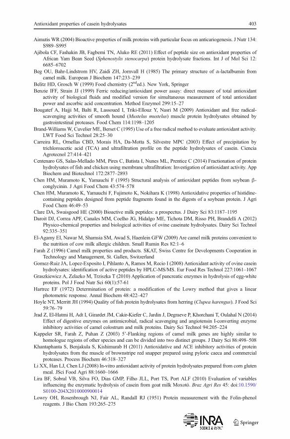
Aimutis WR (2004) Bioactive properties of milk proteins with particular focus on anticariogenesis. J Nutr 134:S989–S995
Ajibola CF, Fashakin JB, Fagbemi TN, Aluko RE (2011) Effect of peptide size on antioxidant properties ofAfrican Yam Bean Seed (Sphenostylis stenocarpa) protein hydrolysate fractions. Int J of Mol Sci 12:6685–6702
Beg OU, Bahr-Lindstrom HV, Zaidi ZH, Jornvall H (1985) The primary structure of α-lactalbumin fromcamel milk. European J Biochem 147:233–239
Belitz HD, Grosch W (1999) Food chemistry (2nded.). New York, SpringerBenzie IFF, Strain JJ (1999) Ferric reducing/antioxidant power assay: direct measure of total antioxidant
activity of biological fluids and modified version for simultaneous measurement of total antioxidantpower and ascorbic acid concentration. Method Enzymol 299:15–27
Bougatef A, Hajji M, Balti R, Lassoued I, Triki-Ellouz Y, Nasri M (2009) Antioxidant and free radical-scavenging activities of smooth hound (Mustelus mustelus) muscle protein hydrolysates obtained bygastrointestinal proteases. Food Chem 114:1198–1205
Brand-WilliamsW, Cuvelier ME, Berset C (1995) Use of a free radical method to evaluate antioxidant activity.LWT Food Sci Technol 28:25–30
Carreira RL, Ornellas CBD, Morais HA, Da-Motta S, Silvestre MPC (2003) Effect of precipitation bytrichloroacetic acid (TCA) and ultrafiltration profile on the peptide hydrolysates of casein. CienciaAgrotecnol 27:414–421
Centenaro GS, Salas-Mellado MM, Pires C, Batista I, Nunes ML, Prentice C (2014) Fractionation of proteinhydrolysates of fish and chicken using membrane ultrafiltration: Investigation of antioxidant activity. AppBiochem and Biotechnol 172:2877–2893
Chen HM, Muramoto K, Yamauchi F (1995) Structural analysis of antioxidant peptides from soybean β-conglycinin. J Agri Food Chem 43:574–578
Chen HM, Muramoto K, Yamauchi F, Fujimoto K, Nokihara K (1998) Antioxidative properties of histidine-containing peptides designed from peptide fragments found in the digests of a soybean protein. J AgriFood Chem 46:49–53
Clare DA, Swaisgood HE (2000) Bioactive milk peptides: a prospectus. J Dairy Sci 83:1187–1195Daroit DJ, Correa APF, Canales MM, Coelho JG, Hidalgo ME, Tichota DM, Risso PH, Brandelli A (2012)
Physico-chemical properties and biological activities of ovine caseinate hydrolysates. Dairy Sci Technol92:335–351
El-Agamy EI, Nawar M, Shamsia SM, Awad S, Haenlein GFW (2009) Are camel milk proteins convenient tothe nutrition of cow milk allergic children. Small Rumin Res 82:1–6
Farah Z (1996) Camel milk properties and products. SKAT, Swiss Centre for Developments Cooperation inTechnology and Management, St. Gallen, Switzerland
Gomez-Ruiz JA, Lopez-Exposito I, Pihlanto A, Ramos M, Recio I (2008) Antioxidant activity of ovine caseinhydrolysates: identification of active peptides by HPLC-MS/MS. Eur Food Res Technol 227:1061–1067
Graszkiewicz A, Zelazko M, Trziszka T (2010) Application of pancreatic enzymes in hydrolysis of egg-whiteproteins. Pol J Food Nutr Sci 60(1):57-61
Hartree EF (1972) Determination of protein: a modification of the Lowry method that gives a linearphotometric response. Annal Biochem 48:422–427
Hoyle NT, Merritt JH (1994) Quality of fish protein hydrolysates from herring (Clupea harengus). J Food Sci59:76–79
Jrad Z, El-Hatmi H, Adt I, Girardet JM, Cakir-Kiefer C, Jardin J, Degraeve P, Khorchani T, Oulahal N (2014)Effect of digestive enzymes on antimicrobial, radical scavenging and angiotensin I-converting enzymeinhibitory activities of camel colostrum and milk proteins. Dairy Sci Technol 94:205–224
Kappeler SR, Farah Z, Puhan Z (2003) 5’-Flanking regions of camel milk genes are highly similar tohomologue regions of other species and can be divided into two distinct groups. J Dairy Sci 86:498–508
Khantaphanta S, Benjakula S, Kishimurab H (2011) Antioxidative and ACE inhibitory activities of proteinhydrolysates from the muscle of brownstripe red snapper prepared using pyloric caeca and commercialproteases. Process Biochem 46:318–327
Li XX, Han LJ, Chen LJ (2008) In-vitro antioxidant activity of protein hydrolysates prepared from corn glutenmeal. JSci Food Agri 88:1660–1666
Lira BF, Sobral VB, Silva FO, Dias GMP, Filho JLL, Port TS, Port ALF (2010) Evaluation of variablesinfluencing the enzymatic hydrolysis of casein from goat milk Moxotó. Braz Agri Res 45: doi:10.1590/S0100-204X2010000900014
Lowry OH, Rosenbrough NJ, Fair AL, Randall RJ (1951) Protein measurement with the Folin-phenolreagents. J Bio Chem 193:265–275
Antioxidant properties of casein hydrolysates 403

Magjeed NA (2005) Corrective effect of camel milk on some cancer biomarkers in blood of rats intoxicatedwith aflatoxin B1. J Saudi Chem Soc 9:253–263
Mao XY, Cheng X, Wang X, Wu SJ (2011) Free-radical-scavenging and anti-inflammatory effect of yak milkcasein before and after enzymatic hydrolysis. Food Chem 126:484–490
Meisel H (2005) Biochemical properties of peptides encrypted in bovine milk proteins. Curr Med Chem 12:1905-1919
Mohamed A (1993) Characterization of camel milk β-casein. Ph.D. Thesis, University of Karachi, PakistanRival SG, Boeriu CG, Wicher HJ (2001) Casein and casein hydrolysates. II. antioxidative properties and
relevance to lipoxygenase inhibition. J Agri Food Chem 49:295–302Rossini K, Norena CPZ, Cladera-Olivera F, Brandelli A (2009) Casein peptides with inhibitory activity on
lipid oxidation in beef homogenates and mechanically deboned poultry meat. LWT - Food Sci Technol42:862–867
Salami M, Yousefi R, Ehsani MR, Dalgalarrondo M, Chobert JM, Haertle T, Razavi SH, SabouryAA N-NA,Moosavi-Movahedi AA (2008) Kinetic characterization of hydrolysis of camel and bovine milk proteinsby pancreatic enzymes. Int Dairy J 18:1097–1102
Salami M, Yousefi R, Ehsani MR, Razavi SH, ChobertJM HT (2009) Enzymatic digestion and antioxidantactivity of the native and molten globule states of camel α-lactalbumin: possible significance for use ininfant formula. Int Dairy J 19:518–523
Salami M, Moosavi-Movahedi AA, Moosavi-Movahedi F, Ehsani MR, Yousefi R, Farhadi M, Niasari-NaslajiA, Saboury AA, Chobert JM, Haertle T (2011) Biological activity of camel milk casein followingenzymatic digestion. J Dairy Res 78:471–478
Saliha SAZ, Dalila A, Chahra S, Saliha BH, AbderrahmaneM (2013) Separation and characterization of majormilk proteins from Algerian Dromedary (Camelus dromedarius). Emir J Food Agri 25:283–290
Thiansilakul Y, Benjakul S, Shahidi F (2007) Antioxidative activity of protein hydrolysate from round scadmuscle using Alcalase and Flavourzyme. J Food Biochem 31:266–287
Tsou MJ, Kao FJ, Tseng CK, Chiang WD (2010) Enhancing the anti-adipogenic activity of soy protein bylimited hydrolysis with flavourzyme and ultrafiltration. Food Chem 122:243–248
404 D. Kumar et al.