Embed Size (px)
Citation preview
Calcium chloride linked camel milk derived casein nanoparticles forthe delivery of sorafenib in hepatocarcinoma cellsAASTHA MITTAL1; NEELAM MAHALA1; KOWTHAVARAPU VENKATA KRISHNA2; UMA S. DUBEY1,*; SUNIL KUMAR DUBEY2,3,*
1Department of Biological Sciences, Birla Institute of Technology and Science, Pilani Campus, Pilani (BITS-PILANI), 333031, India
2Department of Pharmacy, Birla Institute of Technology and Science, Pilani Campus, Pilani (BITS-PILANI), 333031, India
3R&D Healthcare Division Emami, Ltd., Kolkata, 700056, India
Key words: Camel milk derived casein nanoparticles, Sorafenib, UV spectroscopy, FTIR, HepG2 cells, Apoptosis
Abstract: Sorafenib, a multikinase inhibitor used for the treatment of hepatocellular carcinoma, is limited by its low oral
bioavailability. To overcome this drawback, we have developed novel camel milk casein-derived nanoparticles as a drug
delivery system. Camel milk casein is not only biocompatible on oral administration but is actually a dietary protein of
pharmaceutical relevance. Casein is used because of its amphiphilic nature, self-assembling property, ability to show
sustained release, and capability of encapsulating both hydrophilic and hydrophobic drugs. In this study, camel milk
casein nanoparticles loaded with sorafenib were developed and characterized. Characterization of casein nanoparticles
was done by dynamic light scattering (DLS), zeta potential analysis, scanning light microscopy (SEM), and FTIR. The
drug content in nanoparticle and drug-protein binding studies were conducted by UV spectroscopy. The cytotoxicity
and cellular uptake efficiency studies were performed in HepG2 cell lines. It was observed that the cytotoxic effect of
sorafenib loaded camel milk casein nanoparticles was more than free sorafenib in HepG2 cells. This work suggests
camel milk casein as a suitable drug delivery molecule for sorafenib. In the future, it may also be used in enhancing
the efficacy and specific distribution of other water-insoluble anticancer drugs.
Introduction
Nanotechnology has a very high potential in the diagnosis,treatment, and management of cancer (Gmeiner and Ghosh,2015; Din et al., 2017). The characteristics properties ofnanoparticles (NPs) like small size, customized surface,improved solubility, multi-functionality, and administrationthrough different routes are of utmost importance (Krishnaet al., 2019). These features overcome the disadvantages ofusing conventional chemotherapeutic agents (Singh andLillard, 2009; Patra et al., 2018; Hejmady et al., 2020).Different nanocarriers such as liposomes, micelles, polymericnanomaterials, mesoporous silica, metallic nanoparticles havebeen used in drug delivery for improving an array ofbiomedical applications (Krishna et al., 2020). Among variouskinds of protein NPs, milk whey proteins, alpha-lactalbumin,b-lactoglobulin, caseins, etc., have been investigated for thedelivery of drugs, nutraceuticals, and bioactive peptides.These GRAS natural derivatives have characteristics like
biocompatibility, amphiphilicity, wide availability, lack oftoxicity, non-antigenicity, and low cost, making them suitablefor drug delivery, safety, and improved therapeutic potentialof anticancer drugs (Lohcharoenkal et al., 2014).
Casein nanoparticles have a high drug loading capacity andare able to deliver both hydrophilic and hydrophobic drugmolecules. The pH-dependent behavior of casein micellesmakes it beneficial for the controlled release of substancesadministered orally. Furthermore, the unfolded structure ofcaseins makes them easily accessible for proteolysis, ensuringgood release by proteolytic enzymes in the gastrointestinaltract. Additionally, caseins can penetrate the cell membrane inan energy-independent fashion, which can enhance cellularuptake on oral administration. Physiological degradationproducts of casein produce immunomodulatory peptides,antioxidants, and ACE inhibitors (which optimize bloodpressure regulation). All of these features make casein apromising candidate for encapsulation matrix for anticancerdrugs (Głąb and Boratyński, 2017; Sahu et al., 2008).
Camel milk has been traditionally consumed worldwidefor its nutritional benefits and medicinal properties. It alsohas therapeutic and prophylactic benefits against cancer(Dubey et al., 2016; Al haj and Al Kanhal, 2010). Camel
*Address correspondence to: Sunil Kumar Dubey,[email protected]; Uma S. Dubey, [email protected]: 24 January 2021; Accepted: 29 March 2021
BIOCELL echT PressScience2022 46(1): 127-136
Doi: 10.32604/biocell.2021.015932 www.techscience.com/journal/biocell
This work is licensed under a Creative Commons Attribution 4.0 International License, which permitsunrestricted use, distribution, and reproduction in any medium, provided the original work is properlycited.
milk proteins have more thermostability and pH hydrolysisresistance (Atri et al., 2011). Camel milk casein is present inthe micellar form, is easily biodegradable, has anamphiphilic character, is an antioxidant, and has amolecular chaperone-like activity (Kumar et al., 2016).Earlier, also camel casein has been used as an effective nanovehicle for curcumin, and its application has been suggestedin functional health food formulations (Esmaili et al., 2011).Camel milk casein has the potential to bind with both polarand non-polar compounds and serve as a suitablenanocarrier for other physiological compounds, includingdrugs. Therefore, in our study, we have synthesized camelmilk casein nanoparticles and used them as a carrier forencapsulating the drug sorafenib, which is an FDA-approved drug against human hepatocellular carcinoma(HCC). HCC is the most common type of primary livercancer and the most common cause of death in people withcirrhosis (Balogh et al., 2016).
Kinases play a very important role in tumor cell signaling,angiogenesis, and apoptosis (Krishna et al., 2021). Sorafenib is avery promising anticancer drug with the properties of amultikinase inhibitor. It acts against several solid tumors bypreventing cell proliferation, tumor growth, and angiogenesis(Jindal et al., 2019). However, sorafenib is a small-moleculedrug, having poor aqueous solubility leading to lowbioavailability. As it is a small-molecule drug, it may distributethroughout the whole body and be rapidly metabolized in thebody leading to adverse effects (Babos et al., 2018). Camelmilk is known to reduce drug-induced hepatic and renaltoxicity, whereas casein-derived peptides are importantpharmaceutically active molecules in addition to beingimmunomodulatory molecules (Głąb and Boratyński, 2017;Kumar et al., 2016). Therefore, in this study, we have exploredthe use of casein-derived nanoparticles from camel milk forthe encapsulation of sorafenib. For this, we have initiallydeveloped and characterized camel casein nanoparticles. Laterwe studied the cellular uptake and cytotoxicity of sorafenibloaded casein nanoparticles in the HepG2 cell line.
Materials and Methods
Chemicals and reagentsSDS-PAGE molecular weight marker, MTT (3-(4,5-dimethylthiazol-2-yl)-2,5-diphenyltetrazolium Bromide, DAPI(4’,6-diamidino-2-phenylindole) were purchased from Sigma(Bangalore, India), 2-Benzothiazolyl)-7-(diethylamino) coumarin(C6) was purchased from TCI Chemicals (India) Pvt. Ltd. Allorganic solvents were of HPLC grade.
Camel milk sample collectionCamel milk samples were collected from ‘Sarika Raika MilkBhandar’, Jaipur, India. The sample was collected underaseptic conditions, stored at −20°C in a sterile containeruntil further use.
Isolation and purification of casein protein from camel milkIsolation of casein from camel (Camelus dromedarius) milk as wasdone according to the process described earlier (Salami et al., 2011;Kumar et al., 2016) with slight modification. Briefly, fat wasremoved from the milk by centrifugation at 8000 rpm for
30 min at 4°C. The supernatant containing skimmed milk wascollected for further use. Whey and casein were separated byacid precipitation with 1 N HCl at pH 4.6 and centrifugation at12000 rpm for 30 min at 4°C. The precipitated casein waswashed thrice with distilled water. The precipitated casein pelletwas kept at −20°C until further use and solubilized according toa reported method (Madadlou et al., 2009) briefly described asfollows. Casein solution (5%) was prepared by adding 10 gcasein protein to 200 mL of deionized water (pH 7.0), stirringat 1000 rpm for 60 min at room temperature, sodium azide(0.03%) was added to prevent any microbial contamination.The solution was stored at 4°C for 10–12 h to allow completehydration. The pH of the solution, which was originally 6.48,was then adjusted to 7, 9, or 12, separately, by slow addition of1 N sodium hydroxide solution with constant stirring.
Polyacrylamide gel electrophoresisSkimmed milk and isolated camel milk casein protein wereanalyzed by SDS-PAGE on 12% gel under reducingcondition by using a Bio-Rad mini gel electrophoresis unitrun at 80 V. Before the electrophoresis, skimmed milk andcasein protein samples were diluted to 2 μg/μL with thesample buffer. Sample denaturation was done for 5 min at100°C, and protein samples were loaded to each well.Proteins in gel were stained by Coomassie Brilliant BlueR-250, and the gel image was viewed by the use of a geldocumentation system (BioRad). Separated proteins werevalidated for identification using the molecular weight marker.
UV visible spectroscopyQuantitative analysis of sorafenib and casein was performedby UV spectroscopy. Stock solutions of casein and sorafenibdrug were prepared in MQ water and methanol,respectively. The UV absorption spectra were recorded on aUV-2700 spectrophotometer (Shimadzu Co., Kyoto, Japan)over a wavelength range of 200–300 nm, and λmax of thesecompounds was estimated from the calibration curve. Thebinding of sorafenib drug with casein micelles was analyzedby UV spectrophotometry.
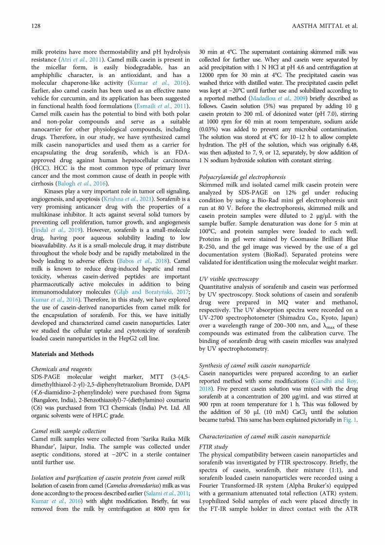
Synthesis of camel milk casein nanoparticleCasein nanoparticles were prepared according to an earlierreported method with some modifications (Gandhi and Roy,2018). Five percent casein solution was mixed with the drugsorafenib at a concentration of 200 µg/mL and was stirred at900 rpm at room temperature for 1 h. This was followed bythe addition of 50 μL (10 mM) CaCl2 until the solutionbecame turbid. This same has been explained pictorially in Fig. 1.
Characterization of camel milk casein nanoparticle
FTIR studyThe physical compatibility between casein nanoparticles andsorafenib was investigated by FTIR spectroscopy. Briefly, thespectra of casein, sorafenib, their mixture (1:1), andsorafenib loaded casein nanoparticles were recorded using aFourier Transformed-IR system (Alpha Bruker’s) equippedwith a germanium attenuated total reflection (ATR) system.Lyophilized Solid samples of each were placed directly inthe FT-IR sample holder in direct contact with the ATR
128 AASTHA MITTAL et al.
crystal. IR spectra were obtained in the spectral region4,000 cm-1 to 500 cm-1, in transmittance mode.
Physical property analysis and scanning electron microscopy ofcamel milk casein nanoparticlesThe prepared casein nanoparticles were characterized for thesize, polydispersity index (PDI), and zeta potential using aZetasizer, Nano series (Nano-ZS, Malvern, UK). The caseinnanoparticle diameter and surface charge/ZP were measuredin water pH (6.2–7.4) at 25°C with a scattering angle of90°C. Morphology was examined by scanning electronmicroscopy (SEM) at a voltage of 20 kV after coating withgold under vacuum. Drops of Nanoparticles samples wereput on slides and were dried under nitrogen gas. Then theseslides were placed on aluminum stubs fixed by double-sidedtape and placed in the vacuum chamber of SEM (ThermoScientificTM Apreo scanning electron microscope). Slideswere seen from 30,000 × to 10,000 × magnifications.
Drug encapsulation efficiencyTo quantify the drug content, 1 mL aliquot of thenanoparticle solution containing the drug wascentrifuged at 10,000 rpm for 20 min at 4°C. Now thepellet was dissolved in methanol to precipitate theprotein. Subsequently, it was vortexed for 1 h, followedby centrifugation at 6000 rpm for 10 min. The drugconcentration in the pellet was determined using a UVspectrophotometer. Results have been presented as means± SE of three independent experiments, each performedin duplicate. The encapsulation efficiency (E.E.) wascalculated by using the following formula:
E.E. (%) = (mass of a drug in nanoparticles/mass of drugused in formulation) × 100.
Cell culture associated studies
Culture of cellsHepG2 cells procured from NCCS, Pune, India, were culturedat 37°C, 5% CO2, in minimum essential medium (Invitrogen)
supplemented with 10% fetal bovine serum (Invitrogen) and1% antibiotics solution–Penicillin (100 U mL−1) andstreptomycin (100 μg mL−1; Invitrogen). Trypsin–EDTAsolution (0.05%) was used for the detachment of cells. Thecells were grown to 70% confluence in tissue culture flasks(Tarson), detached using Trypsin EDTA solution, rinsed inphosphate buffer saline, and finally transferred in completefresh medium. Subsequently, they were analyzed for cellularuptake, viability, and morphological changes.
Cellular internalization studyCoumarin-6 (green-fluorescent dye) entrapped caseinnanoparticles were prepared in the same way as describedabove in synthesis part. HepG2 cells were seeded oncoverslips in six-well plates and treated with the 3 µg/mL ofcoumarin-6 entrapped casein nanoparticles at 37°C for 4and 6 h, respectively. The cells were then washed twice withPBS to remove nanoparticles not taken up by the cells andfixed with methanol for 10 min. Subsequently, they werestained with DAPI (2 µg/mL) for 3–5 min and washed twicewith PBS. Cells were mounted using glycerol and examinedunder a fluorescence microscope (Zeiss Axio Scope A1)using the filters for DAPI and FITC. The merged image wasalso obtained for better visualization.
Cytotoxicity assayMTT (3-(4, 5-dimethylthiazol-2-yl)-2,5-diphenyltetrazoliumBromide) assay was used to determine the cytotoxicity ofthe drug, sorafenib, and its loaded casein nanoparticles.Briefly, HepG2 cells were seeded at a density of 8000cells/well in a 96-well plate. After reaching the 70%confluence, cells were treated with different concentrationsof the drug sorafenib and its loaded casein nanoparticles for24 h. MTT was added to cells and incubated for 4 h at37°C. Subsequently, the media was discarded and 150 μL ofDMSO was added to each well. The color intensity wasmeasured at 570 nm wavelength, using a microplate reader(Multiskan™ Thermo Scientific™). Each determination was
FIGURE 1. Pictorial representation of the synthesis of casein nanoparticles. (A) 5% casein solution with sorafenib drug. (B) Turbid mildlycasein solution upon addition of calcium chloride (CaCl2). (C) Casein nanoparticle.
CAMEL MILK DERIVED CASEIN NANOPARTICLES 129
carried out in triplicate and at least two independentexperiments were carried out. Cell viability was calculated asusing the following formula:
% Cell viability = OD of treated cells/OD of control(without treatment) × 100
DAPI stainingHepG2 cells were implanted on a coverslip in a 6-well plateand incubated with only sorafenib and casein encapsulatedsorafenib, respectively, at 10 µM and 20 µM concentrationfor 24 h. Cells were then washed twice with PBS, fixed withice-cold methanol, and again washed twice with PBS. Next,cells were stained with DAPI (2 μg/mL) and incubated for10 min at room temperature in the dark. The cells werefinally washed with PBS two times and visualized under afluorescence microscope (ZEISS Axio Scope A1microscope). The Apoptotic bodies were observed.
Statistical Analysis
Graphs and statistical analysis of data was performed by usingGraphPad prism5 software. The data are given as the meanvalues ± SEM. Comparisons among inter and intra groupswere analyzed using one-way and two-way ANOVA followedby the Bonferroni post test. P < 0.05 was considered toindicate a statistically significant difference. All experimentswere repeated at least three times performed in triplicates.
Result and Discussion
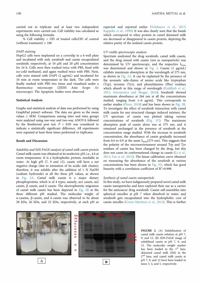
Solubility and SDS PAGE analysis of camel milk casein proteinCamel milk casein was obtained at its isoelectric pH, i.e., 4.6 atroom temperature. It is a hydrophobic protein, insoluble inwater. At high pH (7, 9 and 12), casein will have a netnegative charge (due to ionization of its acidic side chains);therefore, it was soluble after the addition of 1 N NaOH(sodium hydroxide) at all the three pH values, as shownin Fig. 2A. Camel milk casein is a major dietaryphosphoprotein, which is of 4 types, namely, αs1 casein, αs2casein, β casein, and k casein. The electrophoretic migrationof camel milk casein has been depicted in Fig. 2B at thethree different pH studied. The molecular weight ofα-caseins, β-casein, and k-casein was observed to be about28 kDa, 26 kDa, and 22 kDa, respectively, at each pH as
expected and reported earlier (Yelubaeva et al., 2017;Kappeler et al., 1999). It was also clearly seen that the bandswhich correspond to whey protein in camel skimmed milkare decreased or disappeared in casein protein, depicting therelative purity of the isolated casein protein.
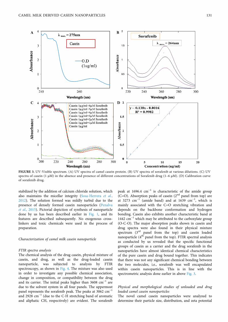
UV-visible spectroscopic analysisSpectrum analysisof the drug sorafenib, camel milk casein,and the drug mixed with casein (not as nanoparticle) wasdetermined by UV spectroscopy, and the respective λmax
was determined and shown in Fig. 3. Casein (1 µg/mL)exhibits maximum absorption at the wavelength of 275 nm,as shown in Fig. 3A. It can be explained by the presence ofthe aromatic side-chains of amino acids like tryptophan(Trp), tyrosine (Tyr), and phenylalanine (Phe) residueswhich absorb in this range of wavelength (Goldfarb et al.,1951; Antosiewicz and Shugar, 2016). Sorafenib showedmaximum absorbance at 264 nm at all the concentrationsstudied, ranging from 1–6 µg/mL. This corresponds toearlier studies (Onur, 2018) and has been shown in Fig. 3B.To investigate the effect of sorafenib interaction with camelmilk casein for any structural changes induced mixture, theUV spectrum of casein was plotted taking variousconcentrations of sorafenib (Fig. 3C). The maximumabsorption peak of casein alone was at 275 nm, and itremained unchanged in the presence of sorafenib at theconcentration range studied. With the increase in sorafenibconcentration, the absorbance of casein gradually increasedfrom 0.6 to 0.9 at the same λmax(275 nm). This suggests thatthe polarity of the microenvironment around Trp and Tyrresidues of casein has been changed by the drug, but thisdoes not cause its conformational change in casein (Li et al.,2011; Yue et al., 2012). The linear calibration curve obtainedon measuring the absorbance of the sorafenib at variousconcentrations has been shown in Fig. 3D, which has goodlinearity with a correlation coefficient of R2=0.998.
Synthesis of camel casein nanoparticleIn this study, we have indigenously prepared novel camel milkcasein nanoparticles and have explored their use as a carrierfor the anticancer drug sorafenib. Casein self-assembles intospherical micelles at pH 7 when dissolved in water, andsorafenib gets encapsulated into the hydrophobic core ofcasein micelles (Corzo-Martínez et al., 2015). This is further
FIGURE 2. (A) Solubilization ofcamel milk casein solution at pH 7,9, and 12. (B) SDS-PAGE image ofsolubilized casein at pH 7, 9, and12. The molecular weight markerhas been loaded in the 1st lane,skimmed camel milk (SM) in the2nd lane, and camel milk casein atpH 7, 9, and 12 have been loaded inlanes 3, 4, and 5, respectively.
130 AASTHA MITTAL et al.
stabilized by the addition of calcium chloride solution, whichalso maintains the micellar integrity (Sosa-Herrera et al.,2012). The solution formed was mildly turbid due to thepresence of densely formed casein nanoparticles (Penalvaet al., 2015). Pictorial depiction of synthesis of nanoparticledone by us has been described earlier in Fig. 1, and itsfeatures are described subsequently. No exogenous cross-linkers and toxic chemicals were used in the process ofpreparation.
Characterization of camel milk casein nanoparticle
FTIR spectra analysisThe chemical analysis of the drug casein, physical mixture ofcasein, and drug, as well as the drug-loaded caseinnanoparticle, was subjected to analysis by FTIRspectroscopy, as shown in Fig. 4. The mixture was also usedin order to investigate any possible chemical association,change in composition, or compatibility between the drugand its carrier. The initial peaks higher than 3608 cm-1 aredue to the solvent system in all four panels. The uppermostpanel represents the sorafenib peak. The peaks at 3062 cm−1
and 2928 cm−1 (due to the C-H stretching band of aromaticand aliphatic CH, respectively) are evident. The sorafenib
peak at 1696.4 cm−1 is characteristic of the amide group(C=O). Absorption peaks of casein (2nd panel from top) areat 3273 cm−1 (amide band) and at 1639 cm−1, which ismainly associated with the C=O stretching vibration anddepends on the backbone conformation and hydrogenbonding. Casein also exhibits another characteristic band at1442 cm−1 which may be attributed to the carboxylate group(O-C-O). The major absorption peaks shown in casein anddrug spectra were also found in their physical mixturespectrum (3rd panel from the top) and casein loadednanoparticle (4th panel from the top). FTIR spectral analysisas conducted by us revealed that the specific functionalgroups of casein as a carrier and the drug sorafenib in thenanoparticles have almost identical chemical characteristicsof the pure casein and drug bound together. This indicatesthat there was not any significant chemical bonding betweenthe two molecules, i.e., sorafenib was well encapsulatedwithin casein nanoparticles. This is in line with thespectrometric analysis done earlier in above Fig. 3.
Physical and morphological studies of unloaded and drugloaded camel casein nanoparticlesThe novel camel casein nanoparticles were analyzed todetermine their particle size, distribution, and zeta potential
FIGURE 3. UV-Visible spectrum. (A) UV spectra of camel casein protein. (B) UV spectra of sorafenib at various dilutions. (C) UVspectra of casein (1 µM) in the absence and presence of different concentrations of Sorafenib drug (1–6 μM). (D) Calibration curveof sorafenib drug.
CAMEL MILK DERIVED CASEIN NANOPARTICLES 131

using the Malvern zeta sizer, shown in Fig. 5 and Tab. 1. Camelmilk casein nanoparticles were found to have an averagehydrodynamic particle size of 192 ± 5.4 nm with apolydispersity index of 0.22, whereas sorafenib-loaded camelcasein nanoparticles had a slightly greater size, i.e., 212 ± 6nm, with a narrow polydispersity index of 0.23 (shown in Tab.1). This indicates that the particles were uniform andmonodispersed. The entrapment efficiency of drug-loadednanoparticles was observed to be 51.5 ± 3.2%. Zeta potential isthe measure of the surface charge of NPs and has animportant effect on the stability of the colloidal system. Thezeta potential of casein nanoparticles was found to be
negatively charged (data are shown in Tab. 1) because of theionized carboxylic groups of sodium caseinate at pH 7. Thenegative surface charge is also indicative of low toxicity ascompared to the positively charged nanoparticle, whichinternalizes the cells by various mechanisms that need to beexplored further (Tang et al., 2018).
The shape of camel casein nanoparticles was analyzedusing scanning electron microscopy (SEM). A smooth surfaceand spherical shape of the nanoparticles were observed as hasbeen shown in Fig. 6. The size of particles ranges between160–220 nm, which is in agreement with the dynamic lightscattering studies done earlier (Penalva et al., 2015).
FIGURE 4. FTIR spectra of (1)sorafenib drug, (2) casein at pH7, (3) a physical mixture ofcasein and drug, and (4) casein-loaded drug nanoparticle (NP).
FIGURE 5. Measurement of nanoparticles. (A) Size and (B) zeta potential of casein nanoparticles by dynamic light scattering (DLS).
TABLE 1
Characterization of nanoparticles
NPs Dh (nm) PDI ZP (mV) EE%
Casein NP 192 ± 5.4 0.21 –23 ± 1.5 –
Sorafenib loaded casein nanoparticle 212 ± 6 0.22 –20 ± 1.2 51.5 ± 3.2Note: Dh, average hydrodynamic diameter; PDI, polydispersity index; ZP, zeta potential; EE, drug encapsulationefficiency. Data represent mean ± SD (N = 3).
132 AASTHA MITTAL et al.
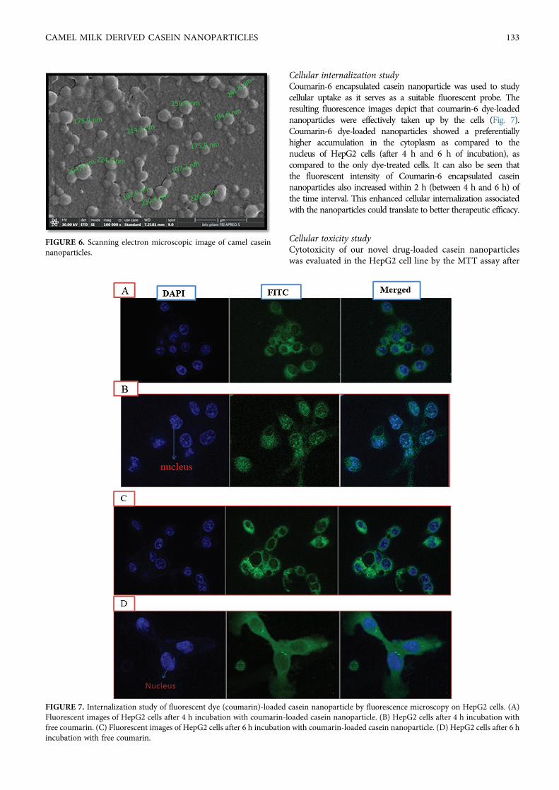
Cellular internalization studyCoumarin-6 encapsulated casein nanoparticle was used to studycellular uptake as it serves as a suitable fluorescent probe. Theresulting fluorescence images depict that coumarin-6 dye-loadednanoparticles were effectively taken up by the cells (Fig. 7).Coumarin-6 dye-loaded nanoparticles showed a preferentiallyhigher accumulation in the cytoplasm as compared to thenucleus of HepG2 cells (after 4 h and 6 h of incubation), ascompared to the only dye-treated cells. It can also be seen thatthe fluorescent intensity of Coumarin-6 encapsulated caseinnanoparticles also increased within 2 h (between 4 h and 6 h) ofthe time interval. This enhanced cellular internalization associatedwith the nanoparticles could translate to better therapeutic efficacy.
Cellular toxicity studyCytotoxicity of our novel drug-loaded casein nanoparticleswas evaluated in the HepG2 cell line by the MTT assay after
FIGURE 6. Scanning electron microscopic image of camel caseinnanoparticles.
FIGURE 7. Internalization study of fluorescent dye (coumarin)-loaded casein nanoparticle by fluorescence microscopy on HepG2 cells. (A)Fluorescent images of HepG2 cells after 4 h incubation with coumarin-loaded casein nanoparticle. (B) HepG2 cells after 4 h incubation withfree coumarin. (C) Fluorescent images of HepG2 cells after 6 h incubation with coumarin-loaded casein nanoparticle. (D) HepG2 cells after 6 hincubation with free coumarin.
CAMEL MILK DERIVED CASEIN NANOPARTICLES 133
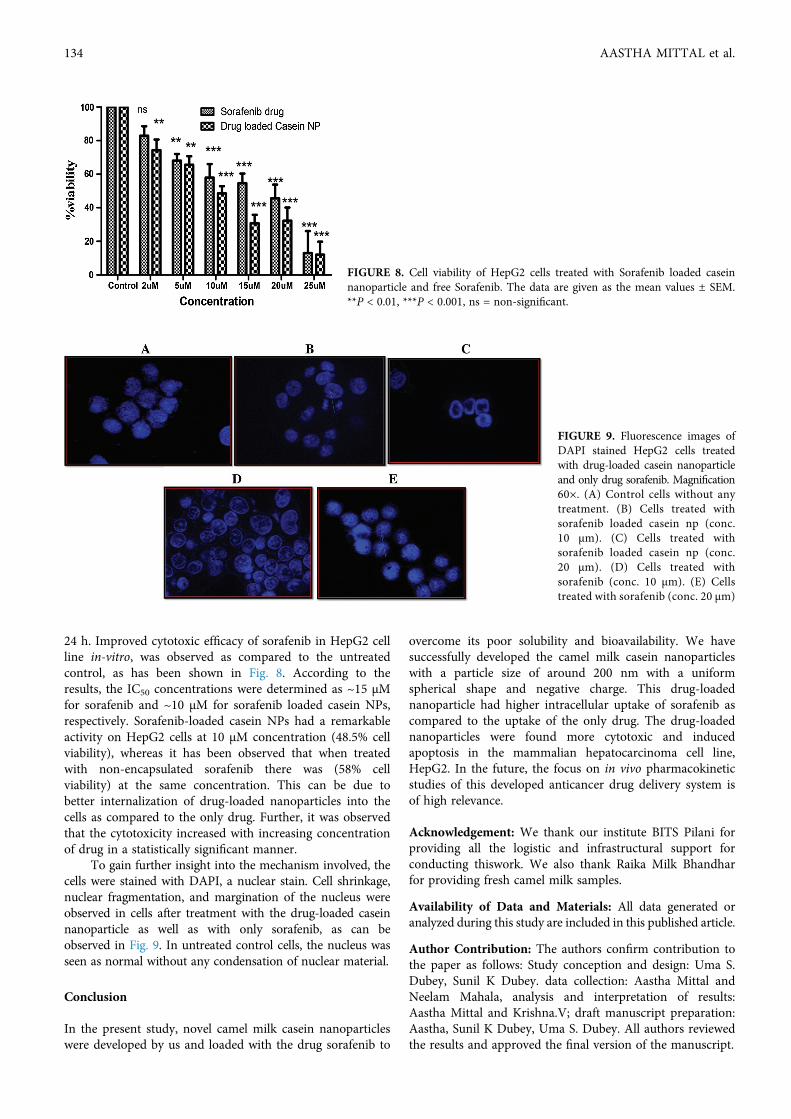
24 h. Improved cytotoxic efficacy of sorafenib in HepG2 cellline in-vitro, was observed as compared to the untreatedcontrol, as has been shown in Fig. 8. According to theresults, the IC50 concentrations were determined as ~15 μMfor sorafenib and ~10 μM for sorafenib loaded casein NPs,respectively. Sorafenib-loaded casein NPs had a remarkableactivity on HepG2 cells at 10 μM concentration (48.5% cellviability), whereas it has been observed that when treatedwith non-encapsulated sorafenib there was (58% cellviability) at the same concentration. This can be due tobetter internalization of drug-loaded nanoparticles into thecells as compared to the only drug. Further, it was observedthat the cytotoxicity increased with increasing concentrationof drug in a statistically significant manner.
To gain further insight into the mechanism involved, thecells were stained with DAPI, a nuclear stain. Cell shrinkage,nuclear fragmentation, and margination of the nucleus wereobserved in cells after treatment with the drug-loaded caseinnanoparticle as well as with only sorafenib, as can beobserved in Fig. 9. In untreated control cells, the nucleus wasseen as normal without any condensation of nuclear material.
Conclusion
In the present study, novel camel milk casein nanoparticleswere developed by us and loaded with the drug sorafenib to
overcome its poor solubility and bioavailability. We havesuccessfully developed the camel milk casein nanoparticleswith a particle size of around 200 nm with a uniformspherical shape and negative charge. This drug-loadednanoparticle had higher intracellular uptake of sorafenib ascompared to the uptake of the only drug. The drug-loadednanoparticles were found more cytotoxic and inducedapoptosis in the mammalian hepatocarcinoma cell line,HepG2. In the future, the focus on in vivo pharmacokineticstudies of this developed anticancer drug delivery system isof high relevance.
Acknowledgement: We thank our institute BITS Pilani forproviding all the logistic and infrastructural support forconducting thiswork. We also thank Raika Milk Bhandharfor providing fresh camel milk samples.
Availability of Data and Materials: All data generated oranalyzed during this study are included in this published article.
Author Contribution: The authors confirm contribution tothe paper as follows: Study conception and design: Uma S.Dubey, Sunil K Dubey. data collection: Aastha Mittal andNeelam Mahala, analysis and interpretation of results:Aastha Mittal and Krishna.V; draft manuscript preparation:Aastha, Sunil K Dubey, Uma S. Dubey. All authors reviewedthe results and approved the final version of the manuscript.
FIGURE 8. Cell viability of HepG2 cells treated with Sorafenib loaded caseinnanoparticle and free Sorafenib. The data are given as the mean values ± SEM.**P < 0.01, ***P < 0.001, ns = non-significant.
FIGURE 9. Fluorescence images ofDAPI stained HepG2 cells treatedwith drug-loaded casein nanoparticleand only drug sorafenib. Magnification60×. (A) Control cells without anytreatment. (B) Cells treated withsorafenib loaded casein np (conc.10 µm). (C) Cells treated withsorafenib loaded casein np (conc.20 µm). (D) Cells treated withsorafenib (conc. 10 µm). (E) Cellstreated with sorafenib (conc. 20 µm)
134 AASTHA MITTAL et al.
Ethics Approval:No specific approval was required for this study.
Funding Statement: The authors received no specific fundingfor this study.
Conflicts of Interest: The authors declare that they have noconflicts of interest.
References
Al haj OA, Al Kanhal HA (2010). Compositional, technological andnutritional aspects of dromedary camel milk. InternationalDairy Journal 20: 811–821. DOI 10.1016/j.idairyj.2010.04.003.
Antosiewicz JM, Shugar D (2016). UV-Vis spectroscopy of tyrosineside-groups in studies of protein structure. Part 2: Selectedapplications. Biophysical Reviews 8: 163–177. DOI 10.1007/s12551-016-0197-7.
Atri MS, Saboury AA, Moosavi-Movahedi AA, Goliaei B, SefidbakhtY et al. (2011). Structure and stability analysis of cytotoxiccomplex of camel α-lactalbumin and unsaturated fatty acidsproduced at high temperature. Journal of BiomolecularStructure and Dynamics 28: 919–928. DOI 10.1080/07391102.2011.10508618.
Babos G, Biró E, Meiczinger M, Feczkó T (2018). Dual drug deliveryof sorafenib and doxorubicin from PLGA and PEG-PLGApolymeric nanoparticles. Polymers 10: 1–12. DOI 10.3390/polym10080895.
Balogh J, Victor III D, Asham EH, Burroughs SG, Boktour M (2016).Hepatocellular carcinoma: A review. Journal of HepatocellularCarcinoma 3: 41–53. DOI 10.2147/JHC.S61146.
Corzo-Martínez M, Mohan M, Dunlap J, Harte F (2015). Effect ofultra-high pressure homogenization on the interactionbetween bovine casein micelles and ritonavir.Pharmaceutical Research 32: 1055–1071. DOI 10.1007/s11095-014-1518-9.
Din FU, Aman W, Ullah I, Qureshi OS, Mustapha O, Shafique S, ZebA (2017). effective use of nanocarriers as drug deliverysystems for treatment of selected tumors. InternationalJournal of Nanomedicine 12: 7291–7309. DOI 10.2147/IJN.S146315.
Dubey US, Lal M, Mittal A, Kapur S (2016). Therapeutic potential ofcamel milk. Emirates Journal of Food and Agriculture 28:164–176. DOI 10.975/ejfa.2015-04-122.
Esmaili M, Ghaffari SM, Moosavi-Movahedi Z, Atri MS, SharifizadehA et al. (2011). Beta casein-micelle as a nano vehicle forsolubility enhancement of curcumin; food industryapplication. LWT-Food Science and Technology 44: 2166–2172. DOI 10.1016/j.lwt.2011.05.023.
Gandhi S, Roy I (2018). Doxorubicin-loaded casein nanoparticles for drugdelivery: Preparation, characterization and in vitro evaluation.International Journal of Biological Macromolecules 121: 6–12.
Głąb TK, Boratyński J (2017). Potential of casein as a carrier forbiologically active agents. Topics in Current Chemistry 375:1–20. DOI 10.1007/s41061-017-0158-z.
Gmeiner WH, Ghosh S (2015). Nanotechnology for cancertreatment. Nanotechnology Reviews 3: 111–122. DOI10.1515/ntrev-2013-0013.
Goldfarb AR, Saidel LJ, Moscovich E (1951). The ultraviolet absorptionspectra of proteins. Journal of Biological Chemistry 193: 397–404.
Hejmady S, Pradhan R, Alexander A, Agrawal M, Singhvi G et al.(2020). Recent advances in targeted nanomedicine aspromising antitumor therapeutics. Drug Discovery Today25: 2227–2244. DOI 10.1016/j.drudis.2020.09.031.
Jindal A, Thadi A, Shailubhai K (2019). Hepatocellular carcinoma:Etiology and current and future drugs. Journal of Clinicaland Experimental Hepatology 9: 221–232. DOI 10.1016/j.jceh.2019.01.004.
Kappeler S, Farah Z, Puhan Z (1999). Sequence analysis of Camelusdromedarius milk caseins. InterJournal of Dairy Research 9:481–486. DOI 10.1017/S0022029997002847.
Krishna KV, Saha RN, Dubey SK (2020). Biophysical, biochemical,and behavioral implications of ApoE3 conjugated donepezilnanomedicine in a Aβ1-42 induced Alzheimer’s disease ratmodel. ACS Chemical Neuroscience 11: 4139–4151. DOI10.1021/acschemneuro.0c00430.
Krishna KV, Wadhwa G, Alexander A, Kanojia N, Saha RN et al.(2019). Design and biological evaluation of lipoprotein-based donepezil nanocarrier for enhanced brain uptakethrough oral delivery. ACS Chemical Neuroscience 10:4124–4135. DOI 10.1021/acschemneuro.9b00343.
Krishna KV, Dubey SK, Singhvi G, Gupta G, Kesharwani P (2021).MAPK pathway: Potential role in glioblastomamultiformurgery. Interdisciplinary Neurosurgery: AdvancedTechniques and Case Management 23: 100901. DOI10.1016/j.inat.2020.100901.
Kumar D, Chatli MK, Singh R, Mehta N, Kumar P (2016). Enzymatichydrolysis of camel milk casein and its antioxidantproperties. Dairy Science and Technology 96: 391–404. DOI10.1007/s13594-015-0275-9.
Li D, Zhu M, Xu C, Ji B (2011). Characterization of the baicalein e-bovine serum albumin complex without or with Cu2+ or Fe3+
by spectroscopic approaches. European Journal of MedicinalChemistry 46: 588–599. DOI 10.1016/j.ejmech.2010.11.038.
Lohcharoenkal W, Wang L, Chen YC, Rojanasakul Y (2014). Proteinnanoparticles as drug delivery carriers for cancer therapy.BioMed Research International 2014: 180549. DOI 10.1155/2014/180549.
Madadlou A, Mousavi ME, Emam-Djomeh Z, Sheehan D, Ehsani M(2009). Alkaline pH does not disrupt re-assembled caseinmicelles. Food Chemistry 116: 929–932. DOI 10.1016/j.foodchem.2009.03.04.
Onur S (2018). Rapid determination and validation of sorafenib viauv-visible method in pharmaceutical formulations. BalıkesirSağlık Bilimleri Dergisi 7: 87–92.
Patra JK, Das G, Fraceto LF, Vangelie E, Campos R et al. (2018).Nano based drug delivery systems: Recent developmentsand future prospects. Journal of Nanobiotechnology 16:1–33. DOI 10.1186/s12951-018-0392-8.
Penalva R, Esparza I, Agüeros M, Gonzalez-Navarro CJ, Gonzalez-Ferrero C, Irache JM (2015). Casein nanoparticles as carriersfor the oral delivery of folic acid. Food Hydrocolloids 44:399–406.
Sahu A, Kasoju N, Bora U (2008). Fluorescence study of thecurcumin-casein micelle complexation and its applicationas a drug nanocarrier to cancer cells. Biomacromolecules 9:2905–2912.
Salami M, Moosavi-Movahedi AA, Moosavi-Movahedi F, Ehsani MR,Yousefi R et al. (2011). Biological activity of camel milk caseinfollowing enzymatic digestion. Journal of Dairy Research 78:471–478. DOI 10.1017/S0022029911000628.
Singh R, Lillard JW Jr (2009). Nanoparticle-based targeted drugdelivery. Experimental & Molecular Pathology 86: 215–223.DOI 10.1016/j.yexmp.2008.12.004.
Sosa-Herrera MG, Lozano-Esquivel IE, Ponce de León-Ramírez YR,Martínez-Padilla LP (2012). Effect of added calcium chloride
CAMEL MILK DERIVED CASEIN NANOPARTICLES 135
on the physicochemical and rheological properties ofaqueous mixtures of sodium caseinate/sodium alginate andrespective oil-in-water emulsions. Food Hydrocolloids 29:175–184. DOI 10.1016/j.foodhyd.2012.02.017.
Tang X, Chen L, Li A, Cai S, Zhang Y et al. (2018). Anti-GPC3 antibody-modified sorafenib-loaded nanoparticles significantly inhibitedHepG2 hepatocellular carcinoma. Drug Delivery 25: 1484–1494. DOI 10.1080/10717544.2018.1477859.
Yelubaeva MY, Buralkhiev BA, Serikbayeva AD, Narmuratova MH,Kenenbay SY (2017). Electrophoretic identification ofcasein in various types of milk. OnLine Journal of BiologicalSciences 17: 348–352.
Yue Q, Shen T, Wang C, Gao C, Liu J (2012). Study on the interactionof bovine serum albumin with ceftriaxone and the inhibitioneffect of zinc (II). International Journal of Spectroscopy 2012:284173. DOI 10.1155/2021/284173.
136 AASTHA MITTAL et al.