Embed Size (px)
Citation preview
antioxidants
Article
Engineering the Unicellular AlgaPhaeodactylum tricornutum for EnhancingCarotenoid Production
Francesco Manfellotto 1,*, Giulio Rocco Stella 2,3 , Angela Falciatore 2,4, Christophe Brunet 1 andMaria Immacolata Ferrante 1,*
1 Stazione Zoologica Anton Dohrn, Villa Comunale, 80121 Naples, Italy; [email protected] Laboratory of Computational and Quantitative Biology, UMR 7238, Centre National de la Recherche
Scientifique (CNRS), Sorbonne Université, Institut de Biologie Paris-Seine, F-75005 Paris, France;[email protected] (G.R.S.); [email protected] (A.F.)
3 Boston Consulting Group, Via Ugo Foscolo 1, 20121 Milano, Italy4 Laboratory of Chloroplast Biology and Light Sensing in Microalgae, UMR 7141, Centre National de la Recherche
Scientifique (CNRS), Sorbonne Université, Institut de Biologie Physico-Chimique, F-75005 Paris, France* Correspondence: [email protected] (F.M.); [email protected] (M.I.F.);
Tel.: +39-081-583-3268 (M.I.F.); Fax: +39-081-764-1355 (M.I.F.)
Received: 23 July 2020; Accepted: 13 August 2020; Published: 16 August 2020�����������������
Abstract: Microalgae represent a promising resource for the production of beneficial natural compoundsdue to their richness in secondary metabolites and easy cultivation. Carotenoids feature among distinctivecompounds of many microalgae, including diatoms, which owe their golden color to the xanthophyllfucoxanthin. Carotenoids have antioxidant, anti-obesity and anti-inflammatory properties, and there is aconsiderable market demand for these compounds. Here, with the aim to increase the carotenoid contentin the model diatom Phaeodactylum tricornutum, we exploited genetic transformation to overexpressgenes involved in the carotenoid biosynthetic pathway. We produced transgenic lines over-expressingsimultaneously one, two or three carotenoid biosynthetic genes, and evaluated changes in pigmentcontent with high-performance liquid chromatography. Two triple transformants over-expressing thegenes Violaxanthin de-epoxidase (Vde), Vde-related (Vdr) and Zeaxanthin epoxidase 3 (Zep3) showed anaccumulation of carotenoids, with an increase in the fucoxanthin content up to four fold. Vde, Vdr andZep3 mRNA and protein levels in the triple transformants were coherently increased. The exact role ofthese enzymes in the diatom carotenoid biosynthetic pathway is not completely elucidated neverthelessour strategy successfully modulated the carotenoid metabolism leading to an accumulation of valuablecompounds, leading the way toward improved utilization of microalgae in the field of antioxidants.
Keywords: diatoms; Phaeodacytlum tricornutum; carotenoids; fucoxanthin; genetic engineering
1. Introduction
Diatoms, one of the major groups of microalgae, represent a potential source for commercial andindustrial applications, because they naturally produce various beneficial substances for human activities,including foodstuffs and pharmaceuticals. Moreover, almost all of biomass can have a profitable use [1].
Diatoms have also been considered for applications such as production of biofuels, biofertilizersand nanomaterials, for industrial waste detoxification [2] and aquaculture feed [3]. Diatoms are richin pharmaceutically active substances such as polyunsaturated fatty acids, vitamins, antioxidants,enzymes, polysaccharides and carotenoids [4]. Among diatoms, Phaeodactylum tricornutum is a verypromising microorganism for application in industrial processes because it grows rapidly, doublingits biomass in a few hours [1]. Its growth is cost-effective, sustainable and can be easily controlled
Antioxidants 2020, 9, 757; doi:10.3390/antiox9080757 www.mdpi.com/journal/antioxidants
Antioxidants 2020, 9, 757 2 of 13
in indoor and outdoor conditions [5]. P. tricornutum is also the best-established diatom molecularmodel system, with cutting edge tools in place to alter gene expression in transformed cells by geneticover-expression, gene silencing and genome editing [6].
We focused our work on carotenoid production enhancement in P. tricornutum. Bioactivity andhuman health benefits of carotenoids are increasingly described [7].
Carotenoids are tetraterpenoids formed by a linear C40 main chain with multiple conjugateddouble bonds [8]. In diatoms, as in all photosynthetic organisms, carotenoids are synthesized via themethylerythritol phosphate (MEP) pathway [9,10]. Carotenoids associate with clorophyll to form thepigment-protein complexes of the photosinthetic apparatus [11].
Diatoms are phylogenetically closer to brown algae than to the green lineage [6] and theircarotenoid profile is different from that found in plants and green algae: fucoxanthin is the mostabundant carotenoid followed by diadinoxanthin (Dd), diatoxanthin (Dt) and β-carotene. Chlorophylla and chlorophyll c form with fucoxanthin the fucoxanthin-chlorophyll protein complex that performslight-harvesting. Dx is known to play an important role in photoprotection [12,13], belonging to theso-called xanthophyll cycle: the diatoxanthin–diadinoxanthin cycle (Dd-Dt). Diatoms also present themore widely distributed violaxanthin–zeaxanthin cycle.
Fucoxantin presents anti-obesity, anti-diabetic and anti-cancer properties. Fucoxanthin and itsmetabolite, fucoxanthinol, are well established as strong antioxidant compounds [14], they have radicalscavenging activities which are 13.5 and 1.7 times higher, respectively, than the activity of α-tocopherol,and fucoxanthin anticancer effect is stronger than that of β-carotene [15]. Dd prevents damage resultingfrom exposure of skin and hair to the UV and visible range of the solar spectrum [16,17].
The biosynthetic carotenoid pigments pathway has been extensively studied in plants and greenalgae; however, diatoms show different metabolic features compared to plants [18] and, as mentioned,use unique pigments, that are not present in other species, for light harvesting and photoprotection [19].
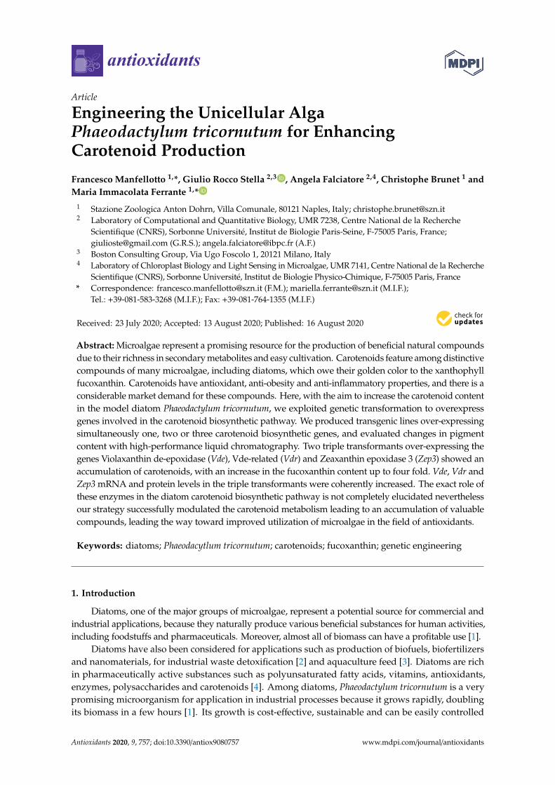
The biosynthetic carotenoid pathway is still not completely understood, and reactions andenzymes from violaxanthin to Dx and fucoxanthin are still hypothetical (Figure 1).
Antioxidants 2020, 9, x FOR PEER REVIEW 3 of 13
Figure 1. Schematic representation of the carotenoid biosynthesis pathway in P. tricornutum, according to the hypothesized pathways proposed by Lohr and Wilhelm 1999 [20] and Dambek et al. 2012 [21]. Dashed lines are used for unknown enzymes. Red arrows represent de-epoxidation reactions and green arrows represent epoxidation reactions operating in the two xanthophyll cycles. GGDS: geranylgeranyl diphosphate synthase; PSY: phytoene synthase; PDS: phytoene desaturase; ZDS: ζ-carotene desaturase; CRTISO: carotene cis/trans isomerase, prolycopene isomerase; LCYB: lycopene cyclase b; LUT: lutein deficient-like; ZEP: zeaxanthin epoxidase; VDE: violaxanthin de-epoxidase. Adapted from M. Bertand et al. (2010) and P. Kuczynska et al. (2015) [9,19].
Attempts to overexpress the 1-deoxy-D-xylulose 5-phosphate synthase (Dxs), the gateway enzyme of the terpenoid pathway, or the Phytoene synthase (Psy), controlling the initial step of the carotenoid biosynthesis, resulted in a moderate increase in fucoxanthin content [22,23].
In the P. tricornutum genome there are seven genes putatively involved in violaxanthin-zeaxanthin and Dd-Dt cycles, four for the de-epoxidase reactions (Violaxanthin de-epoxidase Vde, Vde-like Vdl1 and 2, and the Vde-related, Vdr), which convert violaxanthin in zeaxanthin or Dd in Dt, and three for the reverse reactions (Zeaxanthin epoxidases, Zep1, 2 and 3) [24]. The expansion of these gene families is in contrast with what happens in plants, where only one Vde, one Vdr and one Zep are present. Of the three Zeps genes, Zep1 was found to be non-functional in zeaxanthin epoxidation whereas Zep2 and Zep3 were able to restore zeaxanthin epoxidation and a functional xanthophyll cycle in Arabidopsis thaliana [25]. However, these enzymes exhibited different catalytic activities; for example, ZEP2 exhibited a broader substrate specificity with respect to ZEP3, leading to the
Figure 1. Schematic representation of the carotenoid biosynthesis pathway in P. tricornutum, accordingto the hypothesized pathways proposed by Lohr and Wilhelm 1999 [20] and Dambek et al. 2012 [21].Dashed lines are used for unknown enzymes. Red arrows represent de-epoxidation reactions and greenarrows represent epoxidation reactions operating in the two xanthophyll cycles. GGDS: geranylgeranyldiphosphate synthase; PSY: phytoene synthase; PDS: phytoene desaturase; ZDS: ζ-carotene desaturase;CRTISO: carotene cis/trans isomerase, prolycopene isomerase; LCYB: lycopene cyclase b; LUT: luteindeficient-like; ZEP: zeaxanthin epoxidase; VDE: violaxanthin de-epoxidase. Adapted from M. Bertand et al.(2010) and P. Kuczynska et al. (2015) [9,19].

Antioxidants 2020, 9, 757 3 of 13
Attempts to overexpress the 1-deoxy-d-xylulose 5-phosphate synthase (Dxs), the gateway enzymeof the terpenoid pathway, or the Phytoene synthase (Psy), controlling the initial step of the carotenoidbiosynthesis, resulted in a moderate increase in fucoxanthin content [22,23].
In the P. tricornutum genome there are seven genes putatively involved in violaxanthin-zeaxanthinand Dd-Dt cycles, four for the de-epoxidase reactions (Violaxanthin de-epoxidase Vde, Vde-like Vdl1and 2, and the Vde-related, Vdr), which convert violaxanthin in zeaxanthin or Dd in Dt, and threefor the reverse reactions (Zeaxanthin epoxidases, Zep1, 2 and 3) [24]. The expansion of these genefamilies is in contrast with what happens in plants, where only one Vde, one Vdr and one Zep arepresent. Of the three Zeps genes, Zep1 was found to be non-functional in zeaxanthin epoxidationwhereas Zep2 and Zep3 were able to restore zeaxanthin epoxidation and a functional xanthophyll cyclein Arabidopsis thaliana [25]. However, these enzymes exhibited different catalytic activities; for example,ZEP2 exhibited a broader substrate specificity with respect to ZEP3, leading to the hypothesis thatthis enzyme could be involved in the Dd-Dt cycle [25]. De-epoxidation of the violaxanthin cycle inchlorophytes is catalyzed by VDE. Acting in both xanthophyll cycles, the diatom VDE has been shownto participate in high light acclimation and non-photochemical quenching (NPQ) kinetics [26,27].Vde gene expression is related to photoprotection and strongly induced by high light stress. In vitro,the VDE enzyme can use both violaxanthin and Dd as substrate for de-epoxidation reaction [28,29].Lavaud et al. reported that Vde knock-down lines negatively impact de-epoxidation reactions andpresent Dd-Dt accumulation [27]. The function of the VDL and VDR proteins remains unknown.They probably have a xanthophyll cycle activity in addition to VDE, perhaps differing in localizationand functional role as indicated by the differential light induced expression of the Vdl compared tothe Vde genes [9,24,27]. Vdr is a high light-induced gene and appears to be generally present in allchlorophytes [24]; it supposedly participates in de-epoxidation reactions in addition to VDE at a certainthreshold of light and lumen acidification [30]. VDL2 has been recently overexpressed in the diatomThalassiosira pseudonana, leading to an increase in fucoxanthin and a reduction in Dd-Dt, without anet change in their sum nor in β-carotene, suggesting that VDL2 is required in the step leading fromthe Dd-Dt pigments to fucoxanthin [31]. Reactions from violaxanthin to fucoxanthin and enzymesinvolved are still unknown, but it had been hypothesized that the reactions are catalysed by VDLsproteins. VDE, VDL1, and VDL2 from P. tricornutum have been recently expressed in Escherichia coli,and tested in an in vitro assay commonly used for measuring VDE activity. VDE showed the expectedde-epoxidase activity by converting both violaxanthin to zeaxanthin and Dd to Dt, VDL2 showed nocatalytic activity, whereas VDL1 converted violaxanthin to neoxanthin, suggesting that violaxanthin isits major native substrate [32].
Efforts are needed in order to target the rate limiting steps in the pathway: a strategy in whichmore than one enzyme is overexpressed in the same strain might be more effective, similar to what hasbeen shown for example in yeast, where the simultaneous overexpression of two genes involved inastaxanthin production led to more product than overexpression of the single genes [33].
Our aim was to enhance pigments production in P. tricornutum. Since the exact function ofeach enzyme of the xanthophyll cycle is still unclear, to maximize chances of interfering with thebiosynthetic mechanisms, in parallel with single overexpression strategies already applied in paststudies, we simultaneously tested different double and triple combinations of VDEs and ZEPs genesusing biolistic transformation.
2. Materials and Methods
2.1. Algal Cultures
The P. tricornutum Pt1 strain (Pt1 8.6 CCMP2561) was used in this study. Wild type cells weregrown at 18 ◦C, in a 12 h light/12 h dark photoperiod using white fluorescence neon lamps (PhilipsTL-D 90), at irradiance of 90 µmol m−2 s−1 in ventilated flasks in f/2 medium (Guillard, 1975) [34].
Antioxidants 2020, 9, 757 4 of 13
Transgenic cells were grown under the same condition in selective liquid f/2 medium supplementedwith 50 µg/mL zeocin.
2.2. Plasmids Construction
Total RNA was isolated from P. tricornutum cells using TriPure Isolation Reagent (Sigma-Aldrich,Saint Louise, MO, USA) following manufacturer specifications. Then, cDNA was synthesized witha QuantiTect Reverse Transcription Kit (Qiagen, Hilden, Germany) and was used as template forthe amplification of the transcripts of interest. Transformation vectors for VDEs and ZEPs (Table 1)overexpression were generated by amplifying the cDNA using the primers described in Table S1.DNA fragments amplification was performed using Q5® High-Fidelity DNA Polymerase (NewEngland Biolabs, Ipswich, MA, USA) according to the manufacturer’s instructions. The full-lengthcDNA sequences were cloned in an Invitrogen Entry vector (pENTR) and then in the diatompKSFcpBpAt-C-3HA destination vector, with the Gateway technology [35].
Table 1. Genes selected for overexpression vectors construction.
Gene Description Gene Name Gene ID
Violaxanthin deepoxidase like protein Vde Phatr3_J51703Violaxanthin deepoxidase-like 1 protein Vdl 1 Phatr3_J36048Violaxanthin deepoxidase-like 2 protein Vdl 2 Phatr3_J45846
Zeaxanthin epoxidase like protein Zep 1 Phatr3_J45845Zeaxanthin epoxidase2-like protein Zep 2 Phatr3_J5928Zeaxanthin epoxidase3-like protein Zep 3 Phatr3_J10970
Violaxanthin deepoxidase-related protein Vdr Phatr3_J43240Phytoene desaturase Pds Phatr3_J10438
Lycopene beta cyclase Lcy Phatr3_J8835Phytoene synthase Psy Phatr3_EG02349
Lutein deficient 1-like protein Lut 1 Phatr3_J16586
The plasmids containing the Pds, Lcy, Psy and Lut1 genes were synthesized from cDNA using theprimers described in Table S1. DNA fragments amplification was performed using Q5® High-FidelityDNA Polymerase (New England Biolabs, Ipswich, MA, USA) according to the manufacturer’sinstructions. PCR amplification was performed using a double annealing temperature, first tencycles at the annealing temperature of the primers without tails and then 25 cycles at the annealingtemperature of the entire primer sequence. The amplicons were purified by agarose gel electrophoresisusing a QIAquick® Gel Extraction Kit (Qiagen, Hilden, Germany). PCR products and plasmids weredigested with the restriction enzyme EcoRI (New England Biolabs, Ipswich, MA, USA). The digestedamplicons and plasmids pfcpb-OX [36] were ligated using the T4 DNA Ligase (New England Biolabs,Ipswich, MA, USA) according to the manufacturer’s instructions. We proceeded cloning plasmids inTOP10 competent cells (Thermo Fisher Scientific, Waltham, MA, USA). Plasmids were purified usingQIAprep Miniprep Kit (Qiagen, Hilden, Germany) after overnight growth. All purified plasmids werefurther verified by Sanger sequencing.
2.3. Microparticle Bombardment
The plasmids were introduced into P. tricornutum cells using the Biolistic PDS-1000/He particledelivery system (Bio-Rad Laboratories, Hercules, CA, USA). We co-transformed P. tricornutum cellsusing the pFCPFp-Sh-ble vector conferring zeocin resistance and the carotenoid biosynthesis genes asdescribed in Falciatore et al. (1999) [36]. For single gene transformation, we coated tungsten particleswith 3 µg of resistance vector + 3 µg of the transformation vector. For double gene transformation,we coated tungsten particles with 2 µg of resistance vector + 2 µg for each transformation vector. Fortriple gene transformation we coated tungsten particles with 1.5 µg for each transformation vector.
Antioxidants 2020, 9, 757 5 of 13
2.4. Positive Colony Screening
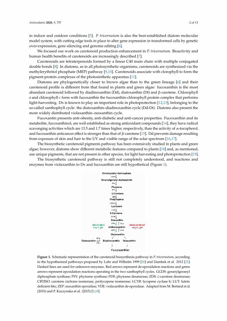
PCR screening to select positive transformants colony was performed on cell lysate as described inFalciatore et al. (1999) [36]. To screen positives transformants, we designed a forward primer annealingto the exogenous fcpB sequence and specific reverse primers, annealing to the carotenoid biosynthesisgenes (Figure 2 and Table S1).
Antioxidants 2020, 9, x FOR PEER REVIEW 5 of 13
Gene Description Gene Name Gene ID Violaxanthin deepoxidase like protein Vde Phatr3_J51703
Violaxanthin deepoxidase-like 1 protein Vdl 1 Phatr3_J36048 Violaxanthin deepoxidase-like 2 protein Vdl 2 Phatr3_J45846
Zeaxanthin epoxidase like protein Zep 1 Phatr3_J45845 Zeaxanthin epoxidase2-like protein Zep 2 Phatr3_J5928 Zeaxanthin epoxidase3-like protein Zep 3 Phatr3_J10970
Violaxanthin deepoxidase-related protein Vdr Phatr3_J43240 Phytoene desaturase Pds Phatr3_J10438
Lycopene beta cyclase Lcy Phatr3_J8835 Phytoene synthase Psy Phatr3_EG02349
Lutein deficient 1-like protein Lut 1 Phatr3_J16586
The plasmids containing the Pds, Lcy, Psy and Lut1 genes were synthesized from cDNA using the primers described in Table S1. DNA fragments amplification was performed using Q5® High-Fidelity DNA Polymerase (New England Biolabs, Ipswich, MA, USA) according to the manufacturer’s instructions. PCR amplification was performed using a double annealing temperature, first ten cycles at the annealing temperature of the primers without tails and then 25 cycles at the annealing temperature of the entire primer sequence. The amplicons were purified by agarose gel electrophoresis using a QIAquick® Gel Extraction Kit (Qiagen, Hilden, Germany). PCR products and plasmids were digested with the restriction enzyme EcoRI (New England Biolabs, Ipswich, MA, USA). The digested amplicons and plasmids pfcpb-OX [36] were ligated using the T4 DNA Ligase (New England Biolabs, Ipswich, MA, USA) according to the manufacturer’s instructions. We proceeded cloning plasmids in TOP10 competent cells (Thermo Fisher Scientific, Waltham, MA, USA). Plasmids were purified using QIAprep Miniprep Kit (Qiagen, Hilden, Germany) after overnight growth. All purified plasmids were further verified by Sanger sequencing.
2.3. Microparticle Bombardment
The plasmids were introduced into P. tricornutum cells using the Biolistic PDS-1000/He particle delivery system (Bio-Rad Laboratories, Hercules, CA, USA). We co-transformed P. tricornutum cells using the pFCPFp-Sh-ble vector conferring zeocin resistance and the carotenoid biosynthesis genes as described in Falciatore et al. (1999) [36]. For single gene transformation, we coated tungsten particles with 3 μg of resistance vector + 3 μg of the transformation vector. For double gene transformation, we coated tungsten particles with 2 μg of resistance vector + 2 μg for each transformation vector. For triple gene transformation we coated tungsten particles with 1.5 μg for each transformation vector.
2.4. Positive Colony Screening PCR screening to select positive transformants colony was performed on cell lysate as described
in Falciatore et al. (1999) [36]. To screen positives transformants, we designed a forward primer annealing to the exogenous fcpB sequence and specific reverse primers, annealing to the carotenoid biosynthesis genes (Figure 2 and Table S1).
Figure 2. Schematic representation of the construct and position of the primers annealing sites used to screen exogenous gene integration.
2.5. Pigment Profile
Figure 2. Schematic representation of the construct and position of the primers annealing sites used toscreen exogenous gene integration.
2.5. Pigment Profile
Pigment profile was analyzed following the protocol of Smerilli et al. (2017) [37]. Samples ofapproximately 15 × 107 cells of P. tricornutum were harvested during the exponential growth phaseand immediately filtered on 25-mm GF/F glass-fiber filters (Whatman™, Whatman International Ltd.,Maidstone, UK). Pigments were extracted with 100% methanol by mechanical grounding, then thehomogenate was filtered onto Whatman 25 mm GF/F filters. A Hewlett Packard series 1100 HPLC(Hewlett Packard, Wilmington, NC, USA), equipped with a C8Kinetex column (2.6 µm diameter;50 mm × 4.6 mm; Phenomenex®, Torrance, CA, USA) was used to separate pigments, which were thusdetected spectrophotometrically at 440 nm using a Hewlett Packard photodiode array detector, modelDAD series1100. Determination and quantification of pigments were performed using pure pigments(D.H.I. Water & Environment; Horsholm, Denmark).
2.6. Gene Expression Profile
The expression levels of target genes were evaluated by qPCR. We designed couples of primers toamplify the endogenous and exogenous genes (Table S1). Total RNA was isolated from approximately5 × 107 transformed cells using TriPure Isolation Reagent (Sigma-Aldrich, Saint Louis, MO, USA).Reverse transcription (RT) was performed using QuantiTect Reverse Transcription Kit (Qiagen, Hilden,Germany). The qPCR experiments were performed in triplicate in a ViiA 7 Real-Time PCR System(Applied Biosystems, Foster City, CA, USA) using Fast SYBR™Green Master Mix (Applied Biosystems,Foster City, CA, USA), following manufacturer instructions. The reference genes used were H4 andRPS [35]. Quantification were made following the ∆-∆-Ct method. The results are mean ± SD of atleast three separate experiments, measuring each parameter by triplicate (n = 3).
2.7. Western Blot Analysis
Protein extraction was performed from 5 × 10⁸ P. tricornutum cells at the exponential growthphase, collected by centrifugation at 2000× g, following the published “TCA protein extraction fromdiatoms” (dx.doi.org/10.17504/protocols.io.bc7rizm6) [38]. Then, 40 ng of total proteins were loadedon 10% polyacrylamide gels and resolved by SDS-PAGE. Proteins were transferred to a nitrocellulosemembrane using a Trans-Blot Semi-Dry Electrophoretic TransferCell (Bio-Rad Laboratories, Hercules,CA, USA) for 1 h at 140 mA in Towbin buffer. Transferred proteins were visualized by Ponceau Sstaining. Blocking was performed in PBS-T buffer supplemented with 5% defatted milk for 1 h at roomtemperature. A rabbit anti-PsbD antibody (Agrisera, Vännäs, Sweden) and a rabbit anti-HA antibody(Agrisera, Vännäs, Sweden) were used for detection of control D2 protein of PSII and HA taggedexogenous proteins, at 1:20,000 dilution in blocking buffer for 1 h. Incubation with the horseradishperoxidase-coupled secondary anti rabbit IgG antibody (Agrisera, Vännäs, Sweden), diluted 1:40,000,lasted for 1 h. Chemoluminescence was detected using the ECL Western Blotting Substrate (Thermo
Antioxidants 2020, 9, 757 6 of 13
Fisher Scientific, Waltham, MA, USA). Membranes were imaged with a Gel Doc XR imaging system(Bio-Rad Laboratories, Hercules, CA, USA) to quantify band intensities by densitometry, usingQuantity-One software (Bio-Rad Laboratories, Hercules, CA, USA).
2.8. Non-Photochemical Quenching of Chlorophyll Fluorescence (NPQ)
Fifteen min. dark-acclimated samples were inserted in a DUAL-PAM (Heinz Walz, Effeltrich,Germany) to estimate NPQ (non-photochemical quenching) [37]. The actinic light was setup at480 µmol photons m−2 s−1 lasting 10 min., and the maximum fluorescence yield was estimated everymin. Fm was obtained applying a saturating pulse of red light (4000 µmol m−2 s−1, lasting 450 ms).NPQ was quantified using the Stern-Volmer expression [37].
3. Results
With the aim to increase carotenoids production, we transformed P. tricornutum cells with singleor multiple combinations of plasmids for overexpression of genes putatively involved in the pigmentsbiosynthetic pathway (Figure 1, Table S2). We obtained over 400 resistant colonies. After isolation,we PCR-screened the resistant colonies to confirm exogenous transgenes integration, using a forwardprimer annealing on the inserted FcpB promoter and a reverse primer annealing on the gene codingsequence in order to discriminate exogenous genes integration (Table S1).
The effect of single and multiple genes integration on the carotenoid content under light irradianceof 90 µmol m−2 s−1 was evaluated by HPLC analysis, in all those cultures which appeared healthy andgrew normally (Tables S2 and S3). In this first pass large screening, where it was not feasible to groweach culture in triplicate, we tested each sample once to identify the most promising strains. For all thesingle overexpressing (OE) strains and for most part of the double OE strains the carotenoid contentwas not substantially different from the content in the wild type strain. From the HPLC screening,one of the double transformants (Vdr/Vde) and four triple transformant lines (Vdr/Vde/Zep3) showed amarked increase in the pigment content (Tables S2 and S3).
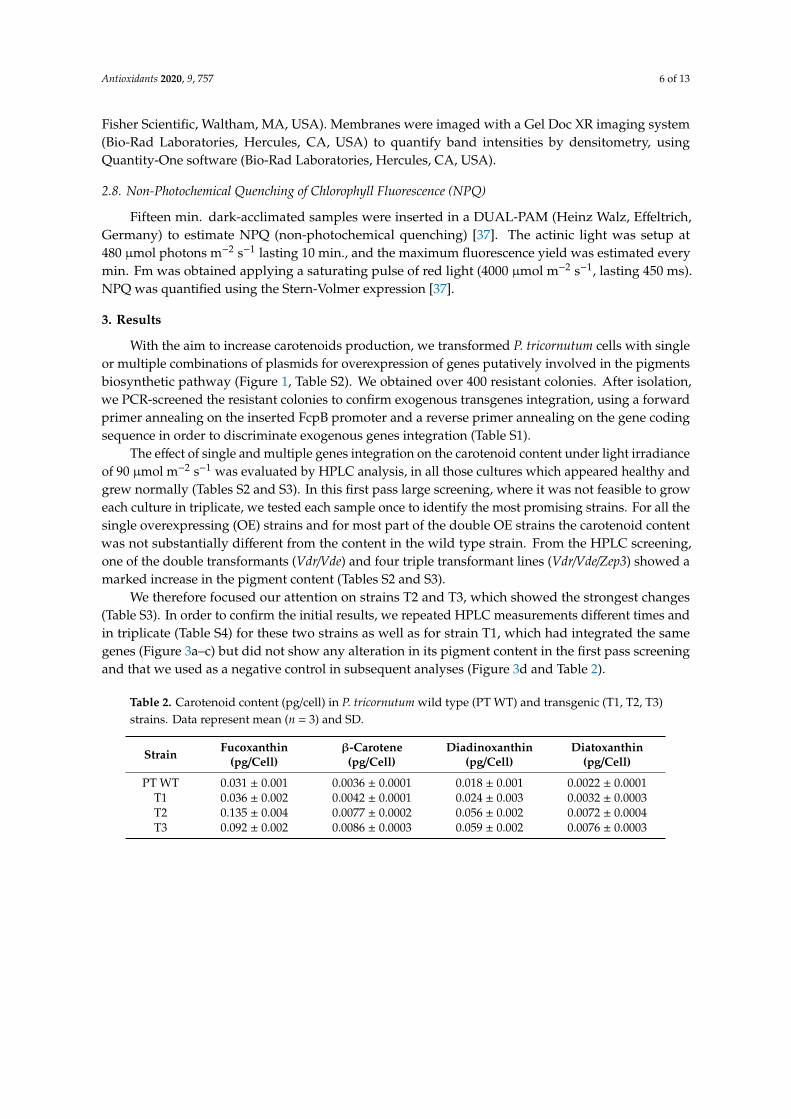
We therefore focused our attention on strains T2 and T3, which showed the strongest changes(Table S3). In order to confirm the initial results, we repeated HPLC measurements different times andin triplicate (Table S4) for these two strains as well as for strain T1, which had integrated the samegenes (Figure 3a–c) but did not show any alteration in its pigment content in the first pass screeningand that we used as a negative control in subsequent analyses (Figure 3d and Table 2).
Table 2. Carotenoid content (pg/cell) in P. tricornutum wild type (PT WT) and transgenic (T1, T2, T3)strains. Data represent mean (n = 3) and SD.
Strain Fucoxanthin(pg/Cell)
β-Carotene(pg/Cell)
Diadinoxanthin(pg/Cell)
Diatoxanthin(pg/Cell)
PT WT 0.031 ± 0.001 0.0036 ± 0.0001 0.018 ± 0.001 0.0022 ± 0.0001T1 0.036 ± 0.002 0.0042 ± 0.0001 0.024 ± 0.003 0.0032 ± 0.0003T2 0.135 ± 0.004 0.0077 ± 0.0002 0.056 ± 0.002 0.0072 ± 0.0004T3 0.092 ± 0.002 0.0086 ± 0.0003 0.059 ± 0.002 0.0076 ± 0.0003

Antioxidants 2020, 9, 757 7 of 13
Antioxidants 2020, 9, x FOR PEER REVIEW 7 of 13
isolation, we PCR-screened the resistant colonies to confirm exogenous transgenes integration, using a forward primer annealing on the inserted FcpB promoter and a reverse primer annealing on the gene coding sequence in order to discriminate exogenous genes integration (Table S1).
The effect of single and multiple genes integration on the carotenoid content under light irradiance of 90 μmol m−2 s−1 was evaluated by HPLC analysis, in all those cultures which appeared healthy and grew normally (Table S2 and S3). In this first pass large screening, where it was not feasible to grow each culture in triplicate, we tested each sample once to identify the most promising strains. For all the single overexpressing (OE) strains and for most part of the double OE strains the carotenoid content was not substantially different from the content in the wild type strain. From the HPLC screening, one of the double transformants (Vdr/Vde) and four triple transformant lines (Vdr/Vde/Zep3) showed a marked increase in the pigment content (Table S2 and S3).
We therefore focused our attention on strains T2 and T3, which showed the strongest changes (Table S3). In order to confirm the initial results, we repeated HPLC measurements different times and in triplicate (Table S4) for these two strains as well as for strain T1, which had integrated the same genes (Figure 3a–c) but did not show any alteration in its pigment content in the first pass screening and that we used as a negative control in subsequent analyses (Figure 3d and Table 2).
Figure 3. Transgene integration and pigments change in P. tricornutum wild type and OE strains T1, T2 and T3. (a); PCR screening for Vdr, using Forward FcpB and reverse VDRint primers, expected fragment of 383 bp. (b); PCR screening for Vde, using Forward FcpB and reverse VDEint primers, expected fragment of 443 bp. (c); PCR screening for Zep3, using Forward FcpB and reverse ZEP3int primers, expected fragment of 848 bp. L; 100 bp ladder. PT WT; P. tricornutum wild type. T1, T2. T3, triple transformants. +, positive control, the vector used for transformation. (d); Fold change in the pigments content normalised for chlorophyll a in transgenic strains with respect to wild type. Data represent mean (n = 3) and SD.
Table 2. Carotenoid content (pg/cell) in P. tricornutum wild type (PT WT) and transgenic (T1, T2, T3) strains. Data represent mean (n = 3) and SD.
Strain Fucoxanthin
(pg/Cell) β-Carotene
(pg/Cell) Diadinoxanthin
(pg/Cell) Diatoxanthin
(pg/Cell)
Figure 3. Transgene integration and pigments change in P. tricornutum wild type and OE strains T1,T2 and T3. (a); PCR screening for Vdr, using Forward FcpB and reverse VDRint primers, expectedfragment of 383 bp. (b); PCR screening for Vde, using Forward FcpB and reverse VDEint primers,expected fragment of 443 bp. (c); PCR screening for Zep3, using Forward FcpB and reverse ZEP3intprimers, expected fragment of 848 bp. L; 100 bp ladder. PT WT; P. tricornutum wild type. T1, T2. T3,triple transformants. +, positive control, the vector used for transformation. (d); Fold change in thepigments content normalised for chlorophyll a in transgenic strains with respect to wild type. Datarepresent mean (n = 3) and SD.
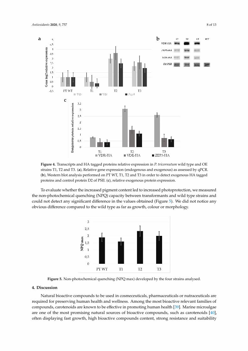
Transformants strains were analysed by quantitative RT-PCR (qPCR). We confirmed the positivecorrelation between carotenoid amount variations and exogenous gene expression. In particular, Zep3,Vde and Vdr were upregulated between 2 and 3 folds in strains T2 and T3 with respect to the wild type,as shown in Figure 4a, while T1 did not show significant variations. To confirm the overexpression ofthe biosynthetic enzymes, we looked at the presence of exogenous proteins by Western blot analysis.Using an anti HA antibody, we detected the exogenous VDR, ZEP3 and VDE HA-tagged proteins of apredicted molecular weight of 84 kDa, 71 kDa and 66 kDa, respectively, in the multiple transgenic linesT1, T2 and T3, albeit with different intensities. The D2 protein of PSII (39 kDa) was used as control fornormalization. Strains T2 and T3 showed a stronger expression of exogenous proteins with respect tothe T1 strain (Figure 4b,c).
Antioxidants 2020, 9, 757 8 of 13
Antioxidants 2020, 9, x FOR PEER REVIEW 8 of 13
PT WT 0.031 ± 0.001 0.0036 ± 0.0001 0.018 ± 0.001 0.0022 ± 0.0001 T1 0.036 ± 0.002 0.0042 ± 0.0001 0.024 ± 0.003 0.0032 ± 0.0003 T2 0.135 ± 0.004 0.0077 ± 0.0002 0.056 ± 0.002 0.0072 ± 0.0004 T3 0.092 ± 0.002 0.0086 ± 0.0003 0.059 ± 0.002 0.0076 ± 0.0003
Transformants strains were analysed by quantitative RT-PCR (qPCR). We confirmed the positive correlation between carotenoid amount variations and exogenous gene expression. In particular, Zep3, Vde and Vdr were upregulated between 2 and 3 folds in strains T2 and T3 with respect to the wild type, as shown in Figure 4a, while T1 did not show significant variations. To confirm the overexpression of the biosynthetic enzymes, we looked at the presence of exogenous proteins by Western blot analysis. Using an anti HA antibody, we detected the exogenous VDR, ZEP3 and VDE HA-tagged proteins of a predicted molecular weight of 84 kDa, 71 kDa and 66 kDa, respectively, in the multiple transgenic lines T1, T2 and T3, albeit with different intensities. The D2 protein of PSII (39 kDa) was used as control for normalization. Strains T2 and T3 showed a stronger expression of exogenous proteins with respect to the T1 strain (Figure 4b,c).
Figure 4. Transcripts and HA tagged proteins relative expression in P. tricornutum wild type and OE strains T1, T2 and T3. (a), Relative gene expression (endogenous and exogenous) as assessed by qPCR. (b), Western blot analysis performed on PT WT, T1, T2 and T3 in order to detect exogenous HA tagged proteins and control protein D2 of PSII. (c), relative exogenous protein expression.
To evaluate whether the increased pigment content led to increased photoprotection, we measured the non-photochemical quenching (NPQ) capacity between transformants and wild type strains and could not detect any significant difference in the values obtained (Figure 5). We did not notice any obvious difference compared to the wild type as far as growth, colour or morphology.
Figure 4. Transcripts and HA tagged proteins relative expression in P. tricornutum wild type and OEstrains T1, T2 and T3. (a), Relative gene expression (endogenous and exogenous) as assessed by qPCR.(b), Western blot analysis performed on PT WT, T1, T2 and T3 in order to detect exogenous HA taggedproteins and control protein D2 of PSII. (c), relative exogenous protein expression.
To evaluate whether the increased pigment content led to increased photoprotection, we measuredthe non-photochemical quenching (NPQ) capacity between transformants and wild type strains andcould not detect any significant difference in the values obtained (Figure 5). We did not notice anyobvious difference compared to the wild type as far as growth, colour or morphology.Antioxidants 2020, 9, x FOR PEER REVIEW 9 of 13
Figure 5. Non-photochemical quenching (NPQ max) developed by the four strains analysed.
4. Discussion
Natural bioactive compounds to be used in cosmeceuticals, pharmaceuticals or nutraceuticals are required for preserving human health and wellness. Among the most bioactive relevant families of compounds, carotenoids are known to be effective in promoting human health [39]. Marine microalgae are one of the most promising natural sources of bioactive compounds, such as carotenoids [40], often displaying fast growth, high bioactive compounds content, strong resistance and suitability for large-scale cultivation. In this context, diatoms are interesting models. However, to address the market challenges, productive costs need to be contained, enhancing the productivity yield of such compounds from microalgal biomass.
The knowledge acquired by sequencing microalgal genomes allows us to predict the metabolic pathways by mapping annotated genes to the database resources, driving the selection of suitable target genes for genetic engineering applications [41]. In recent years, different research has aimed to enhance carotenoids production in the model diatom P. tricornutum overexpressing a single gene in the pigment’s biosynthetic pathway and/or optimizing culture conditions. In one study, overexpression of the Psy gene in P. tricornutum increased fucoxanthin content approximately 1.45 fold with respect to the wild type, although the content of other pigments did not increase [22]. In a similar study, overexpression of Dxs led to an increase in fucoxanthin of 2.4 fold, while overexpression of Psy led to a 1.8-fold increase [22,23]. Different studies using high-silicate medium and modulating red–blue light with an irradiance of 128–200–255 μmol m−2 s−1 also affected the fucoxanthin content [42].
The steps from zeaxanthin to violaxanthin and from Dd to Dt are catalysed by the ZEPs and VDEs gene family members (Figure 1) and are thought to be key players in the carotenoid biosynthetic pathway and found in all photosynthetic organisms. In the present study, by overexpressing Vde, Vdr and Zep3 genes simultaneously in P. tricornutum, we achieved a correlated increase in the amounts of transgenic mRNA, exogenous proteins and carotenoids. Two independent transgenic strains showed a relevant increase in the carotenoids amount, up to a 4-fold increase in fucoxanthin, up to 3-fold in Dd and Dt, and up to 2-fold in the β-carotene content with respect to the wild type. Independent HPLC measurements for these strains taken at different times confirmed the pigment increase, indicating that the phenotype is stable. The increased Dt-Dd in the triple transgenic strains did not induce greater NPQ development with respect to the wild type, probably meaning that the increased Dt molecules are not conjugated to photo-antenna proteins, as already observed under peculiar light conditions in another diatom [43].
These two strains with increased pigment content harbour a combination of genes which are modulated in the light/dark cycle and involved in light acclimation [30,44–46]. In 2020, Kuczynska et al. found that cultures grown under an irradiance of 30 μmol m−2 s−1 and subsequently subjected to light stress with an irradiance of 700 μmol m−2 s−1 displayed a strong expression of Zep3 and Vde genes during the initial days of light adaptation [44]. In 2009, Nymark et al. studied cells acclimated to 35 μmol m−2 s−1 irradiance and later exposed to 500 m−2 s−1 irradiance, and found an up-regulation of Vdr (after 12 h exposure) and Zep3 (after 0.5 h exposure), while Zep1 was down-regulated,
Figure 5. Non-photochemical quenching (NPQ max) developed by the four strains analysed.
4. Discussion
Natural bioactive compounds to be used in cosmeceuticals, pharmaceuticals or nutraceuticals arerequired for preserving human health and wellness. Among the most bioactive relevant families ofcompounds, carotenoids are known to be effective in promoting human health [39]. Marine microalgaeare one of the most promising natural sources of bioactive compounds, such as carotenoids [40],often displaying fast growth, high bioactive compounds content, strong resistance and suitability

Antioxidants 2020, 9, 757 9 of 13
for large-scale cultivation. In this context, diatoms are interesting models. However, to address themarket challenges, productive costs need to be contained, enhancing the productivity yield of suchcompounds from microalgal biomass.
The knowledge acquired by sequencing microalgal genomes allows us to predict the metabolicpathways by mapping annotated genes to the database resources, driving the selection of suitabletarget genes for genetic engineering applications [41]. In recent years, different research has aimed toenhance carotenoids production in the model diatom P. tricornutum overexpressing a single gene in thepigment’s biosynthetic pathway and/or optimizing culture conditions. In one study, overexpressionof the Psy gene in P. tricornutum increased fucoxanthin content approximately 1.45 fold with respectto the wild type, although the content of other pigments did not increase [22]. In a similar study,overexpression of Dxs led to an increase in fucoxanthin of 2.4 fold, while overexpression of Psy led to a1.8-fold increase [22,23]. Different studies using high-silicate medium and modulating red–blue lightwith an irradiance of 128–200–255 µmol m−2 s−1 also affected the fucoxanthin content [42].
The steps from zeaxanthin to violaxanthin and from Dd to Dt are catalysed by the ZEPs andVDEs gene family members (Figure 1) and are thought to be key players in the carotenoid biosyntheticpathway and found in all photosynthetic organisms. In the present study, by overexpressing Vde, Vdrand Zep3 genes simultaneously in P. tricornutum, we achieved a correlated increase in the amounts oftransgenic mRNA, exogenous proteins and carotenoids. Two independent transgenic strains showed arelevant increase in the carotenoids amount, up to a 4-fold increase in fucoxanthin, up to 3-fold in Ddand Dt, and up to 2-fold in the β-carotene content with respect to the wild type. Independent HPLCmeasurements for these strains taken at different times confirmed the pigment increase, indicating thatthe phenotype is stable. The increased Dt-Dd in the triple transgenic strains did not induce greaterNPQ development with respect to the wild type, probably meaning that the increased Dt moleculesare not conjugated to photo-antenna proteins, as already observed under peculiar light conditions inanother diatom [43].
These two strains with increased pigment content harbour a combination of genes which aremodulated in the light/dark cycle and involved in light acclimation [30,44–46]. In 2020, Kuczynska et al.found that cultures grown under an irradiance of 30 µmol m−2 s−1 and subsequently subjected to lightstress with an irradiance of 700 µmol m−2 s−1 displayed a strong expression of Zep3 and Vde genesduring the initial days of light adaptation [44]. In 2009, Nymark et al. studied cells acclimated to35 µmol m−2 s−1 irradiance and later exposed to 500 m−2 s−1 irradiance, and found an up-regulation ofVdr (after 12 h exposure) and Zep3 (after 0.5 h exposure), while Zep1 was down-regulated, suggestingthat these latter two genes encode enzymes with different functions [30]. It has been shown that VDEcan use Dd or violaxanthin as a substrate [27,29], while for VDR and ZEP3 the exact role and thepreferred substrates in P. tricornutum have not been defined but possibly Zep3 encodes the enzyme thatconverts Dd to Dt in response to light stress [30]. We note that the highest pigment content is detectedin the strain with the highest VDR induction, and that a moderate increase in carotenoids was alsoobserved for a double transformant harbouring Vdr and Vde but not Zep3 (Table S3), indicating that alarge part of the phenotype could be linked to the overexpression of the two former enzymes. Vdr,Vde and Zep3 genes expression is related to light-dark cycle, in particular, Zep3 is robustly expressedduring the light phase and under light stress, while Vdr and Vde are normally present at low levelsand become upregulated under light stress [30,44–46]. Zep3 OE may not be influent if the basal levelof the enzyme is already very high. Contrastingly, the ectopic OE of enzymes normally not stronglyexpressed may induce a perturbation in the final steps of the pathway and maybe also affect regulatoryloops that still remain unknown. Other transgenic lines revealed modification in the pigment contentwith a trend towards accumulating more (Table S3), although replicate HPLC measurements would beneeded to confirm the phenotype in those lines. The number of insertions and the genome insertionsite/s for each transgene is not controllable with the methodology chosen for transformation and canvary, leading to variable levels of transcripts in each different OE strain. More detailed examinations of
Antioxidants 2020, 9, 757 10 of 13
the mRNA and protein content in each transformant could help in better understanding the basis forthe changes that we observed in T2 and T3, although this is beyond the scope of the current work.
Here, the increase in the different carotenoids represents an important goal, since thecomplementarity and potential synergy of their bioactivity might enhance the human health benefit ofsuch biomass. Dd, Dt and fucoxanthin display high antioxidant activity [14,47]. Dd and Dt are knownto present a great capacity of scavenging [48], while Dt also displays antiproliferative capacity againstcancer cells [43]. Moreover, fucoxanthin is known to have anti-obesity and anticancer activities [39]while β-carotene is an important pro-vitamin A. Moreover, the growth of the triple transgenic strain isnot limited or impaired compared to the wild type under moderate light conditions, meaning that wesucceed in enhancing the final harvesting of bioactive carotenoids increasing their content per unit ofbiomass without lowering the total biomass production. This is a crucial aspect for industrial scale-up,where energy used for growing is an important limiting factor.
More in-depth analyses will be needed to reveal whether other growth conditions can enhancethe pigment content phenotype; for instance, by using different light intensities or by subjectingcultures to light stress, and whether the presence of more pigments can lead to other alterations in thecell physiology.
5. Conclusions
Our strategy, in which multiple transgenes have been expressed simultaneously in P. tricornutumcells, has successfully led to the generation of transgenic strains displaying an increased content ofcommercially valuable pigments. Future studies should focus on scaling up growth, to extract and testthe pigments produced by the triple Vdr/Vde/Zep3 OE strains for nutraceutics and similar applications.Dedicated investigations with a thorough phenotypic analysis will also contribute to refining ourknowledge of the role of each enzyme in the diatom carotenoid biosynthetic pathway. This study laysthe foundation for implementing genetic engineering aiming to enhance the role of microalgae forbiotechnological applications, and represents a step towards using this microalga as a commerciallysustainable source of these high valuable compounds.
Supplementary Materials: The following are available online at http://www.mdpi.com/2076-3921/9/8/757/s1,Table S1. List and sequence of primers used. Table S2: Transformation results. Table S3: Pigments content of thetransgenic strains measured by HPLC. Table S4: HPLC measurement in triplicates, mean and standard deviationfor strains PT WT, T1, T2 and T3.
Author Contributions: Conceptualization, F.M., A.F. and M.I.F.; methodology, F.M., G.R.S., A.F., C.B. and M.I.F.;validation, F.M.; formal analysis, F.M., A.F., C.B. and M.I.F.; resources, M.I.F.; data curation, F.M.; writing—originaldraft preparation, F.M.; writing—review and editing, F.M., A.F., C.B. and M.I.F.; visualization, F.M.; supervision,M.I.F.; funding acquisition, M.I.F. All authors have read and agreed to the published version of the manuscript.
Funding: This research was funded by the SZN Flagship project MarCan, the European Union’s Horizon 2020research and innovation programme under grant agreement No 654008 (EMBRIC).
Acknowledgments: The authors wish to thank Marianne Jaubert, Ida Orefice, Magdalena Chowaniec and CeciliaBalestra for support.
Conflicts of Interest: The authors declare no conflict of interest. The funders had no role in the design of thestudy; in the collection, analyses, or interpretation of data; in the writing of the manuscript, or in the decision topublish the results.
References
1. Wang, J.-K.; Seibert, M. Prospects for commercial production of diatoms. Biotechnol. Biofuels 2017, 10, 16.[CrossRef] [PubMed]
2. Smol, J.P.; Stoermer, E.F. (Eds.) The Diatoms: Applications for the Environmental and Earth Sciences, 2nd ed.;Cambridge University Press: Cambridge, UK, 2010; ISBN 978-0-521-50996-1.
3. Pudney, A.; Gandini, C.; Economou, C.K.; Smith, R.; Goddard, P.; Napier, J.A.; Spicer, A.; Sayanova, O.Multifunctionalizing the marine diatom Phaeodactylum tricornutum for sustainable co-production of omega-3long chain polyunsaturated fatty acids and recombinant phytase. Sci. Rep. 2019, 9, 1–10. [CrossRef]
Antioxidants 2020, 9, 757 11 of 13
4. Pulz, O.; Gross, W. Valuable products from biotechnology of microalgae. Appl. Microbiol. Biotechnol. 2004, 65,635–648. [CrossRef] [PubMed]
5. Paul Abishek, M.; Patel, J.; Prem Rajan, A. Algae Oil: A Sustainable Renewable Fuel of Future. Biotechnol.Res. Int. 2014, 2014, 1–8. [CrossRef] [PubMed]
6. Falciatore, A.; Jaubert, M.; Bouly, J.-P.; Bailleul, B.; Mock, T. Diatom Molecular Research Comes of Age: ModelSpecies for Studying Phytoplankton Biology and Diversity[OPEN]. Plant Cell 2020, 32, 547–572. [CrossRef][PubMed]
7. Johnson, E.J. The role of carotenoids in human health. Nutr. Clin. Care 2002, 5, 56–65. [CrossRef]8. Farré, G.; Sanahuja, G.; Naqvi, S.; Bai, C.; Capell, T.; Zhu, C.; Christou, P. Travel advice on the road to
carotenoids in plants. Plant Sci. 2010, 179, 28–48. [CrossRef]9. Bertrand, M. Carotenoid biosynthesis in diatoms. Photosynth. Res. 2010, 106, 89–102. [CrossRef]10. Lichtenthaler, F.W.; Peters, S. Carbohydrates as green raw materials for the chemical industry. Comptes
Rendus Chim. 2004, 7, 65–90. [CrossRef]11. Durnford, D.G.; Deane, J.A.; Tan, S.; McFadden, G.I.; Gantt, E.; Green, B.R. A Phylogenetic Assessment of the
Eukaryotic Light-Harvesting Antenna Proteins, with Implications for Plastid Evolution. J. Mol. Evol. 1999,48, 59–68. [CrossRef]
12. Gelzinis, A.; Butkus, V.; Songaila, E.; Augulis, R.; Gall, A.; Büchel, C.; Robert, B.; Abramavicius, D.;Zigmantas, D.; Valkunas, L. Mapping energy transfer channels in fucoxanthin–chlorophyll protein complex.Biochim. Biophys. Acta (BBA) Gen. Subj. 2015, 1847, 241–247. [CrossRef] [PubMed]
13. Latowski, D.; Goss, R.; Bojko, M.; Strzałka, K. Violaxanthin and diadinoxanthin de-epoxidation in variousmodel lipid systems. Acta Biochim. Pol. 2012, 59, 101–103. [CrossRef] [PubMed]
14. Sachindra, N.M.; Sato, E.; Maeda, H.; Hosokawa, M.; Niwano, Y.; Kohno, M.; Miyashita, K. RadicalScavenging and Singlet Oxygen Quenching Activity of Marine Carotenoid Fucoxanthin and Its Metabolites.J. Agric. Food Chem. 2007, 55, 8516–8522. [CrossRef] [PubMed]
15. Neumann, U.; Derwenskus, F.; Flister, V.F.; Schmid-Staiger, U.; Hirth, T.; Bischoff, S.C. Fucoxanthin, ACarotenoid Derived from Phaeodactylum tricornutum Exerts Antiproliferative and Antioxidant Activities InVitro. Antioxidants 2019, 8, 183. [CrossRef] [PubMed]
16. Talero, E.; García-Mauriño, S.; Ávila-Román, J.; Rodríguez-Luna, A.; Alcaide, A.; Motilva, V. BioactiveCompounds Isolated from Microalgae in Chronic Inflammation and Cancer. Mar. Drugs 2015, 13, 6152–6209.[CrossRef]
17. Haguet, Q.; Bonnet, A.; Bérard, J.-B.; Goldberg, J.; Joguet, N.; Fleury, A.; Thiéry, V.; Picot, L. Antimelanomaactivity of Heterocapsa triquetra pigments. Algal Res. 2017, 25, 207–215. [CrossRef]
18. Bowler, C.; Allen, A.E.; Badger, J.H.; Grimwood, J.; Jabbari, K.; Kuo, A.; Maheswari, U.; Martens, C.;Maumus, F.; Otillar, R.P.; et al. The Phaeodactylum genome reveals the evolutionary history of diatomgenomes. Nature 2008, 456, 239–244. [CrossRef]
19. Kuczynska, P.; Jemiola-Rzeminska, M.; Strzalka, K. Photosynthetic Pigments in Diatoms. Mar. Drugs 2015,13, 5847–5881. [CrossRef]
20. Lohr, M.; Wilhelm, C. Xanthophyll synthesis in diatoms: Quantification of putative intermediates andcomparison of pigment conversion kinetics with rate constants derived from a model. Planta 2001, 212,382–391. [CrossRef]
21. Dambek, M.; Eilers, U.; Breitenbach, J.; Steiger, S.; Büchel, C.; Sandmann, G. Biosynthesis of fucoxanthin anddiadinoxanthin and function of initial pathway genes in Phaeodactylum tricornutum. J. Exp. Bot. 2012, 63,5607–5612. [CrossRef]
22. Kadono, T.; Kira, N.; Suzuki, K.; Iwata, O.; Ohama, T.; Okada, S.; Nishimura, T.; Akakabe, M.; Tsuda, M.;Adachi, M. Effect of an Introduced Phytoene Synthase Gene Expression on Carotenoid Biosynthesis in theMarine Diatom Phaeodactylum tricornutum. Mar. Drugs 2015, 13, 5334–5357. [CrossRef] [PubMed]
23. Eilers, U.; Bikoulis, A.; Breitenbach, J.; Büchel, C.; Sandmann, G. Limitations in the biosynthesis of fucoxanthinas targets for genetic engineering in Phaeodactylum tricornutum. J. Appl. Phycol. 2016, 28, 123–129. [CrossRef]
24. Coesel, S.; Oborník, M.; Varela, J.; Falciatore, A.; Bowler, C. Evolutionary Origins and Functions of theCarotenoid Biosynthetic Pathway in Marine Diatoms. PLoS ONE 2008, 3, e2896. [CrossRef] [PubMed]
25. Eilers, U.; Dietzel, L.; Breitenbach, J.; Büchel, C.; Sandmann, G. Identification of genes coding for functionalzeaxanthin epoxidases in the diatom Phaeodactylum tricornutum. J. Plant Physiol. 2016, 192, 64–70. [CrossRef][PubMed]

Antioxidants 2020, 9, 757 12 of 13
26. Goss, R.; Jakob, T. Regulation and function of xanthophyll cycle-dependent photoprotection in algae.Photosynth. Res. 2010, 106, 103–122. [CrossRef]
27. Lavaud, J.; Materna, A.C.; Sturm, S.; Vugrinec, S.; Kroth, P.G. Silencing of the violaxanthin de-epoxidase genein the diatom Phaeodactylum tricornutum reduces diatoxanthin synthesis and non-photochemical quenching.PLoS ONE 2012, 7, e36806. [CrossRef]
28. Yamamoto, H.Y.; Higashi, R.M. Violaxanthin de-epoxidase. Lipid composition and substrate specificity. Arch.Biochem. Biophys. 1978, 190, 514–522. [CrossRef]
29. Olchawa-Pajor, M.; Bojko, M.; Strzałka, W.; Strzałka, K.; Latowski, D. Violaxanthin conversion by recombinantdiatom and plant de-epoxidases, expressed in Escherichia coli - comparative analysis. Acta Biochim. Pol.2019, 66, 249–255. [CrossRef] [PubMed]
30. Nymark, M.; Valle, K.C.; Brembu, T.; Hancke, K.; Winge, P.; Andresen, K.; Johnsen, G.; Bones, A.M. AnIntegrated Analysis of Molecular Acclimation to High Light in the Marine Diatom Phaeodactylum tricornutum.PLoS ONE 2009, 4, e7743. [CrossRef] [PubMed]
31. Gaidarenko, O.; Yee, D.P.; Hildebrand, M. Enhanced triacylglycerol (TAG) and protein accumulationin transgenic diatom Thalassiosira pseudonana with altered photosynthetic pigmentation. bioRxiv 2020.[CrossRef]
32. Dautermann, O.; Lyska, D.; Andersen-Ranberg, J.; Becker, M.; Fröhlich-Nowoisky, J.; Gartmann, H.;Krämer, L.C.; Mayr, K.; Pieper, D.; Rij, L.M.; et al. An algal enzyme required for biosynthesis of the mostabundant marine carotenoids. Sci. Adv. 2020, 6, eaaw9183. [CrossRef] [PubMed]
33. Hara, K.Y.; Morita, T.; Mochizuki, M.; Yamamoto, K.; Ogino, C.; Araki, M.; Kondo, A. Development ofa multi-gene expression system in Xanthophyllomyces dendrorhous. Microb. Cell Fact. 2014, 13, 175.[CrossRef] [PubMed]
34. Guillard, R.R.L. Culture of Phytoplankton for Feeding Marine Invertebrates. In Culture of Marine InvertebrateAnimals: Proceedings—1st Conference on Culture of Marine Invertebrate Animals Greenport; Smith, W.L.,Chanley, M.H., Eds.; Springer: Boston, MA, USA, 1975; pp. 29–60, ISBN 978-1-4615-8714-9.
35. Siaut, M.; Heijde, M.; Mangogna, M.; Montsant, A.; Coesel, S.; Allen, A.; Manfredonia, A.; Falciatore, A.;Bowler, C. Molecular toolbox for studying diatom biology in Phaeodactylum tricornutum. Gene 2007, 406,23–35. [CrossRef] [PubMed]
36. Falciatore, A.; Casotti, R.; Leblanc, C.; Abrescia, C.; Bowler, C. Transformation of Nonselectable ReporterGenes in Marine Diatoms. Mar. Biotechnol. 1999, 1, 239–251. [CrossRef] [PubMed]
37. Smerilli, A.; Balzano, S.; Maselli, M.; Blasio, M.; Orefice, I.; Galasso, C.; Sansone, C.; Brunet, C. Antioxidantand Photoprotection Networking in the Coastal Diatom Skeletonema marinoi. Antioxidants 2019, 8, 154.[CrossRef]
38. Santin, A.; Ruggiero, A.; Manfellotto, F.; Ferrante, M. TCA Protein Extraction from Diatoms. Available online:https://dx.doi.org/10.17504/protocols.io.bc7rizm6 (accessed on 23 July 2020).
39. Galasso, C.; Gentile, A.; Orefice, I.; Ianora, A.; Bruno, A.; Noonan, D.M.; Sansone, C.; Albini, A.; Brunet, C.Microalgal Derivatives as Potential Nutraceutical and Food Supplements for Human Health: A Focus onCancer Prevention and Interception. Nutrients 2019, 11, 1226. [CrossRef]
40. Sansone, C.; Brunet, C. Promises and Challenges of Microalgal Antioxidant Production. Antioxidants 2019, 8,199. [CrossRef]
41. Wee, K.M.; Rogers, T.N.; Altan, B.S.; Hackney, S.A.; Hamm, C. Engineering and Medical Applications ofDiatoms. J. Nanosci. Nanotechnol. 2005, 5, 88–91. [CrossRef]
42. Yi, Z.; Su, Y.; Cherek, P.; Nelson, D.R.; Lin, J.; Rolfsson, O.; Wu, H.; Salehi-Ashtiani, K.; Brynjolfsson, S.; Fu, W.Combined artificial high-silicate medium and LED illumination promote carotenoid accumulation in themarine diatom Phaeodactylum tricornutum. Microb. Cell Fact. 2019, 18, 1–11. [CrossRef]
43. Chandrasekaran, R.; Barra, L.; Carillo, S.; Caruso, T.; Corsaro, M.M.; Dal Piaz, F.; Graziani, G.; Corato, F.;Pepe, D.; Manfredonia, A.; et al. Light modulation of biomass and macromolecular composition of thediatom Skeletonema marinoi. J. Biotechnol. 2014, 192, 114–122. [CrossRef]
44. Kuczynska, P.; Jemiola-Rzeminska, M.; Nowicka, B.; Jakubowska, A.; Strzalka, W.; Burda, K.; Strzalka, K.The xanthophyll cycle in diatom Phaeodactylum tricornutum in response to light stress. Plant Physiol. Biochem.2020, 152, 125–137. [CrossRef] [PubMed]
Antioxidants 2020, 9, 757 13 of 13
45. Annunziata, R.; Ritter, A.; Fortunato, A.E.; Manzotti, A.; Cheminant-Navarro, S.; Agier, N.; Huysman, M.J.J.;Winge, P.; Bones, A.M.; Bouget, F.-Y.; et al. bHLH-PAS protein RITMO1 regulates diel biological rhythmsin the marine diatom Phaeodactylum tricornutum. Proc. Natl. Acad. Sci. USA 2019, 116, 13137. [CrossRef][PubMed]
46. Chauton, M.S.; Winge, P.; Brembu, T.; Vadstein, O.; Bones, A.M. Gene Regulation of Carbon Fixation, Storage,and Utilization in the Diatom Phaeodactylum tricornutum Acclimated to Light/Dark Cycles. Plant Physiol.2012, 161, 1034–1048. [CrossRef] [PubMed]
47. Mikami, K.; Hosokawa, M. Biosynthetic pathway and health benefits of fucoxanthin, an algae-specificxanthophyll in brown seaweeds. Int. J. Mol. Sci. 2013, 14, 13763–13781. [CrossRef]
48. Sommella, E.; Conte, G.M.; Salviati, E.; Pepe, G.; Bertamino, A.; Ostacolo, C.; Sansone, F.; Prete, F.D.;Aquino, R.P.; Campiglia, P. Fast Profiling of Natural Pigments in Different Spirulina (Arthrospira platensis)Dietary Supplements by DI-FT-ICR and Evaluation of their Antioxidant Potential by Pre-ColumnDPPH-UHPLC Assay. Molecules 2018, 23, 1152. [CrossRef]
© 2020 by the authors. Licensee MDPI, Basel, Switzerland. This article is an open accessarticle distributed under the terms and conditions of the Creative Commons Attribution(CC BY) license (http://creativecommons.org/licenses/by/4.0/).


![New Insight into Phaeodactylum tricornutum Fatty Acid Metabolism… · acid metabolism of P. tricornutum were obtained from labeling experiments. Incubation of the diatom with [14C]acetate](https://img.dokumen.tips/doc/110x75/5e99fb3cf9afa538077bed2b/new-insight-into-phaeodactylum-tricornutum-fatty-acid-acid-metabolism-of-p-tricornutum.jpg)