Embed Size (px)
Citation preview

REVIEW ARTICLE
Endovascular Treatment of Spinal ArteriovenousLesions: Beyond the Dural Fistula
A. PatsalidesJ. KnopmanA. Santillan
A.J. TsiourisH. Riina
Y.P. Gobin
SUMMARY: During the past few decades, there have been significant advances in the understandingof spinal vascular lesions, mainly because of the evolution of imaging technology and selective spinalangiography techniques. In this article, we discuss the classification, pathophysiology, and clinicalmanifestations of spinal vascular lesions other than DAVFs and provide a review of the endovascularapproach to treat these lesions.
ABBREVIATIONS: ASA � anterior spinal artery; AVF � arteriovenous fistula; AVM � arteriovenousmalformation; DAVF � dural arteriovenous fistula; MMA � middle meningeal artery; n-BCA �n-butyl 2-cyanoacrylate; PSA � posterior spinal artery; PVA � polyvinyl alcohol; SAH � subarach-noid hemorrhage; SVM � spinal vascular malformation
Spinal arteriovenous lesions are rare entities, with severeconsequences if untreated. During the past few decades,
there have been significant advances in the understanding ofthese lesions, mainly because of selective spinal angiography.Increased knowledge of the angioarchitecture and pathophys-iology of SVMs has lead to the development of endovascularembolization for these lesions, first described by Dr Djindjianin France and Drs DiChiro and Doppman in the United States.Further evolution in imaging technology and interventionaland surgical techniques allows us to manage these lesionsmore efficiently, in the context of a multidisciplinary ap-proach. In this article, we present the classification and patho-physiology of spinal arteriovenous lesions and the approach toendovascular treatment. This article does not discuss thepathophysiology and management of spinal DAVFs, whichwas discussed in a prior review article in this journal.1
ClassificationSpinal arteriovenous lesions represent a group of diverse dis-eases with variable taxonomy. Early classification was purelydescriptive and based on histology, without the necessary un-derstanding of the pathophysiology of each different entity.With the evolution of spinal angiography, our ability to exam-ine the angioarchitecture of these lesions has improved signif-icantly and has led to more accurate classification systemsbased on topographic and anatomic criteria. However, despitethe advancements in the classification of SVMs, there is still anelement of subjective judgment, and some lesions will defy anyclassification system. Based on hemodynamic criteria, thereare 2 distinct categories: 1) the spinal AVFs with a direct shuntbetween the artery and vein, and 2) the AVMs with the pres-ence of a nidus (a network of abnormal vessels) between theartery and vein (Table).2 Capillary telangiectasias and cavern-
ous hemangiomas are vascular malformations without arte-riovenous shunt surgery and are not amenable to endovascu-lar therapy.3
Pathophysiology of Spinal Arteriovenous LesionsSpinal arteriovenous lesions may be associated with myelopa-thy (sensory and motor deficits, bladder and bowel dysfunc-tion), radicular pain or deficit, back pain, or spinal columndeformity. Hemorrhage, venous hypertension, arterial steal,and mass effect are the possible mechanisms for spinal corddamage, and their importance varies for each type of lesion.Hemorrhage can occur in the cord parenchyma and/or sub-arachnoid space, leading to acute onset of neurologic deficits.The risk of hemorrhage is greater in spinal cord AVMs. Largeand giant spinal cord AVFs, cervical DAVFs, and intracranialDAVFs with perimedullary venous drainage may also presentwith hemorrhage, whereas small spinal cord AVFs and tho-racic and lumbar DAVFs are less likely to bleed.4,5 Spinal ar-tery and intranidal aneurysms are associated with a high riskfor hemorrhage.6 Rarely, spinal AVMs with intracranial ve-nous drainage may lead to intracranial hemorrhage.7 Venoushypertension is typically associated with arteriovenous lesionswith perimedullary venous drainage.
The classic lesion associated with venous hypertension isthe spinal DAVF, but this phenomenon can be seen with anylesion that has perimedullary venous drainage, such as the pialAVF of the spinal cord or certain intracranial dural AVFs. The
From the Division of Interventional Neuroradiology, Departments of Radiology and Neuro-surgery (A.P., J.K., A.S., H.R., Y.P.G), and Department of Radiology (A.J.T.), New YorkPresbyterian Hospital/Weill Cornell Medical College, New York, New York.
Please address correspondence to Athos Patsalides, MD, MPH, Division of InterventionalNeuroradiology, Departments of Neurosurgery and Radiology, New York PresbyterianHospital/Weill Cornell Medical College, 525 E 68th St, Box 99, New York, NY 10065;e-mail: [email protected]
Indicates open access to non-subscribers at www.ajnr.org
DOI 10.3174/ajnr.A2190
Topographic classification of spinal arteriovenous lesions
ClassificationA) AVM
1) Intramedullary (also known as type II or glomus-type AVM)2) Pial3) Epidural4) Intra- and extramedullary (also known as type III, intradural-extradural,
juvenile AVM, or metameric AVM)B) AVF
1) Pial AVF (also known as type IV, spinal cord AVF, ventral intraduralAVF, or perimedullary AVF)a) Smallb) Largec) Giant
2) Dural AVF (also known as type I or dorsal intradural AVF)3) Epidural AVF (also known as extradural AVF)
798 Patsalides � AJNR 32 � May 2011 � www.ajnr.org

pressure in the perimedullary veins is abnormally increaseddue to the direct arteriovenous shunt, and this increased pres-sure is transmitted to the intrinsic veins of the cord due to thelack of valves, resulting in “arterialization” of these veins, withthickened and tortuous walls, decreased intramedullary arte-riovenous pressure gradient, decreased tissue perfusion, andhypoxia of the spinal cord.8 In addition, loss of autoregulationof the intrinsic cord vessels leads to cord edema and disruptionof the blood-cord barrier.9 Because the conus is the lowest partof the spine in the upright position, venous hypertension usu-ally predominates there, aided by a valveless venous system.The pressure in the draining veins varies with arterial pressure,resulting in exacerbated symptoms during exercise. Venoushypertension can be confirmed with angiography of the arteryof Adamkiewicz by demonstrating severe prolongation of thevenous phase.10 Lesions with high-flow arteriovenous shuntsmay lead to steal of arterial blood from adjacent normal spinalcord tissue.11 Lesions in the dorsal aspect of the cord that arefed by the ASA are also prone to arterial steal because of thelow potential for collateral arterial supply to the normal cordtissue. Mass effect is a rare mechanism for myelopathy. Largeaneurysms12 and large dilated veins/varices like the ones seenwith giant spinal cord AVFs may compress the spinal cord ornerve roots.
Clinical Manifestations
Spinal Cord AVMsSpinal cord AVMs represent 20%–30% of SVMs.2 They arehigh-flow lesions supplied by �1 branch of the ASA and/orPSA with a discrete nidus. Analogous to brain AVMs, theyrepresent a focal network of arteriovenous shunts that draininto the spinal veins. Associated aneurysms of the feeding ar-teries and the nidus are common.6
Spinal cord AVMs are evenly distributed along the spinalcord axis and may be located within the parenchyma (in-tramedullary) (Fig 1), the surface of the spinal cord (pial) (Fig2), the epidural space (epidural) (Fig 3), or they may have amore complex anatomy with both intramedullary and ex-tramedullary components without respect to tissue boundar-ies. The conus medullaris AVM13 represents a distinct typelocated on the conus medullaris or cauda equina and can ex-tend along the filum terminale.
Spinal cord AVMs typically appear in childhood or earlyadulthood, with sudden onset of symptoms due to hemor-rhage or compression-induced myelopathy. Patients maypresent with motor and/or sensory deficits, bladder and boweldisturbances, and pain. Most patients have partial improve-ment after the initial event, but new events are likely and resultin progressive deterioration of spinal cord function. Arterialsteal and venous hypertension may occur and, though lesscommon, can result in progressive myelopathy. Conus med-ullaris AVMs frequently produce radiculopathy and myelop-athy at the same time.13
Pial AVFThe pial AVF (Fig 4) is characterized by a single or a few intra-dural direct arteriovenous shunts without an intervening ni-dus. It is usually located on the pial surface of the cord. Arterialsupply originates from �1 arterial feeder from the ASA or
PSA, and the shunt drains into the spinal cord veins. The pialAVFs are subdivided into small (type I), large (type II), andgiant (type III), according to the size and flow of the directshunt.14 Small AVFs correspond to a single slow-flow shuntbetween a nondilated ASA and a slightly dilated spinal veinand are located in the anterior aspect of the conus medullarisor the filum terminale. Small spinal cord AVFs of the conusmedullaris can easily be confused with DAVFs.
Large AVFs correspond to a single or a few shunts, withgreater flow than the small AVFs and ampullary dilation of thedraining vein. They are usually located in the posterolateralaspect of the conus medullaris and are supplied by �1 mildlydilated feeder from the PSA. Large AVFs can also occur ante-riorly, in which case the feeder is a branch of the ASA. Regard-less of the location, large AVFs have many arterial feeders thatconverge to 1 or a few shunts. Giant AVFs have a single or a fewhigh-flow shunts with �1 dilated arterial feeder from the ASAand the PSA. The arterial feeders converge to a single shuntdraining into massively dilated arterialized draining veins. Gi-ant AVFs are more prevalent in the conus medullaris regionand can be associated with complex vascular malformationsyndromes.15 They can also be seen in the cervical and thoraciclevels of the spinal cord.
The large and giant spinal cord AVFs usually present inchildhood and adolescence with a variety of clinical scenarios.Acute onset of symptoms can occur secondary to SAH, whileprogressive motor and sensory deterioration and sphincterdisturbance usually result from vascular steal, venous hyper-tension, or mass effect on the spinal cord and/or nerve rootsfrom the dilated veins. SAHs usually occur from venous rup-ture.16 The mass effect on the cord or nerve roots from dilatedveins explains the sometimes asymmetric nature of deficits.
Small spinal cord AVFs present later in life, with progres-sive neurologic deficits related to venous hypertension, whileSAH is rare. Hematomyelia has also been observed after rup-ture of the anterior spinal vein, which is subpial in location.17
For all these lesions, spinal angiography is essential to es-tablish the diagnosis, characterize the shunt, and decide thetreatment approach.
Epidural AVFThe epidural AVF (Fig 5) is a rare lesion associated with sig-nificant neurologic morbidity. It represents an abnormalshunt between an artery and an epidural vein/venous plexus.Neurologic symptoms occur by mass effect on the spinal cordand/or nerve roots from the enlarged draining veins, arterialsteal if there is diversion of a large quantity of blood from theASA, and/or by venous hypertension if the shunt drainsthough spinal veins causing resistance to venous outflow. Epi-dural AVFs are primarily described in case reports and smallcase series,18-20 with the cervical spine representing the mostcommon location.21 The arterial feeders that supply the epi-dural AVF are usually branches of the costocervical and thy-rocervical trunk; the vertebral, intercostal, and lumbar arter-ies; and arteries that supply the sacrum and the pelvis.Therapeutic options include surgery and embolization, withthe latter the treatment of choice, provided the shunt and theproximal draining vein can be occluded.22 Liquid embolic ma-terials are the agents of choice. The transvenous approach canalso be used in cases of incomplete transarterial embolization
REVIEWA
RTICLE
AJNR Am J Neuroradiol 32:798 – 808 � May 2011 � www.ajnr.org 799
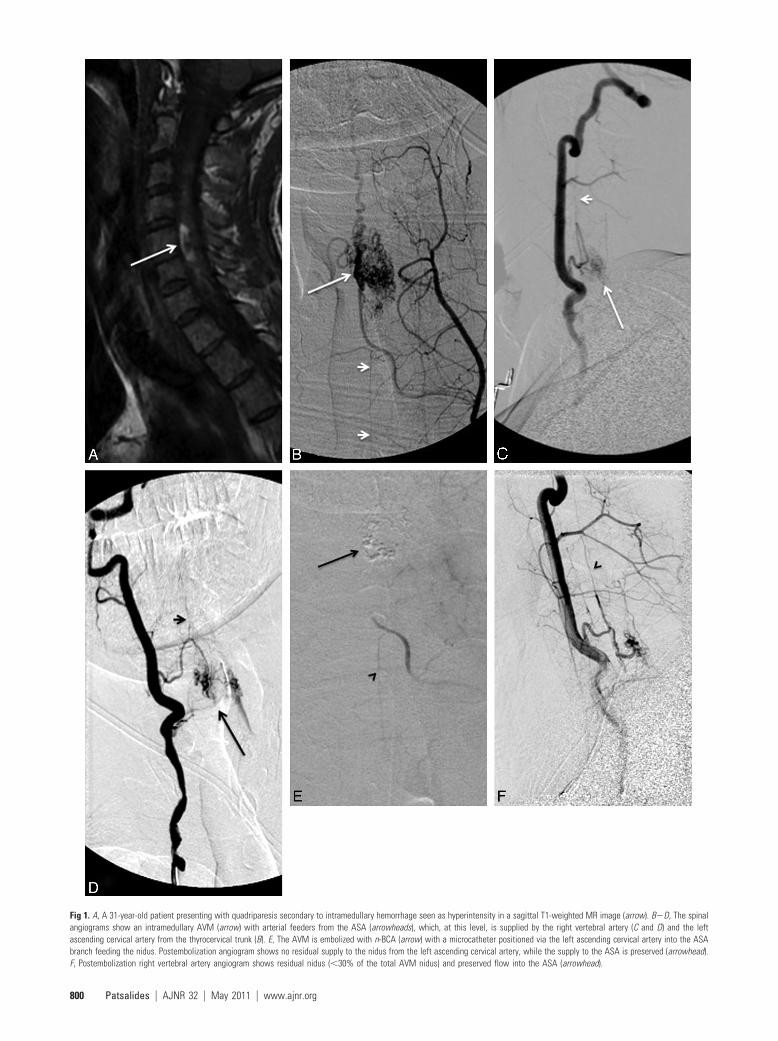
Fig 1. A, A 31-year-old patient presenting with quadriparesis secondary to intramedullary hemorrhage seen as hyperintensity in a sagittal T1-weighted MR image (arrow). B�D, The spinalangiograms show an intramedullary AVM (arrow) with arterial feeders from the ASA (arrowheads), which, at this level, is supplied by the right vertebral artery (C and D) and the leftascending cervical artery from the thyrocervical trunk (B). E, The AVM is embolized with n-BCA (arrow) with a microcatheter positioned via the left ascending cervical artery into the ASAbranch feeding the nidus. Postembolization angiogram shows no residual supply to the nidus from the left ascending cervical artery, while the supply to the ASA is preserved (arrowhead).F, Postembolization right vertebral artery angiogram shows residual nidus (�30% of the total AVM nidus) and preserved flow into the ASA (arrowhead).
800 Patsalides � AJNR 32 � May 2011 � www.ajnr.org

and when the cord is not draining through the vein to beoccluded.23,24
Intramedullary-Extramedullary AVM andComplex AngiomatosisIntramedullary-extramedullary or intradural-extraduralAVMs are extremely rare lesions that correspond to complexAVMs extending along a discrete somite level and involving�1 structure of spinal cord, dura, vertebrae, paravertebral softtissues, or skin. Complete involvement of an AVM along asomite level has been described in Cobb syndrome, but theincomplete types are more common. Diffuse angiomatosis,such as Rendu-Osler-Weber disease, may be seen in associa-tion with spinal cord AVMs.25-27 Intramedullary-extramedullary AVMs are typically seen in children and youngadults and present with pain and progressive myelopathy sec-ondary to mass effect, hemorrhage, and arterial steal. Multiplefeeders over several vertebral levels are common.
Intracranial DAVF with Spinal PerimedullaryVenous DrainageThe intracranial DAVF with perimedullary and spinal venousdrainage (Fig 6)—classified as type V in the Djindjian-Merlandclassification28—represents a distinct category of intracranial le-sions with spinal cord symptoms. These lesions receive arterialsupply from meningeal branches of the internal carotid, externalcarotid, and the vertebral arteries, with venous drainage aroundthe brain stem and into the pial veins in the anterior and posteriorsurface of the spinal cord. They are usually seen in men in theirthird-to-seventh decades of life, and because of the venous drain-age pattern, they may cause venous hypertension of the spinalcord. The typical clinical findings include ascending myelopathy,sphincter disturbances, bulbar signs like difficulty swallowing,and autonomic dysfunction.29 These lesions highlight the impor-tance of complete and meticulous angiographic evaluation fromthe cranium to the sacrum in cases of venous hypertension of thespinal cord.
Fig 2. A, A 50-year-old patient with massive SAH (arrow), Hunt and Hess grade IV. B, Reconstructed image from arotational angiogram shows an aneurysm (arrow) supplied by the ASA. The same arterial feeder supplies a pial AVM (blockarrow) distal to the aneurysm. There is duplication of the ASA distally to the branch supplying the aneurysm and pial AVM.C, A Magic 1.2F microcatheter (Balt, Montmorency, France) is advanced over a Mirage 0.08-inch guidewire (ev3) into theASA branch feeding the aneurysm and the pial AVM. D, Superselective angiography, with the microcatheter into the sulcalbranch of the ASA, shows the aneurysm, the pial AVM, and the abnormal venous drainage of the pial AVM (arrows). E,The aneurysm and AVM are embolized with n-BCA, and the glue cast is shown on reformatted 3D CT images (arrow). Thepatient was temporarily weaker after the procedure, but motor function returned to baseline within a few days.
AJNR Am J Neuroradiol 32:798 – 808 � May 2011 � www.ajnr.org 801

Spinal AngiographyDespite the advances of noninvasive vascular imaging, conven-tional catheter spinal angiography remains the definitive test forthe diagnosis and classification of spinal arteriovenous lesionsdue to its superior spatial resolution and image quality. Perform-ing spinal angiography with the patient under general anesthesiaimproves the quality of the study because the patient remainscomfortable during a potentially long study and apnea can beused to reduce motion artifacts when evaluating the thoracicspine. Angiography is performed via a 5F sheath placed in thecommon femoral artery. An aortogram can be obtained withpower injection via a pigtail high-flow catheter positioned in thedescending aorta in the midthoracic level (10 mL/s for total vol-ume of 35 mL). The individual thoracic and lumbar segmentalarteries must be selectively catheterized and evaluated. The verte-bral, deep cervical, and ascending cervical arteries must be cath-eterized for evaluation of the cervical cord, and the internal iliacand iliolumbar arteries should be catheterized for abnormalitiesin the lumbosacral region. At our institution, the catheters mostcommonly used for catheterization of the segmental arteries areCobra-2, VS2, and Simmons-2 (Terumo Medical, Somerset,New Jersey).
The major drawback of conventional angiography is the smallrisk of major complications due to its invasive nature and the useof iodinated contrast. For these reasons, only experienced inter-ventional neuroradiologists should perform spinal angiography.Spinal angiography can be a long and tedious procedure if thelesion is not identified quickly, for it can take several hours be-cause all arteries supplying the spine and spinal cord—from theskull base to the sacrum—may need to be studied. The degree ofdifficulty is higher in patients with atheromatous disease and os-tial narrowing of segmental arteries. The retrograde bilateral fem-oral injection or bilateral femoral reflux is a useful technique that
helps in identifying lesions of the lumbar or lower thoracic level.30
With bilateral 5F femoral sheaths, 40 mL of contrast is injectedinto each femoral artery at a rate of 20 mL/s, resulting in opacifi-cation of the dorsal aspect of the aorta up to the T10 level. Thus,the lower thoracic and the lumbar arteries are well opacified,while the visceral arteries receive minimal contrast. If findings ofthe study are negative, the mid- and upper thoracic segmentalarteries should be selectively catheterized, but catheterization ofthe lower thoracic and lumbar segmental arteries, as well as theright and left hypogastric arteries, is most likely unnecessary.
Spinal angiography should also be used to evaluate the ve-nous drainage after injection of the artery of Adamkiewicz. Inpatients with severe venous hypertension and myelopathy in-volving the thoracolumbar area, the venous drainage is pro-longed or absent. If venous hypertension is demonstrated dur-ing spinal angiography, the cause— usually a DAVF—must befound. Improvement of venous drainage after treatment is agood prognostic factor.31
Endovascular TreatmentTreatment planning for spinal vascular arteriovenous lesionsis based on the hemodynamics of the lesion, location in theaxial and longitudinal plane, and the angioarchitecture. Em-bolization is the treatment of choice for many arteriovenousanomalies. However, surgery continues to play a key role, anda multidisciplinary approach is essential. An important con-sideration before any intervention is a patient’s preoperativeneurologic status. As with all inherent disease processes of thespinal cord, postoperative function is highly related to preop-erative presentation, and maximum functional results are ob-tained in patients treated early before advanced deterioration.Partial results can still be obtained in patients with severe neu-rologic impairment.
Fig 3. A, Epidural AVM supplied by the left T7 intercostal artery (arrow). Note the venous drainage into dilated epidural veins (block arrow). B, Selective angiogram via a microcatheterplaced into the arterial feeder shows no supply to the ASA or PSA. C, The AVM is embolized with n-BCA with the glue cast shown in the unsubtracted image.
802 Patsalides � AJNR 32 � May 2011 � www.ajnr.org

General Principles for EmbolizationTo decrease the risk of vascular injury to the spinal cord, onemust clearly define the vascular anatomy before any attempt atembolization. The ASA supplies the anterior two-thirds of thespinal cord, including most of the gray matter and the corti-cospinal and spinothalamic tracts, whereas the PSAs supplythe dorsal one-third of the spinal cord. More arterial anasto-moses exist in the PSAs, suggesting that the possibility for suf-ficient collateral circulation is higher after occlusion of thePSA rather than the ASA. Thus, a posterior radiculomedullaryartery supplying the PSA may be occluded without significantclinical consequences or, at most, with the potential risk forposterior column syndrome. Conversely, occlusion of an an-terior radiculomedullary artery supplying the ASA carries asignificant risk for spinal cord ischemia.
Before embolization, it is advisable to check the levelsabove and below for anastomoses that would supply the ASAterritory. Particular attention is required during embolizationof a lesion with arterial steal because partial occlusion of thefeeding artery will lead to diminution of the arterial steal andthe possible appearance of normal spinal arteries; one shouldbe careful to avoid inadvertent embolization of these arteries.
Another point of concern is the rich pial perimedullarynetwork connecting the ASA and PSAs and the possibility ofembolization of the ASA when performing an embolization ina posterior radiculomedullary artery. Embolization in wedgeflow carries some risks: Because of the wedge flow, there can bea change in the usual flow patterns and embolic material mayreach arteries not opacified in the pre-embolization angio-
Fig 4. A, Angiogram of the left T10 intercostal artery shows a pial AVF supplied by the artery of Adamkiewicz (arrow).An aneurysm is seen at the level of the fistula (block arrow). B, Angiogram with a large FOV shows the extensive venousdrainage into spinal perimedullary veins (arrows). C, A microcatheter was advanced to the arterial feeder just proximal tothe aneurysm (arrow). The fistula and aneurysm are treated with n-BCA embolization (block arrow). D, The ASA remainspatent after the embolization, while the fistula, aneurysm, and abnormal spinal perimedullary venous drainage areobliterated.
AJNR Am J Neuroradiol 32:798 – 808 � May 2011 � www.ajnr.org 803

gram. Occlusion of the venous drainage of a large arterio-venous shunt also has increased risks; in an AVM, this mightlead to increased pressure in the nidus and subsequent hem-orrhage, whereas in an AVF, this may lead to increased venoushypertension. Last, too proximal an occlusion of an arterialfeeder to an arteriovenous shunt is usually ineffective becauseother arterial anastomoses may subsequently be recruited tosupply the shunt but may also lead to increased arterial stealbecause blood will now be diverted to the shunt. At the sametime, the ability to access the shunt for further embolization isdiminished.
Lesion-Specific TechniqueIntramedullary AVM. Despite the variable classification
schemes, there is generally consensus among the medical com-munity regarding the need to treat intramedullary AVMs to alterthe natural history and decrease the risk of future hemorrhage.The prognosis for untreated spinal cord AVMs is poor, with 36%of patients younger than 40 years of age developing severe impair-ment after 3 years of evolution.32 In the series of Hurth et al,33
13%, 20%, and 57% of patients had severe clinical deteriorationafter 5, 10, and 20 years, respectively. Thus, there is a strong indi-cation to treat spinal cord AVMs or at least modify their natural
Fig 5. A, A 69-year-old man with progressive bilateral lower extremity weakness. Sagittal T2-weighted MR image shows intramedullary edema in the lower thoracic spine and the conusmedullaris (arrow) with flow voids surrounding the cauda equina and conus medullaris (arrowheads), consistent with venous hypertension. B, Catheter angiogram shows an AVF in the sacralregion (block arrow), supplied by the iliolumbar arteries bilaterally (arrows). C, The arteriovenous shunt drains into the sacral epidural veins (arrowheads, B and C), which in turn communicatewith dilated spinal veins (arrows), causing venous hypertension. D, This epidural AVF is embolized with Onyx with obliteration of the feeding arteries, the arteriovenous shunt, and epiduralveins. There is no residual communication with the spinal veins. E, MR imaging of the lumbar spine obtained 7 months after the embolization shows marked improvement of theintramedullary edema and perimedullary flow voids, correlating with near-complete resolution of the patient’s symptoms.
804 Patsalides � AJNR 32 � May 2011 � www.ajnr.org

history. The current available treatments include embolization,surgery, or both. Surgical obliteration can be associated with ahigh intraprocedural risk of neurologic injury, especially in le-sions located in the anterior cord. Reports of microsurgical oblit-
eration are infrequent and are predominantly limited to smallcase series.34-37 In patients in whom angiographic access is diffi-cult, as in cases of an AVM with a long endovascular route or anunstable catheter position, surgery offers a viable option. A pos-
Fig 6. A, Sagittal T2-weighted MR image in a 67-year-old patient with progressive bilateral leg weakness, gait instability,and tingling in both hands shows increased signal intensity in the spinal cord (arrows) and perimedullary flow voids,consistent with venous hypertension. B, Cerebral angiogram shows a dural AVF with venous drainage into the petrous,anterior medullary, and spinal perimedullary veins (arrows). C and D, The fistula is supplied by the meningo-hypophysealtrunk of the right internal carotid artery (arrow, C) and the petrous branch of the right MMA (arrow, D). E, A microcatheteris positioned in the MMA, just proximally to the fistula (arrow), and embolization is then performed with Onyx. F, Thepostembolization angiogram shows obliteration of the fistula.
AJNR Am J Neuroradiol 32:798 – 808 � May 2011 � www.ajnr.org 805

terior superficial location makes a lesion more amenable to saferesection; however, anterior lesions can be still treated definitivelywith surgery, especially in the cervical region where collateralblood flow to the spinal cord can arise inferiorly. Filum terminalelesions are also amenable to surgical treatment. The basis of sur-gical resection of spinal AVMs, like cranial AVMs, rests in bipolarcoagulation and ligation of arterial feeders to the lesion, whilepreserving venous drainage so as not to increase transmural pres-sure and the risk of intraoperative hemorrhage. Associated ve-nous or arterial aneurysms may be removed with the nidus orshrunk with bipolar cautery.
Embolization plays an important role in the managementof intramedullary spinal AVMs either as a primary treatmentor as an adjunct to surgery. Even a partial embolization canimprove the patient’s prognosis.38,39 Modern microcatheterspermit selective catheterization of the anterior or posteriorarteries feeding the AVM, and the embolization can be per-formed with particles or liquid embolic material, each with itsown advantages and disadvantages. Coils are not used in theselesions because they require relatively rigid microcathetersthat cannot be safely navigated through the spinal arteries. Asa result, coils can only achieve proximal embolization, whichmay lead to the development of collateral flow to the niduswhile preventing safe access for future embolizations.
Particles have the advantage of a stepwise embolization andthe ability to follow the results clinically and angiographicallyduring the procedure, but they have the serious disadvantageof long-term recanalization. Treatment with particle emboli-zation requires annual angiographic control and additionalembolization in cases of recanalization. In a series by Biondi etal40 of thoracic AVMs embolized with particles, clinical im-provement was noted in 57% of patients after the first embo-lization and in 63% of patients after the last embolization.Multiple embolizations were required because the AVM re-canalization rate was 80%. Even though embolization withparticles is not a definitive treatment, it may be considered apalliative treatment because it alters the natural history andprovides a good clinical outcome. The long-term resultsshowed persistence of clinical improvement after treatment,even if the AVM recurs. This is probably explained by the factthat repeat embolizations prevent prolonged exposure of thespinal cord to the AVM.
Liquid agents have the advantage of achieving more per-manent occlusion with a very low recanalization rate but withthe concurrent risk of inadvertent embolization of normalperforating arteries that may not be visible on angiography. Ingeneral, a liquid agent should be used when feasible, especiallywhen embolization is the primary or sole treatment. The liq-uid agent should be delivered within or as close to the nidus aspossible. In a large series of spinal AVM and AVF emboliza-tions by using n-BCA,41 there was good clinical outcome in83% of patients (43% improved, 15% remained asymptom-atic, 25% stable). Embolization failed to stop the clinical evo-lution of the disease in 4% of patients. Permanent morbidityrelated to embolization was seen in 13% of patients, but only4% of patients had severe deficits, all secondary to emboliza-tion through the ASA. Anatomic obliteration was achieved in16% of lesions, and embolization reduced the lesion size bymore than half in 86% of cases. There was no recanalizationdetected during follow-up (mean, 5.6 years), and recurrent
hemorrhage was seen in 4% of patients, all with cervical cordAVMs reduced to �50% of the original size. In this series,children recovered better than adults, and there was no statis-tically significant correlation between the degree of lesionobliteration and clinical outcome.
Onyx (ev3, Irvine, California) has also been used to treatspinal cord AVMs. In a series of 17 patients with symptomaticintramedullary AVMs (15 exclusively intramedullary and 2with additional perimedullary components) treated exclu-sively by Onyx, total obliteration was achieved in 6 patients;subtotal obliteration (tiny remnant), in 5; and partial obliter-ation, in 5.42 The procedure was aborted in 1 patient. Clinicalimprovement during follow-up (mean, 24.3 months) wasnoted in 14 patients, and clinical deterioration, in 3 patients,including the 1 treated surgically. There were 3 intraproce-dural complications (2 extradural hematomas and 1 SAH)without neurologic consequences.
At our institution, embolization for intramedullary AVMis performed with the patient under general anesthesia andsystemic heparin. A 5F guide catheter is placed at the ostium ofthe segmental artery, and a microcatheter is advanced into theradiculomedullary branch that supplies the AVM. Flow-directed microcatheters are usually more appropriate thanstiffer braided microcatheters. For lesions supplied by theASA, the microcatheter should be positioned inside or as closeto the nidus as possible, preferably in a sulcal artery beyond thelongitudinal axis of the ASA. This will minimize the chance ofinadvertent embolization of a normal ASA branch. Similarprinciples are applied for lesions supplied by a feeder from thePSA, even though the consequences of accidentally occludingnormal PSA branches are not as dramatic. For AVMs suppliedby both ASA and PSA feeders, the PSA should be treated firstbecause of less procedure-related risk. Embolization is usuallyperformed with n-BCA mixed with ethiodized oil with 1:2 to1:3 ratios, and the radiodensity of the embolic material is en-hanced with tantalum powder. For high-flow shunts, a higherconcentration of n-BCA can be used. Embolization of falseand flow-related aneurysms is important to prevent recurrenthemorrhage.43 The patient is monitored in the neurologic in-tensive care unit for 24 – 48 hours. Systemic heparin is contin-ued for 24 hours (low-dose protocol with target partial throm-boplastin time of 50 – 60 seconds) to prevent thrombosis of thenormal spinal vessels.
Intramedullary-Extramedullary AVM andComplex AngiomatosisGiven the complex architecture of these lesions involving bothintradural and extradural compartments and the fact that nor-mal cord parenchyma is often intermingled within the actualAVM, these lesions are the most difficult to treat. Optimaltherapy for these lesions has not been established, and surgeryand embolization may be used alone or in combination.Achieving a complete cure in these types of vascular malfor-mations is extremely difficult and likely associated with in-creased morbidity. Thus, treatment is palliative and should betargeted to relieving symptoms that may be caused by a hema-toma, arterial steal, venous hypertension, or direct mass effect.Simple surgical interventions have included ligation of a feed-ing artery,5 and decompressive laminectomy; however, thesemodalities are incomplete at best and offer controversial ben-
806 Patsalides � AJNR 32 � May 2011 � www.ajnr.org

efit. Although rare, there have been case reports in the litera-ture of complete surgical cure by using combined angio-graphic embolization and extensive surgical resection.6-8
Embolization can be performed with particles if usedpreoperatively.
Pial AVFsPial AVFs represent a heterogeneous group of lesions, and thetherapeutic approach depends largely on the individual lesionangioarchitecture. Treatment must be performed early, aimedat surgical or endovascular obliteration of the fistula at thepoint of arteriovenous connection. Embolization can also beused as an adjunct to surgery.44 Regardless of treatment, com-plete and permanent occlusion of the fistula is the only way toprevent long-term recurrence. Thus, liquid embolic materialsare the agents of choice, and PVA particles should be used onlyfor presurgical embolization. In small AVFs, superselectivecatheterization and placement of the microcatheter inside thefistula for effective embolization is difficult because the arte-rial feeder is a distal branch of a thin ASA.45 For large AVFs,embolization is seldom efficient because of the multiple feed-ing arteries, some of which are transmedullary or perimedul-lary branches that cannot be safely catheterized. Embolizationhas been reported in selected cases. Oran et al46 reported suc-cessful glue embolization in 4/5 patients with small AVFs, fol-lowed by clinical improvement in 3 patients. Cho et al47 re-ported successful complete embolization in 3/5 patients(partial embolization in 1 patient) with small AVFs and 0/2patients (partial embolization in 2 patients) with large AVFs.Clinical improvement or stability was seen in 2/4 patients withsmall AVFs (1 worsened) and in one-half of patients with largeAVF (1 worsened). One patient treated with PVA demon-strated recurrent fistula 3 months after the embolization. Onthe other hand, microsurgical treatment plays a curative role,especially in lesions located on the dorsal/dorsolateral cord.
The goal of surgical treatment is to interrupt the connec-tion between the arterial and venous system while preservingthe ASA branches. Superselective catheterization of the arte-rial feeder is more feasible in giant AVFs due to their increaseddiameter and the high flow through the shunt. The massivelydilated draining veins increase the risk of intraoperative hem-orrhage and the difficulty of surgical treatment. Thus, embo-lization is the treatment of choice in these lesions. The majorchallenge of embolization is placing the embolization materialin the proper position without venous migration. Ricolfiet al16 reported their results of endovascular treatment of 12patients with giant AVFs of the spinal cord, by using balloonsor particles. Treatment with nondetachable balloons was cu-rative in 5/6 patients; embolization failed secondary to spasmin 1 patient. Treatment with detachable balloons was curativein 3 patients; partial embolization was achieved in 1 patient. In1 patient who was treated with a detachable balloon, the bal-loon migrated into the venous site causing clinical deteriora-tion. The fistula was completely occluded in 2 patients treatedwith gelatin sponge particles. Clinical improvement was seenin 6/10 patients with complete occlusion of the fistula. In aseries of 35 patients with pial AVFs, Mourier et al5 reportedtheir results in 22 giant AVFs treated with detachable balloons.Complete occlusion of the fistula was achieved in 15 patients,all with clinical improvement posttreatment. Of 6 patients
with partial occlusion, 4 remained unchanged and 2 worsenedclinically posttreatment. There was 1 periprocedural death.
Detachable and nondetachable balloons are not availablein the United States today, and the use of particles alone is notindicated because the particles may pass through the shuntinto the venous circulation where they may cause inadvertentthrombosis or pulmonary embolism. Instead, coils alone orcombined with liquid embolic materials should be the agentsof choice. In giant AVFs, coils can be placed in the fistula to actas a frame for the liquid embolic material and prevent migra-tion through the high-flow shunt into the venous side. Thistechnique requires the use of a microcatheter with an innerdiameter large enough to allow deployment of coils. The em-bolization should occur at the fistula and proximal drainingveins to prevent development of inaccessible collateral feedersand recanalization of the fistula. Progressive retrogradethrombosis of the veins draining the fistula may result in tran-sient worsening of symptoms. This can be prevented byadministration of systemic heparin for 24 – 48 hourspostembolization.
ConclusionsThis article represents an overview of spinal vascular lesionswith reference to the indications, contraindications, and tech-niques of endovascular treatment. While knowledge of thevascular anatomy and the pathophysiology of each spinal vas-cular lesion is necessary to achieve safe and effective treatment,a multidisciplinary approach with endovascular and surgicalspecialists working closely together is paramount to achievethe best possible results.
References1. Krings T, Geibprasert S. Spinal dural arteriovenous fistulas. AJNR Am J Neu-
roradiol 2009;30:639 – 482. Krings T, Mull M, Gilsbach JM, et al. Spinal vascular malformations. Eur Radiol
2005;15:267–783. Barnwell SL, Dowd CF, Davis RL, et al. Cryptic vascular malformations of the
spinal cord: diagnosis by magnetic resonance imaging and outcome of sur-gery. J Neurosurg 1990;72:403– 07
4. Rosenblum B, Oldfield EH, Doppman JL, et al. Spinal arteriovenousmalformations: a comparison of dural arteriovenous fistulas and intraduralAVM’s in 81 patients. J Neurosurg 1987;67:795– 802
5. Mourier KL, Gobin YP, George B, et al. Intradural perimedullary arterio-venous fistulae: results of surgical and endovascular treatment in a series of 35cases. Neurosurgery 1993;32:885–91; discussion 891
6. Biondi A, Merland JJ, Hodes JE, et al. Aneurysms of spinal arteries associatedwith intramedullary arteriovenous malformations. I. Angiographic and clin-ical aspects. AJNR Am J Neuroradiol 1992;13:913–22
7. Di Chiro G, Doppman JL. Endocranial drainage of spinal cord veins. Radiology1970;95:555– 60
8. Hurst RW, Kenyon LC, Lavi E, et al. Spinal dural arteriovenous fistula: thepathology of venous hypertensive myelopathy. Neurology 1995;45:1309 –13
9. Jellema K, Tijssen CC, van Gijn J. Spinal dural arteriovenous fistulas: a con-gestive myelopathy that initially mimics a peripheral nerve disorder. Brain2006;129:3150 – 64
10. Merland JJ, Riche MC, Chiras J. Intraspinal extramedullary arteriovenous fis-tulae draining into the medullary veins. J Neuroradiol 1980;7:271–320
11. Djindjian M, Djindjian R, Hurth M, et al. Steal phenomena in spinal arterio-venous malformations. J Neuroradiol 1978;5:187–201
12. el Mahdi MA, Rudwan MA, Khaffaji SM, et al. A giant spinal aneurysm withcord and root compression. J Neurol Neurosurg Psychiatry 1989;52:532–35
13. Spetzler RF, Detwiler PW, Riina HA, et al. Modified classification of spinal cordvascular lesions. J Neurosurg 2002;96:145–56
14. Gueguen B, Merland JJ, Riche MC, et al. Vascular malformations of the spinalcord: intrathecal perimedullary arteriovenous fistulas fed by medullary arter-ies. Neurology 1987;37:969 –79
15. Nakstad PH, Hald JK, Bakke SJ. Multiple spinal arteriovenous fistulas in
AJNR Am J Neuroradiol 32:798 – 808 � May 2011 � www.ajnr.org 807

Klippel-Trenaunay-Weber syndrome treated with platinum fibre coils. Neu-roradiology 1993;35:163– 65
16. Ricolfi F, Gobin PY, Aymard A, et al. Giant perimedullary arteriovenous fistu-las of the spine: clinical and radiologic features and endovascular treatment.AJNR Am J Neuroradiol 1997;18:677– 87
17. Rodesch G, Hurth M, Alvarez H, et al. Angio-architecture of spinal cord arte-riovenous shunts at presentation: clinical correlations in adults and chil-dren—the Bicetre experience on 155 consecutive patients seen between 1981–1999. Acta Neurochir (Wien) 2004;146:217–26; discussion 226 –27. Epub 2004Feb 23
18. Arnaud O, Bille F, Pouget J, et al. Epidural arteriovenous fistula with perimed-ullary venous drainage: case report. Neuroradiology 1994;36:490 –91
19. Clarke MJ, Patrick TA, White JB, et al. Spinal extradural arteriovenous malfor-mations with parenchymal drainage: venous drainage variability and impli-cations in clinical manifestations. Neurosurg Focus 2009;26:E5
20. Weingrad DN, Doppman JL, Chretien PB, et al. Paraplegia due to posttrau-matic pelvic arteriovenous fistula treated by surgery and embolization: casereport. J Neurosurg 1979;50:805–10
21. Kawabori M, Hida K, Yano S, et al. Cervical epidural arteriovenous fistula withradiculopathy mimicking cervical spondylosis. Neurol Med Chir (Tokyo)2009;49:108 –13
22. Willinsky R, terBrugge K, Lasjaunias P, et al. The variable presentations ofcraniocervical and cervical dural arteriovenous malformations. Surg Neurol1990;34:118 –23
23. Szajner M, Weill A, Piotin M, et al. Endovascular treatment of a cervical para-spinal arteriovenous malformation via arterial and venous approaches. AJNRAm J Neuroradiol 1999;20:1097–99
24. Willinsky R, terBrugge K, Montanera W, et al. Spinal epidural arteriovenousfistulas: arterial and venous approaches to embolization. AJNR Am J Neuro-radiol 1993;14:812–17
25. Djindjian M, Djindjian R, Rey A, et al. Intradural extramedullary spinal arte-rio-venous malformations fed by the anterior spinal artery. Surg Neurol1977;8:85–93
26. Djindjian R, Hurth M, Houdart R. Medullary angiomas, segmentary or gener-alized vascular dysplasias and phakomatoses [in French]. Rev Neurol (Paris)1969;121:109 –11
27. Djindjian R, Hurth M, Houdart R. Spinal angiomas, segmentary vascular dys-plasia and phacomatosis [in French]. Rev Neurol (Paris) 1971;124:121– 42
28. Houdart E, Gobin YP, Casasco A, et al. A proposed angiographic classificationof intracranial arteriovenous fistulae and malformations. Neuroradiology1993;35:381– 85
29. Ricolfi F, Manelfe C, Meder JF, et al. Intracranial dural arteriovenous fistulaewith perimedullary venous drainage: anatomical, clinical and therapeuticconsiderations. Neuroradiology 1999;41:803–12
30. Melki JP, Riche MC, Reizine D, et al. Simultaneous bifemoral retrograde arte-riography under pressure: first stage in angiographic exploration of the spineand spinal cord [in English, French]. J Neuroradiol 1986;13:62–70
31. Gobin YP, Rogopoulos A, Aymard A, et al. Endovascular treatment of intra-cranial dural arteriovenous fistulas with spinal perimedullary venous drain-age. J Neurosurg 1992;77:718 –23
32. Aminoff MJ, Logue V. The prognosis of patients with spinal vascular malfor-mations. Brain 1974;97:211–18
33. Hurth M, Houdart R, Djindjian R, et al. Arteriovenous malformations of thespinal cord: clinical, anatomical and therapeutic consideration: a series of 150cases. Progr Neurol Surg 1978;9:238 – 66
34. Bostrom A, Krings T, Hans FJ, et al. Spinal glomus-type arteriovenousmalformations: microsurgical treatment in 20 cases. J Neurosurg Spine 2009;10:423–29
35. Connolly ES Jr, Zubay GP, McCormick PC, et al. The posterior approach to aseries of glomus (type II) intramedullary spinal cord arteriovenous malfor-mations. Neurosurgery 1998;42:774 – 85, discussion 785– 86
36. Tai PA, Tu YK, Liu HM. Surgical treatment of spinal arteriovenous mal-formations: vascular anatomy and surgical outcome. J Formos Med Assoc2001;100:389 –96
37. Zozulya YP, Slin’ko EI, Al-Qashqish II. Spinal arteriovenous malformations:new classification and surgical treatment. Neurosurg Focus 2006;20:E7
38. Djindjian R, Cophignon J, Rey A, et al. Superselective arteriographic emboli-zation by the femoral route in neuroradiology: study of 50 cases. II. Emboli-zation in vertebromedullary pathology. Neuroradiology 1973;6:132– 42
39. Doppman JL, Di Chiro G, Ommaya A. Obliteration of spinal-cord arterio-venous malformation by percutaneous embolisation. Lancet 1968;1:477
40. Biondi A, Merland JJ, Reizine D, et al. Embolization with particles in thoracicintramedullary arteriovenous malformations: long-term angiographic andclinical results. Radiology 1990;177:651–58
41. Rodesch G, Hurth M, Alvarez H, et al. Embolization of spinal cord arterio-venous shunts: morphological and clinical follow-up and results—review of69 consecutive cases. Neurosurgery 2003;53:40 – 49; discussion 49 –50
42. Corkill RA, Mitsos AP, Molyneux AJ. Embolization of spinal intramedullaryarteriovenous malformations using the liquid embolic agent, Onyx: a single-center experience in a series of 17 patients. J Neurosurg Spine 2007;7:478 – 85
43. Konan AV, Raymond J, Roy D. Transarterial embolization of aneurysms as-sociated with spinal cord arteriovenous malformations: report of four cases.J Neurosurg 1999;90:148 –54
44. Hida K, Iwasaki Y, Goto K, et al. Results of the surgical treatment of perimed-ullary arteriovenous fistulas with special reference to embolization. J Neuro-surg 1999;90:198 –205
45. Riche MC, Melki JP, Merland JJ. Embolization of spinal cord vascular malfor-mations via the anterior spinal artery. AJNR Am J Neuroradiol 1983;4:378 – 81
46. Oran I, Parildar M, Derbent A. Treatment of slow-flow (type I) perimedullaryspinal arteriovenous fistulas with special reference to embolization. AJNRAm J Neuroradiol 2005;26:2582– 86
47. Cho KT, Lee DY, Chung CK, et al. Treatment of spinal cord perimedullaryarteriovenous fistula: embolization versus surgery. Neurosurgery 2005;56:232– 41, discussion 232– 41
808 Patsalides � AJNR 32 � May 2011 � www.ajnr.org