Embed Size (px)
Citation preview

1531
Roundabout 4 (Robo4) is a transmembrane receptor that belongs to the Robo family of neural cell adhesion mol-
ecules. Robo4 has been shown to play a role in endothelial cell (EC) migration, proliferation, angiogenesis, and stabilization of the vasculature.1–5 Robo4 is expressed specifically in ECs6,7 in the developing embryo,1 placenta,6 normal adult tissues,1,8 and tumors.6,8 Recently, shear stress was reported to suppress Robo4 gene expression.9
See cover imageWe previously reported that a 3-kb fragment of the 5′-flank-
ing region of the human Robo4 gene contains information
for EC-specific expression in vivo.10 We demonstrated that the Robo4 promoter is activated by the transcription factors GA-binding protein (GABP) and SP1 through an E26 transfor-mation-specific (ETS)-binding site at −119 and 2 SP1-binding sites at −42 and −153, respectively.10,11 Although GABP and SP1 are essential for Robo4 promoter activation, both factors are known to be expressed ubiquitously. In keeping with these data, previous studies exploring the transcriptional regulation of other EC-specific genes, including von Willebrand factor, PECAM-1 (platelet endothelial cell adhesion molecule; CD31), vascular endothelial growth factor receptors 1 and -2, Tie2, and E-selectin, have consistently implicated a role for noncell-type–specific
© 2014 American Heart Association, Inc.
Arterioscler Thromb Vasc Biol is available at http://atvb.ahajournals.org DOI: 10.1161/ATVBAHA.114.303818
Objective—The molecular basis of endothelial cell (EC)–specific gene expression is poorly understood. Roundabout 4 (Robo4) is expressed exclusively in ECs. We previously reported that the 3-kb 5′-flanking region of the human Robo4 gene contains information for lineage-specific expression in the ECs. Our studies implicated a critical role for GA-binding protein and specificity protein 1 (SP1) in mediating overall expression levels. However, these transcription factors are also expressed in non-ECs. In this study, we tested the hypothesis that epigenetic mechanisms contribute to EC-specific Robo4 gene expression.
Methods and Results—Bisulfite sequencing analysis indicated that the proximal promoter of Robo4 is methylated in non-ECs but not in ECs. Treatment with the DNA methyltransferase inhibitor 5-aza-2′-deoxycytidine increased Robo4 gene expression in non-ECs but not in ECs. Proximal promoter methylation significantly decreased the promoter activity in ECs. Electrophoretic mobility shift assays showed that DNA methylation of the proximal promoter inhibited SP1 binding to the −42 SP1 site. In DNase hypersensitivity assays, chromatin condensation of the Robo4 promoter was observed in some but not all nonexpressing cell types. In Hprt (hypoxanthine phosphoribosyltransferase)-targeted mice, a 0.3-kb proximal promoter directed cell-type–specific expression in the endothelium. Bisulfite sequencing analysis using embryonic stem cell–derived mesodermal cells and ECs indicated that the EC-specific methylation pattern of the promoter is determined by demethylation during differentiation and that binding of GA-binding protein and SP1 to the proximal promoter is not essential for demethylation.
Conclusions—The EC-specific DNA methylation pattern of the Robo4 proximal promoter is determined during cell differentiation and contributes to regulation of EC-specific Robo4 gene expression. (Arterioscler Thromb Vasc Biol. 2014;34:1531-1538.)
Key Words: DNA methylation ◼ endothelial cells ◼ epigenomics
Received on: March 22, 2013; final version accepted on: May 8, 2014.From the Graduate School of Pharmaceutical Sciences, Osaka University, Osaka, Japan (Y.O., N.F., T.T., Y.N., K.S., Y.K., H.N., A.S., M.S., J.Z., K.I.,
N.H., M.K., Y.M., S.N., T.D.); Center for Vascular Biology Research and Division of Molecular and Vascular Medicine, Beth Israel Deaconess Medical Center, Boston, MA (L.Y., W.C.A.); Department of Pathology, Center for Excellence in Vascular Biology, Harvard Medical School, Boston, MA (A.S.T., G.G.-C.); Department of Material Sciences, Massachusetts Institute of Technology, Boston (A.S.T.); and Stem Cell Project, Tokyo Metropolitan Institute of Medical Science, Tokyo, Japan (K.K.).
*These authors contributed equally to this article.The online-only Data Supplement is available with this article at http://atvb.ahajournals.org/lookup/suppl/doi: 10.1161/ATVBAHA.114.303818/-/DC1.Correspondence to Yoshiaki Okada, PhD, Osaka University, Graduate School of Pharmaceutical Sciences, 1-6 Yamadaoka, Suita, Osaka 565-0871,
Japan. E-mail [email protected] or William C. Aird, MD, Beth Israel Deaconess Medical Center, Molecular and Vascular Medicine, RN-237, 330 Brookline Ave, Boston, MA. E-mail [email protected]
Endothelial Cell–Specific Expression of Roundabout 4 Is Regulated by Differential DNA Methylation of the
Proximal PromoterYoshiaki Okada,* Nobuaki Funahashi,* Toru Tanaka, Yuji Nishiyama, Lei Yuan,
Keisuke Shirakura, Alexis S. Turjman, Yoshihiro Kano, Hiroki Naruse, Ayano Suzuki, Miki Sakai, Jiang Zhixia, Kenji Kitajima, Kenji Ishimoto, Nobumasa Hino, Masuo Kondoh,
Yohei Mukai, Shinsaku Nakagawa, Guillermo García-Cardeña, William C. Aird, Takefumi Doi
by guest on June 4, 2018http://atvb.ahajournals.org/
Dow
nloaded from
by guest on June 4, 2018http://atvb.ahajournals.org/
Dow
nloaded from
by guest on June 4, 2018http://atvb.ahajournals.org/
Dow
nloaded from
by guest on June 4, 2018http://atvb.ahajournals.org/
Dow
nloaded from
by guest on June 4, 2018http://atvb.ahajournals.org/
Dow
nloaded from
by guest on June 4, 2018http://atvb.ahajournals.org/
Dow
nloaded from
by guest on June 4, 2018http://atvb.ahajournals.org/
Dow
nloaded from
by guest on June 4, 2018http://atvb.ahajournals.org/
Dow
nloaded from
by guest on June 4, 2018http://atvb.ahajournals.org/
Dow
nloaded from
by guest on June 4, 2018http://atvb.ahajournals.org/
Dow
nloaded from
by guest on June 4, 2018http://atvb.ahajournals.org/
Dow
nloaded from
by guest on June 4, 2018http://atvb.ahajournals.org/
Dow
nloaded from
by guest on June 4, 2018http://atvb.ahajournals.org/
Dow
nloaded from
by guest on June 4, 2018http://atvb.ahajournals.org/
Dow
nloaded from

1532 Arterioscler Thromb Vasc Biol July 2014
transcription factors, such as SP1, ETS family proteins, nuclear factor-κB, and GATA proteins.12 Although some transcription factors are enriched in ECs (eg, Vezf1 [vascular endothelial zinc finger], HoxA9 [homeobox], GATA2, and KLF2 [Krüppel-like factor]), none has been shown to be restricted to the endothelium.
Collectively, these findings argue against the existence of an endothelial-specific master regulator that mediates differentia-tion and expression of multiple differentiation markers, as MyoD (myogenic differentiation) does in skeletal myocytes.13 One pos-sibility is that EC-specific gene expression is mediated by the cooperative activity of multiple noncell-type–specific transcrip-tion factors. Alternatively, unique post-translational modifications
or alternative spliced transcripts may play a role. Finally, there is increasing evidence that epigenetic mechanisms contribute to cell-type–specific expression of genes in the endothelium.14,15
Epigenetic control of gene transcription involves DNA methylation, histone modification, and chromatin remodeling. DNA methylation refers to the addition of a methyl group to the 5′-position of cytosine to create 5-methyl-cytosine. DNA methylation at cytosine residues occurs almost exclusively in the context of the CpG sequence in the vertebrate genome. DNA methylation is a repressive mark that is associated with transcriptional silencing.16 It has been implicated in many cel-lular processes, including X chromosome inactivation, genomic imprinting, embryonic development, lineage specification, and cancer pathogenesis.15 Two general mechanisms have been pro-posed for CpG methylation–mediated gene suppression. First, DNA methylation may inhibit the binding of transcription fac-tors to CpG dinucleotide–containing cis-regulatory elements. Second, DNA methylation results in the recruitment of methyl CpG–binding proteins. These proteins recruit large chromatin-modifying complexes and promote chromatin condensation that represses transcription by reducing DNA accessibility.
In this study, we hypothesized that EC-specific Robo4 gene expression is regulated by epigenetic mechanisms. We
Nonstandard Abbreviations and Acronyms
EC endothelial cell
GABP GA-binding protein
HCAEC human coronary artery endothelial cells
HCASmC human coronary artery smooth muscle cells
HEK human embryonic kidney
NHDF normal human dermal fibroblasts
Robo4 roundabout4
Figure 1. Bisulfite sequencing of the human roundabout 4 (Robo4) promoter. Summary of the 3-kb human Robo4 promoter bisulfite sequencing results for 10 cloned polymerase chain reaction products prepared with bisulfite-treated genomic DNA from 2 endothelial cell types (human coronary artery endothelial cells [HCAEC] and human umbilical vein endothelial cells [HUVEC]) and 3 nonendothelial cell types (human coronary artery smooth muscle cells [HCASmC], normal human dermal fibroblasts [NHDF], and human embryonic kidney [HEK] 293). Each graph indicates the CpG position in the promoter and the percentage of methylated CpG.
by guest on June 4, 2018http://atvb.ahajournals.org/
Dow
nloaded from

Okada et al DNA Methylation Regulates Robo4 Gene Suppression 1533
compared the DNA methylation pattern of the Robo4 promoter between EC and non-EC cell types and found that the proxi-mal promoter region is methylated only in non-ECs. This cell-type–specific methylation inhibited the binding of SP1 to the −42 SP1 site and suppressed Robo4 gene expression. Finally, using transgenic mice and an ES cell differentiation system, we demonstrated that the 0.3-kb proximal promoter, includ-ing 11 CpG sites, contained the information for EC-specific gene expression and that the EC-specific methylation pat-tern of the promoter is determined during cell differentiation. Collectively, these data support a novel model of EC-specific gene expression that involves DNA methylation–mediated inhibition of SP1 activity in non-ECs.
Materials and MethodsMaterials and Methods are available in the online-only Supplement.
ResultsMethylation Pattern of the Robo4 Promoter in ECs and Non-ECsThe upstream promoter (−3000 to +1) of the human Robo4 gene contains a total of 37 CpG sites and no typical CpG islands (Figure I in the online-only Data Supplement). To
investigate the methylation pattern of the endogenous Robo4 promoter in ECs (human coronary artery ECs [HCAEC] and human umbilical vein ECs) and non-ECs (human coronary artery smooth muscle cells [HCASmC], normal human der-mal fibroblasts, and human embryonic kidney [HEK] 293), the methylation status of 37 CpG sites in the 3-kb promoter was analyzed by bisulfite sequencing (Figure 1; Figure II in the online-only Data Supplement). In ECs, CpG methylation was restricted to an upstream region between −2583 and −2034. In contrast, non-ECs demonstrated heavy methylation not only in the upstream region but also at the proximal promoter (11 CpG sites between −287 and −43). Analysis of reduced representa-tion bisulfite sequencing from the ENCODE (Encyclopedia of DNA Elements) consortium revealed similar differences in Robo4 promoter methylation across a wide range of ECs and non-ECs (Figure III in the online-only Data Supplement). Thus, the 300-bp upstream promoter of Robo4 is differentially methylated in expressing and nonexpressing cell types.
Effects of DNA Methylation on Robo4 Gene Expression in ECs and Non-ECsWe next asked whether cell-type–specific methylation of the Robo4 promoter contributes to EC-specific expression
Figure 2. Effects of DNA methylation on roundabout 4 (Robo4) gene expression in endothelial cells (ECs) and non-ECs. A, ECs and non-ECs were treated with the DNA methyltransferase inhibitor 5-aza-2′-deoxycytidine (5-AC). The expression levels of Robo4 and GAPDH (as an internal control) were measured using real-time polymerase chain reaction with cDNAs prepared from human coronary artery endothelial cells (HCAEC), human coronary artery smooth muscle cells (HCASmC), normal human dermal fibroblasts (NHDF), and human embryonic kidney (HEK) 293 treated with or without 10 μmol/L 5-AC for 4 days. Copy numbers were calculated from standard curves prepared by measuring known amounts of plasmids encoding Robo4 or GAPDH. The Robo4 expression level was normalized to that of GAPDH. The data are represented as mean±SE. (n=3, Student t test, *P<0.05). B, Schematic representation of a nonmethylated control luciferase vector (pGL-3-Me−) and the Robo4 promoter–luciferase constructs with or without methylation of 14 CpG sites (pGL3-Robo4-Me+ or -Me−, respectively). The CpG sites in the proximal promoter (−928 [StuI site] to +10) were methylated by methyltransferase SssI. C, pGL3-Robo4-Me−, Me+, or pGL3-Me− was transfected into HCAEC. Luciferase activity was measured 48 hours after the transfection. The data are represented as mean±SE (n=5, Student t test, *P<0.05).
by guest on June 4, 2018http://atvb.ahajournals.org/
Dow
nloaded from

1534 Arterioscler Thromb Vasc Biol July 2014
of Robo4. To investigate the effects of promoter methyla-tion on Robo4 gene expression, ECs (HCAEC) and non-ECs (HCASmC, normal human dermal fibroblasts, and HEK293) were treated with the DNA methyltransferase inhibitor 5-aza-2′-deoxycytidine, and expression levels of the Robo4 mRNA were measured by real-time reverse transcription polymerase chain reaction. Treatment with 5-aza-2′-deoxycytidine did not affect Robo4 mRNA expression in HCAEC but significantly increased mRNA levels in HCASmC, normal human dermal fibroblasts, and HEK293 cells (by 4-fold, 7-fold, and 30-fold, respectively; Figure 2A). These results suggest that DNA methylation of the Robo4 promoter serves to silence expression in non-ECs.
To determine the effect of CpG methylation on promoter activity, we performed transient transfection assays using Robo4 promoter–luciferase plasmids in which the proximal promoter (CpG sites from −826 to −43) was either unmethyl-ated or methylated in vitro using Sss1 methylase (Figure 2B). As shown in Figure 2B, methylation of the proximal promoter resulted in a significant reduction of Robo4 promoter activity in HCAEC. Thus, DNA methylation of the proximal promoter suppresses Robo4 gene expression.
Effect of DNA Methylation on GABP and SP1 Binding to the Robo4 PromoterOne mechanism underlying the silencing effect of DNA meth-ylation on gene expression is direct inhibition of transcription factor binding. We have previously shown that GABP and SP1 bind to an ETS site at −119 and SP1 sites at −42 and −153. All 3 DNA elements are flanked by CpG sites (Figure 3A). In addition, the −42 SP1 site has a CpG dinucleotide internal to the consensus motif. To investigate whether DNA methylation affects the binding of GABP and SP1 to their cis-elements, elecrophoretic mobility shift assay was performed using oli-gonucleotide probes in which these CpG sites were methyl-ated or unmethylated (Figure 3A). Methylation of the −153
SP1 probe did not affect SP1 binding to the −153 SP1 site (Figure 3B, left). Furthermore, in competition assays, cold unmethylated and methylated probes were effective in inhibit-ing binding of SP1 to the wild-type (WT) probe (Figure 3B, right). Similarly, methylation of the −119 ETS probe had no effect on GABP binding (Figure 3C). In contrast, methylation of the −42 SP1 probe significantly inhibited SP1 binding, as evidenced by a loss of binding to the radiolabeled methylated probe (Figure 3D, left) and lack of competition of SP1 binding to a WT unmethylated probe (Figure 3D, right). These results suggest that DNA methylation suppresses Robo4 gene expres-sion in non-ECs by inhibiting SP1 binding to the −42 SP1 site.
Chromatin Condensation of the Robo4 Promoter in ECs and Non-ECsA second mechanism underlying the silencing effect of DNA methylation on gene expression is indirect inhibition of tran-scription factor binding through chromatin condensation. To investigate whether chromatin condensation plays a role in inhibiting Robo4 gene expression in non-ECs, we performed DNase hypersensitivity assays using micrococcal nuclease digestion of genomic DNA from HCAEC, HCASmC, and HEK293 cells (Figure 4). The chromatin was highly condensed around −2.5 kb in both ECs and non-ECs, which correlates with dense methylation in this region (Figure 1). In the proxi-mal promoter region, chromatin condensation was low in ECs. Surprisingly, a similar pattern was observed in HCASmC. In contrast, chromatin condensation was higher in HEK293 cells. These data argue against a consistent role for chromatin con-densation in silencing Robo4 gene expression in non-ECs.
EC-Specific Gene Expression of a 0.3-kb Proximal Promoter Fragment of Robo4 In VivoOur data suggest that the immediate 0.3-kb upstream promoter of Robo4 constitutes a differentially methylated region in ECs
Figure 3. Effect of DNA methylation on the binding of SP1 and GA-binding protein (GABP) to the promoter. A, Oligonucle-otides used for the probes. Underlined and boxed sequences indicate the transcription factor binding sites and CpG sites, respec-tively. To prepare the methylated probes, cytosines of the CpG sites in both strands were methylated. B, Left, Results of elec-rophoretic mobility shift assay performed with a 32P-labeled nonmethylated (wild-type [WT]) or methylated (Me) −153 SP1 probe using recombinant SP1 (rSP1) protein. Right, Results of a competition assay per-formed with the −153 SP1 WT probe and competitors (a cold WT or Me probe). C and D, The same experiments as shown in B were performed with the −119 ETS and −42 SP1 probes using recom-binant GABP (rGABP) and SP1 protein, respectively.
by guest on June 4, 2018http://atvb.ahajournals.org/
Dow
nloaded from
Okada et al DNA Methylation Regulates Robo4 Gene Suppression 1535
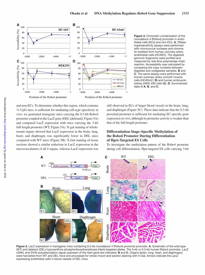
and non-ECs. To determine whether this region, which contains 11 CpG sites, is sufficient for mediating cell-type specificity in vivo, we generated transgenic mice carrying the 0.3-kb Robo4 promoter coupled to the LacZ gene (DEL [deletant]; Figure 5A) and compared LacZ expression with mice carrying the 3-kb full-length promoter (WT; Figure 5A). X-gal staining of whole-mount organs showed that LacZ expression in the brain, lung, heart, and diaphragm was significantly lower in DEL mice compared with WT mice (Figure 5B). X-Gal staining of tissue sections showed a similar reduction in LacZ expression in the microvasculature of all 4 organs, whereas LacZ expression was
still observed in ECs of larger blood vessels in the brain, lung, and diaphragm (Figure 5C). These data indicate that the 0.3-kb proximal promoter is sufficient for mediating EC-specific gene expression in vivo, although its promoter activity is weaker than that of the full-length promoter.
Differentiation Stage–Specific Methylation of the Robo4 Promoter During Differentiation of Hprt-Targeted ES CellsTo investigate the methylation pattern of the Robo4 promoter during cell differentiation, Hprt-targeted ES cells carrying 3-kb
Figure 4. Chromatin condensation of the roundabout 4 (Robo4) promoter in endo-thelial cells (ECs) and non-ECs. A, DNase hypersensitivity assays were performed with micrococcal nuclease and chroma-tin isolated from human coronary artery endothelial cells (HCAEC). The digested genomic fragments were purified and measured by real-time polymerase chain reaction. Accessibility was calculated by comparing the copy numbers between digested and undigested samples. B and C, The same assays were performed with human coronary artery smooth muscle cells (HCASmC; B) and human embryonic kidney (HEK) 293 cells (C). D, Summarized data of A, B, and C.
Figure 5. LacZ expression in transgenic mice containing 0.3-kb roundabout 4 (Robo4) proximal promoter. A, Schematic of the wild-type (WT) and deletant (DEL) hypoxanthine phosphoribosyltransferase (Hprt)-targeted alleles. The 3-kb or 0.3-kb human Robo4 promoter, LacZ cDNA, and SV40 polyadenylation signal upstream of the Hprt gene are indicated. B and C, Organs (brain, lung, heart, and diaphragm) were harvested from WT and DEL mice and processed for whole mount and section staining with X-Gal. Arrows indicate the LacZ-expressing endothelial cells in blood vessels of DEL mice.
by guest on June 4, 2018http://atvb.ahajournals.org/
Dow
nloaded from

1536 Arterioscler Thromb Vasc Biol July 2014
Robo4 promoter-LacZ were differentiated into mesodermal (Flk-1+ [fetal liver kinase]) cells and endothelial (CD31+) cells. The methylation patterns of the promoter in undifferentiated ES cells, Flk-1+ cells, and CD31+ cells were analyzed by bisulfite sequenc-ing (Figure 6; Figure IV in the online-only Data Supplement). In undifferentiated ES cells, a high degree of methylation was observed throughout the promoter. DNA methylation progres-sively decreased during cell differentiation. Importantly, the 11 CpG sites in the proximal promoter were completely unmethyl-ated in the Flk-1+ and CD31+ cells (Figure 6; Figure IV in the online-only Data Supplement). These findings suggest that the EC-specific methylation pattern of the Robo4 promoter is deter-mined by demethylation during cell differentiation.
To investigate the mechanism involved in demethylation of the proximal promoter during cell differentiation from ES cells into Flk-1+ cells, we analyzed whether GABP and SP1 binding to the promoter contributes to demethylation. Two ES cell lines containing the 3-kb Robo4 promoter with mutations either at the −119 ETS site or at the 2 SP1 sites (−153 and −142 SP1 sites) were differentiated, and methylation patterns of the proximal promoter in Flk-1+ cells were analyzed. In both mutant promoters, the proximal region showed a normal hypomethylated pattern similar to that of the WT promoter
(Figure 6B and 6C). These results indicate that binding of GABP and SP1 to the proximal promoter is not essential for promoter demethylation.
Robo4 Expression and DNA Methylation in ECs Exposed to Shear StressOur data raise the interesting question of whether DNA methylation plays a physiological role in mediating tem-poral changes in Robo4 expression in ECs. A previous study by Bicknell et al showed that laminar shear stress suppresses Robo4 gene expression.9 We asked whether suppression was regulated through the methylation of the proximal promoter. Human umbilical vein ECs were grown under static conditions or exposed to laminar shear stress (20 dyn/cm2) for 24 hours and harvested for RNA and genomic DNA. In real-time reverse transcription polymerase chain reaction assays, laminar flow resulted in a significant reduction of Robo4 and an induction of KLF2 mRNA levels (Figure VA in the online-only Data Supplement). However, shear stress did not induce proxi-mal promoter methylation in ECs (Figure VB in the online-only Data Supplement). These findings suggest that DNA
Figure 6. Differentiation stage–specific DNA methylation of the roundabout 4 (Robo4) promoter in targeted ES cells. A, Targeted ES cells containing the wild-type Robo4 promoter were differentiated, and Flk-1+ and CD31+ cells were separated by magnetic activated cell sorting. The methylation pattern of the targeted Robo4 promoter in undifferentiated and differentiated cells was analyzed by bisulfite sequencing. B and C, Targeted embryonic stem (ES) cells containing the Robo4 promoters with either the −119 ETS mutation (B) or the −42 and −153 SP1 double mutation (C) were differentiated into fetal liver kinase (Flk)-1+ cells. The methylation patterns of the targeted Robo4 promoters were analyzed by bisulfite sequencing.
by guest on June 4, 2018http://atvb.ahajournals.org/
Dow
nloaded from

Okada et al DNA Methylation Regulates Robo4 Gene Suppression 1537
methylation is not involved in mediating shear stress–dependent repression of Robo4.
DiscussionRobo4 is selectively expressed in ECs. Thus, an understand-ing of its transcriptional control may provide insights into mechanisms of EC-specific gene expression. We have previ-ously shown that GABP and SP1 positively regulate Robo4 promoter activity. However, these transcription factors are also expressed in other cell types. Thus, other mechanisms must be involved in mediating cell-type–specific expression of Robo4. Recent studies from the Marsden laboratory have demonstrated an important role for DNA methylation in medi-ating EC-specific expression of several genes, including those encoding endothelial nitric oxide synthase, vascular endothe-lial cadherin, CD31, von Willebrand factor, and intercellular adhesion molecule-2.14,17 The results of the present study sup-port a similar role for DNA methylation in governing cell-type–specific expression of Robo4.
The methylation pattern of the 5′-flanking region of Robo4 differed between ECs and non-ECs. This effect was most pro-nounced in the immediate 0.3-kb proximal promoter, in which all 11 CpG sites were heavily methylated in non-ECs but com-pletely unmethylated in ECs. Studies in which the Robo4 pro-moter was either methylated or demethylated demonstrated a functional role for DNA methylation in repressing Robo4 expression in non-ECs. In Hprt-targeted mice, the 0.3-kb prox-imal promoter (containing the 11 CpG sites) region retained information for EC-specific gene expression. Taken together, these findings strongly suggest that differential DNA meth-ylation in ECs and non-ECs contributes to cell-type–specific expression of Robo4. Two general mechanisms have been pro-posed for CpG methylation–mediated gene suppression. First, methylated CpG sites with or without methyl CpG–binding proteins prevent the binding of transcription factors, such as AP (activating protein)-2, HIF (hypoxia inducible factor)-1, and c-Myc (v-myc myelocytomatosis viral oncogene homo-log), to their binding motifs in the promoters.18–20 Second, methyl CpG–binding proteins recruit large chromatin-modi-fying complexes that reduce DNA accessibility by changing chromatin structure.
We first investigated whether DNA methylation interferes with binding of transcription factors to the Robo4 promoter. In elecrophoretic mobility shift assay, DNA methylation had no effect on GABP binding to the −119 ETS or SP1 binding to the −153 SP1 site. In contrast, DNA methylation inhibited bind-ing of SP1 to the −42 SP1 site. Because −42 SP1, but not to the −153 SP1, site contains an internal CpG site, methylation-dependent inhibition of transcription factor binding seems to be more effective when the SP1 and CpG sites overlap. In any event, our data suggest that DNA methylation at the −42 SP1 site plays a role in repressing Robo4 expression in non-ECs.
We next investigated whether DNA methylation of the pro-moter induces chromatin condensation, which is associated with inhibition of transcription factor binding to the promoter. Our DNase hypersensitivity assays indicated that chromatin is strongly condensed at −2.5 kb, where highly methylated CpG sites were observed in both ECs and non-ECs. In contrast,
chromatin condensation of the proximal promoter region, while low in ECs, was variable in non-ECs. Thus, chromatin condensation is not a universal requirement for Robo4 gene suppression in non-ECs.
We have shown that the proximal promoter of Robo4 is demethylated during ES cell differentiation into Flk-1+ cells. We repeated these assays using ES cells targeted with a Robo4 promoter containing mutated GABP- or SP1-binding sites. The WT and mutant promoters demonstrated compa-rable differentiation-dependent demethylation. Thus, binding of GABP and SP1 to the proximal promoter does not seem to mediate demethylation in differentiating ECs. Additional studies are required to determine the underlying mechanism.
It is interesting to speculate that DNA methylation is dynamically regulated in ECs and contributes to physiologi-cal modulation of Robo4 expression. A previous study dem-onstrated that Robo4 expression is inhibited by shear stress. We confirmed these findings but were unable to demonstrate flow-mediated changes in Robo4 promoter methylation. Thus, other mechanisms are likely to be responsible for Robo4 gene repression under shear stress conditions. Perhaps this is not surprising because the methylation status of DNA is a rela-tively stable mark and may change over longer time scales than those used in the present experiments.
A comparison of LacZ expression in Hprt-targeted mice car-rying the 3-kb and 0.3-kb upstream promoter regions of Robo4 indicates that although the shorter DNA fragment is sufficient for EC-specific expression, DNA sequences between 0.3 and 3 kb are necessary for full activity in the microvasculature. We identified enhancer elements at −2.5 kb (REn1 [Robo4 enhancer element]) and −2.9 kb (REn2) that are essential for maximal pro-moter activation.10 Because REn1 is located in the region where DNA methylation and chromatin condensation are observed, REn1 may contribute to activation of the Robo4 promoter through epigenetic control.
In summary, our study supports a model in which EC-specific expression of Robo4 is mediated, at least in part, by DNA methylation–dependent inhibition of SP1 binding to −42 SP1 in non-ECs. Furthermore, the data suggest the exis-tence of an EC-specific regulator that induces demethylation of Robo4 (and perhaps, by extension, other EC-specific genes) during cell differentiation.
AcknowledgmentsWe thank Dr Naoki Mochizuki for his technical support and helpful suggestions. We also thank Dr Sarah Bronson for gifting BK4 cells.
Sources of FundingThis work was supported by MEXT KAKENHI, JSPS KAKENHI, a Health and Labor Sciences Research Grant from the Ministry of Health, Labor, and Welfare of Japan, Takeda Science Foundation, the Uehara Memorial Foundation, Senri Life Science Foundation, Suzuken Memorial Foundation, and Daiichi-Sankyo Foundation of Life Science.
DisclosuresNone.
by guest on June 4, 2018http://atvb.ahajournals.org/
Dow
nloaded from

1538 Arterioscler Thromb Vasc Biol July 2014
References 1. Huminiecki L, Gorn M, Suchting S, Poulsom R, Bicknell R. Magic round-
about is a new member of the roundabout receptor family that is endo-thelial specific and expressed at sites of active angiogenesis. Genomics. 2002;79:547–552.
2. Huminiecki L, Bicknell R. In silico cloning of novel endothelial-specific genes. Genome Res. 2000;10:1796–1806.
3. Park KW, Morrison CM, Sorensen LK, Jones CA, Rao Y, Chien CB, Wu JY, Urness LD, Li DY. Robo4 is a vascular-specific receptor that inhibits endothelial migration. Dev Biol. 2003;261:251–267.
4. Seth P, Lin Y, Hanai J, Shivalingappa V, Duyao MP, Sukhatme VP. Magic roundabout, a tumor endothelial marker: expression and signaling. Biochem Biophys Res Commun. 2005;332:533–541.
5. Wang B, Xiao Y, Ding BB, Zhang N, Yuan Xb, Gui L, Qian KX, Duan S, Chen Z, Rao Y, Geng JG. Induction of tumor angiogenesis by Slit-Robo signaling and inhibition of cancer growth by blocking Robo activity. Cancer Cell. 2003;4:19–29.
6. Suchting S, Heal P, Tahtis K, Stewart LM, Bicknell R. Soluble Robo4 receptor inhibits in vivo angiogenesis and endothelial cell migration. FASEB J. 2005;19:121–123.
7. Bedell VM, Yeo SY, Park KW, Chung J, Seth P, Shivalingappa V, Zhao J, Obara T, Sukhatme VP, Drummond IA, Li DY, Ramchandran R. round-about4 is essential for angiogenesis in vivo. Proc Natl Acad Sci U S A. 2005;102:6373–6378.
8. Jones CA, London NR, Chen H, et al. Robo4 stabilizes the vascular net-work by inhibiting pathologic angiogenesis and endothelial hyperperme-ability. Nat Med. 2008;14:448–453.
9. Mura M, Swain RK, Zhuang X, et al. Identification and angiogenic role of the novel tumor endothelial marker CLEC14A. Oncogene. 2012;31:293–305.
10. Okada Y, Yano K, Jin E, Funahashi N, Kitayama M, Doi T, Spokes K, Beeler DL, Shih SC, Okada H, Danilov TA, Maynard E, Minami T, Oettgen P, Aird WC. A three-kilobase fragment of the human Robo4
promoter directs cell type-specific expression in endothelium. Circ Res. 2007;100:1712–1722.
11. Okada Y, Jin E, Nikolova-Krstevski V, Yano K, Liu J, Beeler D, Spokes K, Kitayama M, Funahashi N, Doi T, Janes L, Minami T, Oettgen P, Aird WC. A GABP-binding element in the Robo4 promoter is necessary for endothelial expression in vivo. Blood. 2008;112:2336–2339.
12. Minami T, Aird WC. Endothelial cell gene regulation. Trends Cardiovasc Med. 2005;15:174–184.
13. Weintraub H, Tapscott SJ, Davis RL, Thayer MJ, Adam MA, Lassar AB, Miller AD. Activation of muscle-specific genes in pigment, nerve, fat, liver, and fibroblast cell lines by forced expression of MyoD. Proc Natl Acad Sci U S A. 1989;86:5434–5438.
14. Shirodkar AV, St Bernard R, Gavryushova A, Kop A, Knight BJ, Yan MS, Man HS, Sud M, Hebbel RP, Oettgen P, Aird WC, Marsden PA. A mechanistic role for DNA methylation in endothelial cell (EC)-enriched gene expression: relationship with DNA replication timing. Blood. 2013;121:3531–3540.
15. Yan MS, Matouk CC, Marsden PA. Epigenetics of the vascular endothe-lium. J Appl Physiol (1985). 2010;109:916–926.
16. Herman JG, Baylin SB. Gene silencing in cancer in association with pro-moter hypermethylation. N Engl J Med. 2003;349:2042–2054.
17. Chan Y, Fish JE, D’Abreo C, Lin S, Robb GB, Teichert AM, Karantzoulis-Fegaras F, Keightley A, Steer BM, Marsden PA. The cell-specific expres-sion of endothelial nitric-oxide synthase: a role for DNA methylation. J Biol Chem. 2004;279:35087–35100.
18. Comb M, Goodman HM. CpG methylation inhibits proenkephalin gene expression and binding of the transcription factor AP-2. Nucleic Acids Res. 1990;18:3975–3982.
19. Wenger RH, Kvietikova I, Rolfs A, Camenisch G, Gassmann M. Oxygen-regulated erythropoietin gene expression is dependent on a CpG methyl-ation-free hypoxia-inducible factor-1 DNA-binding site. Eur J Biochem. 1998;253:771–777.
20. Prendergast GC, Ziff EB. Methylation-sensitive sequence-specific DNA binding by the c-Myc basic region. Science. 1991;251:186–189.
Cell-type–specific transcription factors or tissue-specific combinations of noncell-type–specific transcription factors are thought to regulate cell-type–specific gene expression. Although the regulation of various endothelial cell (EC)–specific genes has been studied, the transcription factors and their combinations that regulate EC-specific gene expression have not been fully identified. To identify the transcription factors that regulate EC-specific gene expression, we previously investigated the regulation of the Robo4 gene and identified SP1 and GA-binding protein as essential regulators for Robo4 promoter activation. However, because these factors are known to be expressed in other tissues, we could not explain the mechanism that induces Robo4 gene expression only in ECs. In this study, we hypothesized that cell-type–specific gene expression was regulated by epigenetics, as well as transcription factors, and succeeded in demonstrating the importance of DNA methylation for EC-specific Robo4 gene expression.
Significance by guest on June 4, 2018http://atvb.ahajournals.org/
Dow
nloaded from

Shinsaku Nakagawa, Guillermo García-Cardeña, William C. Aird and Takefumi DoiZhixia, Kenji Kitajima, Kenji Ishimoto, Nobumasa Hino, Masuo Kondoh, Yohei Mukai,
JiangShirakura, Alexis S. Turjman, Yoshihiro Kano, Hiroki Naruse, Ayano Suzuki, Miki Sakai, Yoshiaki Okada, Nobuaki Funahashi, Toru Tanaka, Yuji Nishiyama, Lei Yuan, Keisuke
Methylation of the Proximal PromoterSpecific Expression of Roundabout 4 Is Regulated by Differential DNA−Endothelial Cell
Print ISSN: 1079-5642. Online ISSN: 1524-4636 Copyright © 2014 American Heart Association, Inc. All rights reserved.
Greenville Avenue, Dallas, TX 75231is published by the American Heart Association, 7272Arteriosclerosis, Thrombosis, and Vascular Biology
doi: 10.1161/ATVBAHA.114.3038182014;34:1531-1538; originally published online May 22, 2014;Arterioscler Thromb Vasc Biol.
http://atvb.ahajournals.org/content/34/7/1531World Wide Web at:
The online version of this article, along with updated information and services, is located on the
http://atvb.ahajournals.org/content/suppl/2014/05/22/ATVBAHA.114.303818.DC1Data Supplement (unedited) at:
http://atvb.ahajournals.org//subscriptions/
at: is onlineArteriosclerosis, Thrombosis, and Vascular Biology Information about subscribing to Subscriptions:
http://www.lww.com/reprints
Information about reprints can be found online at: Reprints:
document. Question and AnswerPermissions and Rightspage under Services. Further information about this process is available in the
which permission is being requested is located, click Request Permissions in the middle column of the WebCopyright Clearance Center, not the Editorial Office. Once the online version of the published article for
can be obtained via RightsLink, a service of theArteriosclerosis, Thrombosis, and Vascular Biologyin Requests for permissions to reproduce figures, tables, or portions of articles originally publishedPermissions:
by guest on June 4, 2018http://atvb.ahajournals.org/
Dow
nloaded from

1
MATERIALS AND METHODS
Cell culture
Human coronary artery endothelial cells (HCAEC), human umbilical vein endothelial cells
(HUVEC), human coronary artery smooth muscle cells (HCASmC), and human dermal
fibroblasts (NHDF) were purchased from Lonza (Basel, Switzerland). Primary ECs, HCASmC,
and NHDF were cultured in EGM-2-MV, SmGM-2, and FGM-2 media, respectively. Human
embryonic kidney cells (HEK293 cells) were cultured in DMEM supplemented with 10% fetal
bovine serum (FBS), 100 IU/ml penicillin, and 100 μg/ml streptomycin. All cells were cultured
at 37ºC under 5% CO2.
Bisulfite sequencing
Genomic DNA was extracted using the ISOGEN reagent (Nippon Gene, Tokyo, Japan). The
resulting DNA (3 g) was treated with a bisulfite reagent using the MethylEasy Xceed Rapid
DNA Bisulfite Modification Kit (Human Genetic Signatures, Sydney, Australia). Robo4
promoter fragments were amplified by PCR using the bisulfite-treated DNA and region-specific
primers (sequences are shown in Table SI). The resulting fragments were cloned into the
pCR2.1 vector using a TOPO TA cloning kit (Invitrogen), and the resulting plasmids were
transfected into DH5 cells. Plasmids were prepared from 10 randomly picked colonies, and
DNA sequences were analyzed. In all of the clones, 100% C to T conversions at non-CpG sites
were observed, indicating efficient sodium bisulfite reactions.
Electrophoretic mobility shift assay (EMSA)
Recombinant SP1 and GABP (GABPα, β, and γ subunits) were prepared using the TNT Quick
coupled transcription/translation system (Promega, Madison, WI) and 1 g of expression
vectors. EMSA was performed as described previously.1 Briefly,
32P-labeled oligonucleotide
probes spanning GABP or SP1 binding sites with or without cytosine methylation were mixed
with 2–4 l recombinant SP1 or GABP for 40 min at 4ºC. The resulting protein-DNA
complexes were analyzed by gel electrophoresis using 4% native polyacrylamide gel and 0.5
TBE buffer at 120 V. Oligonucleotide sequences for the probes are shown in Table SI.
Treatment of cells with 5-aza-2′-deoxycytidine (5-AC) and real-time RT-PCR
HCAEC, HCASmC, NHDF, and HEK293 cells were treated in cell culture media containing 10
M 5-AC (Sigma-Aldrich, St. Louis, MO) for 4 days. The media were replaced with fresh
media containing 5-AC 2 days after beginning the culture. Total RNA was prepared using the
RNeasy Mini Kit (Qiagen, Hilden, Germany). To generate cDNA, 0.5 or 1 g of total RNA was

2
reverse transcribed with Superscript III reverse transcriptase (Invitrogen). Real-time PCR
measurements were performed using the cDNA, primers (shown in Table S1), and QuantiTect
SYBR Green PCR Kit (Qiagen). Copy numbers were calculated from the standard curve
prepared by measuring known amounts of plasmids including target sequences. The expression
level of the Robo4 was normalized against GAPDH. Data were collected from at least 3
independent experiments.
Preparation of plasmid and targeting vectors
Preparation of the Robo4 promoter-reporter construct, pGL3-Robo4 and expression vectors for
SP1 and GABP has been previously described.1 To generate the Hprt-targeting vector,
pGL3-Robo4 was digested with BamHII and NheI. The fragment containing the 0.3-kb
promoter was purified and cloned into the pSDK-lacZ vector containing lacZ cDNA to generate
pRobo4-Del5-lacZ. pRobo4-Del5-lacZ was then digested with PmeI and NotI. The resulting
transgenic cassette was purified and cloned into the Hprt-targeting vector, pMP8II.
Transient transfection assay using methylated plasmid
The Robo4 promoter-reporter construct contains many CpG sites in the promoter, the luciferase
reporter, and in the vector backbone. To assess the role of Robo4 proximal promoter-specific
methylation in the absence of both reporter and vector backbone methylation, a non-methylated
Robo4-promoter-reporter construct (NM-pGL3-Robo4) was prepared by amplifying
pGL3-Robo4 in Dam and Dcm methylase deficient E. coli strain SCS110 (Agilent Technologies,
Santa Clara, CA). To prepare the methylated pGL3-Robo4 (M-pGL3-Robo4) in which the CpG
sites in the proximal promoter were methylated, NM-pGL3-Robo4 was digested with StuI and
XhoI. The resulting short promoter fragment was methylated by SssI (New England Biolabs,
Ipswitch, MA) and cloned into the StuI-XhoI site of NM-pGL3-Robo4 to generate
M-pGL3-Robo4. The NM-pGL3-Robo4, M-pGL3-Robo4, and pGL3 as a control were
transfected into HCAEC, and promoter activities were evaluated as described previously.1 Data
was collected from 5 independent assays.
Generation and analysis of Hprt-targeted mice
Generation of Hprt-targeted mice containing the wild type Robo4 promoter-lacZ was described
previously. 1 To generate ES cells containing the 0.3-kb truncated Robo4 promoter (Del)-lacZ,
the targeting vector was linearized by digesting with SalI, and electroporated into Hprt-deficient
BK4 ES cells. Homologous recombinants were selected in ES cell culture medium containing
HAT (Sigma-Aldrich). The targeted ES cell clones were used for generating Robo4
promoter-lacZ chimeric mice. Chimeric males were bred to C57BL/6 females to obtain agouti

3
offspring. Mouse lines were generated from two independent ES clones. Organs from the
generated mice were fixed with the PBS containing 2% formaldehyde and 0.2% glutaraldehyde,
and stained with the PBS containing 0.02% NP-40, 0.01% SDS, 2 mM MgCl2, 5 mM K3Fe(CN)6,
5 mM K4Fe(CN)6, 1% X-gal at 37 ºC for 20 h.
DNase hypersensitivity assay
DNase hypersensitivity assays were performed as described previously.2 HCAEC, HCASmC,
and HEK293 cells were lysed on ice in Lysis buffer (10 mM Tris-HCl (pH 7.4), 10 mM NaCl, 3
mM MgCl2, 0.5% Nonidet P-40, 150 μM spermine, 500 μM spermidine) and centrifuged. The
pelleted nuclei were washed with Wash buffer (10 mM Tris-HCl (pH 7.4), 15 mM NaCl, 60 mM
KCl, 150 μM spermine, 150 μM spermidine) and resuspended in Digestion buffer (wash buffer
containing 1 mM CaCl2). The resulting suspension was digested with or without 7 units of
micrococcal nuclease (TAKARA, Shiga, Japan) for 5 min at 25°C. The reaction was stopped by
adding Stop buffer (20 mM EDTA, 2 mM EGTA, 1% SDS) followed by incubation on ice for
10 min. Proteins in the resulting samples were digested by proteinase K. The DNA was
extracted by phenol-chloroform extraction and ethanol precipitation, and resuspended in TE (pH
8.0). Real-time PCR analyses were performed in triplicate with the resulting DNA samples and
region-specific primers (sequences are shown in Table SI). Copy numbers of each promoter
fragment was calculated from the standard curve prepared by measuring serial dilutions of
pGL3-Robo4.The percentage of accessibility was calculated by comparing the copy numbers of
promoter fragments in the digested and undigested DNA samples.
In vitro ES cell differentiation
Generation of Hprt locus-targeted ES cells (Robo4 promoter-lacZ and Robo4(ETSmut)-lacZ)
were described previously.1, 3
The ES cells containing the Robo4 promoter with SP1 double
mutation were generated using the plasmid pGL3-SP1(1,2)mut1 and the same method used for
Robo4(ETSmut)-lacZ as previously described.3 To prepare Flk-1
+ or ECs, these targeted ES
cells were seeded onto OP9 cells and cultured for 5 days in MEM supplemented with 20%
FBS, 0.1 mM non-essential amino acids, 2 mM L-glutamine, 100 IU/ml penicillin, and 100
g/ml streptomycin. Flk-1+ cells were purified from the differentiated cells by MACS using an
anti-mouse Flk1 antibody (BD Pharmingen, San Diego, CA). The resulting Flk1
+ cells were
seeded on collagen IV-coated plates (Becton Dickinson, Franklin Lakes, NJ) and cultured for 3
days inMEM supplemented with 50 ng/ml human VEGF165 (R&D systems, Minneapolis,
MN), 0.5 mM 8-bromo cAMP (Nacalai Tesque, Kyoto, Japan), 10% FBS, and 50 M
2-mercaptoethanol. CD31+ ECs were purified by MACS using an anti-mouse CD31
antibody
(BD Pharmingen). Undifferentiated ES cells, Flk-1+ cells, and CD31
+ cells were used for the

4
bisulfite sequencing analysis,
REFERENCES
1. Okada Y, Yano K, Jin E, Funahashi N, Kitayama M, Doi T, Spokes K, Beeler DL,
Shih SC, Okada H, Danilov TA, Maynard E, Minami T, Oettgen P, Aird WC. A
three-kilobase fragment of the human robo4 promoter directs cell type-specific
expression in endothelium. Circ Res. 2007;100:1712-1722
2. Fish JE, Yan MS, Matouk CC, St Bernard R, Ho JJ, Gavryushova A, Srivastava D,
Marsden PA. Hypoxic repression of endothelial nitric-oxide synthase transcription
is coupled with eviction of promoter histones. J Biol Chem. 2010;285:810-826
3. Okada Y, Jin E, Nikolova-Krstevski V, Yano K, Liu J, Beeler D, Spokes K,
Kitayama M, Funahashi N, Doi T, Janes L, Minami T, Oettgen P, Aird WC. A
gabp-binding element in the robo4 promoter is necessary for endothelial expression
in vivo. Blood. 2008;112:2336-2339

SUPPLEMENTAL MATERIAL
Table SI. Sequences of the Oligonucleotides.
Bisulfite sequencing
-802 to +16 forward 5'-GATGGGATGGGTTTAAAAAGAAATATAT-3'
reverse 5'-ACCCATAACTACTCTCAACCCTATATC-3'
-1259 to -817 forward 5'-ATTTTTATTTGGTAGGTTGTTAGTTTGTGGTTG-3'
reverse 5'-TTTTAAACCCATCCCATCACTAAAACTATTAAA-3'
-2297 to -1252 forward 5'-GTAGATTGTTGATAGTGATATTTTTGATAAGTTG-3'
reverse 5'-AACCACAAACTAACAACCTACCAAA-3'
-3030 to -2347 forward 5'-AGGGGGAGTTAGAAAATATAAAATAT-3'
reverse 5'-ACATTAATTTTAAAAACACTAATTAAATAC-3'
EMSA (Methylated oligonucleotides were used for the methylated probes.)
-153 SP1 probe sense 5'-GTTTCTCGCCTCTGGTCTCCTCCCAGTTCTCCAAG-3'
antisense 5'-CTTGGAGAACTGGGAGGAGACCAGAGGCGAGAAAC-3'
-119 ETS probe sense 5'-GGGCCAGGCAGGAAGCATCGGTTTC-3'
antisense 5'-GAAACCGATGCTTCCTGCCTGGCCC-3'
-42 SP1 probe sense 5'-CCACCACCGGCCCACCCCGCCCCTCCTTCCC-3'
antisense 5'-GGGAAGGAGGGGCGGGGTGGGCCGGTGGTGG-3'
Real-time PCR
Robo4 forward 5'-TTATGGCTCCCTCATCGCTG-3'
reverse 5'-GAGGCTGTCTGAGCTGGAAC-3'
GAPDH forward 5'-TGCACCACCAACTGCTTAGC-3'
reverse 5'-GGCATGGACTGTGGTCATGAG-3'
KLF2 forward 5'-CTTTCGCCAGCCCGTGCCGCG-3'
reverse 5'-AAGTCCAGCACGCTGTTGAGG-3'
DNase hypersensitivity assay
-149 to -10 forward 5'-TCTGGTCTCCTCCCAGTTCTCCAAG-3'
reverse 5'-CGAGCACTTTGTCCTGCTGCTCTG-3'
-1337 to -1217 forward 5'-GTTTGTAGAGACCATGGTGTTTC-3'
reverse 5'-CTTCGGTGCCAGCCACAGAC-3'
-2387 to -2242 forward 5'-CTAGCGTCTTTCTGGATTGTGGAG-3'
reverse 5'-CAAAGCCTCCAAGACTGTCTGACTC-3'
-2990 to -2857 forward 5'-CTAGGGATGAAGGAAGGCACTG-3'
reverse 5'-CACAAACTAAGGAAGAGCCGAC-3'

Figure SI. Analysis of the distribution of CpG sites in the Robo4 promoter. In the
top panel, locations of the CpG sites are indicated with lollypops. In the bottom panel,
the moving average of the observed/expected (Obs/Exp) CpG and %G+C were
calculated as described in a previous report.1 Each point on the graph represents the
average values for 10 adjacent 100-bp windows. Values for Obs/Exp CpG and %G+C
are plotted as a continuous line and a broken line, respectively. The typical definition of
a CpG island is a region in which the Obs/Exp CpG and %G+C are greater than 0.6 and
50, respectively.

Figure SII. Bisulfite sequencing of the human Robo4 promoter. 3-kb human Robo4
promoter bisulfite sequencing results for 10 cloned PCR products prepared with
bisulfite-treated genomic DNA from 2 endothelial cell types (HCAEC and HUVEC)
and 3 non-endothelial cell types (HCASmC, NHDF, and HEK293). Open and closed
circles indicate non-methylated and methylated CpGs, respectively.

Figure SIII. DNA methylation patterns of the Robo4 proximal promoter in various
cell-types. The methylation of the Robo4 proximal promoter and Pol II binding were
analyzed using data generated by the ENCODE consortium. The data includes the
promoter methylation pattern in ECs (HUVEC) and non-ECs, including aortic smooth
muscle cells (AoSMC), neonatal dermal fibroblasts (NHDF-neo), HEK293, human
embryonic stem cells (H1-hESC), fetal lung fibroblasts (IMR90), mammary epithelial
cells (HMEC), small airway epithelial cells (SAEC), skeletal striated muscle cells
(SkMC), bronchial epithelial cells (NHBE), skin fibroblasts (BJ), and astrocytes
(NH-A). Levels of methylation are color coded, where blue represents non-methylated,
purple represents partial methylation, and orange represents full methylation, as
identified by Methyl 450K Bead Arrays. Pol II chromatin immunoprecipitation-seq data
in HUVEC is shown at the bottom. A black arrow denotes the transcription start site.

Figure SIV. Bisulfite sequencing of the human Robo4 promoter in ES cells and
Flk-1+ cells. (A) The targeted ES cells containing the wild type Robo4 promoter were
differentiated, and Flk-1+ and CD31
+ cells were separated by MACS. The methylation
pattern of the targeted Robo4 promoter in undifferentiated and differentiated cells was
analyzed by bisulfite sequencing. Open and closed circles indicate non-methylated and
methylated CpG, respectively. (B and C) Targeted ES cells containing the mutated
Robo4 promoters with either the -119 ETS mutation (B) or the -42 and -153 SP1 double
mutation (C) were differentiated into Flk-1+ cells. The methylation patterns of the
targeted Robo4 promoters were analyzed by bisulfite sequencing.

Figure SV. Bisulfite sequencing of the human Robo4 promoter in shear stress
treated ECs. (A) The expression of KLF2 and Robo4 mRNA in HUVECs treated with
or without shear stress was measured by real-time RT-PCR. The data are presented as
the mean ± SE. (n=3, Student’s t-test, * p < 0.05). (B) DNA methylation patterns of
Robo4 proximal promoter in HUVECs treated with or without shear stress were
analyzed by bisulfite sequencing.
SUPPLEMENTAL FIGURE SV METHODS
Preparation of genomic DNA and RNA samples from HUVEC exposed to shear
stress.
HUVEC were plated to confluence and cultured for 24 h in M199 medium containing
20% fetal calf serum, 1% L-glutamine, 1% endothelial cell growth supplement (Alfa
Aesar, Lancashire, United Kingdom), 1% heparin, 100 IU/ml penicillin, and 100
μg/ml streptomycin. Cells were then exposed to constant laminar shear stress (20
dynes/cm2) or left under static (no flow) conditions for 24 h, and subsequently used for
preparations of genomic DNA and RNA for bisulfite-sequencing and real-time RT-PCR
analyses, respectively. Primer sequences are shown in Table SI.
REFERENCES
1. Gardiner-Garden M, Frommer M. Cpg islands in vertebrate genomes. J Mol Biol.
1987;196:261-282